Article

Multi-Target Anti-Aging Mechanisms of Multani Mitti (Fuller’s Earth): Integrating Enzyme Inhibition and Molecular Docking for Cosmeceuticals

1
Doctoral Program in Sciences, Specialization in Applied Cellular and Molecular Biology, Universidad de La Frontera, Temuco 4811230, Chile
2
Department of Zoology, Government College University Faisalabad, Faisalabad 38000, Pakistan
3
Laboratorio de Investigación en Salud de Precisión, Departamento de Procesos Diagnósticos y Evaluación, Facultad de Ciencias de la Salud, Universidad Católica de Temuco, Manuel Montt 056, Temuco 4780000, Chile
4
Center of Molecular Biology and Pharmacogenetics, Department of Basic Sciences, Faculty of Medicine, Universidad de la Frontera, Temuco 4811230, Chile
5
Escuela de Tecnología Médica, Facultad de Salud, Universidad Santo Tomás, Temuco 4780000, Chile
*
Author to whom correspondence should be addressed.
Cosmetics 2025, 12(3), 124; https://doi.org/10.3390/cosmetics12030124
Submission received: 28 April 2025 / Revised: 5 June 2025 / Accepted: 9 June 2025 / Published: 13 June 2025

Abstract

:
The growing demand for natural anti-aging ingredients necessitates scientific validation of traditional cosmetic materials. Multani Mitti (MM), a clay widely used in South Asian traditional skincare, lacks comprehensive chemical and biological characterization. This study employed a multi-analytical approach to investigate MM’s anti-aging potential through chemical analysis, enzyme inhibition studies, and in silico evaluations. Five commercial MM samples were pooled and analyzed using instrumental neutron activation analysis (INAA) and Gas Chromatography–Mass Spectrometry (GC-MS). INAA revealed silicon as the predominant inorganic constituent (169.3742 mg/g), while GC-MS identified 13 bioactive compounds, with Beta-sitosterol (15.45% area), Docosanamide (12.36% area), and Cyclohexasiloxane (9.80% area) being the most abundant. MM demonstrated significant enzyme inhibition against key aging-related enzymes, with notably strong effects on hyaluronidase (IC50: 18 μg/mL) and tyrosinase (IC50: 27 μg/mL), outperforming standard inhibitors. The antioxidant activity showed moderate effectiveness (IC50: 31.938 μg/mL) compared to ascorbic acid (IC50: 8.5 μg/mL). Molecular docking studies of identified compounds against hyaluronidase (PDB: 1FCV) and tyrosinase (PDB: 3NQ1) revealed Beta-sitosterol and Benzyl-piperazine-carboxamide as the most promising candidates, showing strong binding affinities (−8.5 and −8.6 kcal/mol, respectively) and favorable ADMET profiles. This comprehensive characterization provides the first scientific evidence supporting MM’s traditional use in skincare and identifies specific compounds that may contribute to its anti-aging properties, warranting further investigation for modern cosmetic applications.

1. Introduction

The skin, particularly facial skin, serves as a significant reflection of an individual’s overall health. Maintaining a clear and radiant complexion requires consistent skincare practices [1]. Historically, people have been conscious of their skincare routines, adapting them to their specific skin types. Even today, natural remedies remain popular, especially in rural and hilly regions where people continue to use plant extracts such as Aloe vera, tulsi, rose, and neem for their cosmetic benefits [2]. In many parts of Asia, particularly in Pakistan, India, Bangladesh, and Nepal, a unique clay known as MM has long been employed as a cost-effective and efficient skincare solution. Originating from Multan city in Pakistan, MM is prized for its purifying and beautifying properties, with particular emphasis on its anti-aging effects. This clay has been incorporated into skincare routines as a natural solution for maintaining youthful skin, commonly used in face masks [3,4,5]. The powdered form and its application as a face mask are illustrated in Figure 1. MM, also known as ‘Fuller’s Earth’, has been revered for its healing properties for centuries. Scientifically, it is composed of minerals such as montmorillonite, smectite, and bentonite clay [6]. The historical use of clay for therapeutic purposes dates back to ancient Egyptian, Mesopotamian, Chinese, and Indian civilizations, underscoring its enduring significance. Additionally, deposits of Fuller’s Earth are found in various regions globally, including the Americas, France, and Mexico [7,8,9]. The term ‘Fuller’s Earth’ originates from its historical use in the textile industry, where it played a crucial role in cleaning and thickening woolen fabrics—a process known as ‘fulling’. The individuals who specialized in this task were referred to as ‘fullers’, which gave rise to the name ‘Fuller’s Earth’ [10,11].
The mineral-rich composition of clay provides exceptional absorbent properties, making it versatile for medical, dietary, and cosmetic applications. Among clays, MM stands out for its remarkable ability to purify the skin, making it particularly beneficial for oily and sensitive skin types [12]. Due to its superior absorbent properties, MM effectively removes dead skin cells and excess oil from the skin and scalp. These properties, combined with its anti-inflammatory qualities, make it an excellent choice for both skincare routines and hair care. Furthermore, it tones the skin, minimizes the appearance of wrinkles, and serves as an effective deep pore cleanser, leaving the complexion refreshed and radiant [13,14,15,16].
Beyond its cleansing abilities for skin and hair care application in Pakistan, the practice of geophagy—specifically the consumption of MM clay—is widespread among women and children. Pregnant women are often advised to ingest this clay to address deficiencies in essential minerals [4]. Research has also explored developing a reagent for latent fingerprint visualization using MM [10]. Due to its myriad benefits for both skin and hair care—as well as its perceived health advantages—MM has become a highly sought-after cosmetic product. It is commonly marketed as an affordable and readily available cosmetic ingredient with a long history of traditional topical use in Ayurvedic practices, supported by widespread consumer acceptance in South Asian markets [17,18,19,20]. However, comprehensive scientific safety evaluations remain limited. In 2020, the market value of MM was estimated at USD 25.3 million, with a projected compound annual growth rate (CAGR) of 6.3% from 2022 to 2028. This progression reflects an increasing demand for natural beauty products and underscores MM’s reputation for enhancing skin complexion while minimizing blemishes, solidifying its significance in the skincare industry [21,22].
Despite its widespread use in cosmetics and traditional medicine practices, the biological mechanisms underlying its effects on the skin remain largely unexplored. As skincare solutions increasingly prioritize antioxidant, anti-aging, and antimicrobial properties, this research aims to evaluate the antioxidant and enzyme inhibitory activities of MM and identify its potential therapeutic applications in the management of aging-related conditions. Previous studies have indicated that MM lacks significant antimicrobial activity [23], prompting us to shift our focus towards its potential antioxidant and anti-aging properties.
Skin aging is closely associated with the activity of enzymes such as elastase, collagenase, hyaluronidase, and tyrosinase [24]. Elastase and collagenase degrade elastin and collagen, respectively, which are vital structural proteins that maintain skin elasticity and firmness; their overactivity leads to wrinkles and loss of skin integrity [25]. Hyaluronidase breaks down hyaluronic acid, a key molecule responsible for skin hydration and volume, contributing to dryness and sagging when excessively active [26]. Tyrosinase catalyzes melanin production, and its overexpression can cause hyperpigmentation and uneven skin tone [27]. Inhibiting these enzymes can, therefore, slow down the degradation of skin matrix components and regulate pigmentation, making enzyme inhibitors promising targets for anti-aging therapies and cosmetic applications [28].
While a variety of well-established inhibitors—such as EGCG for collagenase and elastase, tannic acid for hyaluronidase, and kojic acid for tyrosinase—are routinely used in laboratory assays and cosmetic formulations, there remains significant interest in identifying new, naturally derived alternatives [29,30]. Given Multani Mitti’s widespread use in traditional skincare, our study aimed to systematically evaluate its potential as a natural source of enzyme inhibitors relevant to skin aging. For comparative purposes and to validate our experimental approach, these known inhibitors were included as positive controls in all assays. This allowed us to directly benchmark the inhibitory effects of MM clay against industry standards, highlighting its promise as a multifunctional, natural ingredient for anti-aging cosmetic applications.
Building on the importance of enzyme inhibition in skin aging and the search for natural alternatives, this study begins with a comprehensive chemical analysis to elucidate the complete organic and inorganic composition of MM powder using Gas Chromatography–Mass Spectrometry (GC-MS) and instrumental neutron activation analysis (INNA). We will then evaluate the inhibitory activity of these compounds against enzymes linked to the aging process of the skin—namely, elastase, collagenase, hyaluronidase, and tyrosinase—through both in vitro assays and in silico molecular modeling techniques. These methods not only allow for active pinpoint compounds but also provide valuable insights into their underlying molecular mechanisms. The outcomes of this research will contribute to understanding MM’s potential as a cosmeceutical agent that could guide the development of innovative antioxidant and anti-aging skincare products.

2. Materials and Methods

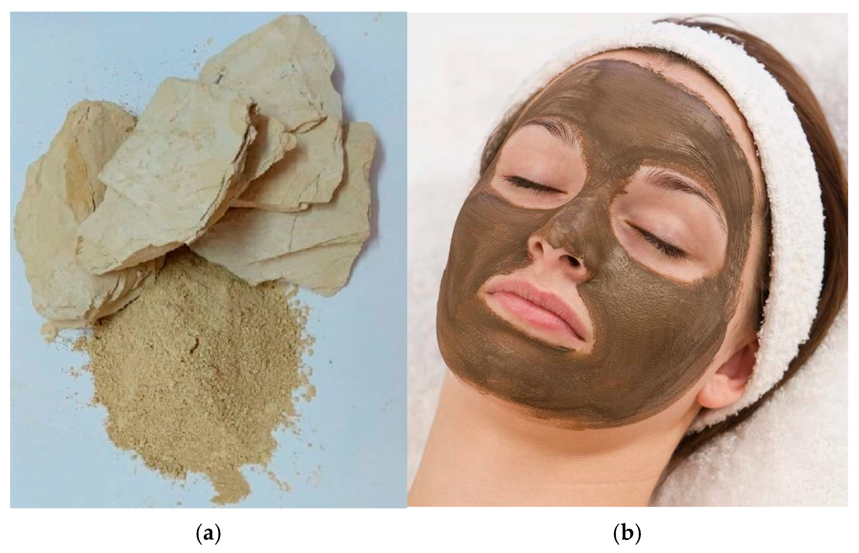
2.1. Sample Collection

The soil sample was procured in a dried powdered form from 5 different well-known ISO-certified cosmetics brands suppliers (https://www.daraz.pk/tag/multani-mitti/; accessed on 8 March 2024, https://saeedghani.pk/products/multani-mud-powder-100gm?_pos=1&_sid=622ee6225&_ss=r; accessed on 8 March 2024, https://karachipansar.pk/product/multani-mitti; accessed on 8 March 2024, https://pansarionline.pk/natural-multani-mud/; accessed on 8 March 2024, and https://somethingorganic.pk/product/multani-mitti-powder/; accessed on 8 March 2024) of Pakistan. They represent the actual products consumers use in cosmetic applications, making our findings directly relevant to real-world usage. It was packaged and marketed as MM powder, one of the top-selling products in this category. All these were mixed to make a single sample for a further overall investigation. These commercial samples were pooled to minimize batch-to-batch variability and obtain a representative composite reflecting the general MM composition available in the market. This approach follows established analytical chemistry practices for improving detection limits in GC-MS analysis and aligns with our primary objective to evaluate the collective anti-aging potential of MM as an ingredient class rather than promote specific brands.

2.2. INAA Methodology

The INAA was performed using the protocol adapted from Waheed et al. [31]. The sample preparation involved placing it in a pre-cleaned polyethylene capsule before irradiation in a Miniature Neutrons Source Reactor (MNSR). After irradiation and appropriate cooling, the sample underwent gamma-ray spectrometry with a high-purity germanium detector. Data acquisition and analysis utilized specialized software tools, ensuring appropriate corrections were made. The results were computed based on dry weight as the reference, with error propagation addressing various uncertainties.

2.3. GCMS Analysis

The analysis of the MM followed the outlined by Profumo et al. [32], with necessary adjustments for its inorganic nature. The dried powder was suspended separately in methanol and dichloromethane (DCM) to extract any polar, non-polar, and medium-polarity organic compounds. After vortexing and centrifugation, the supernatant was analyzed using an Agilent GC 7890A coupled with an Agilent MS 5975C (Seoul, Republic of Korea). Compounds were separated using a DB-5MS column (30 m × 0.25 mm, 0.25 μm film thickness), with helium as the carrier gas at a flow rate of 1 mL/min. The injector was set to 275 °C, with an oven temperature starting at 65 °C, held for 2 min, then increased by 7 °C per minute until reaching 280 °C for an additional minute. The total analysis duration was 40 min. The mass spectrometer was configured to scan within a mass range of 35–500 m/z, with both the ion source and transfer line temperatures maintained at 250 °C. The GC-MS data were analyzed using the National Institute of Standards and Technology (NIST) library to identify organic compounds present in the MM powder. The identification of the organic compounds was based on the relative abundance and retention times of the detected compounds, offering detailed insight into the organic composition of the MM sample.

2.4. Antioxidant Assay

The DPPH (2,2-diphenyl-1-picrylhydrazyl) free radical scavenging assay was performed according to the method outlined by Brand-Williams et al. [33], with adaptations for soil samples. The MM sample was prepared by thoroughly mixing 5 g of air-dried powder with 50 mL of 80% methanol and agitating the mixture for 2 h. The solution was centrifuged at 4000 rpm for 15 min and filtered through a 0.45 μm membrane filter. Antioxidant potential was evaluated using a modified DPPH radical scavenging assay. The test compound was serially diluted in methanol to achieve concentrations ranging from 10 to 100 μg/mL (10, 20, 40, 80, and 100 μg/mL). Each dilution (100 μL) was combined with 3.9 mL of 0.1 mM DPPH methanolic solution. Following thorough homogenization, the reaction mixture was incubated at ambient temperature under light-protected conditions for 30 min. Subsequently, the absorbance was measured spectrophotometrically at λ = 517 nm. Ascorbic acid, serving as a positive control, was prepared at concentrations ranging from 1 to 10 μg/mL. The percentage of inhibition was calculated using the following formula:
Inhibition % = Acontrol − Asample/Acontrol × 100
Acontrol = absorbance of the DPPH blank solution only.
Asample = absorbance of the DPPH test solutions combined with soil samples at various concentrations, adjusted for their individual absorption levels.
The radical scavenging activity was expressed as a percentage, and the half-maximal inhibitory concentration (IC50) was determined. To ensure reproducibility, all experiments were conducted in triplicate.
Ascorbic acid was utilized as a positive control to evaluate and compare the antioxidant activity of the soil samples. A stock solution of ascorbic acid was prepared in 96% ethanol at a concentration of 100 μg/mL. From this stock solution, a series of dilutions was conducted to achieve final concentrations of 10, 20, 40, 80, and 100 μg/mL of ascorbic acid.
The assay was performed as described above for the soil sample, ensuring that the concentration ranges of ascorbic acid were comparable to those of the soil sample to facilitate meaningful analysis.

2.5. Collagenase Enzyme Inhibitory Assay

The collagenase inhibitory assay was conducted following the procedure outlined by Elgamal et al. [34], with minor modifications. The MM solution (30 μL) was prepared in various concentrations ranging from 15.63 to 1000 μg/mL. To each concentration, 10 μL of collagenase enzyme from Clostridium histolyticum (0.1 mg/mL) was added. The mixture was then combined with 60 μL of tricine buffer, which consisted of 50 mM tricine, 10 mM calcium chloride, and 400 mM sodium chloride at pH 7.5. The mixture was incubated for 20 min at 37 °C.
The assay preparation began with a blank solution comprising 10 μL of the enzymatic component, 80 μL of phosphate buffer, and 30 μL of MM solution. Subsequently, all reaction mixtures, except for the blank, were supplemented with 20 μL of N-[3-(2-furyl) acryloyl]-Leu-Gly-Pro-Ala (FALGPA) substrate at a concentration of 1 mM. Following a 15 min incubation period, spectrophotometric measurements were conducted at 335 nm utilizing a microplate reader.
For comparative analysis, EGCG served as the positive control, while a mixture of 10 μL of enzyme and 90 μL phosphate buffer constituted the negative control. The extent of collagenase inhibition was quantified and expressed as a percentage, calculated using Equation (1).

2.6. Elastase Enzyme Inhibitory Assay

The elastase inhibitory assay was conducted following the procedure practiced by Elgamal et al. [34], with minor modifications. Porcine pancreatic elastase (10 μL) was combined with 25 μL of MM solution at concentrations ranging from 15.63 to 1000 μg/mL and Tris-HCl buffer (100 mM, pH 8.0). The mixture was incubated at 37 °C for 5 min. Subsequently, 20 μL of substrate solution (4.4 mM succinyl-Ala-Ala-Ala-p-nitroanilide in Tris-HCl buffer) was added to initiate the reaction. Absorbance was recorded at 410 nm.
EGCG served as the positive control, and distilled water was used as the negative control. The percentage of elastase inhibition was measured using Equation (1).

2.7. Hyaluronidase Enzyme Inhibitory Assay

The hyaluronidase inhibitory potential of MM powder was evaluated using a modified version of the protocol described by Sklirou et al. [35], with minor adjustments. The assay involved preparing a concentration gradient of MM solution (25–250 μg/mL, a low concentration in comparison to collagenase and elastase because of higher sensitivity) in 10% DMSO. The enzymatic reaction was initiated by combining 50 μL of this solution with 10 μL of hyaluronidase enzyme, followed by a 10 min incubation at 37 °C. Subsequently, 20 μL of calcium chloride (12.5 mM) was added, and the mixture was further incubated for an additional 10 min at 37 °C.
Afterwards, 50 μL sodium hyaluronate was introduced, followed by a 40 min incubation at 37 °C. Color development was achieved through the sequential addition of 10 μL sodium hydroxide (0.9 M) and 20 μL sodium borate (0.2 M), followed by heating at 100 °C for 3 min. The chromogenic reaction was completed by adding 50 μL of p-dimethylaminobenzaldehyde (PDMAB, 67 mM). Absorbance measurements were taken at 585 nm, with tannic acid serving as the reference standard and distilled water used as the negative control. The percentage of hyaluronidase inhibition was calculated using Equation (1), providing a quantitative assessment of the inhibitory activity of MM powder.

2.8. Tyrosinase Enzyme Inhibitory Assay

The evaluation of tyrosinase inhibitory activity was conducted using a modified protocol based on the methodology outlined by Fikry et al. [36]. The experimental procedure involved preparing MM solutions across a concentration gradient of 25–250 μg/mL, a range selected based on preliminary optimization and established protocols to accurately capture enzyme sensitivity. A reaction mixture was formulated by combining 20 μL of the prepared MM solution with 10 μL of mushroom tyrosinase (50 units/mL) in an aqueous medium, supplemented with 80 μL of phosphate buffer (pH 6.8). This mixture underwent a 5 min incubation at 37 °C.
Following this initial incubation, 90 μL of L-DOPA solution (2 mg/mL) was added to the reaction vessel, initiating the enzymatic conversion of L-DOPA to dopachrome. The reaction was allowed to progress for an additional 20 min at 37 °C. Quantification of the resultant dopachrome was achieved through spectrophotometric analysis at 475 nm. To ensure experimental validity, a phosphate buffer was used as a blank control, while kojic acid served as the positive control. The extent of tyrosinase inhibition was expressed as a percentage and calculated using Equation (1), providing a quantitative measure of the inhibitory potential of MM powder on tyrosinase activity.

2.9. Statistical Analysis

Experimental rigor was ensured by conducting all assays in triplicate, with each replicate treated as an independent experiment. The resultant data are presented as mean values ± standard deviations (SDs), providing a measure of statistical dispersion. Statistical analyses and graphical representations were performed using GraphPad Prism software (version 10).
To elucidate the relationship between inhibitor concentration and biological response, dose–response curves were constructed. These curves facilitated the determination of the half-maximal inhibitory concentration (IC50), a key parameter quantifying the effectiveness of each tested compound in inhibiting the target enzyme activity.

2.10. Molecular Docking Studies

This study involves the molecular docking of compounds part of this MM Clay Powder based on the results of in vitro enzymatic assays with low IC50, i.e., against two target proteins: tyrosinase and hyaluronidase. Molecular docking studies were conducted using PyRx 0.8 software to evaluate the binding patterns and ligand–receptor interactions of these compounds with the target proteins to understand their potential role in delaying aging.
  • Collection and Optimization of Active Compounds
The MM Clay Powder showed significant inhibition against the tyrosinase and hyaluronidase enzymes. To identify the key inhibitory compounds, molecular docking studies were conducted to assess the binding affinity and interactions of these compounds with the amino acids in the active sites of these enzymes. Approximately 56 organic and metallic compounds identified by GC-MS and INNA analysis were employed as ligands to explore their potential binding interactions with the active site amino acids of the selected proteins. The chemical structures of these compounds were retrieved in .sdf format from the PubChem database [37] (https://pubchem.ncbi.nlm.nih.gov; accessed on 3 May 2024). The energy of these compounds was minimized before the molecular docking study. These molecular docking studies aimed to gain insights into the possible therapeutic role of Fuller’s Earth components in the management of aging.
  • Retrieval and Preparation of Receptor Proteins
Three-dimensional (3D) structural models of tyrosinase and hyaluronidase were obtained from the Protein Data Bank (PDB) for the in silico molecular docking analyses. The specific structures utilized were tyrosinase (PDB identifier: 3NQ1) and hyaluronidase (PDB identifier: 1FCV), both retrieved in .pdb format from the PDB repository (https://www.rcsb.org; accessed on 7 May 2024). Prior to docking simulations, the protein structures underwent a series of preparatory steps. These included the removal of co-crystallized ligands and solvent molecules, the addition of hydrogen atoms to account for physiological pH, and the implementation of 3D protonation. Subsequently, energy minimization was performed to optimize the protein conformations, ensuring their suitability for docking analyses. The exploration of potential anti-aging drug candidates was facilitated through molecular docking studies.
  • Molecular Docking
PyRx software, as described by Dallakyan et al. [38], was employed to simulate the binding interactions between the ligands of interest and the active sites of the prepared receptor proteins. Visualization of the interactions between the receptor proteins and key active compounds was performed using Discovery Studio 21.1.0.0 software [39].
  • Drug Scanning through Pharmacokinetics Parameters
Evaluating druggability is essential for assessing the potential of a compound as a drug candidate. The druggability of the top compounds was analyzed using SwissADME [40]. Additionally, the ADMETlab 2.0 online server was employed to evaluate pharmacokinetic and pharmacodynamic properties, including absorption, distribution, metabolism, excretion, and toxicity (ADMET) profiling. Key parameters such as blood–brain barrier permeability, carcinogenicity, skin sensitization, Ame’s toxicity, and Caco-2 permeability were examined to predict the clinical potential of the compounds. Furthermore, the compounds were assessed against Lipinski’s Rule of Five (Ro5), which specifies that a drug candidate should have a molecular mass under 500 g/mol, no more than 5 hydrogen bond donors, no more than 10 hydrogen bond acceptors, a logP value of 5 or less, and a molecular refractivity index between 40 and 130 [41]. Compounds that met these criteria were considered suitable for further development as potential drug candidates.

3. Results

3.1. INAA Analysis

A total of twenty essential major, minor, and trace elements (Si, Al, Mn, Zn, Ca, V, Rb, Cr, Ba, Co, Sr, Cs, Sn, Fe, Mo, K, Mg, Na, Ti, and Se) were determined in MM clay using the INAA technique. The results of the INAA analysis are presented in Table 1, with concentrations reported on a dry weight basis as averages of multiple determinations. The composition of MM clay reveals the following order for major elements: Si > Al > Mn > Zn > Ca > V > Rb > Cr > Ba. This is followed by minor elements Co and Sr and trace levels of Cs, Sn, Fe, Mo, K, Na, Mg, Ti, and Se. Silicon exhibited the highest concentration at 169.3742 mg/g, while selenium was found to have the lowest concentration at 0.1355 mg/g.
The precision of the INAA results varies among the different elements. Uncertainty values indicate good precision for several elements, with relative uncertainties of less than 10% for Si, Al, Mn, Ca, Rb, Co, Fe, Mo, K, Mg, Na, and Ti. Conversely, higher relative uncertainties were observed for Zn, V, Ba, Sr, Cs, Sn, and Se; these ranged from approximately 11% to 22%. Detection limits for the elements also varied significantly: they ranged from as low as 0.098 mg/g for Fe to as high as 110 mg/g for Na. This variation in detection limits reflects the differing sensitivities of the INAA technique across various elements. Each of these elements plays a specific biological role. For instance, silicon is important for connective tissue health, manganese acts as an antioxidant, zinc is crucial for immune function, and iron is essential for oxygen transport. The presence and concentrations of these elements in MM clay could potentially influence its applications and effects in various contexts [42,43,44,45].
Table 1. Instrumental neutron activation analysis (INAA) of MM clay samples: elemental composition and concentrations.
ElementSymbolPubChem ID/CIDConcentration (mg/g)Uncertainty (±)Detection LimitBiological Role
01SiliconSi5461123169.374220.510.0Connective tissue health [42]
02AluminumAl5359268118.293118.08.5Cellular transport [46]
03ManganeseMn23930123.670113.03.05Antioxidant [43]
04ZincZn23994130.122422.519.0Immune function [44]
05CalciumCa546034196.785315.07.0Bone structure, signaling [47]
06VanadiumV2399091.40832233.5Insulin mimetic [48]
07RubidiumRb535769679.57906.5015.5Potassium substitute [49]
08ChromiumCr2397675.27749.85.85Glucose metabolism [50]
09BariumBa535545772.58901785Bone density [51]
10CobaltCo2397414.62531.51.12Vitamin B12 component [52]
11StrontiumSr535932714.62532.329.5Bone formation [51]
12CesiumCs53546184.86610.850.91Cellular fluid balance [53]
13TinSn53524262.98420.660.72Metabolic activity [54]
14IronFe239251.94650.150.098Oxygen transport [45]
15MolybdenumMo239321.53240.222.00Enzyme cofactor [55]
16PotassiumK54622221.29580.220.19Electrolyte balance [56]
17MagnesiumMg54622240.34140.0450.22Enzyme cofactor [57]
18SodiumNa53605450.28390.025110Nerve function [58]
19TitaniumTi239630.26450.0420.27Biocompatible metal [59]
20SeleniumSe63269700.13550.0310.27Antioxidant [60]
Values expressed in mg/g unless otherwise specified.

3.2. GCMS Analysis

The evaluation of the chemical structure and composition of clay samples can reveal various biological potentials of different clay types used in skincare and medicinal applications. To the best of our knowledge, a comprehensive GC-MS-based metabolic characterization revealing the presence of various bioactive compounds in MM, a clay used extensively in traditional skincare, has not yet been reported. Therefore, we conducted a GC-MS analysis for this study.
In the MM sample, 13 peaks were identified, each corresponding to bioactive compounds (shown in Figure 2). These peaks were analyzed by comparing their retention times, molecular weights, and molecular formulas to those of known compounds listed in the NIST library (see Table 2). The analysis revealed a diverse array of compounds, including alcohols (e.g., 3-Heptanol, 1-Octanol), silicon-containing compounds (e.g., Cyclohexasiloxane), alkanes (e.g., Nonadecane, Pentacosane, Heptacosane), amides (e.g., Docosanamide), steroids (e.g., Ganaxolone, Beta-sitosterol), and plasticizers (e.g., bis(2-ethylhexyl) phthalate). The highest concentration was observed for Beta-sitosterol (15.45% area), followed by Docosanamide (12.36% area) and Cyclohexasiloxane (9.80% area). This diverse chemical profile aligns with the reported skincare benefits of MM, providing a scientific basis for understanding its effects on skin health. However, the presence of these compounds in MM clay is intriguing, as some may have originated from natural processes while others could be anthropogenic additions. For instance, long-chain alkanes (Nonadecane through Tetrapentacontane) are commonly found in soil environments and could result from microbial degradation of organic matter. Beta-sitosterol, a plant-derived sterol, might have accumulated in the clay through natural depositional processes. The presence of Cyclohexasiloxane and bis(2-ethylhexyl) phthalate suggests potential contamination or deliberate addition during processing, though their source remains uncertain. This diverse chemical profile provides initial insights into MM’s composition, warranting further investigation into the origins and safety of these compounds in skincare applications.

3.3. Antioxidant Assay

The DPPH free radical scavenging assay was employed to assess the antioxidant activity of MM powder, with ascorbic acid serving as a positive control. The results revealed that MM powder exhibited moderate antioxidant activity, with an IC50 value of 31.938 μg/mL. In comparison, ascorbic acid demonstrated superior antioxidant capacity, with a lower IC50 value of 8.5 μg/mL (see Table 3). The concentration-dependent increase in DPPH scavenging activity is evident in Figure 3, where both MM powder and ascorbic acid display a typical dose–response curve. Although MM possesses some free radical scavenging ability, its potency is notably lower than that of ascorbic acid, which is a well-known antioxidant. The reduced antioxidant efficacy of MM powder may be attributed to its inorganic nature, which limits its ability to donate electrons or hydrogen atoms to neutralize free radicals as effectively as organic antioxidants [61]. Despite its moderate antioxidant activity, the potential of MM powder for skincare applications remains of interest, particularly when considering its other beneficial properties in conjunction with its antioxidant capacity.

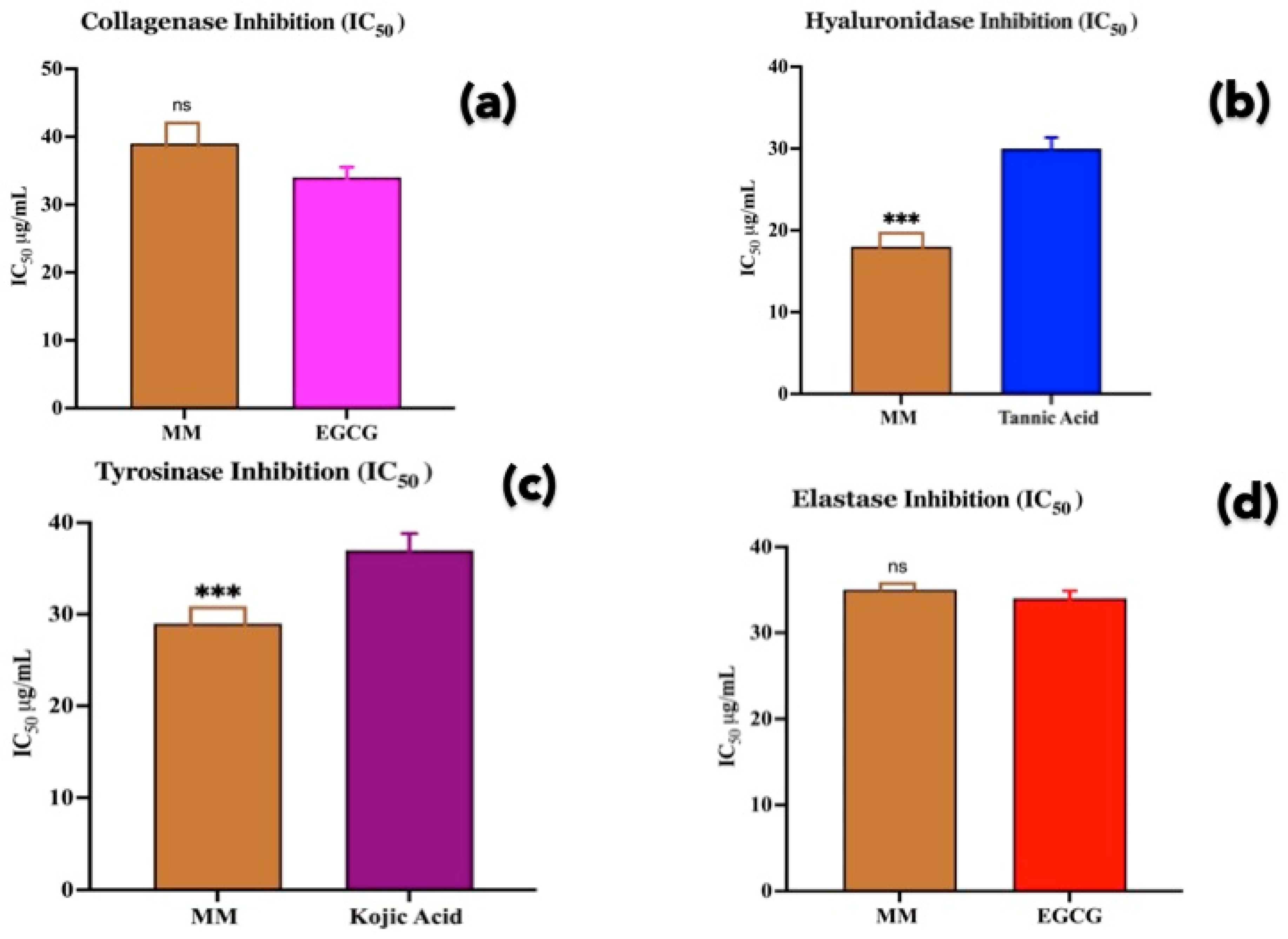
3.4. Enzyme Inhibition Activity

MM was evaluated for its in vitro inhibitory activity against four enzymes involved in skin aging and remodeling: collagenase, hyaluronidase, tyrosinase, and elastase [34]. As illustrated in Figure 4, MM demonstrates varying degrees of inhibition across these enzymes. For collagenase inhibition, MM exhibited moderate inhibitory activity with an IC50 of 38 μg/mL, which was slightly less potent than the reference inhibitor EGCG (IC50 34 μg/mL); it demonstrated strong inhibition of hyaluronidase with an IC50 of 18 μg/mL, surpassing the efficacy of tannic acid (IC50 28 μg/mL). Notably, in the tyrosinase assay, MM displayed superior inhibition with an IC50 of 27 μg/mL, outperforming kojic acid (IC50 36 μg/mL). Lastly, for elastase, MM showed comparable inhibition to EGCG, with an IC50 of 35 μg/mL for MM and 34 μg/mL for EGCG. These results suggest that MM possesses significant enzyme inhibitory activities, particularly against hyaluronidase and tyrosinase, indicating its strong potential in skincare applications aimed at combating signs of aging and hyperpigmentation. Its performance was especially impressive in inhibiting hyaluronidase and tyrosinase, where it outperformed the standard inhibitors. While MM showed moderate to good inhibition across all tested enzymes, its inorganic nature does not appear to hinder its efficacy, as evidenced by its superior performance in some assays compared to organic reference compounds.

3.5. Molecular Docking Studies

Given the strong inhibitory activity of MM powder against tyrosinase and hyaluronidase enzymes, molecular docking studies were conducted to identify the key inhibitory compounds present in MM and asses their binding affinity and interactions with the amino acids in the active sites of these enzymes. The 3D structures of tyrosinase and hyaluronidase enzymes were retrieved from the PDB database and used as the receptor or target proteins. Approximately 33 organic and metallic compounds identified by GC-MS and INNA analysis were employed as ligands to explore their potential binding interactions with the amino acids in the active sites of the selected proteins. These molecular docking studies aimed to gain insights into the possible therapeutic role of MM components in the management of skin aging and related conditions. Among these various organic and metallic compounds, silicon-containing compounds were unable to bind with the receptors.

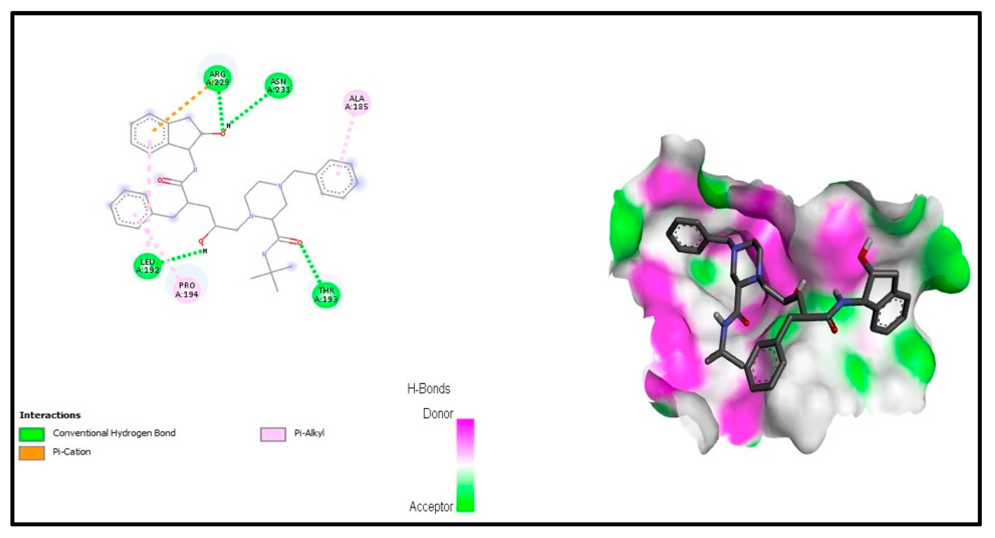
3.5.1. Interaction Analysis

The PyRx virtual screening tool was utilized to conduct docking studies, while BIOVIA Discovery Studio was employed to visualize the 2D interactions between ligands and receptors [62]. Leading drug candidates were selected based on their binding affinities and the amino acid involved in interactions. PyRx facilitated the evaluation of the ligand occupancy within the binding pocket of the target proteins through conformation scores. From the initial set of compounds, the top five compounds were individually selected for docking against each receptor protein based on their minimum binding scores and interacting amino acids (see Table 4).

3.5.2. Anti-Tyrosinase Activity

Among these compounds, Benzyl-piperazine-carboxamide exhibited significant interactions with specific amino acids of the tyrosinase protein, including PheB:258, AspA:36, ProB:51, LysA:30, ProA:145, ThrA:137, GluA:31, ProB:52, and LeuA:27, demonstrating a binding S-score of −8.6 kcal/mol (see Figure 5). The complex revealed a conventional hydrogen bond with LysA:30, ProB:51, and GluA:31, as well as Pi–sigma interactions with ProA:145, ProB:52, and LeuA:27. These Pi–sigma interactions, which encompass Pi–alkyl and Pi–sulfur interactions, facilitate the drug’s intercalation into the receptor’s binding pocket by promoting charge transfer [63].
Similarly, the second-best compound, Bis(2-ethylhexyl) phthalate (S-score of −8.0 kcal/mol), exhibited interactions with the amino acids AsnB:249, GlnB:242, HisB:279, IleB:243, LysB:281 at the binding site of tyrosinase (see Figure 6). The molecular docking results revealed diverse interaction patterns between various compounds and 3NQ1. Particularly, ProA:67 and GluA:71 emerged as common interacting residues for both Ganaxolone and Beta-sitosterol, suggesting their significance in the binding pocket. TrpA:68 also appeared in multiple complexes, interacting with both compounds. Notably, different compounds showed distinct binding preferences: benzyl-piperazine-carboxamide primarily interacted with proline residues (ProB:51, ProA:145, ProB:52) and lysine (LysA:30), while bis(2-ethylhexyl) phthalate formed interactions with asparagine and glutamine residues (AsnB:249, GlnB:242). Heptacosane displayed a simpler interaction pattern, primarily engaging with PheB:124, AspB:123, and LysB:150. Beta-sitosterol demonstrated the most extensive interaction network, involving multiple amino acid types including phenylalanine, methionine, and tyrosine residues. The presence of ValA:276 in both Ganaxolone and Beta-sitosterol complexes suggests its potential importance in ligand binding. The diversity of interacting residues across different compounds indicates a versatile binding site capable of accommodating various molecular structures through multiple interaction points (see Figure 7).

3.5.3. Anti-Hyaluronidase Activity

For the hyaluronidase target protein, the top compound, Beta-sitosterol, exhibited a binding affinity of −8.5 kcal/mol and interacted with the amino acids LysA:45, PheA:20, ProA:100, IleA:99, IleA:99, ProA:105, and AspA:101 within the active pocket (see Figure 8). Similarly, the second-best compound, Benzyl-piperazine-carboxamide, demonstrated interaction with residues ArgA:229, AsnA:231, AlaA:185, LeuA:192, ProA:194, and ThrA:193 at the active site (see Figure 9). This complex also revealed four conventional hydrogen bonds with ArgA:229, AsnA:231, LeuA:192, and ThrA:193. The remaining compounds exhibited strong binding interactions with the amino acids of the hyaluronidase active site (see Figure 10).
Among the top-selected compounds, Benzyl-piperazine-carboxamide and Beta-sitosterol demonstrated strong interactions with both receptor proteins, specifically tyrosinase and hyaluronidase.

3.5.4. Druggability Analyses

The selected compounds were evaluated for their drugability using Lipinski’s Rule of Five (Ro5), which serves as a guideline to determine their potential as active drugs in humans [64]. All compounds demonstrated general adherence to Ro5 parameters, with minor variations noted in Table 5. Further analysis using ADMETLab 2.0 revealed comprehensive absorption, distribution, metabolism, excretion, and toxicity (ADMET) profiles [65]. The ADMET profiling showed that Beta-sitosterol and Benzyl-piperazine-carboxamide exhibited particularly favorable characteristics, with Beta-sitosterol demonstrating moderate plasma protein binding (86.9%), acceptable hepatotoxicity (0.573), and Pfizer Rule compliance. Both compounds showed suitable Caco-2 permeability values (−5.122 and −5.246, respectively), though skin sensitization predictions suggested careful consideration for topical formulation (Table 5 and Table 6).
Molecular docking analysis revealed that Beta-sitosterol and Benzyl-piperazine-carboxamide demonstrated the strongest binding interactions with both target proteins. Beta-sitosterol showed a high binding affinity with 1FCV (−8.5 kcal/mol) and formed multiple stable interactions (sixteen interactions with 3NQ1 and seven with 1FCV). Similarly, Benzyl-piperazine-carboxamide exhibited strong binding energies (−8.6 kcal/mol with 3NQ1 and −8.1 kcal/mol with 1FCV) with stable amino acid interactions. While other compounds showed promising binding energies, their ADMET profiles were less favorable. Based on the combined evaluation of the molecular docking results, drug-likeness parameters, and ADMET profiles, Beta-sitosterol and Benzyl-piperazine-carboxamide emerged as the most promising compounds from the MM clay sample, warranting further experimental validation for therapeutic applications. Beta-sitosterol is a phytosterol already used in anti-aging studies.

4. Discussion

In the contemporary industrial era, characterized by vehicular emissions, environmental pollutants, and UV radiation, there is an increasing desire among individuals to maintain clear, youthful-looking skin [66]. Skin aging is an inherent and inevitable process that affects various regions of the body, with the skin being particularly noticeable. The effects and perception of skin aging can vary between individuals and across cultures. Societal emphasis on preserving a youthful appearance can greatly influence psychological well-being and self-esteem on a global scale. To counteract the signs of aging and promote a more youthful appearance, numerous anti-aging products and treatments have been developed [67]. While synthetic cosmeceuticals offer several benefits, they may also present side effects and can be costly. As a result, there has been a surge in popularity for natural or homemade products as cosmeceuticals, primarily due to their accessibility and perceived lack of side effects. This trend has significantly contributed to the growth of the aging-related cosmeceuticals market, which now has become one of the largest consumer markets [68].
MM, a clay-based product widely used in South Asia and other regions for cosmetic purposes, has garnered considerable attention in recent years [69]. Despite its extensive use in traditional practices, the scientific basis for its efficacy and safety has remained largely unexplored. This study aims to bridge this knowledge gap by conducting a comprehensive analysis of MM’s composition and potential biological activities.
INNA analysis was employed to investigate the inorganic constituents of MM, revealing its metallic element composition [70]. This analysis is crucial for understanding the clay’s absorption properties and potential interactions with skin. Notably, the presence of elements, such as silicon (Si) and tin (Sn), was confirmed; however, their direct involvement in the biological activities of the clay remains unclear.
GC-MS analysis of MM revealed a complex composition of 13 distinct organic compounds. Key findings include silicon-containing compounds (Cyclohexasiloxane), which may contribute to texture and moisture retention [71]; organic acids and alcohols that could provide pH balancing, exfoliating, and antioxidant properties [72]; alkanes, like Nonadecane, Pentacosane, Heptacosane, which may impart sensory qualities [73]; and amides that could contribute to emollient effects. Additionally, the analysis detected steroids, like Ganaxolone and a phytosterol (Beta-sitosterol) compound, which may come from plants decomposed over time or added intentionally to make this product more effective [74]. The detection of bis(2-ethylhexyl) phthalate, a common plasticizer, raises concerns regarding potential contamination sources or intentional additions [75].
The study evaluated the potential anti-aging properties of MM through in vitro enzyme inhibition assays targeting key enzymes involved in skin aging: collagenase, elastase, hyaluronidase, and tyrosinase [30]. The results demonstrated promising enzyme inhibition activity against all four enzymes, with the strongest activity observed against hyaluronidase, followed by tyrosinase and elastase, while the least activity was noted against collagenase. These findings suggest that MM may have significant potential in mitigating various aspects of skin aging.
Molecular docking studies were conducted to elucidate the potential mechanisms of action of the compounds identified in MM. Interestingly, the inorganic components, including Si and Sn, could not be docked with the target proteins, suggesting that their direct involvement in enzyme inhibition is unlikely. In contrast, the organic constituents demonstrated significant binding affinities, often surpassing those of known inhibitors, such as kojic acid and EGCG. Notably, Benzyl-piperazine-carboxamide and Beta-sitosterol exhibited strong interactions with both tyrosinase and hyaluronidase receptor proteins.
Despite these promising findings regarding enzyme inhibition, MM demonstrated only moderate antioxidant activity in the DPPH assay. From our INAA elemental analysis, several minerals are known to contribute significantly to antioxidant activity. Manganese (123.6701 mg/g) serves as a direct antioxidant cofactor and is essential for superoxide dismutase enzyme function, which neutralizes reactive oxygen species [76]. Selenium (0.1355 mg/g), though present in lower concentrations, is a critical component of glutathione peroxidase, a major cellular antioxidant enzyme [77]. Zinc (130.1224 mg/g) also plays a crucial role in antioxidant defense systems by supporting catalase and superoxide dismutase activities, while chromium (75.2774 mg/g) contributes to cellular protection against oxidative stress [78,79].
Among the organic compounds identified through GC-MS analysis, Beta-sitosterol (36.110 min RT, 77% SI) emerges as the primary contributor to antioxidant activity. This phytosterol is well-documented for its potent free radical scavenging properties and ability to inhibit lipid peroxidation [80]. Docosanamide (24.170 min RT, 94% SI) also contributes to the antioxidant profile through its fatty acid amide structure, which can neutralize free radicals [81]. Additionally, Benzyl-piperazine-carboxamide (25.705 min RT, 94% SI) may contribute to the overall antioxidant capacity through its aromatic ring system that can stabilize free radicals [82]. This result, combined with a previously reported lack of significant antimicrobial activity [23], suggests that while MM possesses certain beneficial properties, it may not serve as a comprehensive solution for all skincare needs.
The diverse chemical profile of MM aligns with its reported skincare benefits, providing a scientific basis for understanding its effects on skin health. Its primary advantages appear to be related to oil absorption, improvement of skin texture, and mild anti-aging effects. The clay’s ability to absorb excess oil and impurities from the skin surface, combined with its gentle exfoliating properties, likely contributes to the perceived ‘glowing’ effect reported by users [83].
While Multani Mitti (MM) exhibits moderate antioxidant activity—likely attributable to bioactive compounds, such as Beta-sitosterol, and essential inorganic elements, like manganese and selenium—its antimicrobial properties appear limited based on our findings [84,85]. This suggests that, although MM contributes beneficially to skin health through enzyme inhibition and antioxidant effects, it may not fully address all the factors involved in skin aging and microbial protection. To develop a more comprehensive cosmeceutical formulation, supplementation with well-characterized antioxidants (e.g., vitamin C at 0.5–2%, ferulic acid at 0.1–1%) and antimicrobial agents (such as tea tree oil at 0.5–2%, zinc oxide at 1–5%) could be advantageous [86,87]. These compounds have documented efficacy at these concentrations in enhancing skin protection and complementing clay-based ingredients. Notably, traditional and homemade skincare practices often combine MM with plant powders, like turmeric or neem, which provide additional antioxidant and antimicrobial benefits [88]. Incorporating such synergistic actives can optimize MM’s overall efficacy, creating multifunctional formulations that leverage both its traditional appeal and scientifically supported enhancements.
The organic compounds identified in this study that exhibited strong enzyme inhibition have the potential to be developed into individual anti-aging products. However, the holistic effects of MM, including its oil absorption and skin-brightening properties, suggest that complete powder may offer unique benefits that extend beyond those of its individual components.
This study provides valuable insights into the composition and potential mechanisms of action of MM in skincare. While it demonstrates promising anti-aging properties, particularly through enzyme inhibition, its moderate antioxidant activity and lack of antimicrobial effects indicate areas for improvement. Future research should focus on optimizing MM-based formulations by incorporating additional beneficial compounds to create a more comprehensive, natural, and cost-effective skincare solution. Such an approach could leverage the traditional benefits of MM while addressing its limitations, potentially resulting in an enhanced cosmeceutical product with minimal side effects.

5. Conclusions

The use of MM in cosmeceuticals has gained significant popularity due to its beneficial properties. Our study demonstrated that this clay exhibits moderate antioxidant activity (IC50: 31.938 μg/mL) and significant inhibitory effects on hyaluronidase (IC50: 18 μg/mL) and tyrosinase (IC50: 27 μg/mL), outperforming standard inhibitors. It also showed moderate activity against collagenase and elastase—enzymes crucial in skin aging. These effects are likely attributed to synergistic actions of identified components, particularly Beta-sitosterol and Benzyl-piperazine-carboxamide, which demonstrated strong binding affinities (−8.5 to −8.6 kcal/mol) to target enzymes. ADMET analysis confirmed favorable safety profiles for these compounds. Our findings support MM’s incorporation into anti-aging formulations, with future research focusing on the in vivo validation of topical applications and toxicity profiling.

Author Contributions

Conceptualization, M.J.I., P.L. and L.A.S.; methodology, M.J.I.; validation, M.J.I. and P.L.; formal analysis, M.J.I.; investigation, M.J.I.; resources, V.B. and L.A.S.; data curation, P.L.; writing—original draft preparation, M.J.I.; writing—review and editing, P.L., V.B. and L.A.S.; visualization, M.J.I. and L.A.S.; supervision, P.L., V.B. and L.A.S.; project administration, L.A.S.; funding acquisition, L.A.S. All authors have read and agreed to the published version of the manuscript.

Funding

This research was funded by Dirección de Investigación, Universidad de La Frontera, Temuco, Chile, grant number PP24-0003.

Institutional Review Board Statement

Not applicable.

Informed Consent Statement

Not applicable.

Data Availability Statement

The data that support the findings of this research are available from the corresponding author upon reasonable request.

Conflicts of Interest

The authors declare no conflicts of interest.

References

  1. Ahmed, I.A.; Mikail, M.A. Diet and skin health: The good and the bad. Nutrition 2024, 119, 112350. [Google Scholar] [CrossRef] [PubMed]
  2. Pal, R.S.; Pal, Y.; Wal, P. In-House Preparation and Standardization of Herbal Face Pack. Open Dermatol. J. 2017, 11, 72–80. [Google Scholar] [CrossRef]
  3. Kumar, P. Multani mitti—Is it more than a placebo? J. Pak. Assoc. Dermatol. 2019, 29, 345–348. [Google Scholar]
  4. Waheed, S.; Faiz, Y.; Rahman, S.; Siddique, N. Toxic element composition of multani mitti clay for nutritional safety. J. Radioanal. Nucl. Chem. 2012, 295, 143–150. [Google Scholar] [CrossRef]
  5. Pal, R.S.; Pal, Y.; Wal, P.; Wal, A.; Saraswat, N. Composition and Quality Standards of Naturally Derived Anti Tan Medicine. Open Biol. J. 2021, 9, 40–46. [Google Scholar] [CrossRef]
  6. Waheed, S.; Siddique, N.; Faiz, Y. Rare Earth and High Field-Strength Elements in the Multani Mitti Clay: A Study Using INAA. Geostand. Geoanalytical Res. 2013, 37, 197–205. [Google Scholar] [CrossRef]
  7. Hosterman, J.W.; Patterson, S.H. Bentonite and Fuller’s Earth Resources of the United States; U.S. Government Publishing Office: Washington, DC, USA, 1992. [CrossRef]
  8. Patterson, S.H. Fuller’s Earth and Other Industrial Mineral Resources of the Meigs-Attapulgus-Quincy District, Georgia and Florida; U.S. Government Publishing Office: Washington, DC, USA, 1974; p. 828.
  9. Idoine, N.E.; Raycraft, E.R.; Shaw, R.A.; Hobbs, S.F.; Deady, E.A.; Everett, P.; Evans, E.J.; Mills, A.J. World Mineral Production; British Geological Survey: Nottingham, UK, 2016. [Google Scholar]
  10. Thakur, P.; Garg, R.K. New developing reagent for latent fingermark visualization: Fuller’s earth (Multani Mitti). Egypt. J. Forensic Sci. 2016, 6, 449–458. [Google Scholar] [CrossRef]
  11. Bhola, K.L. Fuller’s Earth in India. Trans. Indian Ceram. Soc. 2014, 5, 104–124. [Google Scholar] [CrossRef]
  12. Klein, C.; Dutrow, B. Manual of Mineral Science; Wiley: Hoboken, NJ, USA, 2007. [Google Scholar]
  13. Kora, A.J. Cosmetic and religious applications of natural nanoclay: An Indian scenario. In Nanoclay-Based Sustainable Materials; Elsevier: Amsterdam, The Netherlands, 2024; pp. 429–447. [Google Scholar]
  14. Tiwari, D.; Prakash, K.; Gupta, P.; Shukla, T.; Yadav, P. Formulation And Evaluation of Face Scrub In Modern Pharmaceutics: A Herbal Formulation. Int. J. Pharm. Sci. 2024, 2, 579–588. [Google Scholar] [CrossRef]
  15. Bhavani, M.S.; Naveena, C.H.; Nagamani, P.; Sowmya, B. Formulation and Evaluation of Herbal Face Cream. Int. J. Pharm. Sci. Rev. Res. 2023, 83, 74–78. [Google Scholar] [CrossRef]
  16. Kumar, R. Komal Formulation and evaluation of herbal face pack. Asian J. Pharm. Res. 2021, 11, 9–12. [Google Scholar] [CrossRef]
  17. Gomes, C.; Silva, J. Minerals and clay minerals in medical geology. Appl. Clay Sci. 2007, 36, 4–21. [Google Scholar] [CrossRef]
  18. Kamble, Y.; Choudhary, G.; Kishore, K.; Sonawale, N.; Choudhary, A.; Baviskar, R. Formulation and evaluation of multi herbal face pack for glowing skin. World J. Pharm. Res. 2023, 12, 1393–1401. Available online: https://www.wjpr.net/abstract_file/21843 (accessed on 8 March 2024).
  19. Tiwari, D.S.; Singh, S.; Kumari, A.; Yadav, S.; Bind, D. The role of ayurvedic herbs in skin rejuvenation by face mask: Ficus religiosa and beyond. Int. J. Pharm. Res. Dev. 2024, 6, 143–150. [Google Scholar] [CrossRef]
  20. Dalavi, P.R.K.; Ziya, K. Formulation and Evaluation of Herbal Face Pack. Int. J. Sci. Res. Technol. 2025, 2, 236–243. [Google Scholar] [CrossRef]
  21. Eximpedia. Find the Multani Mitti Export Opportunity: Foreign Market Demand Statistics. Available online: https://www.eximpedia.app/blog/multani-mitti-export-from-india (accessed on 8 March 2024).
  22. Journal, D. Fuller’s Earth Market Share, Revenue, and Forecast 2023–2030. Available online: https://www.digitaljournal.com/pr/news/fuller-s-earth-market-share-nbsp-revenue-nbsp-and-forecast-2023-2030 (accessed on 8 March 2024).
  23. Muralidharan, N.P.; Dhanasekaran, L. Antimicrobial Activity of Fuller’s Earth, Turmeric, and Sandalwood Against Streptococcus Mutans, Micrococci and Coagulase Negative Staphylococci—An In Vitro Study. J. Pharm. Res. Int. 2021, 33, 65–73. [Google Scholar] [CrossRef]
  24. Park, D.-B.; Lee, Y.-J.; Kim, W.-S.; Kim, Y.-T. Anti-skin Aging and Antihypertensive Activities of Different Solvent Extracts from Sargassum thunbergii. Korean J. Fish. Aquat. Sci. 2025, 58, 140–145. [Google Scholar] [CrossRef]
  25. Hussein, R.S.; Bin Dayel, S.; Abahussein, O.; El-Sherbiny, A.A. Influences on Skin and Intrinsic Aging: Biological, Environmental, and Therapeutic Insights. J. Cosmet. Dermatol. 2025, 24, e16688. [Google Scholar] [CrossRef]
  26. Jung, H. Hyaluronidase: An overview of its properties, applications, and side effects. Arch. Plast. Surg. 2020, 47, 297–300. [Google Scholar] [CrossRef]
  27. Gendreau, I.; Angers, L.; Jean, J.; Pouliot, R. Pigmented Skin Models: Understand the Mechanisms of Melanocytes. In Regenerative Medicine and Tissue Engineering; Andrades, J.A., Ed.; Intech: Houston, TX, USA, 2013. [Google Scholar]
  28. He, X.; Gao, X.; Guo, Y.; Xie, W. Research Progress on Bioactive Factors against Skin Aging. Int. J. Mol. Sci. 2024, 25, 3797. [Google Scholar] [CrossRef]
  29. Hong, Y.H.; Jung, E.Y.; Noh, D.O.; Suh, H.J. Physiological effects of formulation containing tannase-converted green tea extract on skin care: Physical stability, collagenase, elastase, and tyrosinase activities. Integr. Med. Res. 2014, 3, 25–33. [Google Scholar] [CrossRef] [PubMed]
  30. Jiratchayamaethasakul, C.; Ding, Y.; Hwang, O.; Im, S.-T.; Jang, Y.; Myung, S.-W.; Lee, J.M.; Kim, H.-S.; Ko, S.-C.; Lee, S.-H. In vitro screening of elastase, collagenase, hyaluronidase, and tyrosinase inhibitory and antioxidant activities of 22 halophyte plant extracts for novel cosmeceuticals. Fish. Aquat. Sci. 2020, 23, 62–72. [Google Scholar] [CrossRef]
  31. Waheed, S.; Rahman, S.; Faiz, Y.; Siddique, N. Neutron activation analysis of essential elements in Multani mitti clay using miniature neutron source reactor. Appl. Radiat. Isot. 2012, 70, 2362–2369. [Google Scholar] [CrossRef]
  32. Profumo, A.; Gorroni, A.; Guarnieri, S.A.; Mellerio, G.G.; Cucca, L.; Merli, D. GC-MS qualitative analysis of the volatile, semivolatile and volatilizable fractions of soil evidence for forensic application: A chemical fingerprinting. Talanta 2020, 219, 121304. [Google Scholar] [CrossRef]
  33. Brand-Williams, W.; Cuvelier, M.E.; Berset, C. Use of a free radical method to evaluate antioxidant activity. LWT—Food Sci. Technol. 1995, 28, 25–30. [Google Scholar] [CrossRef]
  34. Elgamal, A.M.; El Raey, M.A.; Gaara, A.; Abdelfattah, M.A.O.; Sobeh, M. Phytochemical profiling and anti-aging activities of Euphorbia retusa extract: In silico and in vitro studies. Arab. J. Chem. 2021, 14, 103159. [Google Scholar] [CrossRef]
  35. Sklirou, A.D.; Angelopoulou, M.T.; Argyropoulou, A.; Chaita, E.; Boka, V.I.; Cheimonidi, C.; Niforou, K.; Mavrogonatou, E.; Pratsinis, H.; Kalpoutzakis, E.; et al. Phytochemical Study and In Vitro Screening Focusing on the Anti-Aging Features of Various Plants of the Greek Flora. Antioxidants 2021, 10, 1206. [Google Scholar] [CrossRef]
  36. Fikry, E.; Mahdi, I.; Bugra Ortaakarsu, A.; Tawfeek, N.; Adhiambo Ochieng, M.; Ben Bakrim, W.; Ao Abdelfattah, M.; Omari, K.W.; Mahmoud, M.F.; Sobeh, M. Dermato-cosmeceutical properties of Pseudobombax ellipticum (Kunth) Dugand: Chemical profiling, in vitro and in silico studies. Saudi Pharm. J. 2023, 31, 101778. [Google Scholar] [CrossRef] [PubMed]
  37. PubChem, National Library of Medicine. Available online: https://pubchem.ncbi.nlm.nih.gov/ (accessed on 8 March 2024).
  38. Dallakyan, S.; Olson, A.J. Small-molecule library screening by docking with PyRx. Methods Mol. Biol. 2015, 1263, 243–250. [Google Scholar] [CrossRef]
  39. Shaweta, S.; Akhil, S.; Utsav, G. Molecular Docking studies on the Anti-fungal activity of Allium sativum (Garlic) against Mucormycosis (black fungus) by BIOVIA discovery studio visualizer 21.1.0.0. Ann. Antivir. Antiretrovir. 2021, 5, 028–032. [Google Scholar] [CrossRef]
  40. Daina, A.; Michielin, O.; Zoete, V. SwissADME: A free web tool to evaluate pharmacokinetics, drug-likeness and medicinal chemistry friendliness of small molecules. Sci. Rep. 2017, 7, 42717. [Google Scholar] [CrossRef] [PubMed]
  41. Mustafa, G.; Majid, M.; Ghaffar, A.; Yameen, M.; Samad, H.A.; Mahrosh, H.S. Screening and molecular docking of selected phytochemicals against NS5B polymerase of hepatitis c virus. Pak. J. Pharm. Sci. 2020, 33, 2317–2322. [Google Scholar] [CrossRef] [PubMed]
  42. Gotz, W.; Tobiasch, E.; Witzleben, S.; Schulze, M. Effects of Silicon Compounds on Biomineralization, Osteogenesis, and Hard Tissue Formation. Pharmaceutics 2019, 11, 117. [Google Scholar] [CrossRef] [PubMed]
  43. Carver, P.L. Essential Metals in Medicine: Therapeutic Use and Toxicity of Metal Ions in the Clinic; De Gruyter: Berlin, Germany, 2019. [Google Scholar]
  44. Bonaventura, P.; Benedetti, G.; Albarede, F.; Miossec, P. Zinc and its role in immunity and inflammation. Autoimmun. Rev. 2015, 14, 277–285. [Google Scholar] [CrossRef] [PubMed]
  45. Duck, K.A.; Connor, J.R. Iron uptake and transport across physiological barriers. Biometals 2016, 29, 573–591. [Google Scholar] [CrossRef]
  46. Exley, C.; Mold, M.J. The binding, transport and fate of aluminium in biological cells. J. Trace Elem. Med. Biol. 2015, 30, 90–95. [Google Scholar] [CrossRef]
  47. Wang, L.; You, X.; Zhang, L.; Zhang, C.; Zou, W. Mechanical regulation of bone remodeling. Bone Res. 2022, 10, 16. [Google Scholar] [CrossRef]
  48. Szklarzewicz, J.; Jurowska, A.; Hodorowicz, M.; Kazek, G.; Głuch-Lutwin, M.; Sapa, J. Ligand role on insulin-mimetic properties of vanadium complexes. Structural and biological studies. Inorganica Chim. Acta 2021, 516, 120135. [Google Scholar] [CrossRef]
  49. Zheng, S.; Cheng, S.; Xiao, S.; Hu, L.; Chen, Z.; Huang, B.; Liu, Q.; Yang, J.; Chen, Q. Partial replacement of K by Rb to improve electrochemical performance of K3V2(PO4)3 cathode material for potassium-ion batteries. J. Alloys Compd. 2020, 815, 152379. [Google Scholar] [CrossRef]
  50. Vincent, J.B. New Evidence against Chromium as an Essential Trace Element. J. Nutr. 2017, 147, 2212–2219. [Google Scholar] [CrossRef]
  51. Panahifar, A.; Chapman, L.D.; Weber, L.; Samadi, N.; Cooper, D.M.L. Biodistribution of strontium and barium in the developing and mature skeleton of rats. J. Bone Miner. Metab. 2019, 37, 385–398. [Google Scholar] [CrossRef] [PubMed]
  52. Osman, D.; Cooke, A.; Young, T.R.; Deery, E.; Robinson, N.J.; Warren, M.J. The requirement for cobalt in vitamin B(12): A paradigm for protein metalation. Biochim. Biophys. Acta Mol. Cell Res. 2021, 1868, 118896. [Google Scholar] [CrossRef]
  53. Wang, J.; Zhuang, S. Removal of cesium ions from aqueous solutions using various separation technologies. Rev. Environ. Sci. Bio/Technol. 2019, 18, 231–269. [Google Scholar] [CrossRef]
  54. Zaborowska, M.; Kucharski, J.; Wyszkowska, J. Biological activity of soil contaminated with cobalt, tin, and molybdenum. Environ. Monit. Assess. 2016, 188, 398. [Google Scholar] [CrossRef]
  55. Mayr, S.J.; Mendel, R.R.; Schwarz, G. Molybdenum cofactor biology, evolution and deficiency. Biochim. Biophys. Acta Mol. Cell Res. 2021, 1868, 118883. [Google Scholar] [CrossRef]
  56. Shrimanker, I.; Bhattarai, S. Electrolytes. Available online: https://www.ncbi.nlm.nih.gov/books/NBK541123/ (accessed on 8 March 2024).
  57. Uwitonze, A.M.; Razzaque, M.S. Role of Magnesium in Vitamin D Activation and Function. J. Osteopath. Med. 2018, 118, 181–189. [Google Scholar] [CrossRef] [PubMed]
  58. Wang, J.; Ou, S.W.; Wang, Y.J. Distribution and function of voltage-gated sodium channels in the nervous system. Channels 2017, 11, 534–554. [Google Scholar] [CrossRef] [PubMed]
  59. Grover, T.; Pandey, A.; Kumari, S.T.; Awasthi, A.; Singh, B.; Dixit, P.; Singhal, P.; Saxena, K.K. Role of titanium in bio implants and additive manufacturing: An overview. Mater. Today Proc. 2020, 26, 3071–3080. [Google Scholar] [CrossRef]
  60. Wang, N.; Tan, H.Y.; Li, S.; Xu, Y.; Guo, W.; Feng, Y. Supplementation of Micronutrient Selenium in Metabolic Diseases: Its Role as an Antioxidant. Oxidative Med. Cell. Longev. 2017, 2017, 7478523. [Google Scholar] [CrossRef]
  61. Ge, X.; Cao, Z.; Chu, L. The Antioxidant Effect of the Metal and Metal-Oxide Nanoparticles. Antioxidants 2022, 11, 791. [Google Scholar] [CrossRef]
  62. Saeed, M.; Shoaib, A.; Tasleem, M.; Alabdallah, N.M.; Alam, M.J.; Asmar, Z.E.; Jamal, Q.M.S.; Bardakci, F.; Alqahtani, S.S.; Ansari, I.A.; et al. Assessment of Antidiabetic Activity of the Shikonin by Allosteric Inhibition of Protein-Tyrosine Phosphatase 1B (PTP1B) Using State of Art: An In Silico and In Vitro Tactics. Molecules 2021, 26, 2996. [Google Scholar] [CrossRef] [PubMed]
  63. Ribas, J.; Cubero, E.; Luque, F.J.; Orozco, M. Theoretical study of alkyl-pi and aryl-pi interactions. Reconciling theory and experiment. J. Org. Chem. 2002, 67, 7057–7065. [Google Scholar] [CrossRef] [PubMed]
  64. Benet, L.Z.; Hosey, C.M.; Ursu, O.; Oprea, T.I. BDDCS, the Rule of 5 and drugability. Adv. Drug Deliv. Rev. 2016, 101, 89–98. [Google Scholar] [CrossRef]
  65. Xiong, G.; Wu, Z.; Yi, J.; Fu, L.; Yang, Z.; Hsieh, C.; Yin, M.; Zeng, X.; Wu, C.; Lu, A.; et al. ADMETlab 2.0: An integrated online platform for accurate and comprehensive predictions of ADMET properties. Nucleic Acids Res. 2021, 49, W5–W14. [Google Scholar] [CrossRef] [PubMed]
  66. Chao, J. Skincare for Your Soul: Achieving Outer Beauty and Inner Peace with Korean Skincare; Mango Media Inc.: Balmain, Australia, 2021. [Google Scholar]
  67. Gupta, M.A.; Gilchrest, B.A. Psychosocial aspects of aging skin. Dermatol. Clin. 2005, 23, 643–648. [Google Scholar] [CrossRef]
  68. Younis, M.M.; Ayoub, I.M.; Mostafa, N.M.; El Hassab, M.A.; Eldehna, W.M.; Al-Rashood, S.T.; Eldahshan, O.A. GC/MS Profiling, Anti-Collagenase, Anti-Elastase, Anti-Tyrosinase and Anti-Hyaluronidase Activities of a Stenocarpus sinuatus Leaves Extract. Plants 2022, 11, 918. [Google Scholar] [CrossRef]
  69. Mishra, B.B.; Choudhary, S.K.; Roy, R. Soil and Industry. In The Soils of India; World Soils Book Series; Springer: Cham, Switzerland, 2020; pp. 243–259. [Google Scholar]
  70. Pohl, P.; Stelmach, E.; Welna, M.; Szymczycha-Madeja, A. Determination of the Elemental Composition of Coffee Using Instrumental Methods. Food Anal. Methods 2012, 6, 598–613. [Google Scholar] [CrossRef]
  71. Racles, C.; Dascalu, M.; Bele, A.; Cazacu, M. Reactive and Functional Silicones for Special Applications. In Reactive and Functional Polymers Volume One; Springer: Berlin/Heidelberg, Germany, 2020; pp. 235–291. [Google Scholar]
  72. Chaves, J.O.; de Souza Mesquita, L.M.; Strieder, M.M.; Contieri, L.S.; Pizani, R.S.; Sanches, V.L.; Viganó, J.; Neves Bezerra, R.M.; Rostagno, M.A. Eco-friendly and high-performance extraction of flavonoids from lemon peel wastes by applying ultrasound-assisted extraction and eutectic solvents. Sustain. Chem. Pharm. 2024, 39, 101558. [Google Scholar] [CrossRef]
  73. Li, H.; Zhang, X.; Gao, X.; Shi, X.; Chen, S.; Xu, Y.; Tang, K. Comparison of the Aroma-Active Compounds and Sensory Characteristics of Different Grades of Light-Flavor Baijiu. Foods 2023, 12, 1238. [Google Scholar] [CrossRef]
  74. Baser, B.; Yousaf, B.; Yetis, U.; Abbas, Q.; Kwon, E.E.; Wang, S.; Bolan, N.S.; Rinklebe, J. Formation of nitrogen functionalities in biochar materials and their role in the mitigation of hazardous emerging organic pollutants from wastewater. J. Hazard. Mater. 2021, 416, 126131. [Google Scholar] [CrossRef]
  75. Billings, A.; Jones, K.C.; Pereira, M.G.; Spurgeon, D.J. Plasticisers in the terrestrial environment: Sources, occurrence and fate. Environ. Chem. 2021, 18, 111–130. [Google Scholar] [CrossRef]
  76. Chandra, S.; Roychoudhury, A. Role of Selenium and Manganese in Mitigating Oxidative Damages. In Protective Chemical Agents in the Amelioration of Plant Abiotic Stress; Wiley-Blackwell: Hoboken, NJ, USA, 2020; pp. 597–621. [Google Scholar]
  77. Bizerea, T.O.; Dezsi, S.G.; Marginean, O.; Stroescu, R.; Rogobete, A.; Bizerea-Spiridon, O.; Ilie, C. The Link Between Selenium, Oxidative Stress and Pregnancy Induced Hypertensive Disorders. Clin. Lab. 2018, 64, 1593–1610. [Google Scholar] [CrossRef] [PubMed]
  78. Uttara, B.; Singh, A.V.; Zamboni, P.; Mahajan, R.T. Oxidative stress and neurodegenerative diseases: A review of upstream and downstream antioxidant therapeutic options. Curr. Neuropharmacol. 2009, 7, 65–74. [Google Scholar] [CrossRef]
  79. Singh, V.; Singh, N.; Verma, M.; Kamal, R.; Tiwari, R.; Sanjay Chivate, M.; Rai, S.N.; Kumar, A.; Singh, A.; Singh, M.P.; et al. Hexavalent-Chromium-Induced Oxidative Stress and the Protective Role of Antioxidants against Cellular Toxicity. Antioxidants 2022, 11, 2375. [Google Scholar] [CrossRef]
  80. Dwivedi, J.; Sachan, P.; Wal, P. A Mechanistic Approach on Structural, Analytical and Pharmacological Potential of Beta-sitosterol: A Promising Nutraceutical. Curr. Nutr. Food Sci. 2024, 20, 932–951. [Google Scholar] [CrossRef]
  81. Elufioye, T.O.; Habtemariam, S.; Adejare, A. Chemistry and Pharmacology of Alkylamides from Natural Origin. Rev. Bras. Farmacogn. 2020, 30, 622–640. [Google Scholar] [CrossRef] [PubMed]
  82. Mortada, S.; Karrouchi, K.; Hamza, E.H.; Oulmidi, A.; Bhat, M.A.; Mamad, H.; Aalilou, Y.; Radi, S.; Ansar, M.; Masrar, A.; et al. Synthesis, structural characterizations, in vitro biological evaluation and computational investigations of pyrazole derivatives as potential antidiabetic and antioxidant agents. Sci. Rep. 2024, 14, 1312. [Google Scholar] [CrossRef]
  83. Inglis, K.; Tettoni, L.I. Ayurveda: Asian Secrets of Wellness, Beauty and Balance; Tuttle Publishing: North Clarendon, VT, USA, 2012. [Google Scholar]
  84. Nandi, S.; Nag, A.; Khatua, S.; Sen, S.; Chakraborty, N.; Naskar, A.; Acharya, K.; Calina, D.; Sharifi-Rad, J. Anticancer activity and other biomedical properties of beta-sitosterol: Bridging phytochemistry and current pharmacological evidence for future translational approaches. Phytother. Res. 2024, 38, 592–619. [Google Scholar] [CrossRef]
  85. Yu, Y.; Fan, P.; Li, J.; Wang, S. Preparation of Biocompatible Manganese Selenium-Based Nanoparticles with Antioxidant and Catalytic Functions. Molecules 2023, 28, 4498. [Google Scholar] [CrossRef]
  86. Yasin, M.; Younis, A.; Javed, T.; Akram, A.; Ahsan, M.; Shabbir, R.; Ali, M.M.; Tahir, A.; El-Ballat, E.M.; Sheteiwy, M.S.; et al. River Tea Tree Oil: Composition, Antimicrobial and Antioxidant Activities, and Potential Applications in Agriculture. Plants 2021, 10, 2105. [Google Scholar] [CrossRef]
  87. Yadav, E.; Kumar, S.; Mahant, S.; Khatkar, S.; Rao, R. Tea tree oil: A promising essential oil. J. Essent. Oil Res. 2016, 29, 201–213. [Google Scholar] [CrossRef]
  88. Kumar, P.; Karam, N.; Choudhury, D. Assessment of the Phytochemical Constituents and Metabolites in the Medicinal Plants and Herbal Medicine Used in the Treatment and Management of Skin Diseases. In Herbal Medicine Phytochemistry; Springer: Berlin/Heidelberg, Germany, 2024; pp. 1–41. [Google Scholar]
Figure 1. Powered form of MM (a) and its application as a face mask (b).
Cosmetics 12 00124 g001
Figure 2. GC-MS chromatograms of bioactive compounds in the MM clay sample. Red arrows indicate the observed peaks; blue lines denote baseline signals.
Cosmetics 12 00124 g002
Figure 3. Concentration-dependent DPPH radical scavenging activity of MM powder and ascorbic acid.
Cosmetics 12 00124 g003
Figure 4. Enzyme inhibitory activity of MM powder against (a) collagenase, (b) hyaluronidase, (c) tyrosinase, and (d) elastase compared to that of epigallocatechin gallate (EGCG), tannic acid, and kojic acid, respectively. The results are presented as IC50 values, representing the mean of three independent experiments. *** p < 0.001, ns: not significant vs. positive control.
Cosmetics 12 00124 g004
Figure 5. Two-dimensional interaction and 3D binding pattern of Benzyl-piperazine-carboxamide with tyrosinase as a receptor.
Cosmetics 12 00124 g005
Figure 6. Two-dimensional interaction and 3D binding pattern of Bis(2-ethylhexyl) phthalate with tyrosinase as a receptor.
Cosmetics 12 00124 g006
Figure 7. Two-dimensional interaction and 3D binding pattern with tyrosinase as a receptor: (a) Heptacosane, (b) Ganaxolone, and (c) Beta-sitosterol.
Cosmetics 12 00124 g007
Figure 8. Two-dimensional interaction and 3D binding pattern of Beta-sitosterol with hyaluronidase as a receptor.
Cosmetics 12 00124 g008
Figure 9. Two-dimensional interaction and 3D binding pattern of Benzyl-piperazine-carboxamide with hyaluronidase as a receptor.
Cosmetics 12 00124 g009
Figure 10. Two-dimensional interaction and 3D binding pattern with hyaluronidase as a receptor: (a) Bis(2-ethylhexyl) phthalate, (b) Heptacosane, and (c) Ganaxolone.
Cosmetics 12 00124 g010
Table 2. Bioactive compounds identified in MM clay by GC-MS analysis.
PeakRT (Min)SI (%)Compound NamePubChem IDMolecular FormulaMolecular Weight (g/mol)
108.120833-Heptanol, 1-Octanol957C8H18O130
210.06083Cyclohexasiloxane10911C12H36O6Si6444.92
315.92993Nonadecane15979C19H40268.5
415.99590Pentacosane12406C25H52352.7
516.06091Heptacosane11636C27H56380.7
617.02092Nonacosane12409C29H60408.8
717.13092Tetrapentacontane545963C54H108Br2917.2
823.97582Oleth-213387455C22H44O3356.6
924.17094Docosanamide76468C22H45NO339.6
1025.70594Benzyl-piperazine-carboxamide455963C37H48N4O4612.8
1126.58075Bis(2-ethylhexyl) phthalate8343C24H38O4390.6
1228.65083Ganaxolone6918305C22H36O2332.5
1336.11077Beta-sitosterol222284C29H50O414.7
Table 3. IC50 values of MM powder and ascorbic acid in the DPPH radical scavenging assay.
SampleIC50
MM Powder (Fuller’s Earth)31.938 μg/mL
Ascorbic Acid8.5 μg/mL
Table 4. Binding scores of the top five compounds with tyrosinase (3NQ1) and hyaluronidase (1FCV) protein receptors.
NamePubChem IDBinding Affinity (kcal/mol) with 3NQ1Interacting Amino Acids with 3NQ1Binding Affinity (kcal/mol) with 1FCVInteracting Amino Acids with 1FCV
Benzyl-piperazine-carboxamide455963−8.6PheB:258, AspA:36, ProB:51, LysA:30, ProA:145, ThrA:137, GluA:31, ProB:52 and LeuA:27−8.1ArgA:229, AsnA:231, AlaA:185, LeuA:192, ProA:194 and ThrA:193
Bis(2-ethylhexyl) phthalate8343−8.0AsnB:249, GlnB:242, HisB:279, IleB:243 and LysB:281−5.9TyrA:168, AlaA:185, ArgA:244, SerA:225, TyrA:190 and ArgA:116
Heptacosane11636−8.0PheB:124, AspB:123 and LysB:150−4.6TyrA:227, TrpA:301, TrpA:267, TrA:184,
TyrA:55 and
AspA:111
Ganaxolone6918305−7.2PheA:262, ProA:273, ValA:276, MetA:266, ProA:67, TrpA:68, GluA:71 and ValA:276−7.4SerA:304, AspA:111, AspA:305, Tyra:55, AspA:56, PheA:112, GluA:113, SerA:303 and AspA:305
Beta-sitosterol222284−7.1AsnA:249, PheA:262, MetA:277, tyrA:250, GluA:274, ProA:273, ValA:276, ProA:67, GluA:71, TyrA:72, trpA:269, TrpA:68, ThrA:272, ArgA:70, AspA:275 and MetA:266−8.5LysA:45, PheA:20, ProA:100, IleA:99, IleA:99, ProA:105 and AspA:101
Table 5. Molecular and drug-likeness properties of the top five compounds evaluated through Lipinski’s Rule of Five (3NQ1 and 1FCV).
Molecular Properties
LigandMolecular Mass (≤500 Dalton)Hydrogen Bond Donor (≤5)Hydrogen Bond Acceptor (≤10)No. of Rotatable Bonds (≤10)LogP (≤5)Refractivity (40–130)Violations
Benzyl-piperazine-carboxamide612.8046143.65184.830
Bis(2-ethylhexyl) phthalate390.604164.77116.301
Heptacosane380.7300247.32130.11
Ganaxolone332.521213.29100.290
Beta-sitosterol414.711165.05133.230
Table 6. ADMET-related drug-like parameters of the best-selected compounds.
Compounds
Benzyl-piperazine-carboxamideBis(2-ethylhexyl) phthalateHeptacosaneGanaxoloneBeta-sitosterol
Absorption and Distribution
BBB−−−+++−−−+++−−−
HIA−−−−−−−−−−−−−−−
Caco-2 Permeability−5.246−4.918−5.074−4.806−5.122
Volume of Distribution (Vd)0.0010.943.0090.205−0.244
Plasma Protein Binding (PPB)95.1%98.7%105.5%67.4%86.9%
Excretion
CLplasma4.9285.794.54516.97813.205
T1/20.8650.3243.0310.970.541
Toxicity
Skin Sensitization0.8070.9380.9980.9920.99
Carcinogenicity0.130.3280.1640.9830.688
Human Hepatotoxicity0.9160.0860.4730.8540.573
AMES Toxicity0.1290.0180.0120.1830.139
Drug-Likeness Rules
Acute Aquatic Toxicity Rule00121
Skin Sensitization Rule10020
Lipinski RuleYesYesYesYesYes
Pfizer RuleYesNoNoNoYes
BBB: blood–brain barrier; HIA: Human Intestinal Absorption; CLplasma: plasma clearance; T1/2: half-life. For classification endpoints, prediction probability values are represented by the following symbols: 0–0.1 (−−−) and 0.9–1.0 (+++).
Disclaimer/Publisher’s Note: The statements, opinions and data contained in all publications are solely those of the individual author(s) and contributor(s) and not of MDPI and/or the editor(s). MDPI and/or the editor(s) disclaim responsibility for any injury to people or property resulting from any ideas, methods, instructions or products referred to in the content.

