Article

Photoprotective Effect and Potential Mechanisms of Gardeniae Fructus Extract in UVB-Irradiated HaCaT Cells

1
Skin Health and Cosmetic Development & Evaluation Laboratory, China Pharmaceutical University, Nanjing 211198, China
2
Shanghai Junyu Biotechnology Group Co., Ltd., Shanghai 200050, China
*
Authors to whom correspondence should be addressed.
Cosmetics 2025, 12(2), 72; https://doi.org/10.3390/cosmetics12020072
Submission received: 7 March 2025 / Revised: 3 April 2025 / Accepted: 8 April 2025 / Published: 9 April 2025
(This article belongs to the Special Issue Skin Anti-Aging Strategies)

Abstract

:
Gardeniae Fructus (GF), the desiccative mature fruitage of Gardenia jasminoides J. Ellis (G. jasminoides), is a traditional herbal medicine in China with potential value against skin photodamage. However, the phytochemical basis and mechanisms underlying GF’s anti-photodamage effects remain unclear. In this study, the chemical components in GF extract (GFE) were analyzed using ultra-high-performance liquid chromatography coupled with tandem mass spectrometry (UPLC-MS/MS), and iridoids were identified as the main components. The antioxidant, anti-inflammatory, and barrier-repair effects of GFE in UVB-induced photodamage were assessed through in vitro experiments. Additionally, the potential mechanisms of GFE against skin photodamage were predicted using proteomics and network pharmacology. The results showed that GFE significantly increased the levels of total superoxide dismutase (T-SOD), catalase (CAT), and glutathione peroxidase (GSH-Px) induced by UVB, while decreasing reactive oxygen species (ROS) and malondialdehyde (MDA) contents. GFE also inhibited the secretion of interleukin-6 (IL-6) and interleukin-1β (IL-1β). Additionally, GFE upregulated the expression of filaggrin (FLG), loricrin (LOR), and involucrin (IVL) in 3D epidermal skin models. Proteomic analysis and network pharmacology indicated that the iridoid components identified in GFE ameliorated UVB-induced damage probably by regulating cell cycle-related proteins and signaling pathways, though this requires further experimental confirmation. Overall, the results provide essential evidence to support the development of GFE as a skincare active ingredient.

1. Introduction

The skin is the body’s protective barrier against environmental hazards, one of which, exposure to solar ultraviolet (UV) radiation, is unavoidable. UV can be categorized into three types: UVA (320 to 400 nm), UVB (290 to 320 nm), and UVC (200 to 290 nm). Among them, UVA and UVB account for 95% and 5%, respectively, of solar radiation at the Earth’s surface, while most UVC is absorbed by the ozone layer [1,2]. UV exposure stimulates the production of vitamin D, which is essential in preventing rickets and osteomalacia [3]. However, excessive UV irradiation can cause skin damage. Although UVB accounts for a smaller proportion, it is the primary cause of acute damage to normal human skin [4]. Prolonged exposure to UVB radiation destructs the skin’s antioxidant system via the generation of excessive reactive oxygen species (ROS), which results in oxidative stress damage due to alterations in the activity of multiple antioxidant enzymes, including superoxide dismutase (SOD), catalase (CAT), glutathione peroxidase (GSH-Px), and glutathione reductase (GR) [5,6,7]. Long-term overproduction of ROS can lead to cellular structural and functional damage, triggering the activation of multiple pro-inflammatory cytokines and pathways, which induces skin inflammation [8,9]. Meanwhile, UVB irradiation negatively impacts epidermal morphology and barrier function, causing changes in stratum corneum lipids and keratin structure [10,11,12]. Furthermore, although the epidermis of the skin absorbs almost all UVB, about 5–10% can still reach and affect the upper layer of the dermis [13]. Repetitive irradiation with UVB leads to significant changes in the three-dimensional structure of the skin’s elastic fibers, resulting in decreased elasticity and the formation of wrinkles [14].
In recent years, there has been growing interest in developing functional foods and medicinal products with photoprotective properties for skin applications [15,16]. Gardeniae Fructus (GF), the dried mature fruit of Gardenia jasminoides J. Ellis (G. jasminoides), which belongs to the family Rubiaceae, has been recognized by the Ministry of Health of China as one of the first traditional Chinese medicinal herbs with dual purposes of food and medicine. In China, G. jasminoides is widely distributed in the eastern, central, south-western, and southern provinces. The primary bioactive ingredients in GF are iridoid glycosides, such as geniposide, genipin 1-gentiobioside, geniposidic acid, etc. [17]. Modern pharmacological studies have shown that GF exhibits various beneficial activities, including anti-inflammatory, antioxidant, anti-aging, and neuroprotective effects [18,19,20,21,22]. Additionally, GF is commonly utilized as an active ingredient in cosmetic products with soothing effects. Although GF has been reported to protect cells from UV irradiation, the underlying mechanisms have not been investigated [23,24].
This study aimed to investigate the protective effect of GF extract (GFE) on UVB-damaged HaCaT cells and explore the potential mechanisms of GFE’s anti-photodamage properties. In this study, UPLC-MS/MS was employed to analyze the iridoid compounds in GFE. Subsequently, we evaluated the antioxidant, anti-inflammatory, and skin-barrier-repair effects of GFE on UV-irradiated HaCaT cells and 3D epidermal models. In addition, proteomics and network pharmacology were employed to elucidate the potential targets and mechanisms underlying the anti-photodamage effects of the iridoids identified in GFE.

2. Materials and Methods

2.1. Preparation and Analysis of GFE

GFE was provided by Shanghai Junyu Biotechnology Group Co., Ltd. (Shanghai, China) and prepared by the following steps [25]. GF powder was extracted twice with a 20-times volume of water at 80 °C for 2 h. Two filtrates were combined and decolorized via active carbon. Then, the liquid was purified with macroporous adsorption resin and desorbed by ethanol to enrich iridoids. The extract was filtered with a 0.45 μm microporous filter membrane and lyophilized to obtain GFE.
UPLC-MS/MS analyses were performed using a Vanquish UPLC system with an ACQUITY UPLC BEH Amide (2.1 mm × 100 mm, 1.7 μm; Waters, Milford, MA, USA) coupled to an Orbitrap Exploris 120 mass spectrometer. The MS/MS spectra were acquired by acquisition software (Xcalibur; Thermo Fisher Scientific, Waltham, MA, USA) in the information-dependent acquisition (IDA) mode. Identification of peaks was performed using the database provided by Shanghai BIOTREE biomedical technology Co., Ltd. (Shanghai, China) and the corresponding cleavage law matching method.

2.2. Cell Culture and UVB Irradiation

HaCaT cells were obtained from Zhejiang Meisen Cell Technology Co., Ltd. (Jinhua, China) and cultured in Dulbecco’s modified Eagle’s medium supplemented with 10% fetal bovine serum, 100 units/mL penicillin, and 100 μg/mL streptomycin in a 5% CO2 incubator at 37 °C.
UV crosslinker (Luyor, Shanghai, China) UVB lamps (302 nm) were applied for cell modeling at 10 mJ/cm2 [26]. The HaCaT cells had a thin film of phosphate-buffered saline (PBS) on them and were irradiated by UVB. After irradiation, the medium was replaced with fresh medium and incubated for 24 h.

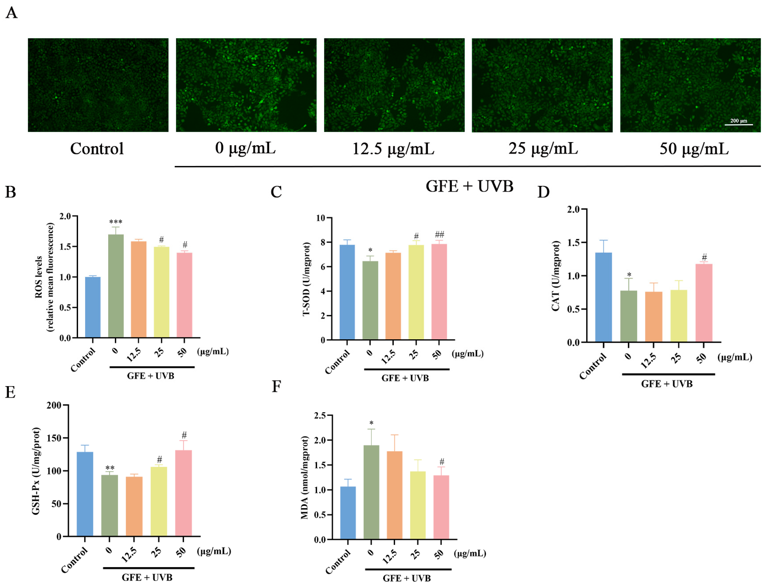
2.3. Detection of Reactive Oxygen Species (ROS), SOD, CAT, GSH-Px, and MDA

The HaCaT cells were treated with different concentrations of GFE (12.5, 25, and 50 μg/mL) and UVB irradiation and then stained by DCFH-DA (Beyotime, Shanghai, China). ROS contents were measured via a fluorescence microplate reader for the same number of cells and observed by fluorescence microscopy.
The levels of SOD, CAT, GSH-Px, and MDA were measured using a commercially available assay kit (Jiancheng Biological Engineering, Nanjing, China).

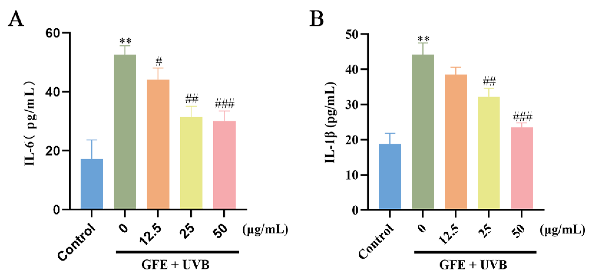
2.4. Enzyme-Linked Immunosorbent Assay

Contents of IL-6 and IL-1β were detected by enzyme-linked immunosorbent assay kits (TransGen Biotech, Beijing, China). HaCaT cells were irradiated by UVB, treated with GFE (12.5, 25, and 50 μg/mL), and analyzed for IL-6 and IL-1β at 24 h.

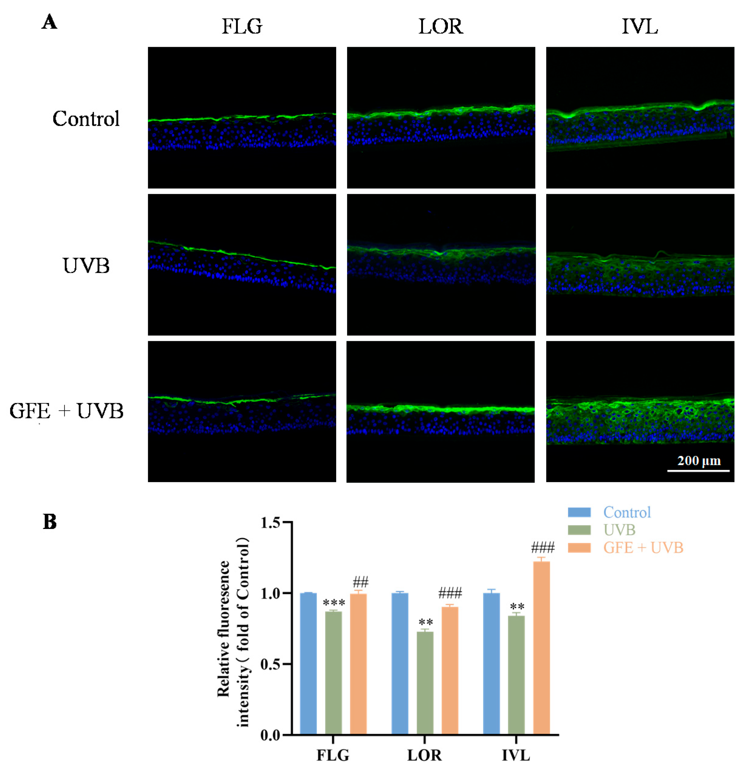
2.5. Immunofluorescence Staining

Epidermal skin models obtained from Regenovo were exposed to UVB (302 nm) at 400 mJ/cm2 and treated with 50 μg/mL GFE for 24 h. Then, each skin model was frozen and cut. The sections were blocked and incubated with primary antibodies, including filaggrin (FLG), loricrin (LOR), and involucrin (IVL) (Abcam, Cambridge, UK), overnight. After washing with PBS/T three times, the sections were incubated with Alexa: 488-conjugated goat anti-rabbit IgG H&L (Abcam, Cambridge, UK) and DAPI (Beyotime Biotechnology, Shanghai, China). The sections were observed under a fluorescent inverted microscope.

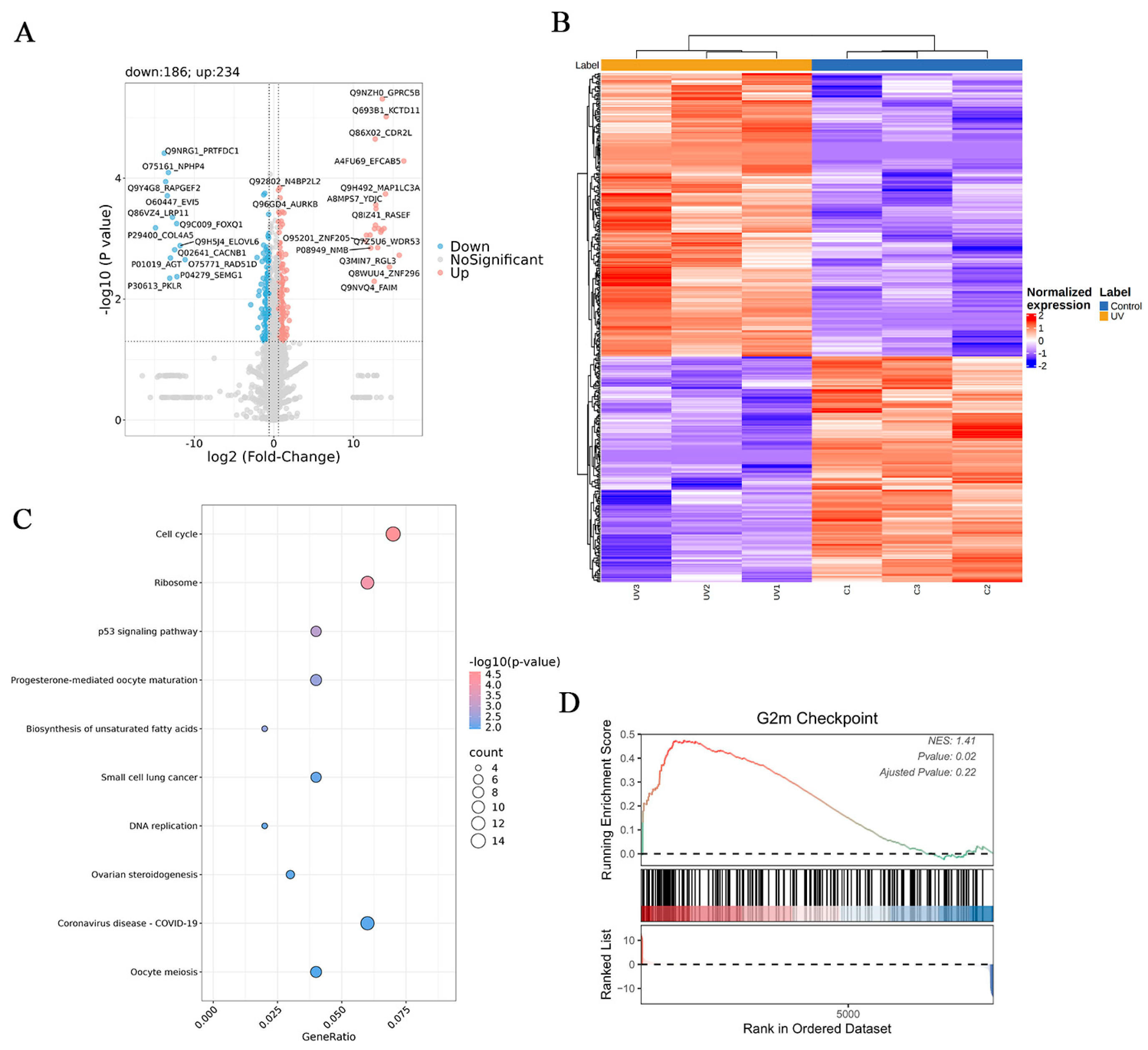
2.6. Proteomic Analysis

The HaCaT cells were treated with 50 μg/mL GFE and irradiated by UVB for 24 h. Then, the HaCaT cells were collected and stored at −80 °C. For each tube containing cell precipitate, the contents were lysed, digested, and desalted. Then, liquid chromatography–mass spectrometry (LC-MS) analysis was performed using a Vanquish Neo UHPLC system coupled to an Orbitrap Astral mass spectrometer (Thermo Scientific, Waltham, MA, USA) for data-independent acquisition (DIA) analysis. Differentially expressed proteins (DEPs) were identified with a p-value less than 0.05 and a fold change greater than 1.5.

2.7. Network Pharmacology

UV photodamage targets were collected from the GeneCards, OMIM, and CTD databases with the keyword “photodamage”. Meanwhile, the targets of the iridoids identified in GFE were predicted via SwissTargetPrediction and the CTD database. The intersected genes were then revealed by Venn diagrams.
The intersected targets were imported into STRING, the species was limited to “Homo sapiens”, and confidence data > 0.7 were used to construct the PPI network. A visual PPI network and an “ingredient–target” network were constructed using Cytoscape 3.9.0 software.
Protein structures were downloaded from the PDB database, and ingredient structures were obtained from the Pubchem database. Then, molecular docking was plotted and displayed with Auto Dock Vina 1.25 and PyMol 2.3.0 [27].

2.8. Statistical Analysis

Statistical analysis was performed using GraphPad Prism 9 software. The student’s t-test was performed for the differential analyses, and p < 0.05 was considered statistically significant.

3. Results

3.1. Analysis of GFE by UPLC-MS/MS

The total positive and negative ion chromatograms of GFE are shown in Figure 1A,B. Ten iridoids were identified in GFE (Figure 1C and Table S1): (1) genipin 1-gentiobioside, (2) geniposidic acid, (3) shanzhiside methyl ester, (4) deacetylasperulosidic acid, (5) genipin, (6) geniposide, (7) feretoside, (8) gardenoside, (9) asperuloside, and (10) shanzhiside.

3.2. Effects of GFE on Antioxidants in HaCaT Cells

UVB irradiation induces oxidative damage and promotes intracellular ROS production. In this study, the intracellular ROS levels in HaCaT cells were stained via DCFH-DA. Compared to the UVB exposure group, the production of ROS was significantly inhibited in the GFE groups (Figure 2A,B). Additionally, the levels of T-SOD, CAT, and GSH-Px were decreased under UVB irradiation, while GFE effectively increased the contents of T-SOD, CAT, and GSH-Px (Figure 2C–E). Figure 2F demonstrates that the contents of MDA were decreased in GFE-treated HaCaT cells.

3.3. Effects of GFE on Anti-Inflammation in HaCaT Cells

Skin oxidative stress can further amplify skin inflammation induced by UVB exposure [28]. In the results, the levels of both IL-6 and IL-1β in the supernatants were increased under UVB irradiation, while treatment with GFE inhibited the secretion of IL-6 and IL-1β (Figure 3).

3.4. Effects of GFE on Barrier Repair in Three-Dimensional Epidermal Models

Filaggrin (FLG), loricrin (LOR), and involucrin (IVL) are the important barrier proteins that regulate skin permeability and prevent water evaporation [29]. Our research showed that UVB exposure diminished barrier function by downgrading the expression of FLG, IVL, and LOR in the three-dimensional epidermal models. After GFE treatment, the barrier functions of the skin models were restored and the expression of barrier-related proteins (FLG, IVL, and LOR) was increased (Figure 4).

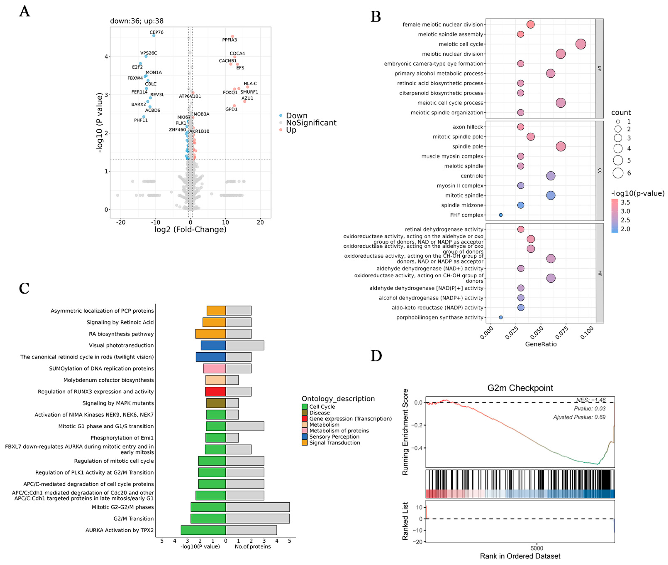
3.5. Proteomic Analysis

DEPs among the control, UV, and UV-GFE groups were analyzed by proteomics. The results revealed that 234 and 186 DEPs increased and decreased between the control group and UV group, respectively (Figure 5A). Subsequently, cluster analysis of these DEPs was performed, and the results were visualized as a heatmap (Figure 5B). The control group and UV group were separated distinctly, suggesting that the identified DEPs were representative. Kyoto Encyclopedia of Genes and Genomes (KEGG) results revealed that UVB irradiation induced cellular photodamage mainly by interfering with the cell cycle (Figure 5C). Additionally, Gene Set Enrichment Analysis (GSEA) showed that UVB treatment could damage epidermal cells by activating the G2/M checkpoint (Figure 5D).
Between the UV-GFE and UV groups, 38 DEPs were upregulated and 36 DEPs were downregulated (Figure 6A). Gene Ontology (GO) analysis (Figure 6B) showed that GFE treatment exerted a significant impact on meiotic nuclear division, meiotic spindle assembly, and the meiotic cell cycle in the biological process (BP) category. Cell components (CCs) that were influenced mostly included axon hillocks, mitotic spindle poles, and spindle poles. In the molecular function (MF) category, the functions of retinal dehydrogenase activity, oxidoreductase activity, and aldehyde dehydrogenase (NAD+) activity were mainly altered. Next, reactome enrichment analysis revealed that the DEPs were enriched in the cell cycle (Figure 6C). Interestingly, the GESA results indicated that GFE treatment significantly downregulated the G2/M checkpoint, which was upregulated by UVB treatment (Figure 6D).

3.6. Network Pharmacology

After removing duplicates, a total of 201 genes associated with photodamage were collected from the GeneCards, OMIM, and CTD databases. Additionally, 203 genes related to iridoids in GFE were predicted using SwissTargetPrediction and the CTD database. A Venn diagram showed that 24 targets were intersected between GFE and photodamage (Figure 7A).
Subsequently, a “component–target” network was constructed and visualized using Cytoscape 3.9.0 (Figure 7B). The red polygon nodes represent the compounds, the green ellipsis nodes represent the genes, and each edge indicates an interaction between them. The network consisted of 31 nodes and 40 edges, with geniposide being associated with the most targets.
In order to predict the potential targets of GFE anti-photodamage, PPI network analysis was performed. The Centiscape analysis of the key targets is shown in Table 1. PPI network results revealed the genes with values above the average for degree centrality (5.64), betweenness centrality (0.055), and closeness centrality (0.497) obtained from the Centiscape analysis, including IL1B, MMP9, TP53, BCL2, and SIRT1.
Based on network pharmacology and proteomics, geniposide and TP53 (p53) were selected for molecular docking. Typically, a binding energy less than −5 kcal/mol signifies an excellent binding affinity. The results indicated that geniposide–p53 had two hydrogen bonds with Leu145 and Glu221 and exhibited a favorable binding activity, with a binding energy of −6.0 kcal/mol.

4. Discussion

Repeated or long-term exposure to UVB can induce skin photodamage due to its penetration of the epidermis and upper dermis, manifesting as sunburn, tanning, immunosuppression, and the release of pro-inflammatory mediators [30]. Keratinocytes are the most predominant cells in the epidermis of the skin. Researchers have revealed that UVB radiation could lead to DNA damage and accumulation of ROS in keratinocytes, resulting in cell death [31]. Additionally, apoptosis of keratinocytes is accompanied by inflammation and disruption of the epidermal barrier [32]. Therefore, it is essential to find a safe and effective treatment to prevent skin photodamage. In recent years, researchers have shown an increasing interest in the application of natural products in the cosmetic industry [33]. For example, ferulic acid, caffeine, and rutin have shown excellent efficacy in skin photoprotection and sunscreen cosmetic applications [34,35,36,37]. GF, with a positive effect on anti-photoaging, has garnered our attention.
Iridoids are a special class of monoterpenoids found in GFE, serving as some of the main chemical components with numerous pharmacological activities, such as antioxidant, anti-inflammatory, and hypoglycemic properties [38,39,40,41]. In this study, ten iridoids in GFE were identified via LC-MS/MS. Among them, geniposide is the main active ingredient in GFE and has the largest content [25]. To further evaluate the effects of GFE against UVB-induced skin damage, a UVB-irradiated HaCaT cell model was established. Oxidative stress and inflammation are the main negative effects caused by UVB. UVB irradiation induced oxidative stress damage in HaCaT cells, which presented as a decrease in antioxidant enzyme (T-SOD, CAT, and GSH-Px) levels and an increase in ROS and lipid peroxides (MDA). GFE treatment effectively combated UVB-induced cellular oxidative damage. Meanwhile, GFE inhibited the IL-6 and IL-1β levels caused by UVB exposure. UVB-induced skin photodamage is accompanied by impairment of the skin barrier, and skin barrier function is closely associated with the terminal differentiation-associated proteins, such as FLG, LOR, and IVL [42]. In this study, UVB-irradiated epidermal models exhibited significant downregulation of FLG, LOR, and IVL expression, while GFE effectively ameliorated the UVB-induced epidermal barrier damage by upregulating the expression of these barrier-related proteins.
Recently, proteomics has served as a crucial tool for uncovering the potential targets in TCM. In our studies, proteomic analysis indicated that UVB radiation affected various cell cycle-related proteins in HaCaT cells. Cell cycle checkpoints are typically regulated by a variety of cell cycle regulatory proteins [43]. The activation of targets in the G2/M checkpoint also confirmed the damage of keratinocyte DNA by UVB radiation, which is one of the main factors affecting DNA synthesis in keratinocytes [44]. Between the UV and UV-GFE groups, reactome enrichment analysis revealed that the UV-GFE group mitigated UVB-induced skin photodamage mainly by affecting the cell cycle. In addition, GFE treatment improved UVB-induced cell cycle arrest at the G2/M phase in HaCaT cells. Combined with the results of pharmacological experiments, GFE might act on multiple targets and cause complex pharmacological changes, resulting in a comprehensive effect on UVB-induced skin damage.
The network pharmacology method is widely utilized to explore the complex relationship between TCM and targets. The results of topological analyses indicated that geniposide was associated with more targets. Geniposide is the most abundant iridoid in GFE, and it can exert anti-photoaging effects by inhibiting oxidative stress in human skin cells [24]. Additionally, it can regulate the G2/M phase and inhibit inflammatory responses to improve apoptosis, suggesting that it might be the main active component in GFE against photodamage [45]. PPI network results revealed that apoptosis- and cell cycle-related targets, such as TP53, CASP3, and BCL2, were essential targets in GFE-mediated anti-photodamage, which corresponded to the proteomic results. TP53 (p53), the key target affecting the cell cycle and apoptosis, might play an essential role in regulating photodamage [46]. In order to demonstrate the binding modes between geniposide and p53, molecular docking was performed. Our research found that the geniposide–p53 complex showed stable binding, with a binding energy of -6.0 kcal/mol. The findings of this study lay a foundation for the further development of GFE in cosmetic raw materials and products capable of repairing damage induced by UVB. The anti-photodamage components and the mechanisms of GFE still need to be further verified.

5. Conclusions

Our study reveals the effects and potential mechanisms of GFE in ameliorating UVB-induced photodamage in HaCaT cells based on pharmacological experiments, proteomics, and network pharmacology. GFE treatment effectively ameliorates UVB-induced skin photodamage by regulating the cell cycle, including the G2/M checkpoint and p53 pathways, indicating its potential as an active ingredient against skin photoaging.

Supplementary Materials

The following supporting information can be downloaded at https://www.mdpi.com/article/10.3390/cosmetics12020072/s1: Table S1: Ten iridoids identified in GFE.

Author Contributions

Conceptualization, J.W., Q.H., and K.Z.; methodology, software, validation, and visualization, K.Z., X.L., and F.Z.; investigation and data curation, J.D.; writing—original draft preparation, K.Z. and J.D.; writing—review and editing, Q.H. and J.W.; project administration, funding acquisition, J.W., Q.H., and J.D. All authors have read and agreed to the published version of the manuscript.

Funding

This research was supported by the 2019 high-level talent introduction scientific research start-up fund of China Pharmaceutical University (3150020056).

Institutional Review Board Statement

Not applicable.

Informed Consent Statement

Not applicable.

Data Availability Statement

The data that support the findings of this study are available from the corresponding author upon reasonable request.

Acknowledgments

The authors are grateful for the research material provided by Shanghai Junyu Biotechnology Group Co., Ltd., located in Shanghai, China, and wish to thank Hangzhou CIRS KEXIN Biotech Co., Ltd. for its participation in the conduction of the studies.

Conflicts of Interest

X.L., F.Z., and J.D. were employed by the Shanghai Junyu Biotechnology Group Co., Ltd. The remaining authors declare that the research was conducted in the absence of any commercial or financial relationships that could be construed as a potential conflict of interest.

References

  1. Wong, Q.Y.A.; Chew, F.T. Defining skin aging and its risk factors: A systematic review and meta-analysis. Sci. Rep. 2021, 11, 22075. [Google Scholar] [CrossRef] [PubMed]
  2. Gan, C.; Liu, T.; Jia, X.; Huang, X.; Qin, X.; Wang, X.; Sheng, J.; Xu, H. ATP supplementation suppresses UVB-induced photoaging in HaCaT cells via upregulation of expression of SIRT3 and SOD2. Ski. Res. Technol. 2023, 29, e13303. [Google Scholar] [CrossRef] [PubMed]
  3. Passeron, T.; Bouillon, R.; Callender, V.; Cestari, T.; Diepgen, T.L.; Green, A.C.; van der Pols, J.C.; Bernard, B.A.; Ly, F.; Bernerd, F.; et al. Sunscreen photoprotection and vitamin D status. Br. J. Dermatol. 2019, 181, 916–931. [Google Scholar] [CrossRef]
  4. de la Coba, F.; Aguilera, J.; Korbee, N.; de Gálvez, M.V.; Herrera-Ceballos, E.; Álvarez-Gómez, F.; Figueroa, F.L. UVA and UVB Photoprotective Capabilities of Topical Formulations Containing Mycosporine-like Amino Acids (MAAs) through Different Biological Effective Protection Factors (BEPFs). Mar. Drugs 2019, 17, 55. [Google Scholar] [CrossRef]
  5. Solano, F. Photoprotection and Skin Pigmentation: Melanin-Related Molecules and Some Other New Agents Obtained from Natural Sources. Molecules 2020, 25, 1537. [Google Scholar] [CrossRef]
  6. Park, J.; Kim, D.; Lee, M.; Han, S.; Jun, W.; Jung, H.M.; Koo, Y.K.; Na, G.H.; Han, S.H.; Han, J.; et al. Enzyme-Treated Caviar Prevents UVB Irradiation-Induced Skin Photoaging. Mar. Drugs 2022, 20, 685. [Google Scholar] [CrossRef]
  7. Md Jaffri, J. Reactive Oxygen Species and Antioxidant System in Selected Skin Disorders. Malays. J. Med. Sci. 2023, 30, 7–20. [Google Scholar] [CrossRef]
  8. Prasanth, M.I.; Sivamaruthi, B.S.; Chaiyasut, C.; Tencomnao, T. A Review of the Role of Green Tea (Camellia sinensis) in Antiphotoaging, Stress Resistance, Neuroprotection, and Autophagy. Nutrients 2019, 11, 474. [Google Scholar] [CrossRef]
  9. Hussain, T.; Tan, B.; Yin, Y.; Blachier, F.; Tossou, M.C.; Rahu, N. Oxidative Stress and Inflammation: What Polyphenols Can Do for Us? Oxidative Med. Cell. Longev. 2016, 2016, 7432797. [Google Scholar] [CrossRef]
  10. Ansary, T.M.; Hossain, M.R.; Kamiya, K.; Komine, M.; Ohtsuki, M. Inflammatory Molecules Associated with Ultraviolet Radiation-Mediated Skin Aging. Int. J. Mol. Sci. 2021, 22, 3947. [Google Scholar] [CrossRef]
  11. Diao, P.; He, H.; Tang, J.; Xiong, L.; Li, L. Natural compounds protect the skin from airborne particulate matter by attenuating oxidative stress. Biomed. Pharmacother. 2021, 138, 111534. [Google Scholar] [CrossRef] [PubMed]
  12. Biniek, K.; Levi, K.; Dauskardt, R.H. Solar UV radiation reduces the barrier function of human skin. Proc. Natl. Acad. Sci. USA 2012, 109, 17111–17116. [Google Scholar] [CrossRef]
  13. Mavrogonatou, E.; Angelopoulou, M.; Rizou, S.V.; Pratsinis, H.; Gorgoulis, V.G.; Kletsas, D. Activation of the JNKs/ATM-p53 axis is indispensable for the cytoprotection of dermal fibroblasts exposed to UVB radiation. Cell Death Dis. 2022, 13, 647. [Google Scholar] [CrossRef]
  14. Mayangsari, E.; Mustika, A.; Nurdiana, N.; Samad, N.A. Comparison of UVA vs UVB Photoaging Rat Models in Short-term Exposure. Med. Arch. 2024, 78, 88–91. [Google Scholar] [CrossRef]
  15. Cavinato, M.; Waltenberger, B.; Baraldo, G.; Grade, C.V.C.; Stuppner, H.; Jansen-Dürr, P. Plant extracts and natural compounds used against UVB-induced photoaging. Biogerontology 2017, 18, 499–516. [Google Scholar] [CrossRef]
  16. Tan, H.; Ren, H.; Chai, J.; Zhai, C.; Li, T.; Zhou, X.; Lee, J.; Li, X.; Zhao, Y. Protective effect of ginseng berry saponin conversion products on skin photodamage caused by UVB in vitro and in vivo. Food Res. Int. 2024, 198, 115379. [Google Scholar] [CrossRef]
  17. Fu, H.; Zhang, Y.; An, Q.; Wang, D.; You, S.; Zhao, D.; Zhang, J.; Wang, C.; Li, M. Anti-Photoaging Effect of Rhodiola rosea Fermented by Lactobacillus plantarum on UVA-Damaged Fibroblasts. Nutrients 2022, 14, 2324. [Google Scholar] [CrossRef]
  18. Guo, S.; Bao, L.; Li, C.; Sun, J.; Zhao, R.; Cui, X. Antiviral activity of iridoid glycosides extracted from Fructus Gardeniae against influenza A virus by PACT-dependent suppression of viral RNA replication. Sci. Rep. 2020, 10, 1897. [Google Scholar] [CrossRef]
  19. Kim, S.H.; Shin, M.R.; Lee, A.R.; Seo, B.I.; Park, H.J.; Roh, S.S. Improvement of Inflammation through Antioxidant Pathway of Gardeniae Fructus 50% EtOH Extract (GE) from Acute Reflux Esophagitis Rats. BioMed Res. Int. 2020, 2020, 4826176. [Google Scholar] [CrossRef]
  20. Nguyen, L.T.H.; Ahn, S.H.; Nguyen, U.T.; Yang, I.J.; Shin, H.M. Geniposide, a Principal Component of Gardeniae Fructus, Protects Skin from Diesel Exhaust Particulate Matter-Induced Oxidative Damage. Evid. -Based Complement. Altern. Med. 2021, 2021, 8847358. [Google Scholar] [CrossRef]
  21. De Tollenaere, M.; Chapuis, E.; Martinez, J.; Paulus, C.; Dupont, J.; Don Simoni, E.; Robe, P.; Sennelier-Portet, B.; Auriol, D.; Scandolera, A.; et al. Gardenia jasminoides Extract, with a Melatonin-like Activity, Protects against Digital Stress and Reverses Signs of Aging. Int. J. Mol. Sci. 2023, 24, 4948. [Google Scholar] [CrossRef] [PubMed]
  22. Zhang, H.; Lai, Q.; Li, Y.; Liu, Y.; Yang, M. Learning and memory improvement and neuroprotection of Gardenia jasminoides (Fructus gardenia) extract on ischemic brain injury rats. J. Ethnopharmacol. 2017, 196, 225–235. [Google Scholar] [CrossRef] [PubMed]
  23. Kato, T.; Hino, S.; Horie, N.; Shimoyama, T.; Kaneko, T.; Kusama, K.; Sakagami, H. Anti-UV activity of Kampo medicines and constituent plant extracts: Re-evaluation with skin keratinocyte system. Vivo 2014, 28, 571–578. [Google Scholar]
  24. Shin, D.; Lee, S.; Huang, Y.H.; Lim, H.W.; Lee, Y.; Jang, K.; Cho, Y.; Park, S.J.; Kim, D.D.; Lim, C.J. Protective properties of geniposide against UV-B-induced photooxidative stress in human dermal fibroblasts. Pharm. Biol. 2018, 56, 176–182. [Google Scholar] [CrossRef]
  25. Dong, J.; Li, B.; Xu, P.; Kong, L.; Zhang, W.; Wu, D.; Tian, Y.; Wang, X. Skin barrier repair efficacy and safety evaluation of an extract from Gardenia jasminoides J. Ellis. Nat. Prod. Res. 2024, 1–7. [Google Scholar] [CrossRef]
  26. Ke, H.; Zhang, X.; Liang, S.; Zhou, C.; Hu, Y.; Huang, Q.; Wu, J. Study on the anti-skin aging effect and mechanism of Sijunzi Tang based on network pharmacology and experimental validation. J. Ethnopharmacol. 2024, 333, 118421. [Google Scholar] [CrossRef]
  27. Zong, K.; Xu, K.; Zhang, X.; Wang, P.; Wang, Z.; Yang, S.; Li, H.; Ke, H.; He, S.; Hu, Y.; et al. Explorating the mechanism of Huangqin Tang against skin lipid accumulation through network pharmacology and experimental validation. J. Ethnopharmacol. 2023, 313, 116581. [Google Scholar] [CrossRef]
  28. Baek, J.; Kim, J.H.; Park, J.; Kim, D.H.; Sa, S.; Han, J.S.; Kim, W. 1-Kestose Blocks UVB-Induced Skin Inflammation and Promotes Type I Procollagen Synthesis via Regulating MAPK/AP-1, NF-κB and TGF-β/Smad Pathway. J. Microbiol. Biotechnol. 2024, 34, 911–919. [Google Scholar] [CrossRef]
  29. Jang, A.Y.; Choi, J.; Rod-In, W.; Choi, K.Y.; Lee, D.H.; Park, W.J. In Vitro Anti-Inflammatory and Skin Protective Effects of Codium fragile Extract on Macrophages and Human Keratinocytes in Atopic Dermatitis. J. Microbiol. Biotechnol. 2024, 34, 940–948. [Google Scholar] [CrossRef]
  30. He, H.; Huang, W.; Xiong, L.; Ma, C.; Wang, Y.; Sun, P.; Shi, D.; Li, L.; Yan, H.; Wu, Y. FUNDC1-mediated mitophagy regulates photodamage independently of the PINK1/Parkin-dependent pathway. Free Radic. Biol. Med. 2024, 225, 630–640. [Google Scholar] [CrossRef]
  31. Tang, Z.; Tong, X.; Huang, J.; Liu, L.; Wang, D.; Yang, S. Research progress of keratinocyte-programmed cell death in UV-induced Skin photodamage. Photodermatol. Photoimmunol. Photomed. 2021, 37, 442–448. [Google Scholar] [CrossRef] [PubMed]
  32. Cheng, W.; Di, F.; Li, L.; Pu, C.; Wang, C.; Zhang, J. Anti-Photodamage Effect of Agaricus blazei Murill Polysaccharide on UVB-Damaged HaCaT Cells. Int. J. Mol. Sci. 2024, 25, 4676. [Google Scholar] [CrossRef]
  33. Milutinov, J.; Pavlović, N.; Ćirin, D.; Atanacković Krstonošić, M.; Krstonošić, V. The Potential of Natural Compounds in UV Protection Products. Molecules 2024, 29, 5409. [Google Scholar] [CrossRef]
  34. Morocho-Jácome, A.L.; Freire, T.B.; de Oliveira, A.C.; de Almeida, T.S.; Rosado, C.; Velasco, M.V.R.; Baby, A.R. In vivo SPF from multifunctional sunscreen systems developed with natural compounds-A review. J. Cosmet. Dermatol. 2021, 20, 729–737. [Google Scholar] [CrossRef]
  35. Peres, D.D.; Sarruf, F.D.; de Oliveira, C.A.; Velasco, M.V.R.; Baby, A.R. Ferulic acid photoprotective properties in association with UV filters: Multifunctional sunscreen with improved SPF and UVA-PF. J. Photochem. Photobiol. B Biol. 2018, 185, 46–49. [Google Scholar] [CrossRef]
  36. Rosado, C.; Tokunaga, V.K.; Sauce, R.; de Oliveira, C.A.; Sarruf, F.D.; Parise-Filho, R.; Maurício, E.; de Almeida, T.S.; Velasco, M.V.R.; Baby, A.R. Another Reason for Using Caffeine in Dermocosmetics: Sunscreen Adjuvant. Front. Physiol. 2019, 10, 519. [Google Scholar] [CrossRef]
  37. Tomazelli, L.C.; de Assis Ramos, M.M.; Sauce, R.; Cândido, T.M.; Sarruf, F.D.; de Oliveira Pinto, C.A.S.; de Oliveira, C.A.; Rosado, C.; Velasco, M.V.R.; Baby, A.R. SPF enhancement provided by rutin in a multifunctional sunscreen. Int. J. Pharm. 2018, 552, 401–406. [Google Scholar] [CrossRef]
  38. Wang, L.; Chen, S.; Liu, S.; Biu, A.M.; Han, Y.; Jin, X.; Liang, C.; Liu, Y.; Li, J.; Fang, S.; et al. A comprehensive review of ethnopharmacology, chemical constituents, pharmacological effects, pharmacokinetics, toxicology, and quality control of gardeniae fructus. J. Ethnopharmacol. 2024, 320, 117397. [Google Scholar] [CrossRef]
  39. Sun, K.X.; Chen, Y.Y.; Li, Z.; Zheng, S.J.; Wan, W.J.; Ji, Y.; Hu, K. Genipin relieves diabetic retinopathy by down-regulation of advanced glycation end products via the mitochondrial metabolism related signaling pathway. World J. Diabetes 2023, 14, 1349–1368. [Google Scholar] [CrossRef]
  40. Li, H.; Yang, D.H.; Zhang, Y.; Zheng, F.; Gao, F.; Sun, J.; Shi, G. Geniposide suppresses NLRP3 inflammasome-mediated pyroptosis via the AMPK signaling pathway to mitigate myocardial ischemia/reperfusion injury. Chin. Med. 2022, 17, 73. [Google Scholar] [CrossRef]
  41. Chen, Y.I.; Cheng, Y.W.; Tzeng, C.Y.; Lee, Y.C.; Chang, Y.N.; Lee, S.C.; Tsai, C.C.; Chen, J.C.; Tzen, J.T.; Chang, S.L. Peroxisome proliferator-activated receptor activating hypoglycemic effect of Gardenia jasminoides Ellis aqueous extract and improvement of insulin sensitivity in steroid induced insulin resistant rats. BMC Complement. Altern. Med. 2014, 14, 30. [Google Scholar] [CrossRef] [PubMed]
  42. Jing, C.; Guo, J.; Li, Z.; Xu, X.; Wang, J.; Zhai, L.; Liu, J.; Sun, G.; Wang, F.; Xu, Y.; et al. Screening and Research on Skin Barrier Damage Protective Efficacy of Different Mannosylerythritol Lipids. Molecules 2022, 27, 4648. [Google Scholar] [CrossRef] [PubMed]
  43. Faurschou, A.; Gniadecki, R.; Calay, D.; Wulf, H.C. TNF-alpha impairs the S-G2/M cell cycle checkpoint and cyclobutane pyrimidine dimer repair in premalignant skin cells: Role of the PI3K-Akt pathway. J. Investig. Dermatol. 2008, 128, 2069–2077. [Google Scholar] [CrossRef]
  44. Chilampalli, C.; Guillermo, R.; Zhang, X.; Kaushik, R.S.; Young, A.; Zeman, D.; Hildreth, M.B.; Fahmy, H.; Dwivedi, C. Effects of magnolol on UVB-induced skin cancer development in mice and its possible mechanism of action. BMC Cancer 2011, 11, 456. [Google Scholar] [CrossRef]
  45. Chen, W.J.; Bao, T.Z.; Chen, K.; Zhu, C.M.; Wan, F.; Tan, Y.L.; Yan, F. [Effects of geniposide on SNP-induced apoptosis of chondrocyte and cell cycle]. China J. Orthop. Traumatol. 2013, 26, 232–235. [Google Scholar]
  46. Liu, F.J.; Chen, X.Y.; Yang, J.; Zhao, Z.; Wang, Q.L.; Li, P.; Jiang, Y.; Li, H.J. Revealing active components and action mechanism of Fritillariae Bulbus against non-small cell lung cancer through spectrum-effect relationship and proteomics. Phytomedicine 2023, 110, 154635. [Google Scholar] [CrossRef]
Figure 1. UPLC-MS/MS total ion chromatograms in the positive (A) and negative (B) ion models of GFE. (C) Molecular structure of constituents.
Cosmetics 12 00072 g001
Figure 2. The effects of GFE on the oxidative stress in HaCaT cells. (A,B) The levels of intercellular ROS were stained with DCFH-DA, photographed by fluorescence microscopy, and measured under a fluorescence microplate reader; scale bar = 50 μm. The levels of (C) T-SOD, (D) CAT, (E) GSH-Px, and (F) MDA were determined by specialized test kits (mean ± SD, n = 3). * p < 0.05, ** p < 0.01, and *** p < 0.001 vs. the control; # p < 0.05 and ## p < 0.01 vs. UVB.
Cosmetics 12 00072 g002
Figure 3. The effects of GFE on inflammation in HaCaT cells. The contents of (A) IL-6 and (B) IL-1β were quantified by ELISA (mean ± SD, n = 3). ** p < 0.01 vs. the control; # p < 0.05, ## p < 0.01, and ### p < 0.001 vs. UVB.
Cosmetics 12 00072 g003
Figure 4. The effects of GFE on the barrier functions in three-dimensional epidermal models. (A) The levels of FLG, LOR, and IVL were stained by immunofluorescence and (B) measured via Imagj J 1.53q. Bar = 50 μm (mean ± SD, n = 3). ** p < 0.01 and *** p < 0.001 vs. the control; ## p < 0.01 and ### p < 0.001 vs. UVB.
Cosmetics 12 00072 g004
Figure 5. Proteomic analysis of the control and UV groups. (A) DEPs between the control group and UV group shown in a volcano plot; (B) cluster analysis of DEPs between the control group and UV group shown in a heatmap; (C) KEGG analysis between the control group and UV group; (D) GSEA between the control group and UV group.
Cosmetics 12 00072 g005
Figure 6. Proteomic analysis of UV and UV-GFE groups. (A) DEPs between the UV-GFE group and UV group shown in a volcano plot; (B) GO enrichment analysis of DEPs between the UV-GFE group and UV group; (C) reactome analysis of DEPs between the UV-GFE group and UV group; (D) GSEA between the UV-GFE group and UV group.
Cosmetics 12 00072 g006
Figure 7. Network pharmacology of GF and photodamage. (A) Venn diagram; (B) ”component–target” network; (C) PPI network; (D) molecular docking of geniposide–p53.
Cosmetics 12 00072 g007
Table 1. Centiscape analysis of the key targets in the PPI network.
No.TargetDegree CentralityBetweenness CentralityCloseness Centrality
1IL1B140.3020573080.7
2MMP9110.1241223460.6
3TP53100.1385101010.636363636
4BCL2100.0792341790.617647059
5SIRT180.1879349620.583333333
Disclaimer/Publisher’s Note: The statements, opinions and data contained in all publications are solely those of the individual author(s) and contributor(s) and not of MDPI and/or the editor(s). MDPI and/or the editor(s) disclaim responsibility for any injury to people or property resulting from any ideas, methods, instructions or products referred to in the content.

