Article

The Oral Intake of Specific Bovine-Derived Bioactive Collagen Peptides Has a Stimulatory Effect on Dermal Matrix Synthesis and Improves Various Clinical Skin Parameters

1
Department of Dermatology, University Hospital of Schleswig-Holstein, Campus Kiel, 24105 Kiel, Germany
2
CRI, Collagen Research Institute, Schauenburgerstr 116, 24118 Kiel, Germany
*
Author to whom correspondence should be addressed.
Cosmetics 2025, 12(2), 79; https://doi.org/10.3390/cosmetics12020079
Submission received: 27 February 2025 / Revised: 3 April 2025 / Accepted: 10 April 2025 / Published: 14 April 2025
(This article belongs to the Special Issue Skin Anti-Aging Strategies)

Abstract

:
Collagen products are widely marketed for skin improvement. This study evaluated the efficacy of VERISOL B in relation to key skin aging parameters. In a double-blind, placebo-controlled trial, 66 women (aged 35–55) were randomized to receive either 2.5 g of bovine-derived bioactive collagen peptides (SCPs) (n = 33) or a placebo (n = 33) daily for 8 weeks. Their eye wrinkle volume, skin elasticity, and hydration were objectively measured at baseline (X0), 4 weeks (X4), and 8 weeks (X8). Additionally, the SCPs’ impact on type I collagen, elastin, and proteoglycan biosynthesis was assessed in human dermal fibroblasts. The SCP supplementation significantly (p < 0.05) reduced their eye wrinkle volume and improved their skin elasticity and hydration within 4 weeks. After 8 weeks of treatment, the positive effects were even more pronounced for all of the clinical parameters measured (p < 0.05). The fibroblast experiments confirmed the SCPs’ stimulatory impact on dermal metabolism (p < 0.05). In conclusion, oral SCP supplementation effectively reduced wrinkles and enhanced skin elasticity and hydration, likely by promoting extracellular matrix biosynthesis.

1. Introduction

The appearance and integrity of the skin deteriorate with age due to the synergistic effects of chronological aging and photoaging, hormonal deficiency, and environmental influences [1]. A decrease in various metabolic activities, such as quantitative and qualitative changes in dermal collagen and elastin, leads to changes in skin texture and the typical signs of aging. The loss of connective tissue during skin aging leads to reduced elasticity, a loss of skin tone, and the progressive development and deepening of facial wrinkles and folds [2].
Facial wrinkles, accompanied by an age-related decrease in the skin’s elasticity and a leathery appearance of the affected skin areas, are the best-known signs of skin aging. Certain facial areas, such as the corners of the eyes, commonly known as crow’s feet, are particularly susceptible to wrinkling [3,4].
The function and healthy appearance of the skin depend on an adequate supply of essential nutrients. The relationship between nutrition and skin has become an issue of current interest worldwide. Intervention studies suggest dietary supplements can modulate or delay skin aging and improve the skin’s integrity [5].
In addition to topical applications, a significant trend in skin care is the consumption of various supplements, such as vitamins, antioxidants, fatty acids, and hydrolyzed proteins, to enhance the appearance and texture of the skin [6,7,8,9,10,11,12,13,14,15,16]. As a result, there is increasing focus on the efficacy of these products, and several clinical studies suggest that dietary supplements can influence skin health [17,18,19,20,21].
Collagen peptides have been used for some time in dietary supplements to improve the skin’s properties and appearance. Collagen peptides are partially absorbed intact from the digestive tract [22,23] and accumulate in skin tissue [24]. In vitro studies have shown that specific collagen peptides have a stimulatory effect on molecules of the dermal extracellular matrix (ECM), such as collagen, elastin, and proteoglycans [25,26,27,28]. The ECM is produced by dermal fibroblasts, cells responsible for synthesizing and maintaining the matrix molecules, thus counteracting age-related degradation processes.
Collagen hydrolysates have antioxidant properties and promote cell regeneration and synthesis of the extracellular matrix in the dermal connective tissue [6]. Additionally, orally administered collagen peptides enhance the skin’s elasticity, hydration, and dermal density [8,29] while also reducing wrinkles and improving the skin’s firmness [30], as described in systematic reviews of the literature on the effects of collagen supplementation on skin health [6,30,31]. The benefits of orally administered collagen peptides include significant improvements in skin elasticity and hydration and wrinkle reduction, as well as wound healing, ulcer treatment, and skin regeneration. The efficacy of collagen peptides has been demonstrated in several clinical trials, with the daily dosages ranging from 2.5 g to 10 g and the treatment periods ranging from 4 to 24 weeks.
Clinical trials on healthy women have shown that daily supplementation of 2.5 g of specific bioactive collagen peptides (SCPs) over 8 weeks significantly improves skin elasticity [32]. Another study [25] showed a positive impact on wrinkle reduction after 8 weeks of SCP treatment. In addition, a significant increase in type I collagen and elastin contents was observed in the skin of the women who had consumed 2.5 g SCPs/day for 8 weeks.
Most of these clinical trials have been conducted using porcine- or fish-derived specific bioactive collagen peptides [25,32,33], but there is also an increasing number of studies on the effects of bovine-derived collagen products on various skin parameters [34,35]. Based on the existing studies on specific products made from porcine-based collagen peptides (VERISOL P) [25,32], the aim of the current study was to investigate the efficacy of a specific bovine-derived bioactive collagen peptide product (VERISOL B) in relation to various clinically relevant skin parameters. Moreover, to gain a better understanding of the potential mechanisms of action, the biosynthesis of dermal matrix molecules in human dermal fibroblast cultures was investigated following the supplementation of this specific bovine-derived product.

2. Materials and Methods

2.1. The Test Product

The test product used in this study (SCPs) was a specific bioactive collagen peptide composition derived from the special enzymatic hydrolysis of bovine type I collagen. The peptide composition was produced and provided by GELITA AG (Eberbach, Germany) and is commercially available under the brand name VERISOL® B. It has a mean molecular weight of approximately 2.0 kDa. It is free of allergens. The recommended dosage ranges from 2.5 g to 5 g per day, with no concerns of overdose. This product is classified as a nutritional supplement and was classified by the United States Food and Drug Administration (FDA) as generally recognized as safe (GRAS) [36].

2.2. In Vitro Tests

We have used the fibroblast cells NHDF (PromoCell, Heidelberg, Germany). The cells were cultured in an optimized medium (HAM’s F12) supplemented with 10% FBS, 20 U/mL penicillin–streptomycin, 50 µg/mL partricin, 0.05 mg/mL ascorbic acid, and 1 mM glutamine. The primary dermal fibroblasts were then seeded into 12-well culture plates following the proteolytic detachment of the adherent cells using Accutase solution and incubated at 37 °C with 5% CO2. The culture medium was refreshed every 48 h, and the cells were maintained until they reached 80% confluence. Subsequently, the regular medium was replaced with medium containing the test product at a concentration of 0.5 mg/mL. Control experiments were conducted by culturing the cells in the initial medium without any additional treatment. To assess the mRNA expression profiles, the fibroblasts were stimulated for 24 h. The biosynthesis of the extracellular matrix molecules was evaluated after a 2-week treatment. The results are expressed relative to those for the untreated control.

2.3. Gene Expression Studies

Gene expression studies were conducted by treating fibroblast monolayer cultures with the test product for 24 h and comparing them to untreated controls. RNA was extracted using the phenol–chloroform method (PeqGoldTriFast, VWR, Erlangen, Germany). The matrix molecule RNA expression was measured semi-quantitatively using RT-PCR (DyNAmo Flash SYBR Green, Thermo Scientific, Waltham, MA, USA) after determining the RNA concentration photometrically.
Each reaction used 10–40 ng of transcribed cDNA (optimized in preliminary experiments), 12.5 µL of distilled water, 5.0 µL of DyNAmo Flash Master Mix, and 1.25 µL of the primers described in Table 1. The reaction profile included 7′ at 95 °C, followed by 35–45 cycles of denaturation (10″ at 95 °C), annealing (15″ at temperatures in Table 1), and elongation (20″ at 72 °C), with a final step at 60 °C for 60″. The RNA expression was determined relative to GAPDH.
The primer annealing temperatures were optimized between 50 and 70 °C using gradient PCR. The products were separated on 2% agarose gel (3 g of agarose in 150 mL of buffer of 0.05 M EDTA, 40 mM Tris, and 57.1 mL of acetic acid), stained with 10 µL of GelRed (41003, BIOTREND Chemikalien GmbH, Cologne, Germany), and analyzed using an Alpha Innotech Fluorchem Imager (Bio-Rad Laboratories GmbH, Munich, Germany). Products were specified using a 50 bp DNA fluoro-ladder (8263.1, Carl Roth GmbH, Karlsruhe, Germany).

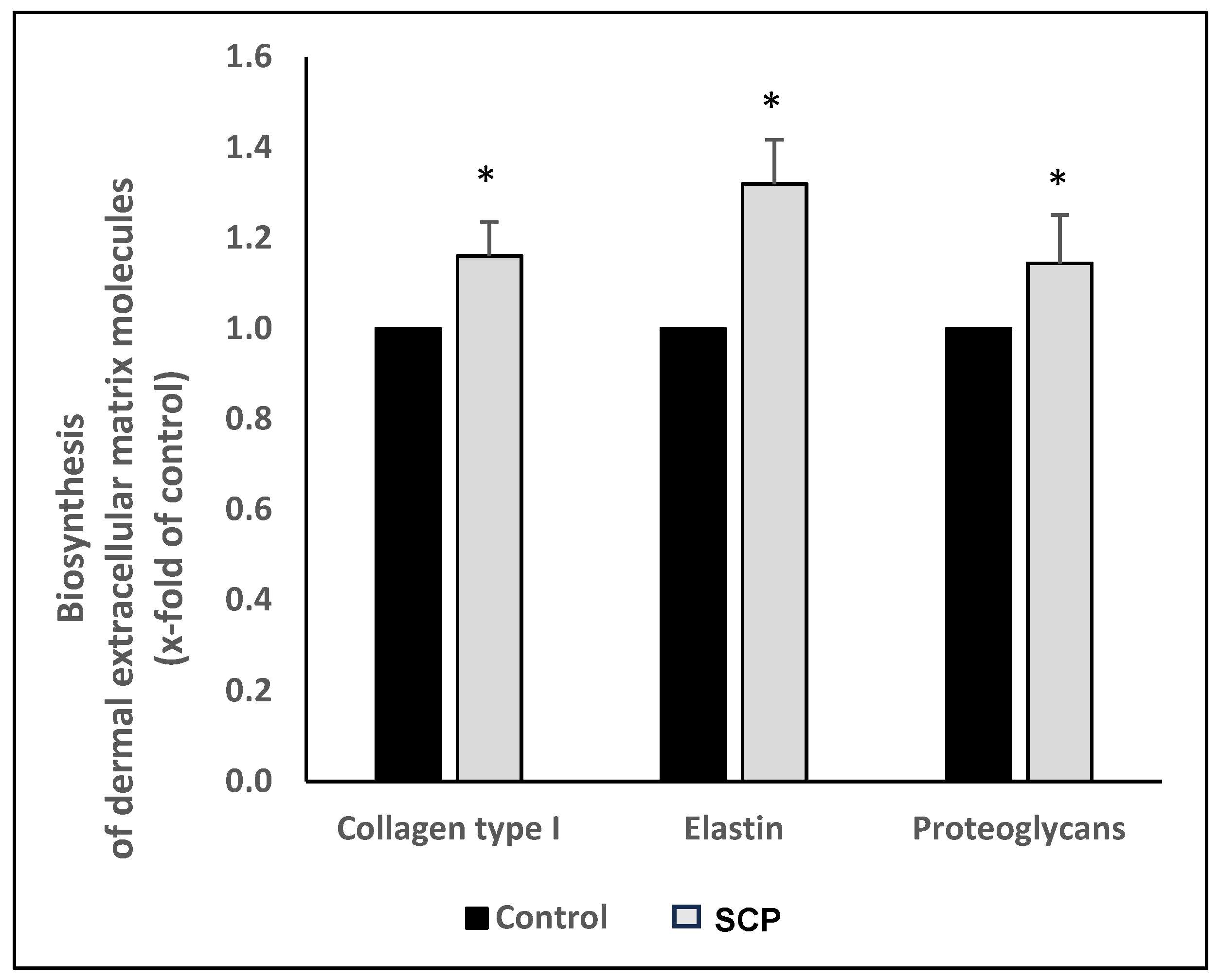
2.4. Measurement of the ECM Macromolecules

Commercially available assay kits were used to quantitatively analyze the extracellular matrix molecules investigated. These methods were used for all of the cell culture studies, including the batch-to-batch analysis.
Newly formed collagen was determined after three weeks of stimulation. Synthesized collagen was isolated using the Sircol assay (tebu-bio, Offenbach, Germany) according to the manufacturer’s instructions. For this purpose, the culture medium was discarded, and the adherent cell layers were digested in 0.1 mg of pepsin solution in 0.5 M acetic acid overnight at 4 °C. The cell suspensions were neutralized by adding 100 µL of acidic neutral reagent, and the synthesized collagen was then separated by adding 200 µL of the isolation and concentration kit solution and vigorously shaking it overnight at 4 °C. After centrifugation (12,000 rpm, 10′) and disposal of the supernatant, the isolated collagen was resuspended in 1 mL of Sircol dye reagent. Following 30 min of shaking and further centrifugation, the collagen pellet was overlaid with 750 µL of cold acid wash reagent. After centrifugation, the supernatant was discarded, and the enriched collagen was dissolved in 250 µL of alkaline solution. From these sample solutions, 200 µL was taken for photometric quantification of the synthesized collagen. The absorbance was measured at 492 nm. The amount was determined from the measured standard collagen solutions.
For the detection of proteoglycans, the Blyscan glycosaminoglycan assay (tebu-bio, Offenbach, Germany) was used. The biosynthesis of the proteoglycans was determined after two weeks of stimulation. Following the manufacturer’s instructions, the cell layers were coated with 1 mL of papain extraction solution after discarding the cell culture medium and incubated for 3 h at 65 °C, with vigorous shaking. The cell suspensions were centrifuged (10,000× g, 10′), and the supernatants were collected. After adding 1 mL of Blyscan dye solution and shaking it (30′), the supernatants were again centrifuged (12,000 rpm, 10′) and discarded. The isolated proteoglycan pellets were resuspended in 500 µL of dissociation solution. A total of 200 µL per sample was subjected to photometric determination at 656 nm and compared to the untreated control experiments.
Elastin synthesis was determined using the Fastin-Elastin assay kit (tebu-bio, Offenbach, Germany). The fibroblasts were stimulated with the test product for 2 weeks or left untreated in the control experiment. After discarding the culture medium, the cell monolayers were dissolved in 250 µL of trypsin. The trypsin solution was removed after centrifugation (3000 rpm, 10′). The cell pellets were dissolved in 100 µL of oxalic acid solution (1 M) and boiled for 1 h in a water bath. After cooling, 300 µL of the elastin precipitation solution was added to precipitate the dissolved elastin within 15 min. After centrifugation (10′), the supernatant was discarded, and the elastin pellet was dissolved in 1 mL of dye solution through shaking for 90 min. Following centrifugation and disposal of the supernatant, the elastin pellet was resuspended in 260 µL of dye dissociation solution. A total of 200 µL of the resolvent was used for the photometric elastin measurements at a wavelength of 492 nm.

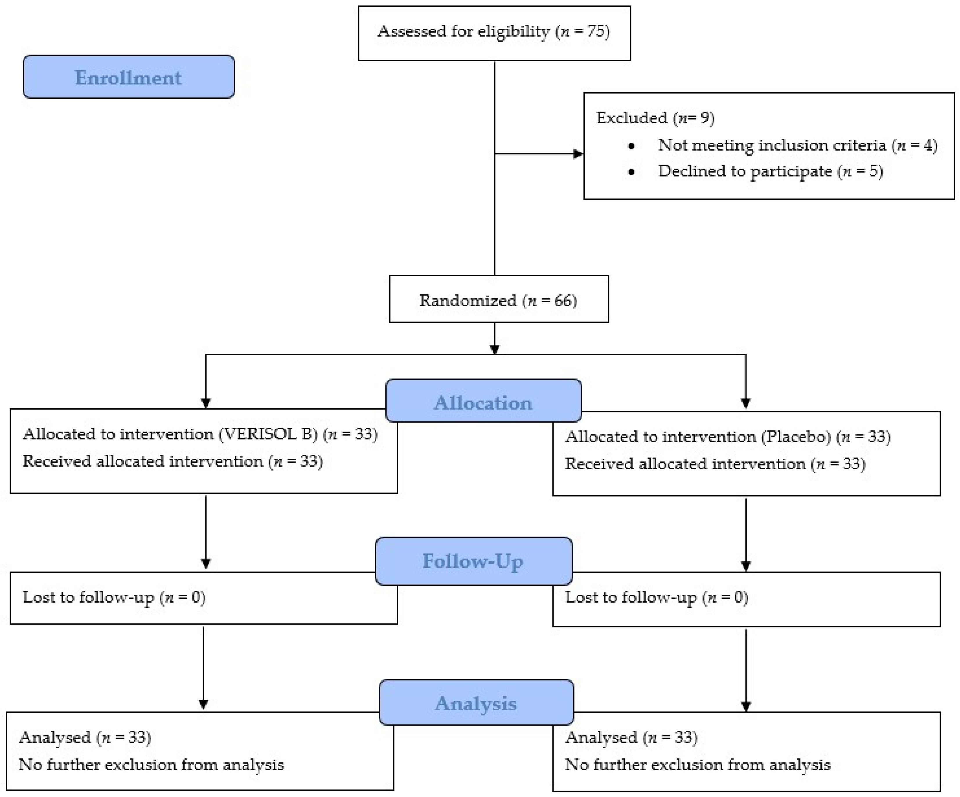
2.5. The Study Design of the Clinical Trial

The trial was a monocentric, double-blind, randomized, placebo-controlled trial (RCT) studying the effects of specific bioactive collagen peptides (SCPs) on various skin parameters and was conducted in accordance with the guidelines for Good Clinical Practice (GCP) and in accordance with the Declaration of Helsinki at the SGS SIT GmbH in Hamburg, Germany. This study’s primary objective was defined as a change in eye wrinkle volume after 8 weeks of treatment. In addition, the effects on the skin’s elasticity and hydration (as secondary objectives) were examined after 4 and 8 weeks.
The examination was approved by the Institutional Review Board of the Ethics Committee of the Hamburg Medical Association (PV5894) and registered in the German Clinical Trials Register (DRKS00036187). All subjects received detailed information listing all relevant individual parameters of the study. After receiving written information, each participant had the opportunity to ask further questions and subsequently signed a consent form. Participants who met the eligibility criteria were randomized (at a 1:1 ratio) into the SCP or placebo group using a web-based random number generator [37].

2.6. The Inclusion and Exclusion Criteria

The inclusion criteria were (1) healthy women aged 35 to 55 (2) of phototypes I to IV (on the Fitzpatrick scale) (3) in a state of good physical and mental health and social well-being (4) who provided personal informed consent to participate in the study, (5) were personally present at the institute on the specified days, and (6) were willing and able to comply with the study rules and the fixed schedule.
The exclusion criteria were any deviation from the above inclusion criteria; acute skin diseases (e.g., atopic eczema, atopic dermatitis, psoriasis); food allergies related to the supplemented products; gastrointestinal diseases or digestive disorders; the use of topical medication on the test sites within 6 weeks prior to study entry, systemic medication containing anti-inflammatory agents or antibiotics within 2 weeks prior to study entry, systemic medication containing corticosteroids and/or antihistamines within 4 weeks prior to study entry, or other systemic medication within 4 weeks prior to study entry; systemic disease in the subject at study entry; pregnancy or lactation; immunological diseases; severe disease or severe diabetes; alcohol and drug abuse within 6 months prior to study entry; participation in other studies involving cosmetic products within 2 weeks before study entry or during the study; participation in a study involving a pharmaceutical preparation and/or the intake of dietary supplements within 4 weeks before study entry; changes in lifestyle or dietary habits during this study besides the use of the test products; treatment with leave-on products or oily or moisturizing skin-cleansing products on the test areas or changes to the usual skin care routine; intense sun or artificial UV exposure (through the use of a solarium) of the test areas within 1 week before study entry or during this study; swimming, sauna use, or intense sports activity within 1 day before the measurements; smoking; a lack of compliance; and an intellectual or mental inability to follow the study instructions.

2.7. Subjects

A total of 66 healthy female subjects with an average age of 46.1 ± 5.6 years were included in this study. All of the participants were randomly assigned into either the treatment group with a daily dose of 2.5 g of the SCPs or the placebo group. In the placebo group, a daily dose of 2.5 g of maltodextrin (Walter GmbH, Olpe, Germany) was supplemented. The samples were packed in individual sachets. Each woman who met the inclusion criteria and was recruited for the trial consumed 2.5 g of a sachet of either the SCPs or the placebo daily over the study period of 8 weeks.
The preparations were taken orally by the subjects at home according to the investigator’s instructions. The powders were to be dissolved in water or another cold liquid, with the exception of milk.
A conditioning period of at least 7 days preceded the start of the oral treatment and data collection. During this time, the test subjects were instructed to refrain from using leave-on products on the test areas and not to change their usual skin care routine. In addition, treatment with dermatological therapeutics on the test areas was prohibited for 6 weeks prior to the start of the trial. During this study, changes in lifestyle or dietary habits, the use of additional nutritional or vitamin supplements, treatment of the test areas with cosmetic and dermatological skin care products, and intensive sun or UV light exposure were also forbidden.

2.8. Product Safety

Collagen peptides are characterized by a very high safety profile. No clinical indications of allergies have been observed to date. No incompatibilities with other diets or medications have ever been described in the medical literature. Collagen peptides were awarded a GRAS status by the American Food and Drug Administration. “GRAS” is an official abbreviation for “Generally Recognized As Safe” [36]. In addition, experimental studies have clearly shown that there is no systemic toxicity [38].
During this study, the tolerability of the SCP treatment was assessed through dermatological examinations and interviews with the participants before, during, and after the study.

2.9. Measurements

The wrinkle area around the left eye (the lateral canthus) was defined as the test site for the eye wrinkle measurements (with 1 test site per body region). Skin elasticity and hydration were measured on the inside of the right forearm. The test areas on the forearms were 5 cm× 5 cm. On each measurement day, the subjects had to expose their uncovered test sites to the climate of the room (21.5 °C; 50% relative humidity) for at least 30 min. Two measurement times were set for the primary objective.
The wrinkle measurements were conducted immediately before the start of the product treatment (X0) and after 8 weeks (X8) of daily product intake. In addition, an interim analysis was performed after 4 weeks (X4). For the secondary objectives, skin elasticity and hydration were examined at X0, X4, and X8.
The compliance of the study participants (dosage and type of intake) and tolerability of the products were assessed after 1, 4, and, again, 6 weeks of administration.
The eye wrinkle volume was measured at the outer corner of the eye (lateral canthus) using the PRIMOS® Compact optical 3-dimensional in vivo measuring device (GF Messtechnik GmbH, Teltow, Germany). Three measurements were performed per test site. The size of the measurement area was 30 mm × 40 mm. Post-baseline measurements were performed using the overlay function. The original images of the reference files at baseline and the corresponding measurement files of the post-baseline measurements were matched using the 3-dimensional matching function for each participant. The height of the images was calculated using the standard procedure with mathematical filters. The eye fold volume (in mm3) of a selected fold was calculated from these height images using the PRIMOS® software. This was carried out for all three images taken per measurement site and at each time. The mean value of the three individual measurements was subsequently calculated for each test site and measurement time point.
Skin elasticity was measured using the Cutometer® MPA 580 (Courage & Khazaka, Cologne, Germany), as described by Segger et al. [39,40]. In brief, the stretching of the skin was recorded in response to negative pressure generated by using a vacuum (350 mbar) over the skin test area. The exposure and non-exposure time was 5 s, with one cycle per measurement. The R5 value (Ur/Ue, instantaneous recovery/elastic deformation) was recorded to analyze the skin’s elasticity. This parameter has been shown to be particularly useful in detecting age-related skin changes [41,42]. The measurements were repeated three times at each test site.
The assessment of the skin’s surface hydration according to electrical capacitance was performed using the Corneometer® CM 825 (Courage & Khazaka), which measures the reactive capacitance of the skin by using the stratum corneum as a dielectric membrane. The measurements are arbitrarily expressed as hydration indices that rise with increasing skin hydration. A total of 15 individual measurements were taken for each application site and the controls.

2.10. The Statistical Analysis

Statistical analyses were performed using SPSS Statistics (IBM SPSS Statistics for Windows, Version 23.0, Armonk, NY, USA: IBM Corp.). Normality was assessed using the one-sample Kolmogorov–Smirnov (KS) test. The hypothesis of a normal distribution was accepted if the KS test yielded p > 0.05. For the cell culture experiments and for the batch-to-batch analysis, the descriptive results were expressed relative to those from the untreated control experiment. For the cell culture experiments, statistical significance was determined based on the KS test results: if normality was confirmed (p > 0.05), a parametric one-sample t-test was applied; otherwise, the 1-sample Wilcoxon signed-rank test was used to detect differences between the SCP treatment and the untreated control. For normally distributed data (proofed using the KS test), a one-way ANOVA with Tukey’s HSD post hoc test was used to compare batches; otherwise, the Kruskal–Wallis test was used with Dunn–Bonferroni correction for pairwise comparisons. In the clinical trial, baseline and intervention-related group differences were analyzed using either the independent t-test (for normally distributed data) or the Mann–Whitney U test (for non-normally distributed data). Differences between the treatment conditions were considered statistically significant if p < 0.05. The data were presented as described in the legends in the figures and tables. The effect size was calculated using Cohen’s d for inter-group differences after an 8-week treatment.

3. Results

3.1. The In Vitro Test

The present in vitro study was designed to investigate the possible impact of bovine-derived SCPs on the biosynthesis of ECM macromolecules for the first time. For these cell culture experiments, primary human dermal fibroblasts were used. In a second experimental approach, different batches of the used product were tested regarding their efficacy.

3.1.1. The Bioactivity of the SCPs

The biosynthesis of the most important molecules in the dermal extracellular matrix was statistically significantly increased (p < 0.05) after the SCP treatment compared to that with the untreated controls (Figure 1).
The amount of collagen type I, elastin, and proteoglycans was statistically significantly increased (p < 0.05) after the SCP supplementation compared to that in the untreated controls (Table 2). The stimulatory effect of SCP treatment on the biosynthesis of ECM molecules was also confirmed at the gene expression level. The mRNA expression levels for type I collagen, elastin, and the proteoglycans decorin and biglycan were also significantly increased (p < 0.05) after the SCP supplementation.

3.1.2. The Batch-to-Batch Analysis of the Test Product

A batch-to-batch analysis of four different SCP batches produced between 2018 and 2024 showed no statistically significant differences between the tested products regarding their efficacy in stimulating matrix biosynthesis. All of the tested batches had a similar pronounced stimulatory effect on synthesizing type I collagen, proteoglycans, and elastin (Table 3).

3.2. The Clinical Trial

The CONSORT (Consolidated Standards of Reporting Trials) flow diagram for this controlled trial showed that 66 Caucasian women between the ages of 35 and 55 were included (Figure 2). The women, with an average age of 46.1 ± 5.6 years (46.7 ± 6.0 for the SCP group; 44.9 ± 5.6 for the placebo), had phototypes I–IV (on the Fitzpatrick scale) and were in generally good condition in terms of their physical and mental health. The participants were randomly assigned into the placebo or SCP group. All measured parameters were compared between the groups to assess the homogeneity of the data at baseline between the SCP and placebo groups. The data showed no statistically significant differences between the two study groups for any of the primary and secondary outcomes at baseline.
The data were evaluated based on the “Per-Protocol” (PP) population. All of the study participants who fulfilled the inclusion criteria and had given informed consent were included after randomization. As all of the recruited women had fulfilled the requirements according to the study protocol and no drop-outs occurred, the PP population was identical to the “intention-to-treat” (ITT) population. The assessment of the tolerability of the product revealed no adverse effects during the evaluation after 1, 4, and 6 weeks. All of the study participants were compliant with the study protocol. There were no discontinuations of the RCT related to product uptake or the study process in general. No inconveniences or adverse reactions were reported.

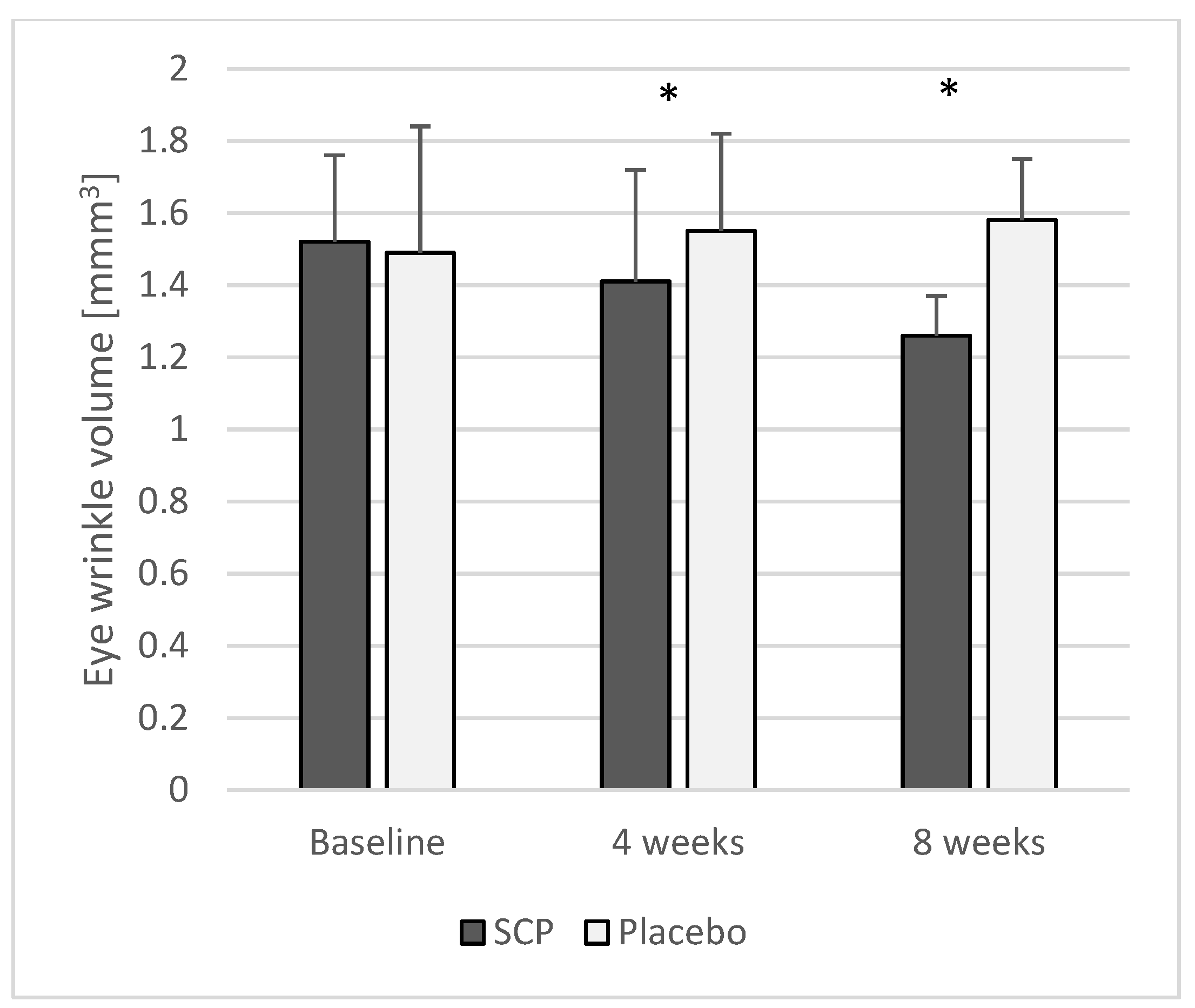
3.2.1. Eye Wrinkle Volume

At baseline, the two treatment groups had no statistically significant difference in their eye wrinkle volume (Table 3). After 4 weeks of the treatment with the SCPs, eye wrinkle volume decreased by approximately 9% (p < 0.05; Cohen’s ≥ 0.8) compared to that in the placebo group (Figure 3, Table 4). At the end of this study, after 8 weeks, eye wrinkle volume was statistically significantly reduced, by 25%, in the participants who had received SCPs compared to that in the placebo group (p < 0.05; Table 4). The analysis of the individual cases revealed a maximum reduction in the eye wrinkle volume of more than 51%.
During the course of this study, there was also a statistically significant (p < 0.05) decrease in the eye wrinkle volume in the SCP group after 4 and 8 weeks.

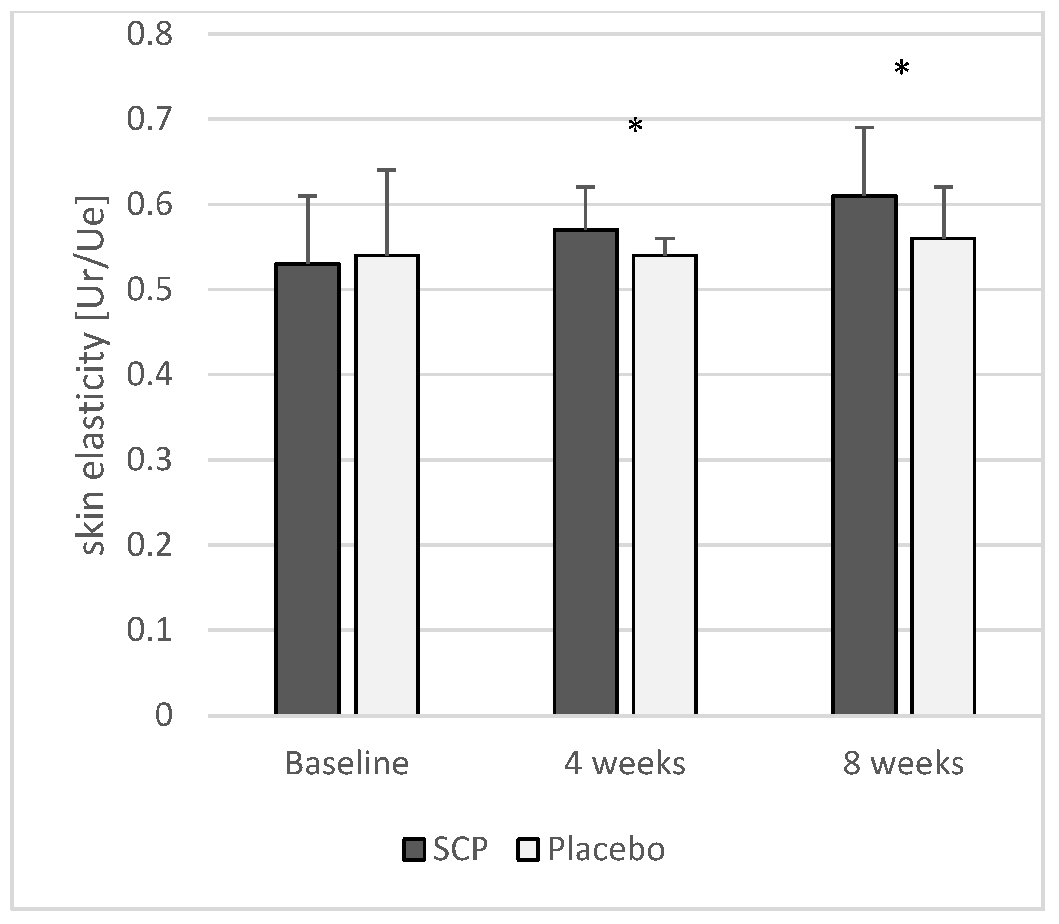
3.2.2. Skin Elasticity

At the beginning of this study, there was no statistically significant difference in skin elasticity between the SCP and placebo groups (Table 3). Compared to that in the placebo, a statistically significant (p < 0.05) improvement in skin elasticity of 6% was observed in the SCP group after 4 weeks of treatment (Figure 4; Table 4). At the end of this study (Figure 4; Table 4), their skin elasticity had increased by 9% compared to that in the placebo group (p < 0.05; Cohen’s d ≥ 0.8).
A statistically significant (p < 0.05) improvement in skin elasticity was observed in the treatment group after 4 and 8 weeks of SCP supplementation. In contrast, no changes in elasticity over time were observed in the placebo group.

3.2.3. Skin Hydration

At the beginning of this study, no statistically significant differences were observed in skin hydration between the SCP and placebo groups (Table 3). After 4 weeks, skin hydration was statistically significantly (p < 0.05) increased in the SCP group compared to that in the control group (Figure 5; Table 4). After 8 weeks, at the end of the study period, skin hydration was significantly (p < 0.05) increased, by 29%, in the treatment group. Compared to that in the control group, the skin hydration in the treatment group was increased by 26% (p < 0.05; Cohen’s d ≥ 0.8) at the end of the investigation (Figure 5; Table 4).
In contrast to the treatment group, no significant changes in hydration were observed in the placebo group during the study period.

4. Discussion

Human skin undergoes a continuous aging process. This chronological or intrinsic aging is characterized by decreased thickness of the skin and a loss of skin elasticity and hydration [9,43,44,45,46,47,48,49]. As the major physiological changes in the skin are known to be located in the dermis, topical products such as creams and lotions may have a limited efficacy and duration. Most of these products do not reach the deeper layer of the skin, explaining their temporary effect on the skin’s appearance.
Over the last decade, orally administered supplements for improving skin health have become increasingly popular. The consumption of products derived from collagen in particular has risen significantly. The effects of such collagen hydrolysates or collagen peptides on skin physiology have been studied by several groups worldwide. Experimental studies have shown that supplementation with specific collagen peptides stimulates the synthesis of the dermal extracellular matrix and provides antioxidant protection [9,50,51,52,53,54,55,56,57,58]. These studies suggest that the intake of collagen peptides can improve the appearance and function of the skin.
Numerous controlled clinical studies have now investigated the effects of orally administered collagen products on various skin parameters [9,10,21,25,32,59,60,61,62]. Mainly, collagen peptides derived from fish, pigs, or cattle are marketed. However, the mechanism of action of collagen peptides is not fully understood to date. It is known that, in addition to the molecular weight distribution of peptides, their specific production process plays a crucial role. The special enzymatic degradation of collagen results in specific collagen peptides that can bind to certain cell receptors, ensuring their high bioactivity. The importance of the production process could help to explain why collagen-derived products differ significantly in their efficacy. A systematic review of orally supplied collagen showed a stimulatory effect on the synthesis of matrix molecules [30]. The products studied differ in the recommended daily dosages, the mean molecular weights of the collagen peptides, and the source of collagen.
To ensure the efficacy of the bovine SCPs used in this clinical trial, the same peptides were tested in in vitro studies on primary human dermal fibroblasts. A batch-to-batch analysis was conducted to assess the reproducibility of the measured effects.
The current cell culture experiments clearly demonstrated a pronounced stimulatory effect of the SCPs on the expression of dermal extracellular matrix macromolecules. After supplementation with the SCPs, a significant increase in the biosynthesis of type I collagen, elastin, and proteoglycans was observed.
The current results are consistent with those of several experimental studies in which human fibroblasts demonstrated a stimulatory effect following collagen peptide supplementation on the extracellular matrix molecules [27,28,54]. Although these studies utilized different collagen peptides from various species, their results consistently showed an increase, primarily in collagen production. In the present study, the stimulatory efficacy of the SCPs was confirmed at both the mRNA and protein levels.
The small differences observed in the efficacy of the different batches confirm the stable production process and constant quality of the SCP samples.
This study investigated the efficacy of 2.5 g/day of SCPs of bovine origin in terms of their effect on skin physiology. The results confirmed a statistically significant decrease in eye wrinkle volume and a significant increase in skin elasticity and hydration after 4 weeks of treatment compared to these values int he placebo. After 8 weeks of SCP supplementation, the effects on the measured skin parameters were even more pronounced.
Skin elasticity is a crucial indicator of skin aging. In a clinical study on post-menopausal women, the decrease in skin elasticity per year was compared with its development in pre-menopausal women [47]. Based on the results, a decrease in skin elasticity of 0.55% per year after the menopause was calculated. It is known that elasticity, along with skin hydration [63,64,65], is mainly influenced by dermal collagen. Marini et al. (2012) described a correlation between skin elasticity and hydration and type I collagen synthesis after oral treatment [66].
In the current study, the skin elasticity was already statistically significantly increased after 4 weeks of treatment compared to that in the placebo (p < 0.05). After 8 weeks of treatment, the skin’s elasticity was increased by 9% compared to that in the placebo. In one participant, a maximum increase in skin elasticity of 51% was observed after 8 weeks of treatment. These results closely resemble the findings of a study that investigated porcine-derived SCPs [32]. Here, an average increase in skin elasticity of 7% was observed. Another clinical study [67] used a combination of collagen peptides with various vitamins. In this study, skin elasticity was improved by 8% after 12 weeks of supplementation compared to that in the placebo. Additionally, the authors reported an increase in skin hydration of 28% in the treatment group and 9% in the placebo group.
In the current clinical trial, a 26% improvement in skin hydration was observed after 8 weeks of the SCP treatment compared to the controls. After a treatment time of only 4 weeks, skin hydration was increased by 18%, indicating that SCP intake seems to have a fast impact on water-binding structures in the dermal tissue. This observed effect was confirmed in in vitro experiments, where a significant increase in proteoglycans was determined. As proteoglycans have a very high water-binding capacity, this might explain the clinically proven results.
Reducing wrinkles, especially in the face, is key to a healthier and younger appearance. Topically applied creams and lotions are often ineffective, as they do not penetrate the deeper layers of the skin [68,69]. Usually, such treatments have only a very short-term effect, if any.
Collagen and elastin are known to be the main components of the dermis responsible for maintaining the skin’s structure, firmness, and elasticity. All of these factors together directly impact wrinkle formation. In particular, the conversion of elastic fibers and the progressive degradation of collagen bundles promote wrinkles.
The current clinical data showed a pronounced reduction in wrinkles in the treatment group within 4 weeks compared to those in the placebo. After 8 weeks, this effect was even more pronounced: wrinkles decreased by an average of 25% after SCP intake compared to those in the untreated controls.
The positive effects on skin moisture and elasticity and the reduction in eye wrinkle volume observed in this study align with comparable clinical studies where similar skin parameters were investigated, although not using bovine-derived collagen peptides. For example, studies performed using a very similar porcine-derived collagen peptide product [25,32] demonstrated comparable positive effects on skin health. In these studies, a daily dose of 2.5 g was also administered. Additionally, a study using fish-derived collagen peptides also showed positive effects [33]. In this study, the daily oral intake of 5 g of the product led to a significant reduction in eye wrinkles, as well as improved skin moisture and elasticity. These data seem to suggest that the clinical effectiveness does not primarily depend on the animal species from which the collagen peptides are derived.

5. Conclusions

The results of the RCT involving healthy women aged 35 to 55 years indicated that a 4-week treatment with bovine-derived SCP led to a statistically significant reduction in eye wrinkle volume, along with an increase in skin elasticity and hydration, compared to these values in the untreated control subjects. After 8 weeks of supplementation, the positive clinical effects were even more pronounced (Cohen’s d ≥ 0.8). Cell culture experiments with human dermal fibroblasts demonstrated that SCP supplementation resulted in a statistically significant increase in collagen, elastin, and proteoglycan biosynthesis compared to that in the controls. Although further studies are needed, it can be speculated that the positive clinical effects on wrinkle volume, skin elasticity, and skin hydration following SCP supplementation observed are due to the stimulatory effect of these bioactive collagen peptides on the synthesis of type I collagen, elastin, and proteoglycans. The analysis of different SCP batches confirmed the product’s stimulatory efficacy and demonstrated its consistent quality. No adverse events were reported in this clinical study, and no side effects were observed. The product was found to be safe and well tolerated. Further studies should continue to investigate its mechanism of action and effects on other ECM macromolecules. Moreover, it would be interesting to investigate whether the sources of collagen peptides derived from different species have an impact on the efficacy of the product further.
In summary, orally administered SCP treatment has been shown to significantly improve the health and appearance of the skin. However, the preclinical and clinical results presented here are specific to the SCPs used in this study and may not necessarily apply to other collagen products.

Author Contributions

Conceptualization, E.P. and S.O.; methodology, E.P. and S.O.; validation, E.P.; formal analysis, S.O.; investigation, E.P.; resources, S.O.; data curation, S.O.; writing—original draft preparation, S.O.; writing—review and editing, E.P., D.Z. and S.O.; visualization, D.Z.; supervision, E.P.; project administration, E.P.; funding acquisition, S.O. All authors have read and agreed to the published version of the manuscript.

Funding

The specific collagen peptides (product name VERISOL B®) were provided by GELITA GmbH, Eberbach Germany. It should be emphasized that the sponsor had no influence on this study’s design, the conduct of this study, or the analysis of the data.

Institutional Review Board Statement

This study was conducted according to the guidelines of the Declaration of Helsinki and approved by the Institutional Review Board of the Ethics Committee of the Hamburg Medical Association (protocol code: PV5894; date of approval: 24 September 2018).

Informed Consent Statement

Informed consent was obtained from all of the subjects involved in this study.

Data Availability Statement

The datasets used and/or analyzed during the current study are available from the corresponding author on reasonable request.

Acknowledgments

We would like to thank all of the participants, as well as the staff of the study center, who supported us with the examination.

Conflicts of Interest

The authors declare no conflicts of interest. S.O. has given scientific lectures that were in part supported by GELITA AG. He is also a coinventor of patents concerning the use of collagen peptides. All of the procedures performed in the studies involving human participants were performed in accordance with the ethical standards of the institutional and/or national research committee and with the 1964 Declaration of Helsinki and its later amendments or comparable ethical standards. The funders had no role in the design of this study; in the collection, analyses, or interpretation of the data; in the writing of the manuscript; or in the decision to publish the results. The authors responsible for the data’s assessment and analysis have neither financial nor competing interests concerning the outcomes of this investigation.

Abbreviations

The following abbreviations are used in this manuscript:
ECMExtracellular Matrix
EDTAEthylenediaminetetraacetic Acid
FCSFetal Calf Serum
GAPDHGlyceraldehyde 3-Phosphate Dehydrogenase
GCPGood Clinical Practice
GRASGenerally Recognized As Safe
ITTIntention-to-Treat
kDKilodalton
KSKolmogorov–Smirnov
PPPer-Protocol
RCTRandomized Controlled Trial
RT-PCRReal-Time Polymerase Chain Reaction
SCPSpecific Collagen Peptide
SPSSStatistical Package for the Social Sciences
TMMelting Temperature
UVUltraviolet

References

  1. Smalls, L.K.; Randall Wickett, R.; Visscher, M.O. Effect of Dermal Thickness, Tissue Composition, and Body Site on Skin Biomechanical Properties. Skin Res. Technol. 2006, 12, 43–49. [Google Scholar] [CrossRef] [PubMed]
  2. Calleja-Agius, J.; Muscat-Baron, Y.; Brincat, M.P. Skin Ageing. Menopause Int. 2007, 13, 60–64. [Google Scholar] [CrossRef]
  3. Takema, Y.; Yorimoto, Y.; Kawai, M.; Imokawa, G. Age-Related Changes in the Elastic Properties and Thickness of Human Facial Skin. Br. J. Dermatol. 1994, 131, 641–648. [Google Scholar] [CrossRef] [PubMed]
  4. Takema, Y.; Yorimoto, Y.; Kawai, M. The Relationship between Age-Related Changes in the Physical Properties and Development of Wrinkles in Human Facial Skin. J. Soc. Cosmet. Chem. 1995, 46, 163–173. [Google Scholar]
  5. Boelsma, E.; Hendriks, H.F.; Roza, L. Nutritional Skin Care: Health Effects of Micronutrients and Fatty Acids. Am. J. Clin. Nutr. 2001, 73, 853–864. [Google Scholar] [CrossRef]
  6. Aguirre-Cruz, G.; León-López, A.; Cruz-Gómez, V.; Jiménez-Alvarado, R.; Aguirre-Álvarez, G. Collagen Hydrolysates for Skin Protection: Oral Administration and Topical Formulation. Antioxidants 2020, 9, 181. [Google Scholar] [CrossRef] [PubMed]
  7. Cho, S. The Role of Functional Foods in Cutaneous Anti-Aging. J. Lifestyle Med. 2014, 4, 8–16. [Google Scholar] [CrossRef]
  8. Czajka, A.; Kania, E.M.; Genovese, L.; Corbo, A.; Merone, G.; Luci, C.; Sibilla, S. Daily Oral Supplementation with Collagen Peptides Combined with Vitamins and Other Bioactive Compounds Improves Skin Elasticity and Has a Beneficial Effect on Joint and General Wellbeing. Nutr. Res. 2018, 57, 97–108. [Google Scholar] [CrossRef]
  9. Liang, J.; Pei, X.; Zhang, Z.; Wang, N.; Wang, J.; Li, Y. The Protective Effects of Long-Term Oral Administration of Marine Collagen Hydrolysate from Chum Salmon on Collagen Matrix Homeostasis in the Chronological Aged Skin of Sprague-Dawley Male Rats. J. Food Sci. 2010, 75, H230–H238. [Google Scholar] [CrossRef]
  10. Lin, P.; Alexander, R.A.; Liang, C.-H.; Liu, C.; Lin, Y.-H.; Lin, Y.-H.; Chan, L.-P.; Kuan, C.-M. Collagen Formula with Djulis for Improvement of Skin Hydration, Brightness, Texture, Crow’s Feet, and Collagen Content: A Double-Blind, Randomized, Placebo-Controlled Trial. J. Cosmet. Dermatol. 2021, 20, 188–194. [Google Scholar] [CrossRef]
  11. Morgado-Carrasco, D.; Gil-Lianes, J.; Jourdain, E.; Piquero-Casals, J. Oral Supplementation and Systemic Drugs for Skin Aging: A Narrative Review. Actas Dermosifiliogr. 2023, 114, 114–124. [Google Scholar] [CrossRef] [PubMed]
  12. Pincemail, J.; Meziane, S. On the Potential Role of the Antioxidant Couple Vitamin E/Selenium Taken by the Oral Route in Skin and Hair Health. Antioxidants 2022, 11, 2270. [Google Scholar] [CrossRef]
  13. Tarshish, E.; Hermoni, K.; Sharoni, Y.; Muizzuddin, N. Effect of Lumenato Oral Supplementation on Plasma Carotenoid Levels and Improvement of Visual and Experiential Skin Attributes. J. Cosmet. Dermatol. 2022, 21, 4042–4052. [Google Scholar] [CrossRef]
  14. Wertz, P.W. Roles of Lipids in the Permeability Barriers of Skin and Oral Mucosa. Int. J. Mol. Sci. 2021, 22, 5229. [Google Scholar] [CrossRef] [PubMed]
  15. Zhang, K.; Zhao, C.; Liu, K.; Feng, R.; Zhao, Y.; Zong, Y.; Du, R. Oral Administration of Deer Bone Collagen Peptide Can Enhance the Skin Hydration Ability and Antioxidant Ability of Aging Mice Induced by D-Gal, and Regulate the Synthesis and Degradation of Collagen. Nutrients 2024, 16, 1548. [Google Scholar] [CrossRef] [PubMed]
  16. Draelos, Z.D. Nutrition and Enhancing Youthful-Appearing Skin. Clin. Dermatol. 2010, 28, 400–408. [Google Scholar] [CrossRef]
  17. Bagheri Miyab, K.; Alipoor, E.; Vaghardoost, R.; Saberi Isfeedvajani, M.; Yaseri, M.; Djafarian, K.; Hosseinzadeh-Attar, M.J. The Effect of a Hydrolyzed Collagen-Based Supplement on Wound Healing in Patients with Burn: A Randomized Double-Blind Pilot Clinical Trial. Burns 2020, 46, 156–163. [Google Scholar] [CrossRef]
  18. Venzin, C.; Jacot, V.; Berdichevsky, A.; Karol, A.A.; Seliktar, D.; von Rechenberg, B.; Nuss, K.M. Biocompatibility of Pegylated Fibrinogen and Its Effect on Healing of Full-Thickness Skin Defects: A Preliminary Study in Rats. J. Biotechnol. Biomater. 2016, 6, online. [Google Scholar] [CrossRef]
  19. Vollmer, D.L.; West, V.A.; Lephart, E.D. Enhancing Skin Health: By Oral Administration of Natural Compounds and Minerals with Implications to the Dermal Microbiome. Int. J. Mol. Sci. 2018, 19, 3059. [Google Scholar] [CrossRef]
  20. Wang, J.; Xu, M.; Liang, R.; Zhao, M.; Zhang, Z.; Li, Y. Oral Administration of Marine Collagen Peptides Prepared from Chum Salmon (Oncorhynchus Keta) Improves Wound Healing Following Cesarean Section in Rats. Food Nutr. Res. 2015, 59, 26411. [Google Scholar] [CrossRef]
  21. Zague, V. A New View Concerning the Effects of Collagen Hydrolysate Intake on Skin Properties. Arch. Dermatol. Res. 2008, 300, 479–483. [Google Scholar] [CrossRef] [PubMed]
  22. Ohara, H.; Matsumoto, H.; Ito, K.; Iwai, K.; Sato, K. Comparison of Quantity and Structures of Hydroxyproline-Containing Peptides in Human Blood after Oral Ingestion of Gelatin Hydrolysates from Different Sources. J. Agric. Food Chem. 2007, 55, 1532–1535. [Google Scholar] [CrossRef]
  23. Iwai, K.; Hasegawa, T.; Taguchi, Y.; Morimatsu, F.; Sato, K.; Nakamura, Y.; Higashi, A.; Kido, Y.; Nakabo, Y.; Ohtsuki, K. Identification of Food-Derived Collagen Peptides in Human Blood after Oral Ingestion of Gelatin Hydrolysates. J. Agric. Food Chem. 2005, 53, 6531–6536. [Google Scholar] [CrossRef]
  24. Oesser, S.; Adam, M.; Babel, W.; Seifert, J. Oral Administration of (14)C Labeled Gelatin Hydrolysate Leads to an Accumulation of Radioactivity in Cartilage of Mice (C57/BL). J. Nutr. 1999, 129, 1891–1895. [Google Scholar] [CrossRef] [PubMed]
  25. Proksch, E.; Schunck, M.; Zague, V.; Segger, D.; Degwert, J.; Oesser, S. Oral Intake of Specific Bioactive Collagen Peptides Reduces Skin Wrinkles and Increases Dermal Matrix Synthesis. Skin Pharmacol. Physiol. 2014, 27, 113–119. [Google Scholar] [CrossRef] [PubMed]
  26. Dumas, M.; Chaudagne, C.; Bonté, F.; Meybeck, A. In Vitro Biosynthesis of Type I and III Collagens by Human Dermal Fibroblasts from Donors of Increasing Age. Mech. Ageing Dev. 1994, 73, 179–187. [Google Scholar] [CrossRef]
  27. Sanchez, A.; Blanco, M.; Correa, B.; Perez-Martin, R.I.; Sotelo, C.G. Effect of Fish Collagen Hydrolysates on Type I Collagen mRNA Levels of Human Dermal Fibroblast Culture. Mar. Drugs 2018, 16, 144. [Google Scholar] [CrossRef]
  28. Zague, V.; do Amaral, J.B.; Rezende Teixeira, P.; de Oliveira Niero, E.L.; Lauand, C.; Machado-Santelli, G.M. Collagen Peptides Modulate the Metabolism of Extracellular Matrix by Human Dermal Fibroblasts Derived from Sun-Protected and Sun-Exposed Body Sites. Cell Biol. Int. 2018, 42, 95–104. [Google Scholar] [CrossRef]
  29. Chai, H.-J.; Li, J.-H.; Huang, H.-N.; Li, T.-L.; Chan, Y.-L.; Shiau, C.-Y.; Wu, C.-J. Effects of Sizes and Conformations of Fish-Scale Collagen Peptides on Facial Skin Qualities and Transdermal Penetration Efficiency. J. Biomed. Biotechnol. 2010, 2010, 757301. [Google Scholar] [CrossRef]
  30. Choi, F.D.; Sung, C.T.; Juhasz, M.L.W.; Mesinkovsk, N.A. Oral Collagen Supplementation: A Systematic Review of Dermatological Applications. J. Drugs Dermatol. 2019, 18, 9–16. [Google Scholar]
  31. Barati, M.; Jabbari, M.; Navekar, R.; Farahmand, F.; Zeinalian, R.; Salehi-Sahlabadi, A.; Abbaszadeh, N.; Mokari-Yamchi, A.; Davoodi, S.H. Collagen Supplementation for Skin Health: A Mechanistic Systematic Review. J. Cosmet. Dermatol. 2020, 19, 2820–2829. [Google Scholar] [CrossRef] [PubMed]
  32. Proksch, E.; Segger, D.; Degwert, J.; Schunck, M.; Zague, V.; Oesser, S. Oral Supplementation of Specific Collagen Peptides Has Beneficial Effects on Human Skin Physiology: A Double-Blind, Placebo-Controlled Study. Skin Pharmacol. Physiol. 2014, 27, 47–55. [Google Scholar] [CrossRef]
  33. Oesser, S.; Schunk, M.; Proksch, E. Positive Effect of Fish-Derived Bioactive Collagen Peptides on Skin Health. Int. J. Nutraceuticals Funct. Foods Nov. Foods 2020, 1, 127–133. [Google Scholar] [CrossRef]
  34. Wang, H. A Review of the Effects of Collagen Treatment in Clinical Studies. Polymers 2021, 13, 3868. [Google Scholar] [CrossRef] [PubMed]
  35. Pu, S.-Y.; Huang, Y.-L.; Pu, C.-M.; Kang, Y.-N.; Hoang, K.D.; Chen, K.-H.; Chen, C. Effects of Oral Collagen for Skin Anti-Aging: A Systematic Review and Meta-Analysis. Nutrients 2023, 15, 2080. [Google Scholar] [CrossRef] [PubMed]
  36. Evaluation of the Health Aspects of Gelatin as a Food Ingredient; Federation of American Societies for Experimental Biology: Bethesda, MD, USA; Life Sciences Research Office, Food and Drug Administration: Washington, DC, USA, 1975.
  37. Urbaniak, G.C.; Plous, S. Research Randomizer (Version 3.0). Available online: https://www.randomizer.org/about/ (accessed on 29 January 2013).
  38. 7 Final Report on the Safety Assessment of Hydrolyzed Collagen. J. Am. Coll. Toxicol. 1985, 4, 199–221. [CrossRef]
  39. Segger, D.; Schönlau, F. Supplementation with Evelle Improves Skin Smoothness and Elasticity in a Double-Blind, Placebo-Controlled Study with 62 Women. J. Dermatol. Treat. 2004, 15, 222–226. [Google Scholar] [CrossRef]
  40. Segger, D.; Matthies, A.; Saldeen, T. Supplementation with Eskimo Skin Care Improves Skin Elasticity in Women. A Pilot Study. J. Dermatol. Treat. 2008, 19, 279–283. [Google Scholar] [CrossRef]
  41. Krueger, N.; Luebberding, S.; Oltmer, M.; Streker, M.; Kerscher, M. Age-Related Changes in Skin Mechanical Properties: A Quantitative Evaluation of 120 Female Subjects. Skin Res. Technol. 2011, 17, 141–148. [Google Scholar] [CrossRef]
  42. Ryu, H.S.; Joo, Y.H.; Kim, S.O.; Park, K.C.; Youn, S.W. Influence of Age and Regional Differences on Skin Elasticity as Measured by the Cutometer. Skin Res. Technol. 2008, 14, 354–358. [Google Scholar] [CrossRef]
  43. Batisse, D.; Bazin, R.; Baldeweck, T.; Querleux, B.; Lévêque, J.-L. Influence of Age on the Wrinkling Capacities of Skin. Skin Res. Technol. 2002, 8, 148–154. [Google Scholar] [CrossRef] [PubMed]
  44. Frei, V.; Perrier, E.; Orly, I.; Huc, A.; Augustin, C.; Damour, O. Activation of Fibroblast Metabolism in a Dermal and Skin Equivalent Model: A Screening Test for Activity of Peptides. Int. J. Cosmet. Sci. 1998, 20, 159–173. [Google Scholar] [CrossRef]
  45. Paul, C.; Maumus-Robert, S.; Mazereeuw-Hautier, J.; Guyen, C.N.; Saudez, X.; Schmitt, A.M. Prevalence and Risk Factors for Xerosis in the Elderly: A Cross-Sectional Epidemiological Study in Primary Care. Dermatology 2011, 223, 260–265. [Google Scholar] [CrossRef] [PubMed]
  46. Nishimori, Y.; Edwards, C.; Pearse, A.; Matsumoto, K.; Kawai, M.; Marks, R. Degenerative Alterations of Dermal Collagen Fiber Bundles in Photodamaged Human Skin and UV-Irradiated Hairless Mouse Skin: Possible Effect on Decreasing Skin Mechanical Properties and Appearance of Wrinkles. J. Investig. Dermatol. 2001, 117, 1458–1463. [Google Scholar] [CrossRef]
  47. Sumino, H.; Ichikawa, S.; Abe, M.; Endo, Y.; Ishikawa, O.; Kurabayashi, M. Effects of Aging, Menopause, and Hormone Replacement Therapy on Forearm Skin Elasticity in Women. J. Am. Geriatr. Soc. 2004, 52, 945–949. [Google Scholar] [CrossRef] [PubMed]
  48. Vuillermoz, B.; Wegrowski, Y.; Contet-Audonneau, J.-L.; Danoux, L.; Pauly, G.; Maquart, F.-X. Influence of Aging on Glycosaminoglycans and Small Leucine-Rich Proteoglycans Production by Skin Fibroblasts. Mol. Cell. Biochem. 2005, 277, 63–72. [Google Scholar] [CrossRef]
  49. Reilly, D.M.; Lozano, J. Skin Collagen through the Lifestages: Importance for Skin Health and Beauty. Plast. Aesthet. Res. 2021, 8, 2. [Google Scholar] [CrossRef]
  50. Zague, V.; de Freitas, V.; da Costa Rosa, M.; de Castro, G.Á.; Jaeger, R.G.; Machado-Santelli, G.M. Collagen Hydrolysate Intake Increases Skin Collagen Expression and Suppresses Matrix Metalloproteinase 2 Activity. J. Med. Food 2011, 14, 618–624. [Google Scholar] [CrossRef]
  51. Matsuda, N.; Koyama, Y.; Hosaka, Y.; Ueda, H.; Watanabe, T.; Araya, T.; Irie, S.; Takehana, K. Effects of Ingestion of Collagen Peptide on Collagen Fibrils and Glycosaminoglycans in the Dermis. J. Nutr. Sci. Vitaminol. 2006, 52, 211–215. [Google Scholar] [CrossRef]
  52. Tanaka, M.; Koyama, Y.; Nomura, Y. Effects of Collagen Peptide Ingestion on UV-B-Induced Skin Damage. Biosci. Biotechnol. Biochem. 2009, 73, 930–932. [Google Scholar] [CrossRef]
  53. Zhang, Z.; Wang, J.; Ding, Y.; Dai, X.; Li, Y. Oral Administration of Marine Collagen Peptides from Chum Salmon Skin Enhances Cutaneous Wound Healing and Angiogenesis in Rats. J. Sci. Food Agric. 2011, 91, 2173–2179. [Google Scholar] [CrossRef]
  54. Lin, P.; Hua, N.; Hsu, Y.-C.; Kan, K.-W.; Chen, J.-H.; Lin, Y.-H.; Lin, Y.-H.; Kuan, C.-M. Oral Collagen Drink for Antiaging: Antioxidation, Facilitation of the Increase of Collagen Synthesis, and Improvement of Protein Folding and DNA Repair in Human Skin Fibroblasts. Oxid. Med. Cell. Longev. 2020, 2020, 8031795. [Google Scholar] [CrossRef] [PubMed]
  55. Prokopová, A.; Pavlačková, J.; Mokrejš, P.; Gál, R. Collagen Hydrolysate Prepared from Chicken By-Product as a Functional Polymer in Cosmetic Formulation. Molecules 2021, 26, 2021. [Google Scholar] [CrossRef] [PubMed]
  56. Brandao-Rangel, M.A.R.; Oliveira, C.R.; da Silva Olímpio, F.R.; Aimbire, F.; Mateus-Silva, J.R.; Chaluppe, F.A.; Vieira, R.P. Hydrolyzed Collagen Induces an Anti-Inflammatory Response That Induces Proliferation of Skin Fibroblast and Keratinocytes. Nutrients 2022, 14, 4975. [Google Scholar] [CrossRef] [PubMed]
  57. Lee, M.; Kim, D.; Park, S.-H.; Jung, J.; Cho, W.; Yu, A.R.; Lee, J. Fish Collagen Peptide (Naticol®) Protects the Skin from Dryness, Wrinkle Formation, and Melanogenesis Both In Vitro and In Vivo. Prev. Nutr. Food Sci. 2022, 27, 423–435. [Google Scholar] [CrossRef]
  58. Wauquier, F.; Boutin-Wittrant, L.; Bouvret, E.; Le Faouder, J.; Roux, V.; Macian, N.; Pickering, G.; Wittrant, Y. Benefits of Circulating Human Metabolites from Fish Cartilage Hydrolysate on Primary Human Dermal Fibroblasts, an Ex Vivo Clinical Investigation for Skin Health Applications. Nutrients 2022, 14, 5027. [Google Scholar] [CrossRef]
  59. Bauza, E.; Oberto, G.; Berghi, A.; Dal, C.F.; Domloge, N. Collagen-like Peptide Exhibits a Remarkable Antiwrinkle Effect on the Skin When Topically Applied: In Vivo Study. Int. J. Tissue React. 2004, 26, 105–111. [Google Scholar]
  60. Maia Campos, P.M.B.G.; Melo, M.O.; Siqueira César, F.C. Topical Application and Oral Supplementation of Peptides in the Improvement of Skin Viscoelasticity and Density. J. Cosmet. Dermatol. 2019, 18, 1693–1699. [Google Scholar] [CrossRef]
  61. Sangsuwan, W.; Asawanonda, P. Four-Weeks Daily Intake of Oral Collagen Hydrolysate Results in Improved Skin Elasticity, Especially in Sun-Exposed Areas: A Randomized, Double-Blind, Placebo-Controlled Trial. J. Dermatol. Treat. 2021, 32, 991–996. [Google Scholar] [CrossRef]
  62. Evans, M.; Lewis, E.D.; Zakaria, N.; Pelipyagina, T.; Guthrie, N. A Randomized, Triple-Blind, Placebo-Controlled, Parallel Study to Evaluate the Efficacy of a Freshwater Marine Collagen on Skin Wrinkles and Elasticity. J. Cosmet. Dermatol. 2021, 20, 825–834. [Google Scholar] [CrossRef]
  63. Varani, J.; Warner, R.L.; Gharaee-Kermani, M.; Phan, S.H.; Kang, S.; Chung, J.H.; Wang, Z.Q.; Datta, S.C.; Fisher, G.J.; Voorhees, J.J. Vitamin A Antagonizes Decreased Cell Growth and Elevated Collagen-Degrading Matrix Metalloproteinases and Stimulates Collagen Accumulation in Naturally Aged Human Skin. J. Investig. Dermatol. 2000, 114, 480–486. [Google Scholar] [CrossRef] [PubMed]
  64. Fisher, G.J.; Wang, Z.Q.; Datta, S.C.; Varani, J.; Kang, S.; Voorhees, J.J. Pathophysiology of Premature Skin Aging Induced by Ultraviolet Light. N. Engl. J. Med. 1997, 337, 1419–1428. [Google Scholar] [CrossRef] [PubMed]
  65. Schwartz, S.R.; Hammon, K.A.; Gafner, A.; Dahl, A.; Guttman, N.; Fong, M.; Schauss, A.G. Novel Hydrolyzed Chicken Sternal Cartilage Extract Improves Facial Epidermis and Connective Tissue in Healthy Adult Females: A Randomized, Double-Blind, Placebo-Controlled Trial. Altern. Ther. Health Med. 2019, 25, 12–29. [Google Scholar]
  66. Marini, A.; Grether-Beck, S.; Jaenicke, T.; Weber, M.; Burki, C.; Formann, P.; Brenden, H.; Schönlau, F.; Krutmann, J. Pycnogenol® Effects on Skin Elasticity and Hydration Coincide with Increased Gene Expressions of Collagen Type I and Hyaluronic Acid Synthase in Women. Skin Pharmacol. Physiol. 2012, 25, 86–92. [Google Scholar] [CrossRef]
  67. Bolke, L.; Schlippe, G.; Gerß, J.; Voss, W. A Collagen Supplement Improves Skin Hydration, Elasticity, Roughness, and Density: Results of a Randomized, Placebo-Controlled, Blind Study. Nutrients 2019, 11, 2494. [Google Scholar] [CrossRef] [PubMed]
  68. Lodén, M. Barrier Recovery and Influence of Irritant Stimuli in Skin Treated with a Moisturizing Cream. Contact Dermat. 1997, 36, 256–260. [Google Scholar] [CrossRef]
  69. Draelos, Z.D. Active Agents in Common Skin Care Products. Plast. Reconstr. Surg. 2010, 125, 719–724. [Google Scholar] [CrossRef]
Figure 1. The stimulation of collagen type I, elastin, and proteoglycan biosynthesis after the SCP treatment compared to that in the untreated controls. The data represent the mean ± SD for n = 12; * p < 0.05.
Cosmetics 12 00079 g001
Figure 2. Flow chart of subject recruitment, randomization, and follow-up. VERISOL B (SCPs).
Cosmetics 12 00079 g002
Figure 3. Statistically significant reductions in eye wrinkle volume after 4 and 8 weeks of treatment with SCPs compared to that in untreated controls. Data represent mean ± SD; * p < 0.05.
Cosmetics 12 00079 g003
Figure 4. Statistically significant increase in skin elasticity after 4 and 8 weeks of treatment with SCPs compared to that in untreated controls. Data represent mean ± SD; * p < 0.05.
Cosmetics 12 00079 g004
Figure 5. Statistically significant increase in skin hydration after 4 and 8 weeks of treatment with SCPs compared to that in untreated controls. Data represent mean ± SD; * p < 0.05.
Cosmetics 12 00079 g005
Table 1. Primer sequences used for RT-PCR analysis.
GeneForward Sequences (5′-3′)Reverse Sequences (3′-5′)Annealing (°C)Accession
GAPDHGCTCTCTGCTCCTCCTGTTCACTCCGACCTTCACCTTCC63.0NG_007073.2
Collagen type IAATGGTGCTCCTGGTATTGCACCAGGTTCACCGCTGTTAC59.0NM_000088
DecorinTGATTTGGGTCTGGACAAAGTGCCCAGTTCTATGACAATC60.0AF_491944.2
BiglycanCCTCCAGGTGGTCTATCTGCCATCAGGATGTGTGGCTGTG58.0AH_002674.2
ElastinAAGGTGGCTGCCAAAGCACTCCTCCAAGTGGGAACTG60.0NM_00501
Table 2. The stimulatory effect on the mRNA expression of different ECM macromolecules after a 24 h treatment with SCP.
mRNA Expressionp-Value
Collagen type I2.06 ± 0.17<0.05
Elastin1.17 ± 0.12<0.05
Decorin1.15 ± 0.21<0.05
Biglycan1.30 ± 0.13<0.05
The data represent the mean ± SD for n = 12 compared to the untreated controls.
Table 3. The consistent stimulatory effect of the different bovine-derived SCP batches on the synthesis of ECM molecules in the human dermal fibroblasts.
Batch No.1234p-Value
Collagen type I1.22 ± 0.091.19 ± 0.061.20 ± 0.151.19 ± 0.09n.s.
Proteoglycans1.19 ± 0.091.17 ± 0.091.18 ± 0.091.22 ± 0.06n.s.
Elastin1.26 ± 0.141.34 ± 0.141.34 ± 0.301.31 ± 0.37n.s.
The different batches were produced between 2018 and 2024 by GELITA. The data represent the mean ± SD for n = 12; n.s. = not statistically significant.
Table 4. Statistically significant improvement in various skin parameters in healthy women after SCP supplementation for 8 weeks.
Groupn X ¯ 0 ± SD X ¯ 4 ± SDp-Value *
(After 4 Weeks)
X ¯ 8 ± SDp-Value *
(After 8 Weeks)
Eye wrinkle volume (mm3)SCPs
Placebo
33
33
1.52 ± 0.24
1.49 ± 0.35
1.41 ± 0.31
1.55 ± 0.27
<0.051.26 ± 0.11
1.58 ± 0.17
<0.05
Skin elasticity
(Ur/Ue)
SCPs
Placebo
33
33
0.53 ± 0.08
0.54 ± 0.10
0.57 ± 0.05
0.54 ± 0.02
<0.050.61 ± 0.08
0.56 ± 0.06
<0.05
Skin hydration (AU)SCPs
Placebo
33
33
38 ± 3.9
37 ± 4.4
46 ± 2.9
39 ± 4.7
<0.0549 ± 4.2
39 ± 5.3
<0.05
The data represent the mean ± SD for 33 study participants. * Statistical significance of group comparison.
Disclaimer/Publisher’s Note: The statements, opinions and data contained in all publications are solely those of the individual author(s) and contributor(s) and not of MDPI and/or the editor(s). MDPI and/or the editor(s) disclaim responsibility for any injury to people or property resulting from any ideas, methods, instructions or products referred to in the content.

