Article

Design and Characterisation of Personal Hygiene Gels Containing a Gypsophila Trichotoma Extract and Xanthium Strumarium Essential Oil

by
Aleksandar Shkondrov
1,*,
Denitsa Momekova
2,
Maya Margaritova Zaharieva
3,
Hristo Najdenski
3,
Ekaterina Kozuharova
1 and
Ilina Krasteva
1
1
Department of Pharmacognosy, Faculty of Pharmacy, Medical University of Sofia, 2 Dunav st., 1000 Sofia, Bulgaria
2
Department of Pharmaceutical Technology and Biopharmaceutics, Faculty of Pharmacy, Medical University of Sofia, 2 Dunav st., 1000 Sofia, Bulgaria
3
The Stephan Angeloff Institute of Microbiology, Bulgarian Academy of Sciences, 26 Acad. G. Bontchev st., 1113 Sofia, Bulgaria
*
Author to whom correspondence should be addressed.
Cosmetics 2025, 12(2), 65; https://doi.org/10.3390/cosmetics12020065
Submission received: 18 February 2025 / Revised: 26 March 2025 / Accepted: 28 March 2025 / Published: 31 March 2025

Abstract

:
The aim was to develop a series of handwashing gels containing a standardised extract of the aerial part of Gypsophila trichotoma as well as an essential oil from Xanthium strumarium fruits. The saponins present in the extract are a suitable alternative to synthetic sulphate surfactants and have better skin tolerance, and with the addition of essential oil, a higher antibacterial activity of the gels thus developed is achieved. The elaborated gels were characterised by good spreadability and a pH close to the physiological for healthy skin (pH 5.5), which is a prerequisite for improved skin tolerability. The dynamic rheological studies showed that the extract fraction increase was not associated with gel viscosity change, and it was only a function of the gelling agent concentration. The gels’ foam-forming ability increased with the increasing extract concentrations. The comparative evaluation of the antibacterial activity of elaborated gels vs. plain gel based on coco glucoside against a panel of selected pathogens demonstrated that the newly developed formulations have superior antibacterial effects. Based on the good technological characteristics and the results of antimicrobial testing of the washing gels thus developed, it can be concluded that they are promising candidates as personal hygiene products.

1. Introduction

According to WHO, infectious diseases are one of the leading causes of mortality worldwide, with the most common transmission of infection occurring through contaminated hands [1]. In this regard, the development of effective personal hygiene products is of paramount importance. On the other hand, the frequent use of hygiene products based on synthetic anionic surfactants has been shown to disrupt the skin barrier function causing skin irritation and sensitivity [2]. Another significant disadvantage of synthetic surfactants as products manufactured from raw petroleum material is their poor biodegradability, raising concerns about environmental and water pollution [3]. All this necessitates the search for alternative agents with optimal biocompatibility, good skin tolerance and biodegradability. Plant-derived products offer a good alternative to synthetic ones. In recent years, one of the most widely studied active substances of plant origin in the cosmetic industry is saponins [4]. Saponins are secondary plant metabolites with a triterpene or steroid structure with pleiotropic pharmacological effects [5]. Triterpene saponins are those that find application in cosmetic products due to their pronounced antioxidant, anti-inflammatory and antibacterial properties [6]. In addition to being active substances, they are attracting increasing interest as excipients in cosmetic products as natural surfactants, emulsifiers and washing agents with excellent biodegradability and biocompatibility [7].
Gypsophila trichotoma Wend. (Caryophyllaceae) is a perennial plant native to Central and South-western Asia, and South-eastern Europe. The plant is listed in the Bulgarian red Data Book [8] because of its rather restricted distribution on the Black Sea coast of Bulgaria, although it is found lately in new locations [9]. Additionally, the ex-situ experiments reveal promising results for introduction into cultivation [10,11]. G. trichotoma accumulates a high percentage of triterpenoid saponins, mainly glycosides of gypsogenin and gypsogenic acid. Extracts from the plant have proven anti-inflammatory, cytotoxic, antioxidant and immunomodulatory effects [12]. Other species of the genus are used in industry as a source for the production of saponins with emulsifying and foaming effects, also used in the food industry [13]. Studies have utilised extraction with methanol of the roots and aerial parts of the species in order to examine their activity or to isolate the compounds contained therein [12]. The roots and the aerial parts of the species contain saponins with gypsogenin and gypsogenic acid as the aglycone. These saponins are substituted at C-3 with a trisaccharide and at C-28 with an oligosaccharide through a fucose residue. The oligosaccharide attached to C-28 may also be substituted by acetyl and/or sulphate groups [14]. The sterols identified in the plant are stigmast-7-en-3-ol, spinasterol (stigmasta-7,22-dien-3-ol), ergost-7-en-3-ol, and β-sitosterol. Additionally, 22,23-dihydrospinasterone has been detected. Flavonoid glycosides such as vitexin, orientin, homoorientin, and hyperoside have been proven in the plant. In addition, the flavone saponarin has been isolated from its aerial parts [15]. Saponarin exhibited hepatoprotective properties and protected against oxidative stress-induced liver damage in animal models. As previously observed, the extracts from the plant’s aerial parts are considered safe and non-toxic [16]. Gypsogenic acid and gypsogenin, found in the plant, had demonstrated cytotoxic effects against various human tumour cell lines, including leukemic and bladder carcinoma cells [17]. These findings highlight the potential of G. trichotoma extracts as a source of bioactive compounds with possible applications. Nevertheless, there are no studies of the extracts from this species as topical or cosmetic applications.
Xanthium strumarium L. (Asteraceae) is a coarse, erect, branching, annual plant reproducing solely by seed [18]. This species tolerates a wide variety of soil types and textures and a soil pH range of 5.2 to 8.0, as well as frequent flooding and saline conditions [19]. It appears in cultivated fields, along beaches, coastal dunes, watercourses, railway embankments, roadsides, field edges, as well as waste places. Since X. strumarium is the most frequently recorded plant in the field borders between the crop land and adjacent territories in Bulgaria’s agricultural areas [20], it is an inexpensive source of bioactive compounds [21]. It has various medicinal applications like a febrifuge drug used by native North Americans [22], or as an agent to treat chronic malaria, leucorrhoea, urinary diseases, small pox, strumous disease, dysentery, insomnia, fever, headache and leprosy in India and Pakistan [23,24,25,26,27,28,29,30]. There are reports concerning its traditional use to cure skin disease, eczema and leukoderma in these regions [26,27], as well as boils and pimples in Nepal [31,32]. X. strumarium has been studied for the composition and biological activities of its essential oil, which, like all essential oils, is produced by steam distillation [33]. The fruit essential oil contains various terpenes, including sesquiterpenes like xanthatin, demonstrating anti-inflammatory, anti-tumour, antimicrobial, and anti-parasitic properties. The essential oil exhibited significant antimicrobial effects, particularly against Staphylococcus aureus. It also showed potent antifungal activity against strains such as Candida albicans, Aspergillus niger, and Fusarium solani [34]. Research indicates that the essential oil possessed insecticidal properties, effectively deterring feeding and inhibiting growth in larvae, with low ovicidal and insecticidal concentrations [35]. While essential oil demonstrates various bioactive properties, specific safety data, including potential toxicity, side effects, and recommended usage guidelines, are limited. Traditional use has reported symptoms such as vomiting, tremors, weak pulse, loss of appetite, and convulsions at high oil doses but only when ingested. Additionally, compounds like xanthatin have shown hepatotoxic effects in animal studies only when given orally [36]. X. strumarium essential oil exhibits promising antimicrobial, antifungal, and insecticidal activities, suggesting potential applications in various fields. In addition, the antibacterial activity of X. strumarium stem and root ethanol extracts is experimentally confirmed. The plant can be collected in large quantities and it is promising for the industrial production of essential oil, proving antiseptic, wound healing, antifungal, antibacterial, epithelising and repellent effects [37]. The X. strumarium essential oil inhibits Gram-positive and Gram-negative bacteria growth [38]. However, due to limited safety data, caution is advisable when considering its use, especially at higher concentrations [39]. According to official regulations, the advisable concentration in topical preparations should be less than 1.5% [40].
The aim of the study was to develop a series of handwashing gels containing a standardised extract of the aerial part of Gypsophila trichotoma as well as an essential oil from Xanthium strumarium fruits.

2. Materials and Methods

2.1. Solvents, Chemicals and Reagents

Methanol, anhydrous Na2SO4, H2SO4, trifluoroacetic acid (TFA) and UHPLC grade solvents (MeCN and H2O) were received from Merck (Darmstadt, Germany). The reference substances gypsogenic acid CRS and gypsogenin CRS were purchased from LGC Standards (Teddington, Middlesex, UK). n-Hexane (GC-MS grade) was from Fisher Scientific (Waltham, MA, USA). Xanthan gum (obtained from Xanthomonas campestris, CAS № 11138-66-2) was purchased from Sigma-Aldrich (St. Louis, MO, USA), and coco glucoside (ECOCERT certified) and glycerine were supplied by Zoya Ltd. (Sofia, Bulgaria).

2.2. Plant Material and Extraction

Aerial parts of Gypsophila tricotoma were gathered from the experimental field of the University of Forestry, Sofia, Bulgaria, in July 2022. The plant identity was confirmed by one of the authors (I.K.). A voucher specimen was deposited at the Herbarium of the Faculty of Pharmacy, Medical University of Sofia (#FF-202/2022). The plant material was dried at room temperature, pulverised, and sieved (3 mm). The powdered substance (800 g) was defatted with dichloromethane (4 × 3 L) by percolation to remove the lipophilic constituents. Then, the defatted plant material was aired in the fume cupboard to remove residual solvent. Afterwards, it was percolated exhaustively with 80% MeOH (7 × 3 L). The methanol extracts were filtered, combined, and the MeOH evaporated under reduced pressure. The viscous extract was lyophilised (−110 °C, 0.125 mbar, 52 h) and named EGT.
Fruits of Xanthium strumarium were gathered in 2022 in Sofia, Bulgaria. The plant species was identified by one of the authors (E.K.). A voucher specimen is currently kept in the Herbarium of the Faculty of Pharmacy at the Medical University of Sofia (#FF-203/2022). The fresh fruits (35 g) were purified from contaminants, crushed, and then immediately distilled in a Clevenger-type apparatus (200 mL H2O) for 6 h. The essential oil (EOXS, 0.2 mL) was dried with anhydrous Na2SO4, and kept at −20 °C until analysis.

2.3. Methods

2.3.1. Phytochemical Analysis

UHPLC Analysis of Saponins in EGT

The EGT (200 mg) was dissolved in 10 mL of 50% ethanol and then hydrolysed with a mixture of 10 mL of 70% H2SO4 and 2 mL of TFA under a reflux condenser for 8 h. The hydrolysate was dried in vacuo, dissolved in 5 mL of 50% MeOH, and loaded onto an SPE cartridge (Strata™, RP C18-e, 600 mg, Phenomenex, CA, USA). The cartridge was washed with 5% MeOH (3 × 6 mL), and then the non-polar compounds from the hydrolysate were eluted with 100% MeOH (3 × 6 mL). The eluate was dried on a rotary evaporator, dissolved in MeOH, and transferred in a 10.0 mL volumetric flask, made up with the same solvent. An aliquot (2 mL) was filtered through a 0.22 μm PVDF membrane and put in an autosampler vial.
For the study of recovery, model solutions of gypsogenic acid CRS and gypsogenin CRS (5 mg/mL) were treated by the same procedure with acids and SPE. The reference substances had more than 95% purity (HPLC). The standard solutions for performing calibration were made in MeOH: 0.6280, 1.364, and 2.7280 mg/mL (gypsogenic acid) and 0.4895, 0.9790, and 1.9580 mg/mL (gypsogenin).
The UHPLC analysis of the hydrolysed extract was performed on a Vanquish™ system (Thermo Fisher Scientific, Bremen, Germany), consisting of a Split Sampler HT, a Binary Pump H, a Column Compartment H, and a Diode Array Detector HL. The software Chromeleon™ (v. 7.3.2, Thermo Fisher Scientific) was used to collect data, construct calibration curves, and perform quantitation.
The chromatographic column was Symmetry® C18 (Waters, Drinagh, Ireland), 4.6 × 75 mm, 3.5 μm. The injection volume was 2 μL. The temperature was 30 °C, and the flow rate was 0.8 mL/min. The mobile phases were (A) 0.5% TFA in H2O and (B) MeCN, pre-filtered through 0.22 μm PVDF membranes. The elution was performed as follows: initial 10% B, to 3 min 10% B (isocratic); to 4 min to 30% B (gradient); to 5 min 30% B (isocratic); and to 22 min to 95% B (gradient). The analytical wavelength was 210 nm [41]. Each injection was administered in triplicate, and the mean value was calculated.

GC-MS Analysis of EOXS

Immediately before the analysis, EOXS was diluted 1:9000 with n-hexane. GC-MS was performed with an Exactive™ Orbitrap™ GC-MS (Thermo Fisher Scientific, Bremen, Germany) system operating at 70 eV, ion source temperature 240 °C, transfer capillary temperature 250 °C, with split injection (1 μL, 20:1 ratio) at 230 °C injector temperature. A Trace Gold TG-5SilMS GC column (30 m × 0.25 mm × 0.25 µm, Thermo Scientific, Germany) was used. The temperature program was as described previously [33]. EI ionisation mode and full MS-SIM scan were used (resolution 600, AGC target ×106, max. IT 200 ms, and a scan range of m/z 50–450). Data collection, peak processing, and compound identification were performed with Xcalibur software (v. 4.2.28.14, Thermo Scientific, Germany). RI and the relative percentage of constituents were calculated by the same software. Peak processing, and acceptance/identification criteria, based on two databases (NIST and Wiley Registry), were as described in [33]

2.3.2. Preparation of EGT Test Solutions

The solutions were prepared by dissolving 10, 20 or 30 g of the EGT in up to 100 g of deionised water, corresponding to 0.4, 0.8, and 1.2% (w/w) saponin concentration, respectively.

2.3.3. Preparation of Liquid Handwashing Gels

A series of handwashing gels were prepared by dissolving their components in deionised water. The gel composition is shown in Table 1.
In brief, the respective quantity of EGT was dissolved in deionised water, followed by the dissolving of the co-surfactant. In an individual glass, xanthan gum was mixed with glycerine to avoid the formation of aggregates, and then this mixture was added to the solution. The essential oil was last added, and the final mixtures were homogenised by gentle stirring to avoid bubble formation. The quantity of the essential oil (EOXS) used for each gel follows the allowed dermal limits for rinse-off products (category 9) [42]. They are not more than 2.3 to 2.9% for sesquiterpenes, which are the main ingredient in the EOXS, according to GC-MS results (see Section 3).

2.3.4. pH Determination

pH values of EGT aqueous solutions at varying (10, 20, or 30%) concentrations or 10% solutions of the corresponding liquid washing gels were evaluated using calibrated in standard buffered solutions (pH 4, pH 7.4 and pH 10) Jenway 3520 pH-meter (Bibby Scientific Ltd., Staffordshire, United Kingdom). Before measurements, the pH electrode was rinsed with distilled water, dried, and immersed into the test solutions, and the corresponding pH values were recorded. For each test solution, the pH value is given as a mean of three measurements (±SD, n = 3) at a temperature of 25 ± 1 °C.

2.3.5. Foam-Forming Ability and Foam Stability

The foam formation ability and foam stability of EGT solution and their corresponding liquid washing gels were investigated using the cylinder shake method as reported previously with minor modifications [43]. Shortly, 15 mL of the tested EGT solutions or 15 mL of 10% solution of prepared gels, dissolved in deionised water, were transferred in 50 mL graduated cylinders and manually “bottom to top” shaken vigorously 30 times. In order to reproduce test conditions, thus the comparability of the results obtained, all test shakes were conducted by the same person. The foam height (cm) was measured immediately after shaking and 5 min afterwards. The foam stability was expressed as R5 value, calculated by Equation (1).
R 5 = h 5 h 0 × 100
where ho—foam height immediately after shaking; h5—foam height after 5 min at rest.
For each formulation, the experiment was conducted in triplicate, and the results were presented as ±SD of three independent experiments.

2.3.6. Cleaning Capacity
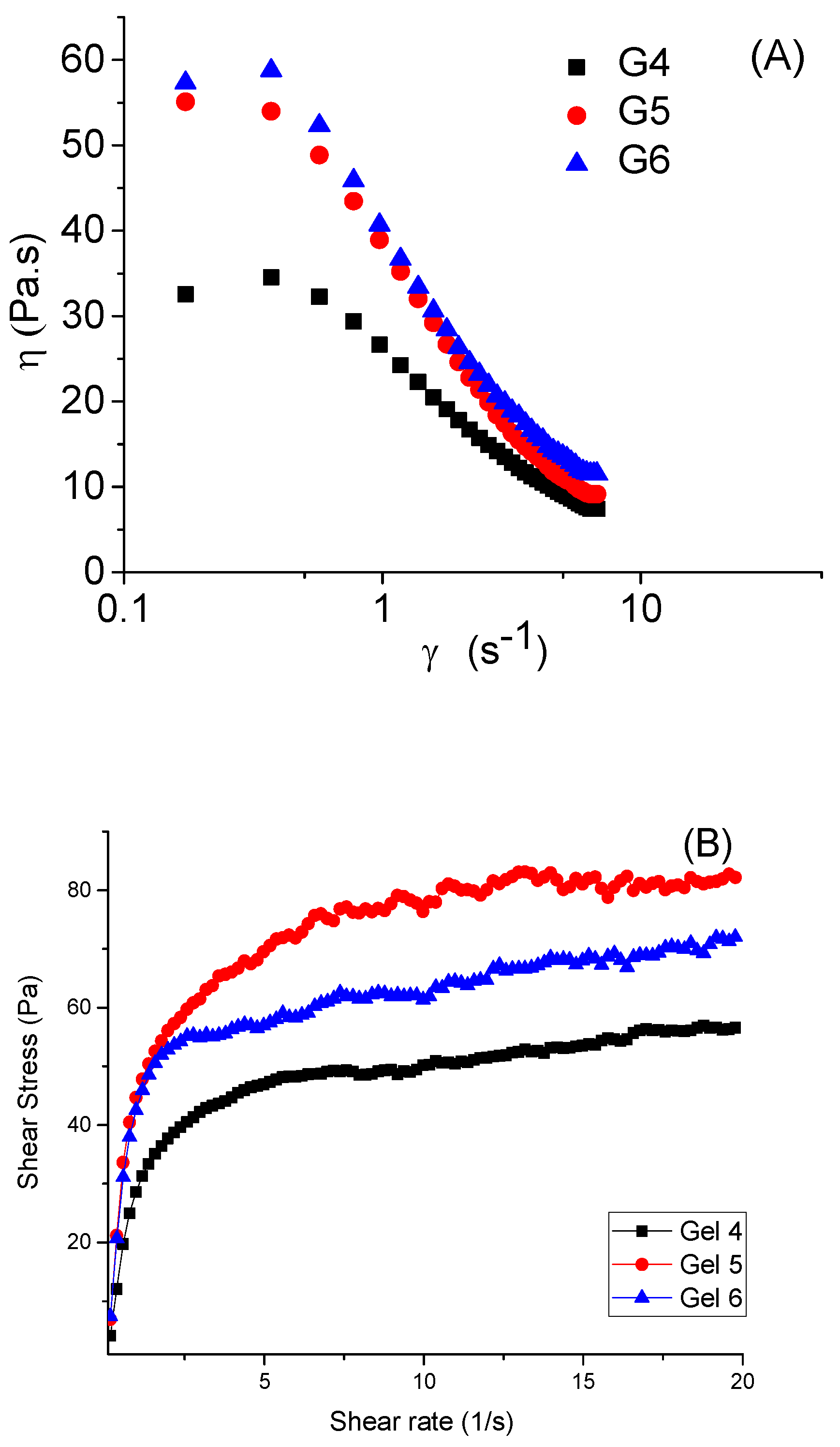
The cleansing capacity of EGT’s solutions and washing gels prepared thereof was investigated following previously described procedures [44] as follows: Cotton wool tissues were soaked in distilled water for 2 h then dried at room temperature (25 ± 1 °C) and accurately weighed. After that, the cotton tissues were immersed for 5 min in 1% coconut oil solution in hexane, then dried at room temperature and weighed again. Next, this cotton wool was soaked in EGT’s or washing gel solutions and agitated for 5 min at a constant agitation speed of 50 rpm in a shaking water bath (IKASH—B 20, Staufen, Germany). After that, cotton wools were washed with distilled water, dried and accurately weighed. All steps were performed at room temperature, whereas each experiment was performed in triplicate. The cleaning capacity of the tested samples was calculated by Equation (2):
C C ( % ) =   w 2 w 3 w 2 w 1 × 100
where w1—initial weight of the cotton wool; w2—weight of the cotton wool with the stimulated dirt; w3—weight of the cotton wool after washing with the tested gels.

2.3.7. Viscosity

The viscosity of the prepared hand wash gels was evaluated with a HAAKE MARS 60 rheometer (Thermo Fisher Scientific, Waltham, MA, USA), equipped with a parallel plate geometry (20 mm diameter) and a thermoelectric Peltier controller. The viscosity values were determined as a function of the shear rate (η = f(γ̇)) by varying the shear rate from 0.01 to 20 1/s (at a controlled ramp speed). Each measurement was carried out at 25 °C for 1 mL sample loaded between the parallel plates with a gap value of 1.2 mm. Three runs for each sample were conducted and the mean value was presented.

2.3.8. Spreadability

The spreadability of the gels developed was measured by a method described elsewhere [45]. In brief, 0.5 g of the tested formulations were spread on a circle of 2 cm diameter marked on a glass plate of a 15 cm diameter, and then a second glass plate (d = 15 cm) was placed above it. Next, a weight of 500 g was placed upon the upper glass plate for 5 min. The circle diameter (in cm) of the spread gel was determined. For each tested formulation, the measurements were conducted in triplicate, and the data were presented as mean ± SD (n = 3).

2.3.9. Antibacterial Activity

The minimal inhibitory concentration (MIC) of the gels tested was evaluated according to ISO 20776-1:2019 [46] against two bacterial strains originating from the American Type Culture Collection (ATCC, Manassas, Virginia, USA): Staphylococcus aureus (ATCC® 29213TM) and Escherichia coli (ATCC® 35218TM). The strains were cultured under aerobic conditions at 37 °C in Trypticase Soy Agar/Broth (TSA—#M1968/TSB—#LQ508, Himedia, India). All experiments were performed in Mueller Hinton broth (MHB, #M0405B, Thermo Scientific, Oxford, Hampshire, UK). Briefly, ten serial gel dilutions were prepared in 96-well plates of round bottoms in a volume of 50 µL, and an equivalent bacterial suspension volume of cell density of 5 × 10⁵/mL was added to each well. The bacterial suspension was prepared from an overnight culture after the cell density was measured by a densitometer at OD600. MHB was used as a negative control. The antibiotics penicillin (MIC = 0.25 mg/mL for S. aureus) and gentamicin (MIC = 2 mg/mL for E. coli) were used as positive controls. Plates were incubated overnight at 37 °C and the MICs were determined after visual evaluation of plate wells. The concentration, fully inhibiting the bacterial growth, was considered as MIC. The same samples were used thereafter to determine the bacteria’s metabolic activity. The cell redox (dehydrogenase, metabolic) bacteria activity was measured by the MTT dye, reduced in living cells by the membrane-located bacterial enzyme NADH–ubiquinone reductase (H+-translocation) to non-soluble violet crystals of formazan. The metabolic activity inhibition in treated bacteria was calculated as a fraction of the untreated control. Briefly, the MTT dye was added to each plate well at a final concentration of 0.5 mg/mL. Plates were incubated at 37 °C for 1 h, and the formazan crystals were dissolved with 2-propanol containing 5% HCOOH. The absorbance was measured at λ = 550 nm/ref. 690 nm on a microplate reader ELx800 (BioTek Instruments, Inc., Winooski, VT, USA). The viable cell percentage was calculated against the untreated control. The statistical evaluation was performed by one-way ANOVA in the GraphPad Prism software (version 6.0.0 for Windows, GraphPad Software, Boston, MA, USA). Each sample was prepared in triplicate. A p-value less than 0.05 was considered statistically significant.

3. Results and Discussion

A HPLC method for the quantitation of total saponins as gypsogenic acid and gypsogenin derivatives was developed and validated, according to the ICH Q2 (R1) [47,48] guideline. It was employed to standardise the EGT, used as a gel ingredient. At the chromatographic conditions established (see Material and Methods), gypsogenic acid had a retention time of 17.05 ± 0.2 min, and gypsogenin had a retention time of 18.66 ± 0.2 min. We examined the system’s suitability, accuracy, linearity, precision, and selectivity during the method validation process. The system was deemed appropriate—unlike HPLC systems, the UHPLC’s low dwell volume ensured better results [48,49]. The blank baseline showed no peaks interacting at the tR of the two analytes with an AUC greater than 0.05%. Both references displayed signal-to-noise (S/N) ratios of more than 10 in the standard solution (0.1% in MeOH). This solution’s agreement was ≤1.5%. The resolution factor (neighbouring peaks) was found to be >2 (Table 2).
The difference between the two subsequent injections was less than 1.4%. A blank (MeOH) was used to test specificity. There were no peaks with AUC > 0.1% interfering with the tR of either analytical marker. There was no indication of co-elution following peak purity tests. The S/N from the slope of the response and the standard deviation (SD) were used to compute the limits of detection and quantification. Gypsogenic acid and gypsogenin have respective limits of detections (LODs) of 0.002 and 0.003 mg/mL. For gypsogenic acid and gypsogenin, the limit of quantification (LOQ) is 0.02 and 0.03 mg/mL, respectively. From 25% to 200% of the compounds’ nominal concentration, the method’s linearity was investigated. MeOH-based model solutions were employed. Linear regression was used to process the calibration data. Following the linear least-square analysis, correlation coefficients (r2) of >0.99 were determined (Table 3).
This illustrates the relationship between the concentration and the AUC of the peaks [49] during the period under study (Figure 1).
Quantitation is possible, and there was no substantial bias, as indicated by the y-intercept (≤0.5%) at the 100% level [47]. Accuracy was within acceptable intervals (Table 4).
The method was evaluated for precision (Table 5), revealing that the correlation between the singular intraday and interday concentrations measured falls within accepted levels [47,49].
When the column thermostat temperature was increased by five °C increments, the method’s robustness was examined. Although it was demonstrated that the separation was improved at the standard temperature of 30 °C, the data obtained were not statistically affected by the increase.
The method developed for gypsogenic acid and gypsogenin was proven linear, accurate, and exact in accordance with ICH recommendations [47]. The validated method was deployed to quantify the amount of total saponins in EGT after acid hydrolysis. The total saponin content of the EGT was determined to be 4%, expressed as a sum of gypsogenic acid and gypsogenin.
Fruits of X. strumarium produced 0.57% (v/fresh w) essential oil. Twenty-five chemicals (98.79% of the sample) were found in the EOXS following GC-MS analysis and database comparison. Terpenes made up 51.98% of the essential oil, with the remaining components accounting for 48.02% of the sample overall. Himachalol (16.13%) and isovalencenol (15.02%) were found to be the primary sesquiterpenes in this group. Notably, this essential oil included a significant quantity of phenol 2,6-di-tert-butyl-p-cresol (14.22%). According to GC-MS, the EOXS was composed mostly of terpenes (52%), followed by hydrocarbons (25%) and other chemicals (23%). These findings coincide with previous reports on Bulgarian EOXS composition [33]. Other research reported a similar essential oil composition, but from different organs. The major compounds in stem essential oil were bornyl acetate (19.5%), limonene (15.0%) and β-selinene (10.1%) [50]. The plant leaves essential oil consisted predominantly of sesquiterpenoids (72.4%), followed by monoterpenes (25.19%). The main components of this essential oil were 1,5-dimethyltetralin (14.27%), eudesmol (10.60%), L-borneol (6.59%), ledene alcohol (6.46%), (-)-caryophyllene oxide (5.36%), isolongifolene, 7,8-dehydro-8a-hydroxy (5.06%), L-bornyl acetate (3.77%), and aristolene epoxide (3.58%) [51].
The aim of this study was the development of eco-friendly biocompatible natural liquid handwashing gels, based on an extract of G. trichotoma, containing 4% saponins, with the addition of the X. strumarium essential oil. Our hypothesis was that the use of saponins contained in the EGT as an alternative to synthetic sulphate surfactants of better skin compatibility, and the EOXS inclusion was aimed at achieving higher antibacterial activity of the gels developed. In this regard, the study was conducted in two stages: foaming characterisation and cleaning properties of crude saponin extract to find the optimal saponin concentrations, and the next stage is the formulation of liquid handwashing gel prepared thereof.

3.1. Characterisation of EGT Solutions

A series of EGT aqueous solutions were prepared by dissolving increasing amounts of lyophilised crude extract (10, 20 and 30%) in deionised water. The prepared solutions corresponding to 0.4, 0.8 and 1.2% concentrations of saponins (Table 6) were subjected to evaluation of pH, foamability, foam stability, and cleaning ability.
As illustrated in Table 6, all three solutions are characterised by slightly acidic pH around the pH-tolerance range of human skin being a prerequisite for skin compatibility [52]. Hence, the EGT solutions could be considered suitable from a topical application perspective, and their application in a final cosmetic formulation offers a light pH adjustment to the desired healthy skin pH. All solutions showed concentration-dependent foam formation ability, with the highest foam formation in the 1.2% saponin solution concentration. Foam stability also increased gradually with saponin concentration. Thus, while S1 forms an unstable foam as the R5 is less than 50%, increasing the saponin concentration in S2 and S3 leads to the formation of metastable foam with an R5 value higher than 50% [53].
An integral part of the saponin solutions evaluation is determining their cleaning properties. Similar to the other properties studied, a concentration-dependent cleaning ability of the solutions was expected, but the results obtained showed that the solution of the lowest tested saponin concentration of 0.4% showed a cleaning ability higher than that of the 0.8% concentration solution (Table 6). These findings correlate well with the previously published data [54]. Unstable foam can clean better than stable one, which could be explained by the variation of liquid fraction and foam stability leading to different combinations of the three cleaning mechanisms: imbibition, wiping and drainage at high liquid fractions and thus different cleaning properties of foams [54].
Based on the results of the conducted studies, all three tested EGT concentrations could be considered as suitable for wash gel formulation.

3.2. Characterisation of Washing Gels

The second stage of this study was the preparation of washing gels based on EGT. Xanthan gum was chosen as a gelling agent due to its excellent gelling properties, biocompatibility, degradability and good toxicological profile [55]. Xanthan gum is an anionic polysaccharide, containing glucuronic and pyruvic acid in its chemical structure, making its aqueous solution acidic; thus, it is considered a good natural gelling agent for dermal formulations [56]. For plausible dermal application, an optimal washing gel should be characterised by optimal pH, viscosity, spreadability, and good foaming and cleaning properties. In accordance with the above statement, we prepared and evaluated a series of gel formulations of varying combinations of total saponin extract, coco glucoside used as an ECOCERT-certified co-surfactant and EOXS (Table 1). The gels were prepared using the cold method and were subjected to detailed characterisation in order to find the best formulation of optimal physicochemical and cleaning properties (Table 7).

3.2.1. Preliminary Evaluation of Washing Gels

At a preliminary step in washing gels preparation, we sought to determine the influence of xanthan gum concentration on the main characteristics of washing gels in order to choose the optimal formulation. For this purpose, a series of gels were prepared (formulation G1 to G3) with increasing concentration of the gelling agent.
The visual evaluation showed that the formulations obtained were translucent brown-coloured gels. The gelation is due to the formation of intra- and intermolecular hydrogen bonds within the molecules of xanthan gum, and the gel viscosity increases in parallel with the increase of the gelling agent concentration. It is evident by the rheological studies conducted, that the increase in the EGT fraction is not accompanied by a change in viscosity, and it is a function solely of the gelling agent concentration.
Another important gel characteristic is their spreadability, as this property is closely related to the uniform application on the skin. It is also considered an important aspect of patient compliance. As evident from the results presented, all three formulations were characterised by good spreadability, although a slight decrease was encountered for G2 and G3 associated with the increased viscosity of these formulations as compared to formulation G1.
The evaluation of important characteristics of washing gels prepared at this preliminary stage (formulations G1 to G3), such as pH, foam stability and cleaning properties, was performed on 10% solutions of the corresponding gels. As shown in Table 7, a slight decrease in the pH values of gel solutions is evident as compared to the analogous solutions of lyophilised extract (formulation S1 to S3), but still, the encountered pH values of gels were within the skin pH tolerance. The decrease in gel pH could be explained by the acidic nature of xanthan gum, donating in solution H+ ions from glucuronic and pyruvic acid residues in its structure.
Similar to pH, a slight decrease in gel foam stability and cleaning properties as compared to the parent total extract solutions was observed. This could be explained by the decreased saponin concentration in gel solutions after dilution.
Based on the results obtained at this preliminary stage, an optimal concentration of the gelling agent, 0.6% xanthan gum, was chosen, and additionally, to optimise the gels’ foaming and cleaning properties, we decided to add a co-surfactant, namely nonionic coco glucoside, in the optimised formulations (G4 to G6). For comparison, a washing gel based solely on coco glucoside was prepared (G7).

3.2.2. Evaluation of Optimised Washing Gels

As evident in Table 7, the addition of coco glucoside is associated with an increase in gels pH as compared to formulations G1 to G), thus all optimised gels were characterised by the same pH as the pH of healthy skin (pH = 5.5), which is a prerequisite for their excellent skin compatibility. The pH increase observed in the pH of the optimised formulation could be explained by the alkaline properties of the coco glucoside solution (pH 11) [57]. Although the high pH of coco glucoside, the increase in the optimised gels pH is slight, probably due to the increased dissociation of H+ ions from acidic residues of xanthan gum in the medium of elevated pH [58].
As shown by the data presented in Table 7, the coco glucoside addition, although it is a viscous liquid, is not associated with an increase in viscosity or spreadability of the formulations as compared to formulations G1 to G3 without the co-surfactant. As the viscosity and rheological properties are two of the most important properties of fluids and play a major role in applications where the flow is a key characteristic, we sought to determine the flow properties of the prepared optimised gels (Figure 2).
The results of the dynamic rheological measurements indicate that the prepared samples at different concentrations of gelling agent behave as shear-thinning pseudoplastic fluids, as after a critical point, the viscosities of the three gels decreased significantly with increased shear rate. The shear stress–shear rate curve of the gels (Figure 2B) shows that with increasing shear rate, an increase in shear stress was observed. The shear stress reached the yield point, after which the solution began to flow. This finding is well-paralleled with other published studies [59,60,61,62].
The shear-thinning behavior observed could be explained by the gradual disruption of intra- and intermolecular hydrogen bonds within the xanthan molecules under increased shear stress and consequent alignment of the polymer chains in the flow direction, thus facilitating uniform gel distribution on the skin during application [58]. The encountered pseudoplastic flow properties correlate well with the good spreadability of the tested formulations observed (Table 7). In addition, as evident from Table 7, all optimised formulations (G4 to G6) are characterised by excellent foam ability and foam stability characteristics. Although some researchers consider high foam formation and foam stability of no significant effect on the formulation cleaning properties [54], the current study, as well as other previously published articles, clearly demonstrates that high foamability is associated with higher cleaning ability [63].
Based on the good physical, foaming and cleaning properties of the optimised washing gels developed, they can be considered as promising candidates for personal hygiene products. This gives us reason to widen the gels’ evaluation by investigating their antibacterial activity.

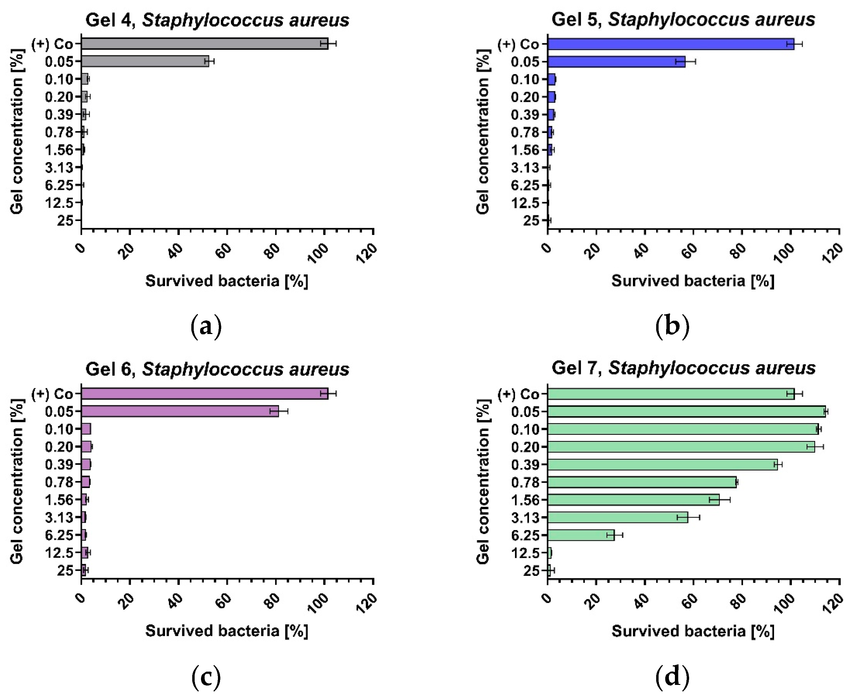
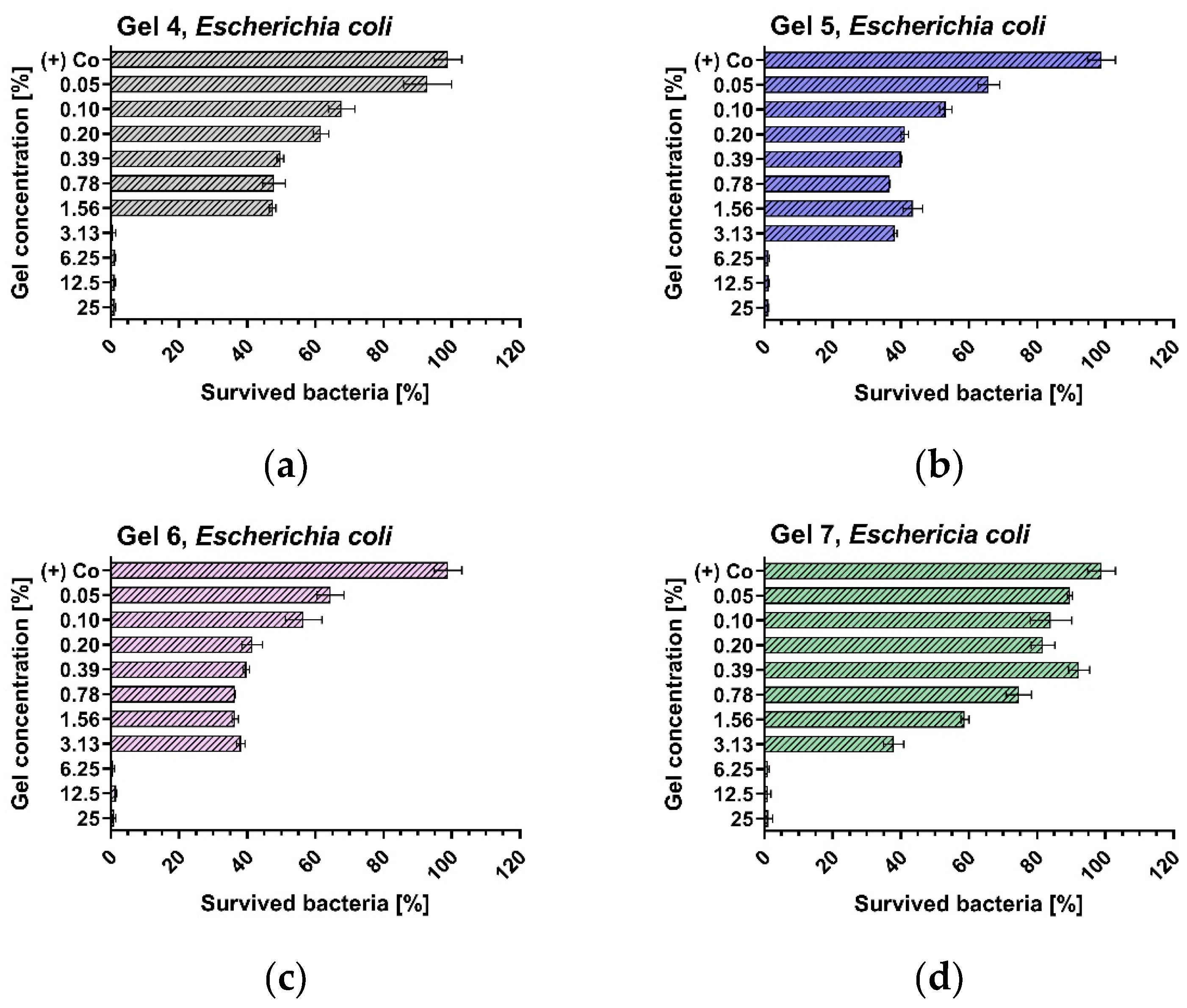
3.3. Antibacterial Activity of Optimised Gels

The antibacterial activity of optimised gels containing increasing concentrations of X. strumarium essential oil (formulations G4–G7) was evaluated in a comparative way vs. plain washing gel based solely on coco glucoside (formulation G7) (Table 8). For the purpose of this investigation, as representatives of the two different types of bacteria regarding the staining and their morphology, we chose one Gram-positive (S. aureus) and one Gram-negative (E. coli) bacterial strain recommended by ISO 20776-1:2019 for antimicrobial evaluation.
Gel 4, 5 and 6 inhibited the growth of Staphylococcus aureus (Figure 3) in concentrations of 0.1%, whereas G7 was less active and showed a MIC of 6.25%.
Inhibition of Escherichia coli strain growth (Figure 4) occurred after exposure to 3.125% of G6 and 6.25% of G4, G5 and G7. The minimal bactericidal concentrations of the four gels ranged from 3.125% to 12.5%, wherein Gel 4 exhibited the strongest activity.
Interestingly, Gel 7, containing only coco glucoside, displayed some antibacterial activity on Gram-negative bacteria, comparable to gels containing EGT and EOXS. This is supposed to be a result of the molecule’s characteristics and its capability to permeate the cell envelope’s outer layer by reacting with proteins [64]. Gels 4, 5, and 6 showed stronger antibacterial activity than Gel 7. All gels exerted bactericidal effects in concentrations over 6.25% on the tested bacterial strains except Gel 4. Gel 4 was characterised by the strongest antibacterial activity, and gel 7—by the lowest. This paradox could be a result of the synergism between the saponins contained in the EGT and the EOXS’s components. It is known that essential oils containing phenols display lower MIC, than essential oils, free of such compounds [65]. In the present case, the EOXS was rich in 2,6-di-tert-butyl-p-cresol (14.22%), according to GC-MS analysis.
These effects could be attributed to two factors. First, EGT contains triterpenoid saponins, reported to lower surface tension, help hydrate particles, and emulsify lipids. In addition, saponins are well-known adjuvants in vaccines, contributing to better absorption and enhanced immunogenicity [66]. Increasing the percentage of EGT and saponins, respectively, enhances the antibacterial effect observed throughout the gels. Secondly, the role of the EOXS is invaluable—the phenol compound content of 2,6-di-tert-butyl-p-cresol is especially high for essential oils. It has reported antiseptic and antibacterial properties [67]. There is an obvious concentration-dependent rise in antibacterial activity when the percentage of EOXS is increased amongst gels. Moreover, the sesquiterpenes, EOXS’s main ingredients, have also proven antibacterial properties [68].

4. Conclusions

A series of novel plant-based handwashing gels containing a defatted extract of G. trichotoma and an essential oil of X. strumarium were formulated. A quantitative method for the determination of triterpenoid saponins was developed and successfully applied to standardise the extract. Essential oil analysis was performed by GC-MS, as sesquiterpenes were the main compounds. All the gels developed were evaluated for technological characteristics (pH, foam-forming ability, foam stability, cleaning capacity, viscosity, and spreadability). The best formulations were examined for their possible antibacterial activity on two bacterial strains (Gram-positive and Gram-negative). An antibacterial effect increase by increasing both the extract and essential oil fraction was observed, and one of the formulations tested exhibited the best antibacterial effect (gel 4). This initial research could serve as the basis for further development of an eco-friendly alternative in personal hygiene.

Author Contributions

Conceptualisation, I.K. and D.M.; methodology, A.S., M.M.Z. and D.M.; software, A.S.; validation, A.S.; formal analysis, M.M.Z., A.S., H.N. and I.K.; investigation, A.S., M.M.Z.; resources, E.K. and I.K.; data curation, I.K.; writing—original draft preparation, A.S., D.M., M.M.Z. and E.K.; writing—review and editing, A.S., M.M.Z., D.M., H.N. and I.K.; visualisation, A.S., D.M. and M.M.Z.; supervision, I.K.; project administration, I.K.; funding acquisition, I.K. All authors have read and agreed to the published version of the manuscript.

Funding

This study was financed by the European Union-NextGenerationEU, through the National Recovery and Resilience Plan of the Republic of Bulgaria, project No BG-RRP-2.004-0004-C01.

Institutional Review Board Statement

Not applicable.

Informed Consent Statement

Not applicable.

Data Availability Statement

All data connected with this research are included in this manuscript. Further information is available from the corresponding author upon reasonable written request.

Conflicts of Interest

The authors declare no conflicts of interest.

Abbreviations

The following abbreviations are used in this manuscript:
EGTDefatted Extract from Gypsophila trichotoma
EOXSEssential Oil from Xanthium strumarium

References

  1. Bloom, D.E.; Cadarette, D. Infectious Disease Threats in the Twenty-First Century: Strengthening the Global Response. Front. Immunol. 2019, 10, 549. [Google Scholar] [CrossRef]
  2. Ma, Y.; Yi, J.; Ma, J.; Yu, H.; Luo, L.; Wu, W.; Jin, L.; Yang, Q.; Lou, T.; Sun, D.; et al. Hand Sanitizer Gels: Classification, Challenges, and the Future of Multipurpose Hand Hygiene Products. Toxics 2023, 11, 687. [Google Scholar] [CrossRef] [PubMed]
  3. Johnson, P.; Trybala, A.; Starov, V.; Pinfield, V.J. Effect of Synthetic Surfactants on the Environment and the Potential for Substitution by Biosurfactants. Adv. Colloid Interface Sci. 2021, 288, 102340. [Google Scholar] [CrossRef]
  4. Nizioł-Łukaszewska, Z.; Bujak, T. Saponins as Natural Raw Materials for Increasing the Safety of Bodywash Cosmetic Use. J. Surfactants Deterg. 2018, 21, 767–776. [Google Scholar] [CrossRef]
  5. Vincken, J.-P.; Heng, L.; de Groot, A.; Gruppen, H. Saponins, Classification and Occurrence in the Plant Kingdom. Phytochemistry 2007, 68, 275–297. [Google Scholar] [CrossRef]
  6. Bildziukevich, U.; Wimmerová, M.; Wimmer, Z. Saponins of Selected Triterpenoids as Potential Therapeutic Agents: A Review. Pharmaceuticals 2023, 16, 386. [Google Scholar] [CrossRef]
  7. Yang, Y.; Leser, M.E.; Sher, A.A.; McClements, D.J. Formation and Stability of Emulsions Using a Natural Small Molecule Surfactant: Quillaja Saponin (Q-Naturale®). Food Hydrocoll. 2013, 30, 589–596. [Google Scholar] [CrossRef]
  8. Petrova, A. Gypsophila trichotoma Wend.a. In Red Data Book of the Republic of Bulgaria, Vol. 1, Plants and Fungi; Peev, D., Golemanski, V., Eds.; Bulgarian Academy of Sciences: Sofia, Bulgaria, 2015. [Google Scholar]
  9. Vladimirov, V.; Aybeke, M.; Matevski, V.; Tan, K. New Floristic Records in the Balkans: 34. Phytol. Balc. 2017, 23, 413–444. [Google Scholar]
  10. Balabanova, V.; Zdraveva, P.; Kozuharova, E.; Krasteva, I.; Nikolov, S. A Possibility for Cultivation and Phytochemical Study of Endangered Gypsophila trichotoma Wend. Comptes Rendus L’académie Bulg. Sci. 2009, 62, 1247–1252. [Google Scholar]
  11. Kozuharova, E.; Balabanova, V.; Boev, V. In-Situ and Ex-Situ Morphological Characters of Gypsophila trichotoma Wend. Comptes Rendus L’académie Bulg. Des Sci. 2011, 64, 523–528. (In Bulgarian) [Google Scholar]
  12. Yotova, M.; Krasteva, I.; Nikolov, S. Triterpenoid Saponins from Genus Gypsophila L. (Caryophyllaceae). In Saponins: Properties, Applications and Health Benefits; Koh, R., Tay, I., Eds.; Nova Science Publishers: New York, NY, USA, 2012; pp. 99–122. [Google Scholar]
  13. Kamali, M.; Talebi, M.; Mottaghipisheh, J.; Sasani, E.; Mirshekari, B.M. An Updated Overview of Gypsophila Species: Phytochemical and Pharmacological Investigations. Fitoterapia 2024, 179, 106230. [Google Scholar] [CrossRef]
  14. Voutquenne-Nazabadioko, L.; Gevrenova, R.; Borie, N.; Harakat, D.; Sayagh, C.; Weng, A.; Thakur, M.; Zaharieva, M.; Henry, M. Triterpenoid Saponins from the Roots of Gypsophila trichotoma Wender. Phytochemistry 2013, 90, 114–127. [Google Scholar] [CrossRef] [PubMed]
  15. Krasteva, I.; Popov, I.; Balabanova, V.; Nikolov, S.; Pencheva, I. Phytochemical Study of Gypsophila trichotoma Wend. (Caryophyllaceae). Quim. Nova. 2008, 31, 1125–1126. [Google Scholar] [CrossRef]
  16. Vitcheva, V.; Simeonova, R.; Krasteva, I.; Yotova, M.; Nikolov, S.; Mitcheva, M. Hepatoprotective Effects of Saponarin, Isolated from Gypsophila trichotoma Wend. on Cocaine-Induced Oxidative Stress in Rats. Redox Rep. 2011, 16, 56–61. [Google Scholar] [CrossRef]
  17. Krasteva, I.; Yotova, M.; Yosifov, D.; Benbassat, N.; Jenett-Siems, K.; Konstantinov, S. Cytotoxicity of Gypsogenic Acid Isolated from Gypsophila trichotoma. Pharmacogn. Mag. 2014, 10, 430–433. [Google Scholar] [CrossRef]
  18. Fan, W.; Fan, L.; Peng, C.; Zhang, Q.; Wang, L.; Li, L.; Wang, J.; Zhang, D.; Peng, W.; Wu, C. Traditional Uses, Botany, Phytochemistry, Pharmacology, Pharmacokinetics and Toxicology of Xanthium strumarium L.: A Review. Molecules 2019, 24, 359. [Google Scholar] [CrossRef]
  19. Strother, J.L. Xanthium. In Flora of North America; Oxford University: New York, NY, USA, 2006. [Google Scholar]
  20. Aneva, I.Y.; Zhelev, P.; Stoyanov, S.S. Alien Species as a Part of Plant Composition in the Periphery of Agricultural Fields. Acta Zool. Bulg. Suppl. 2018, 11, 173–176. [Google Scholar]
  21. Kozuharova, E.; Ionkova, I.; Spadaro, V. Xanthium strumarium—A Potential Cheap Resource of Plant Substances for Medicinal Use. Flora Mediterr. 2019, 29, 93–102. [Google Scholar]
  22. Plants of the World Online. 2024. Available online: https://powo.science.kew.org/taxon/urn:lsid:ipni.org:names:260893-1/general-information (accessed on 1 December 2024).
  23. Arshad, M.; Khan, Q.A. Ethnobotanical Study of Some Medicinal Plants of Rawal Town. Pak. J. Biol. Sci. 2000, 3, 1245–1246. [Google Scholar]
  24. Hussain, K.; Shahazad, A.; Zia-ul-Hussnain, S. An Ethnobotanical Survey of Important Wild Medicinal Plants of Hattar District Haripur, Pakistan. Ethnobot. Leafl. 2008, 2008, 5. [Google Scholar]
  25. Ali, H.; Qaiser, M. The Ethnobotany of Chitral Valley, Pakistan with Particular Reference to Medicinal Plants. Pak. J. Bot. 2009, 41, 2009–2041. [Google Scholar]
  26. Bhardwaj, R.; Dutta, S.; Sharma, K.C. Conserving Biodiversity of Medicinal Plants from Central Aravallis of Rajasthan, India. J. Env. Res. Dev. 2011, 6, 69–75. [Google Scholar]
  27. Yadav, D.K.; Jhariya, M.K.; Kumar, A.; Sinha, R. Documentation and Ethnobotanical Importance of Medicinal Plants Found in Sarguja District. J. Plant Dev. Sci. 2015, 7, 439–446. [Google Scholar]
  28. Ullah, M.; Khan, M.U.; Mahmood, A.; Malik, R.N.; Hussain, M.; Wazir, S.M.; Daud, M.; Shinwari, Z.K. An Ethnobotanical Survey of Indigenous Medicinal Plants in Wana District South Waziristan Agency, Pakistan. J. Ethnopharmacol. 2013, 150, 918–924. [Google Scholar] [CrossRef]
  29. Akhtar, N.; Rashid, A.; Murad, W.; Bergmeier, E. Diversity and Use of Ethno-Medicinal Plants in the Region of Swat, North Pakistan. J. Ethnobiol. Ethnomed. 2013, 9, 25. [Google Scholar] [CrossRef]
  30. Gilani, S.A.; Khan, A.M.; Qureshi, R.A.; Sherwani, S.K.; Ullah Khan, R.; Bokhari, T.Z. Ethnomedicinal Treatment of Common Gastrointestinal Disorders by Indigenous People in Pakistan. Adv. Biores 2014, 5, 42–49. [Google Scholar]
  31. Manandhar, N.P. Native Use of Herbal Drugs for Treatment of Skin Diseases in Nepal. In Plant-Derived Antimycotics; CRC Press: Boca Raton, FL, USA, 2024; pp. 429–439. [Google Scholar]
  32. Kunwar, R.M.; Acharya, R.P.; Chowdhary, C.L.; Bussmann, R.W. Medicinal Plant Dynamics in Indigenous Medicines in Farwest Nepal. J. Ethnopharmacol. 2015, 163, 210–219. [Google Scholar]
  33. Shkondrov, A.; Krasteva, I.; Kozuharova, E.; Ionkova, I. Chemical Composition of Essential Oil in Fruits of Xanthium strumarium L. Biotechnol. Biotechnol. Equip. 2021, 35, 1474–1479. [Google Scholar]
  34. Parveen, Z.; Mazhar, S.; Siddique, S.; Manzoor, A.; Ali, Z. Chemical Composition and Antifungal Activity of Essential Oil from Xanthium strumarium L. Leaves. Indian J. Pharm. Sci. 2017, 79, 316–321. [Google Scholar]
  35. Sarmah, M.; Bhola, R.K. Bio-Activity of Xanthium strumarium Extracts against Tea Mosquito Bug, Helopeltis Theivora. J. Plant Crop. 2014, 42, 40–47. [Google Scholar]
  36. Kejlová, K.; Jírová, D.; Bendová, H.; Gajdoš, P.; Kolářová, H. Phototoxicity of Essential Oils Intended for Cosmetic Use. Toxicol. Vitr. 2010, 24, 2084–2089. [Google Scholar] [CrossRef]
  37. Zazharskyi, V.V.; Brygadyrenko, V.V.; Boyko, O.O.; Bilan, M.V.; Zazharska, N.M. Antibacterial and Anthelmintic Activities of Xanthium strumarium (Asteraceae) Extracts. Regul. Mech. Biosyst. 2024, 15, 129–133. [Google Scholar]
  38. Sharifi-Rad, J.; Hoseini-Alfatemi, S.M.; Sharifi-Rad, M.; Sharifi-Rad, M.; Iriti, M.; Sharifi-Rad, M.; Sharifi-Rad, R.; Raeisi, S. Phytochemical Compositions and Biological Activities of Essential Oil from Xanthium strumarium L. Molecules 2015, 20, 7034–7047. [Google Scholar] [CrossRef] [PubMed]
  39. Sharmeen, J.B.; Mahomoodally, F.M.; Zengin, G.; Maggi, F. Essential Oils as Natural Sources of Fragrance Compounds for Cosmetics and Cosmeceuticals. Molecules 2021, 26, 666. [Google Scholar] [CrossRef]
  40. Es, I.; Khaneghah, A.M.; Akbariirad, H. Global Regulation of Essential Oils. In Essential Oils in Food Processing; John Wiley & Sons, Ltd.: Hoboken, NJ, USA, 2017; pp. 327–338. ISBN 9781119149392. [Google Scholar]
  41. Yücekutlu, A. Determination of Plant Saponins and Some of Gypsophila Species: A Review of the Literature. Hacet. J. Biol. Chem. 2008, 36, 129–135. [Google Scholar]
  42. Available online: https://ifrafragrance.org/safe-use/library (accessed on 12 December 2024).
  43. Pradhan, A.; Bhattacharyya, A. Shampoos Then and Now: Synthetic versus Natural. J. Surf. Sci. Technol. 2014, 30, 59–76. [Google Scholar]
  44. Rai, S.; Kafle, A.; Devkota, H.P.; Bhattarai, A. Characterization of Saponins from the Leaves and Stem Bark of Jatropha curcas L. for Surface-Active Properties. Heliyon 2023, 9, e15807. [Google Scholar]
  45. Dantas, M.G.B.; Reis, S.A.G.B.; Damasceno, C.M.D.; Rolim, L.A.; Rolim-Neto, P.J.; Carvalho, F.O.; Quintans-Junior, L.J.; Almeida, J.R.G.d.S. Development and Evaluation of Stability of a Gel Formulation Containing the Monoterpene Borneol. Sci. World J. 2016, 2016, 7394685. [Google Scholar]
  46. ISO 20776-1:2019 Susceptibility Testing of Infectious Agents and Evaluation of Performance of Antimicrobial Susceptibility Test Devices Part 1: Broth Micro-Dilution Reference Method for Testing the in Vitro Activity of Antimicrobial Agents. Available online: https://www.iso.org/standard/70464.html (accessed on 1 December 2024).
  47. ICH Harmonised Tripartite Guideline. Validation of Analytical Procedures: Text and Methodology Q2 (R1). In Proceedings of the International Conference on Harmonization, Geneva, Switzerland, 1 November 2005; pp. 11–12. [Google Scholar]
  48. Chen, S.; Kord, A. UHPLC Method Development. In Ultra-High Performance Liquid Chromatography and Its Applications; John Wiley & Sons, Ltd.: Hoboken, NJ, USA, 2013; pp. 1–30. ISBN 9781118533956. [Google Scholar]
  49. Swartz, M.E.; Krull, I.S. Analytical Method Development and Validation; CRC Press: Boca Raton, FL, USA, 2018. [Google Scholar]
  50. Esmaeili, A.; Rustaiyan, A.; Akbari, M.T.; Moazami, N.; Masoudi, S.; Amiri, H. Composition of the Essential Oils of Xanthium strumarium L. and Cetaurea solstitialis L. from Iran. J. Essent. Oil Res. 2006, 18, 427–429. [Google Scholar]
  51. El-Gawad, A.A.; Elshamy, A.; El Gendy, A.E.-N.; Gaara, A.; Assaeed, A. Volatiles Profiling, Allelopathic Activity, and Antioxidant Potentiality of Xanthium strumarium Leaves Essential Oil from Egypt: Evidence from Chemometrics Analysis. Molecules 2019, 24, 584. [Google Scholar] [CrossRef]
  52. Lukić, M.; Pantelić, I.; Savić, S.D. Towards Optimal PH of the Skin and Topical Formulations: From the Current State of the Art to Tailored Products. Cosmetics 2021, 8, 69. [Google Scholar] [CrossRef]
  53. Lunkenheimer, K.; Malysa, K. Simple and Generally Applicable Method of Determination and Evaluation of Foam Properties. J. Surfactants Deterg. 2003, 6, 69–74. [Google Scholar] [CrossRef]
  54. Schad, T.; Preisig, N.; Blunk, D.; Piening, H.; Drenckhan, W.; Stubenrauch, C. Less Is More: Unstable Foams Clean Better than Stable Foams. J. Colloid Interface Sci. 2021, 590, 311–320. [Google Scholar] [CrossRef] [PubMed]
  55. Xiao, N.; He, W.; Zhao, Y.; Yao, Y.; Xu, M.; Du, H.; Wu, N.; Tu, Y. Effect of PH and Xanthan Gum on Emulsifying Property of Ovalbumin Stabilized Oil-in Water Emulsions. LWT 2021, 147, 111621. [Google Scholar] [CrossRef]
  56. Badwaik, H.R.; Giri, T.K.; Nakhate, K.T.; Kashyap, P.; Tripathi, D.K. Xanthan Gum and Its Derivatives as a Potential Bio-Polymeric Carrier for Drug Delivery System. Curr. Drug Deliv. 2013, 10, 587–600. [Google Scholar] [CrossRef]
  57. Ćirin, D.; Milutinov, J.; Krstonošić, V. Occurrence of Alkyl Glucosides in Rinse-off Cosmetics Marketed as Hypoallergenic or for Sensitive Skin. Toxicol. Ind. Health 2024, 40, 306–311. [Google Scholar] [CrossRef]
  58. Brunchi, C.-E.; Bercea, M.; Morariu, S.; Dascalu, M. Some Properties of Xanthan Gum in Aqueous Solutions: Effect of Temperature and PH. J. Polym. Res. 2016, 23, 123. [Google Scholar] [CrossRef]
  59. Ali, I.; Shah, L.A. Rheological Investigation of the Viscoelastic Thixotropic Behavior of Synthesized Polyethylene Glycol-Modified Polyacrylamide Hydrogels Using Different Accelerators. Polym. Bull. 2021, 78, 1275–1291. [Google Scholar] [CrossRef]
  60. Li, M.-C.; Wu, Q.; Moon, R.J.; Hubbe, M.A.; Bortner, M.J. Rheological Aspects of Cellulose Nanomaterials: Governing Factors and Emerging Applications. Adv. Mater. 2021, 33, 2006052. [Google Scholar] [CrossRef]
  61. Patel, M.C.; Ayoub, M.A.; Hassan, A.M.; Idress, M.B. A Novel ZnO Nanoparticles Enhanced Surfactant Based Viscoelastic Fluid Systems for Fracturing under High Temperature and High Shear Rate Conditions: Synthesis, Rheometric Analysis, and Fluid Model Derivation. Polymers 2022, 14, 4023. [Google Scholar] [CrossRef]
  62. Lin, H.-R.; Sung, K.C.; Vong, W.-J. In Situ Gelling of Alginate/Pluronic Solutions for Ophthalmic Delivery of Pilocarpine. Biomacromolecules 2004, 5, 2358–2365. [Google Scholar] [CrossRef] [PubMed]
  63. Stubenrauch, C.; Drenckhan, W. Cleaning Solid Surfaces with Liquid Interfaces and Foams: From Theory to Applications. Curr. Opin. Colloid Interface Sci. 2024, 72, 101818. [Google Scholar] [CrossRef]
  64. Nourbakhsh, F.; Kashi, M.E.; Shakeri, A. Natural Products against Gram-Negative Bacteria: Promising Antimicrobials in Future Complementary Medicine. Phytochem. Rev. 2024. [Google Scholar] [CrossRef]
  65. Di Vito, M.; Smolka, A.; Proto, M.R.; Barbanti, L.; Gelmini, F.; Napoli, E.; Bellardi, M.G.; Mattarelli, P.; Beretta, G.; Sanguinetti, M.; et al. Is the Antimicrobial Activity of Hydrolates Lower than That of Essential Oils? Antibiotics 2021, 10, 88. [Google Scholar] [CrossRef]
  66. Fan, J.; Jin, S.; Gilmartin, L.; Toth, I.; Hussein, W.M.; Stephenson, R.J. Advances in Infectious Disease Vaccine Adjuvants. Vaccines 2022, 10, 1120. [Google Scholar] [CrossRef]
  67. Ahmad, A.; Elisha, I.L.; van Vuuren, S.; Viljoen, A. Volatile Phenolics: A Comprehensive Review of the Anti-Infective Properties of an Important Class of Essential Oil Constituents. Phytochemistry 2021, 190, 112864. [Google Scholar] [CrossRef]
  68. Cappiello, F.; Loffredo, M.R.; Del Plato, C.; Cammarone, S.; Casciaro, B.; Quaglio, D.; Mangoni, M.L.; Botta, B.; Ghirga, F. The Revaluation of Plant-Derived Terpenes to Fight Antibiotic-Resistant Infections. Antibiotics 2020, 9, 325. [Google Scholar] [CrossRef]
Figure 1. Calibration curves of gypsogenic acid (a) and gypsogenin (b).
Cosmetics 12 00065 g001
Figure 2. Viscosity (A) and shear stress as a function of shear rate (B) for Gel 4, Gel 5 and Gel 6 at 25 °C.
Cosmetics 12 00065 g002
Figure 3. Dehydrogenase activity of treated Staphyloccocus aureus vs. untreated control.
Cosmetics 12 00065 g003
Figure 4. Dehydrogenase activity of treated Escherchia coli vs. untreated control.
Cosmetics 12 00065 g004
Table 1. Gel composition.
GelEGT (%)Coco Glycoside
(%)
EOXS (%)Xanthan Gum
(%)
Glycerine (%)Deionised Water (%)
G110--0.2782.8
G220--0.4772.6
G330--0.6762.4
G41050.20.6777.2
G52050.50.6773.9
G63051.00.6763.4
G7-5-0.6787.4
Table 2. System suitability, n = 6.
ParameterGypsogenic AcidGypsogenin
Mean peak area ± SD20.091033.9863
Peak area, %RSD2.782.33
Mean tR, min ± SD17.05 ± 0.218.66 ± 0.2
tR, %RSD0.150.21
Mean theoretical plates ± SD477,458 ± 173344,687 ± 132
Resolution, mean ± SD2.0 ± 0.02.2 ± 0.0
Resolution, %RSD0.230.24
Table 3. Linearity of the method, n = 3.
ParameterValue
Gypsogenic AcidGypsogenin
Range of linearity, mg/mL0.2–3.00.1–2.0
Slope13.61734.821
Y-intercept−1.6129−9.3945
Coefficient of correlation r20.99800.9973
Table 4. Accuracy of the method, n = 3.
Analytical MarkerTheoretical Concentration, mg/mLConcentration Found, mg/mL Recovery,
% ± SD
%RSDAverage Recovery, %
Gysogenic acid1.0001.050105.0 ± 3.53.5100.2 ± 1.5
0.5000.501100.2 ± 1.34.1
0.1000.09999.9 ± 3.02.1
Gypsogenin1.0000.99999.9 ± 2.84.0100.5 ± 1.6
0.5000.49899.6 ± 3.22.7
0.1000.102102.0 ± 3.51.5
Table 5. Precision of the method.
Analytical MarkerConcentration of the Standard
Solution, mg/mL
Concentration Found (Intraday) *, mg/mLIntraday
Variance **,
%RSD
Concentration
Found (Interday) *,
mg/mL
Interday Variance ***,
%RSD
Analysis Series 1Analysis Series 2
Gysogenic acid1.00001.05021.04981.41.05012.9
0.50000.50120.50091.60.50021.7
0.10000.99890.99792.10.09982.3
Gypsogenin1.0000.99980.99581.20.99962.5
0.5000.49810.49901.30.49951.9
0.1000.10200.10101.10.10122.2
* n = 3; ** n = 6; *** n = 9.
Table 6. Evaluation of the basic properties of EGT: pH, foamability, and foam stability (R5) ± SD (n = 3).
EGT SolutionEGT
(%)
Saponin Concentration (%)pHFoam Height (cm)R5
(%)
Cleaning Ability
(%)
S1100.46.84.5 ± 0.144 ± 1.190 ± 0.5
S2200.86.75 ± 0.351 ± 2.185 ± 1.3
S3301.26.56 ± 0.263 ± 2.897 ± 0.8
Table 7. Composition and pH, viscosity, foamability, foam stability, and cleaning properties of the washing gels (±SD n = 3).
Formulation
Code
EGT (%)Coco Glucoside (%)EOXS (%)Xanthan Gum (%)pHViscosity (Pa.s)Spreadability (cm)Foam Height (cm)R5 (%)Cleaning Ability (%)
G110--0.25.2 ± 0.624 ± 3.58.6 ± 0.93.8 ± 0.339 ± 2.176 ± 3.2
G220--0.45.0 ± 0.840 ± 3.17.7 ± 0.55.0 ± 0.448 ± 1.482 ± 1.8
G330--0.64.8 ± 0.543 ± 1.17.5 ± 1.26.0 ± 1.151 ± 2.288 ± 2.5
G41050.20.65.8 ± 0.544 ± 2.87.4 ± 0.212.0 ± 1.297 ± 2.198 ± 1.1
G52050.50.65.7 ± 0.545 ± 0.57.3 ± 0.613.0 ± 1.198 ± 0.998 ± 0.6
G63051.00.65.6 ± 0.545 ± 1.27.1 ± 0.815.0 ± 1.398 ± 1.199 ± 0.8
G7-5-0.67.8 ± 0.544 ± 2.17.6 ± 0.613.0 ± 2.195 ± 2.1 97 ± 1.6
Table 8. Minimal inhibitory and bactericidal concentrations.
GelsStaphylococcus aureus, ATCC® 29213TMEscherichia coli, ATCC® 35218 TM
MIC [%]MBC [%]MIC [%]MBC [%]
G40.1003.1253.1253.125
G50.1003.1256.2506.250
G60.1003.1256.2506.250
G76.25012.5006.2506.250
Legend: MIC—minimal inhibitory concentration; MBC—minimal bactericidal concentration.
Disclaimer/Publisher’s Note: The statements, opinions and data contained in all publications are solely those of the individual author(s) and contributor(s) and not of MDPI and/or the editor(s). MDPI and/or the editor(s) disclaim responsibility for any injury to people or property resulting from any ideas, methods, instructions or products referred to in the content.

