Article

Skin Cell Phototoxicity and Photoprotection Study of Agro-Derived Lignin and Nanocellulose

by
Juliana Varella Cruz
1,2,*,
Adriana Solange Maddaleno
3,
Julia Salles Gava
4,
Washington Luiz Esteves Magalhães
5,
Danielle Palma de Oliveira
2,6,
Daniela Morais Leme
1,6,
Montserrat Mitjans
3 and
Maria Pilar Vinardell
3,*
1
Department of Genetics, Federal University of Paraná (UFPR), Curitiba 81531-980, Brazil
2
School of Pharmaceutical Sciences of Ribeirão Preto, University of São Paulo, Ribeirão Preto 14040-900, Brazil
3
Department of Biochemistry and Physiology, Universitat de Barcelona, Av. Joan XXIII, 27-31, 08028 Barcelona, Spain
4
Pharmaceutical Sciences Graduation Program, University of Vila Velha, Vila Velha 29102-920, Brazil
5
Embrapa Florestas, Colombo 83411-000, Brazil
6
National Institute for Alternative Technologies of Detection, Toxicological Evaluation and Removal of Micropollutants and Radioactives (INCT-DATREM), Institute of Chemistry, Araraquara 14800-060, Brazil
*
Authors to whom correspondence should be addressed.
Cosmetics 2025, 12(2), 61; https://doi.org/10.3390/cosmetics12020061
Submission received: 18 February 2025 / Revised: 18 March 2025 / Accepted: 19 March 2025 / Published: 28 March 2025
(This article belongs to the Special Issue Feature Papers in Cosmetics in 2025)

Abstract

:
Lignin, a significant industrial byproduct from paper manufacturing processes, exhibits ultraviolet (UV) radiation absorption properties. Cellulose nanofibers (CNFs) demonstrate universal ligand characteristics and represent an innovative approach for converting industrial waste into value-added products. Given their potential applications in cosmetic formulations, their efficacy and safety parameters, such as their photoprotection mechanisms and phototoxicity, need to be investigated. Therefore, two kraft lignin fractions, LE and R1, along with a kraft-bleached pulp CNF, were evaluated for their phototoxicity and photoprotection mechanisms, both using the HaCaT cell line (immortalized human keratinocytes) as the in vitro model. Phototoxicity assessment involved exposing cells to UVA radiation (4 J/cm2), with the subsequent comparison of cell viability between irradiated and non-irradiated samples. ROS quantification was performed using a 2′,7′-dichlorofluorescein diacetate (DCF-DA) probe, with fluorescence intensity measurements, and was then used to evaluate the photoprotection effect. The results demonstrated that both LE and R1 exhibited concentration-dependent increases in phototoxicity, whereas CNF showed no phototoxic effects under the conditions tested. For photoprotection, LE, R1, and CNF reduced UV-induced ROS production, a result which could be associated with antioxidant properties in the case of the lignin fractions. These findings suggest that both lignin fractions and CNF hold promise for use in renewable and sustainable cosmetic formulations.

1. Introduction

In recent years, consumers have demanded greener and safer products for daily use, such as cosmetics and cleaning products [1,2]. In this context, the search for bio-based materials and nanomaterials has emerged as a means to promote safer products alongside sustainability [3]. Within this context, agriculturally derived lignin and nanocellulose are examples of bio-based materials that have emerged as eco-friendly and safer alternatives for cosmetic applications.
Lignin, a predominant structural component in plant biomass, constitutes a significant proportion of the waste materials generated by pulp and paper manufacturing processes, representing a renewable botanical resource. Although a large amount of lignin is generated annually worldwide as a byproduct of the industry, only a small percentage is converted into commercial products [4]. Lignin presents chemical features that confer the capacity to absorb ultraviolet (UV) rays and have antioxidant and antimicrobial properties, leading to increased interest in its incorporation into cosmetic formulations [5,6].
Studies have demonstrated that lignin derived from different sources exhibits differential sun protection factors (SPFs), ranging from 9.5 to 33.8 [7,8]. Lignin extracted from sugarcane bagasse contains chemical functional groups responsible for UV absorption, with etherified phenolic hydroxyl groups contributing to the absorption of 280–290 nm and carbonyl groups to the absorption of 300–400 nm [9]. Organic acid lignin showed the capacity to enhance SPF formulations by 2.8 to 3.53 [10]. Similarly, only 1% of alkali lignin could double the SPF of sunscreen formulations, and organosolv lignin increased the SPF from 15 to 91.61 [11]. Furthermore, when lignin is combined with synthetic UV filters in sunscreen formulations, it can increase the SPF [11] and reduce the reactive oxygen species (ROS) caused by physical UV filters, such as titanium dioxide (TiO2) [12].
Among the various classifications of nanocellulose, cellulose nanofibers (CNFs) represent a versatile class of nanoparticulate materials with potential applications in both industrial and biomedical fields [13]. CNFs can be added to controlled delivery systems, helping achieve long-lasting effects of chemical products due to their adherence and universal ligand properties [14]. The carrier capabilities of nanocellulose are being widely explored in the cosmetic industry, where its hydrophilic property is also interesting for developing stable dispersions and Pickering emulsion systems [15].
Despite the promising properties of bio-based materials, their use is not inherently free from potential hazards; consequently, toxicological assessments comprising multiple endpoints relevant to their intended applications are necessary to ensure safety. Phototoxicity is a toxic response elicited by topically or systemically administered photoreactive chemicals after exposure to UV radiation [16]. It is important to consider that even chemicals that are not irritants per se can become irritants when exposed to UV light [17]. Considering that skin is the first site of contact for several products, especially cosmetics, and that UV radiation exposure cannot always be controlled or prevented, the assessment of phototoxic potential is an important endpoint in the safety evaluation of lignin and CNF. It has been previously reported that some lignin forms present cytotoxicity only at very high concentrations [3]. Furthermore, a previous study performed in our laboratory reported that kraft lignin is not a skin irritant [8]. However, there remains a lack of information regarding the phototoxic potential of these compounds. CNF has been evaluated in different scenarios concerning human health; nevertheless, some evaluations are lacking, especially for CNFs with different physicochemical properties, such as CNF kraft-bleached pulp [18]. Phototoxicity can be evaluated using photo-cytotoxicity assays, which measure the relative decrease in cell viability when cells are exposed to a chemical in the presence and absence of UV light [19].
Furthermore, the photoprotective properties of lignin require additional investigation to be fully understood. The precise mechanism by which natural compounds act as UV filters remains incompletely elucidated. However, as a polyphenol, lignin exerts photoprotective effects through its antioxidant capacity [20]. UV radiation can induce ROS generation, contributing to skin aging and DNA mutation [21]. Consequently, photoprotection can be evaluated by measuring ROS production.
In this context, the present article aims to investigate the phototoxic potential of kraft lignin fractions (LE and R1) and kraft-bleached pulp CNF and evaluate the photoprotective properties of the lignins. Phototoxicity was evaluated using a modified version of the OECD TG 432 to determine the photoirritation factor (PIF). Additionally, ROS production was assessed to elucidate the photoprotective activity of lignins. All tests were conducted using the HaCaT cell line, an immortalized human keratinocyte model, as keratinocytes are among the most essential and abundant cell types in the skin.

2. Materials and Methods

2.1. Lignin Fractions and Nanocellulose: Production and Tested Concentrations

Two lignin fractions, LE and R1, were tested. LE was an ethanol-soluble kraft lignin fraction obtained from black liquor through sequential acid precipitation. R1 was derived from the LE fraction via an enzymatic reaction using bulk laccase from Myceliophthora thermophila in a 100 nM sodium acetate buffer reaction medium at pH 4.5 [8]. Working suspensions of LE and R1 were prepared in DMSO, with final concentrations ranging from 0.12 to 125 µm/mL.
The production details and full characterization of the kraft-bleached pulp CNF selected for this study can be found in previous publications [22,23,24,25]. CNFs were derived from bleached eucalyptus kraft pulp (Suzano Papel e Celulose) through a fibrillation process involving mechanical compression and shear forces to break down cellulose fibers to the nanometric scale (ranging from 0.1 to 100 nm in width and extending up to several micrometers in length) [24]. The resulting CNF, with dimensions below 100 nm, primarily consisted of cellulose, hemicellulose, and lignin. CNF suspensions were prepared using ultrapure water, with tested concentrations ranging from 0.97 to 125 µm/mL.

2.2. Cell Culture

The immortalized human keratinocyte HaCaT cell line obtained from Eucellbank, Celltec UB, Universitat de Barcelona, Barcelona, Spain, was used for both phototoxicity and photoprotection evaluations. HaCaT cells were cultured in DMEM medium supplemented with 10% fetal bovine serum (FBS), L-glutamine 2 mM (Glu), and penicillin–streptomycin solution (10,000 U/mL penicillin and 10 mg/mL streptomycin) at 37 °C, 5% CO2. Subcultures were made when cells reached around 80% confluence.

2.3. Phototoxicity Evaluation

Phototoxicity was assessed following OECD TG 432, with modifications. The HaCaT cell line was used in place of the 3T3 mouse embryonic fibroblast cell line, and in addition to neutral red dye, MTT was incorporated as a second cell viability indicator. Briefly, when the HaCaT cells reached approximately 80% confluence, they were harvested with trypsin/EDTA, seeded into 96-well plates at a 10 × 104 cells/mL cell density, and incubated for 24 h at 37 °C, 5% CO2. Two 96-well plates were pre-incubated with the CNF or lignin solution in PBS for 1 h. One plate was exposed to a dose of 4 J/cm2 UVA, while the other was kept in the dark. UVA irradiation was performed using a UVA Actínic BL 15W/10 FAM/10X25BOX (Royal Philips Electronics, Eindhoven, The Netherlands), with a 315–400 nm spectral range [26]. The treatment medium was replaced with a culture medium, and after 24 h, cell viability was determined by neutral red uptake (NRU) assay and MTT assay. After 3 h of incubation, absorbance for both assays was measured at a wavelength of 550 nm using a microplate reader. Cell viability for each sample was compared to solvent controls (ultrapure water or DMSO), and the percentage of inhibition was calculated. To predict the phototoxic potential, the cell viability obtained in the presence and absence of UVA radiation was compared [27].
When possible, the photoirritant factor (PIF) was calculated by dividing the IC50 obtained under UVA-irradiated conditions by the IC50 obtained under non-irradiated conditions. The PIF value allows the classification of chemical substances.
According to OECD TG 432, a substance is considered non-phototoxic when the PIF is <2.0, equivocally phototoxic when the PIF is >2 and <5, and phototoxic if the PIF is <5.

2.4. Determination of Photoprotection Effect by Measuring Intracellular Reactive Oxygen Species (ROS)

For the ROS assessment, the HaCaT epithelial cells were seeded at a density of 6 × 10⁴ cells/well in a 24-well plate with DMEM 10% FBS, 1% Glu, and 1% antibiotic, followed by subsequent incubation for 24 h with 5% CO2 at 37 °C. The cells were exposed to the tested concentrations of LE, R1, or CNF in DMEM for 1 h and were kept in the incubator with 5% CO2 at 37 °C. Then, the HaCaT cells were treated with 10 µM of dichlorodihydrofluorescein diacetate (DCF-DA) solution in PBS for 40 min under atmospheric humidity with 5% CO2 at 37 °C for permeabilization through the cell membrane into the cytoplasm. Once internalized, the probe was deacetylated to 2′,7′-dichlorofluorescein (DCF) by the action of intracellular esterase. After the incubation time, the excess probe was discarded, and the cells were washed with PBS (×2), 500 µL of PBS was applied to all wells, and a fluorescence reading was performed at excitation and emission wavelengths of 480 and 530 nm, respectively, to determine the baseline signal of the probe. Next, the cells were treated with LE, R1, or CNF in PBS, and the plates were irradiated at a dose of 10 J/cm2 with UVA, and an immediate fluorescence reading was taken again. The negative control for the cells was the N-acetylcysteine (NAC) at 5 mM. All tested concentrations of LE, R1, and CNF were compared to NAC and cells without any treatment, which received only UVA irradiation and worked as a positive control for ROS production. The control plate was kept in the dark for further comparison with the UVA-irradiated plate. ROS production was assessed for both kraft lignin (LE and R1) and CNF at three different concentrations: 3.9, 7.81, and 15.2 µg/mL for kraft lignin and 31.25, 62.5, and 125 µg/mL for CNF.
In order to normalize the fluorescence results, the amount of protein resulting from each well was quantified with the Bradford method adapted for a 96-well plate. A total of 250 μL of lysis solution (PBS + Triton 0.05%) was applied to each well, followed by subsequent freezing at −80 °C for 15 min. The samples were centrifuged at 1200× g for 15 min at 4 °C (Nahita Blue, high-speed centrifuge), and 10 μL of the supernatant was mixed with 10 μL of formic acid and 200 μL of Biorad reagent, with a final absorbance reading at 595 nm after an incubation period of 10 min. A calibration curve was made with 37.5–750 μg/mL BSA. The results were expressed according to the relative fluorescence produced.

3. Results

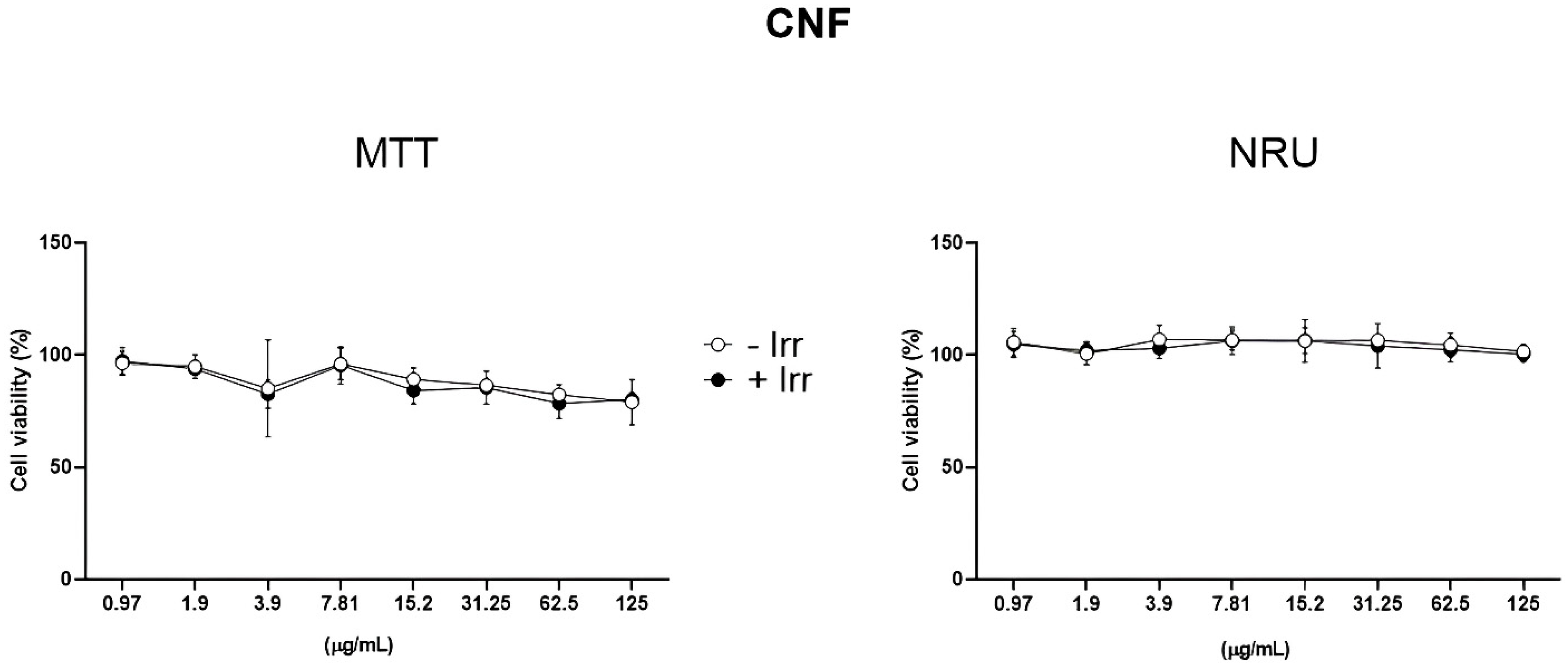
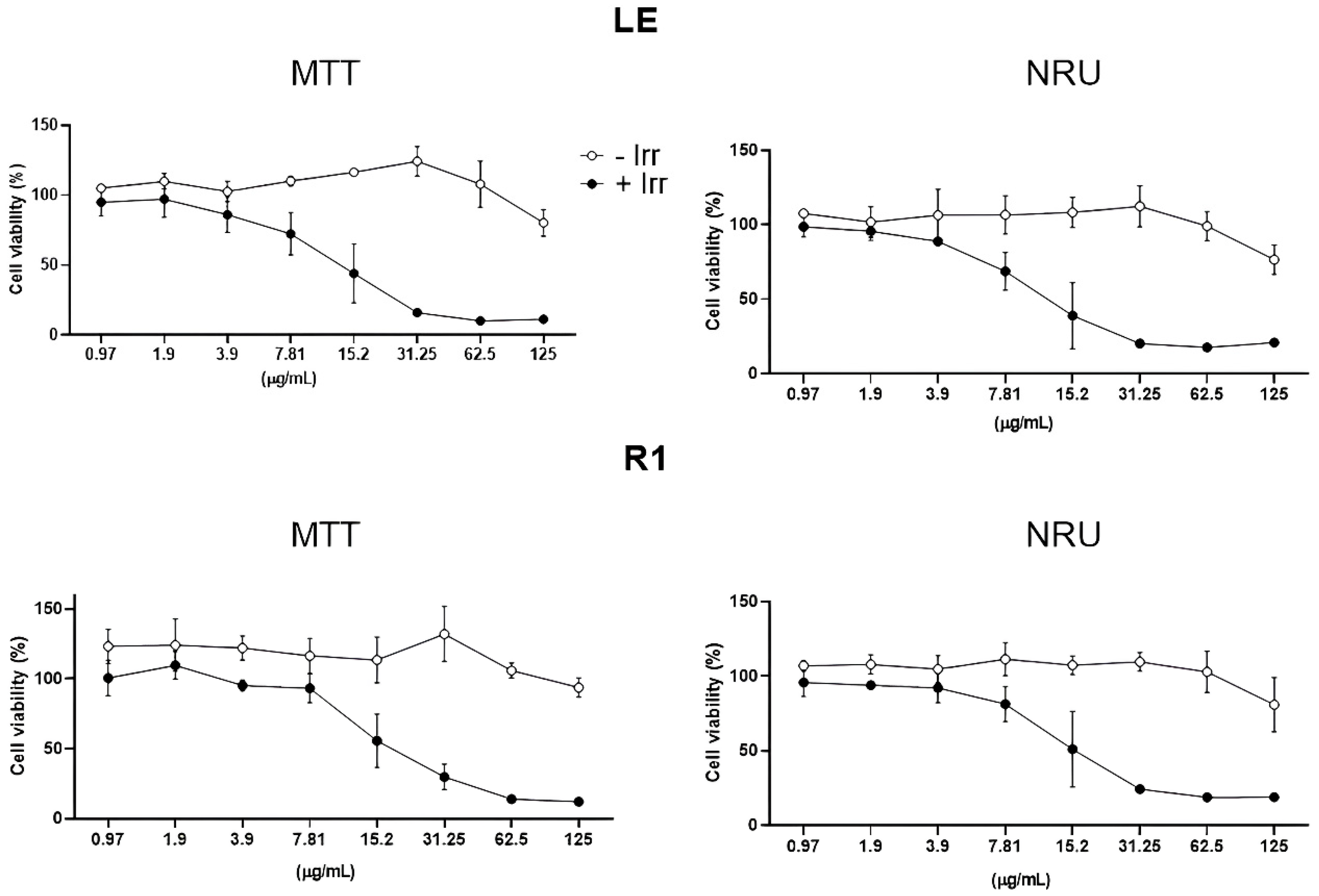
3.1. Phototoxicity Evaluation

While, under non-irradiation conditions, the viability of the HaCaT cells was unchanged after exposure to the tested lignins (LE and R1), a concentration-dependent decrease in cell viability was observed under the irradiation condition for both cell viability markers employed (MTT and NR) (Figure 1). CNF treatment did not affect the viability of the HaCaT cells in any of the tested conditions (irradiation and non-irradiation) (Figure 2). No differences were observed between negative, positive, and solvent controls; the graphics can be observed in the Supplementary Materials (Figures S1 and S2).
At the concentrations assessed (0.12 to 7.8 µg/mL), cell viability was higher than 80% for both types of kraft lignin. For this reason, the calculation of the PIF was not feasible. Therefore, higher lignin concentrations, ranging from 0.97 to 125 µg/mL, were tested to determine the IC50.
When the concentration increased, cell viability decreased after UVA irradiation (Figure 3). Under UVA irradiation, the IC50 determined was 0.015 µg/mL and 0.019 µg/mL for LE and R1, respectively. Both lignins presented an IC50 > 125 µg/mL without UVA irradiation. Therefore, LE presented a PIF of 9.99 and 8.73 for the MTT and NRU test, respectively, and R1 presented a PIF of 6.36 with the MTT assay and 5.59 with the NRU assay (Table 1). Thus, when the lignin concentration increased, the phototoxicity response also increased.

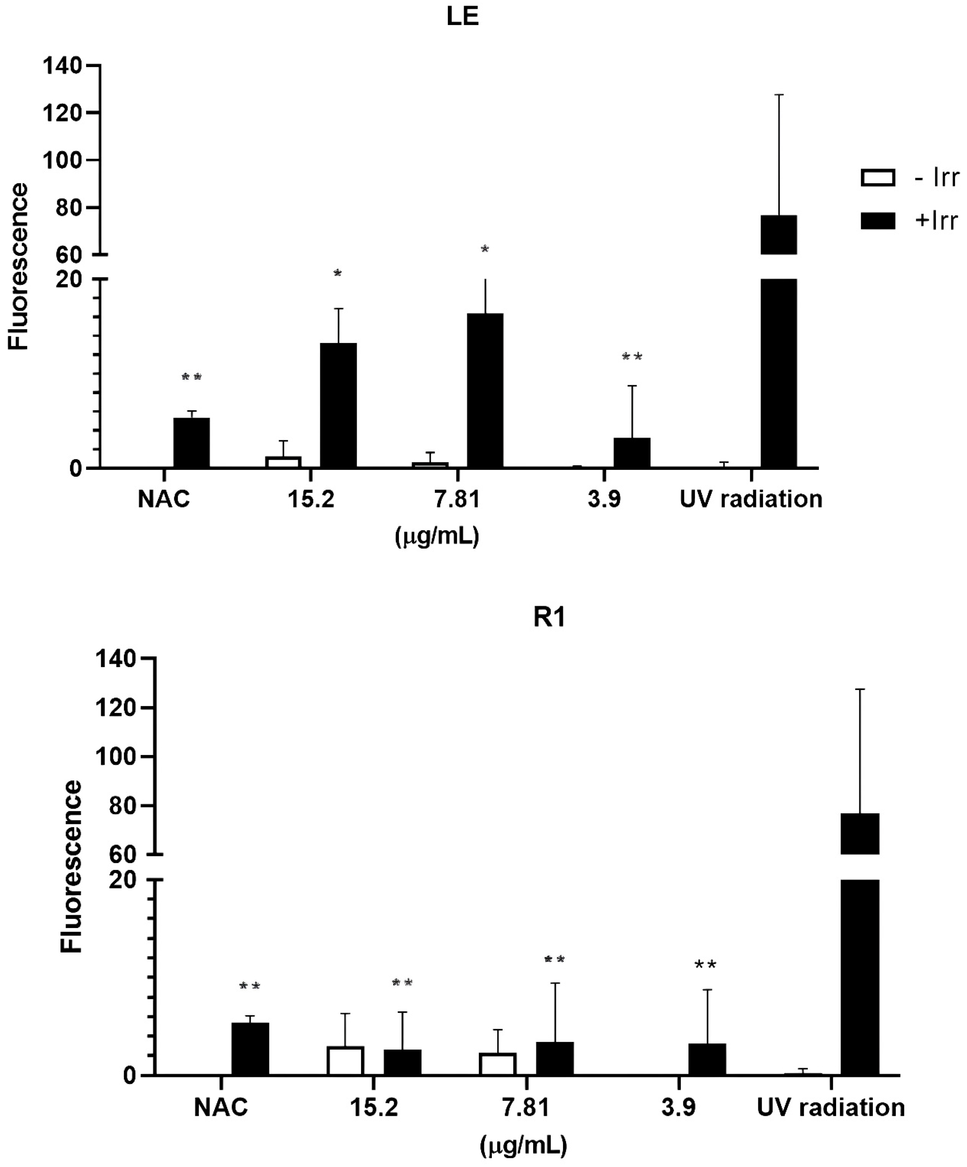
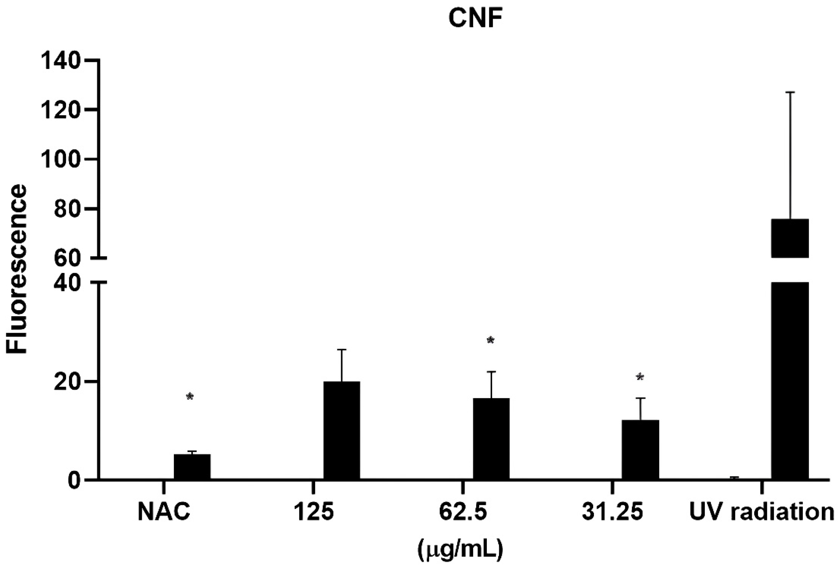
3.2. Evaluation of the Photoprotection Effect Measured by ROS Production

LE, R1, and CNF significantly decreased the production of ROS induced by UVA irradiation in all the tested concentrations (Figure 4).

4. Discussion

In recent years, there has been growing interest in incorporating lignin into cosmetic products, such as sunscreens [8]. Lignin has demonstrated UV filter properties and antioxidant and antimicrobial activities [5]. On the other hand, CNFs are recognized as universal ligands, promoting stability and working as controlled delivery systems [14].
Although lignin has been considered an ingredient for cosmetics due to its UV absorption capacity, few studies have been conducted on its safety. Overall, studies regarding lignin cytotoxicity have been published, but there is still no evidence of phototoxicity. In this study, the cytotoxicity of lignin from sugarcane bagasse was evaluated using HaCaT cells, and a dose-dependent response was reported at higher concentrations (375–1500 µg/mL) [7]. Kraft lignin showed a similar pattern at high concentrations but was more cytotoxic. Gordobil et al., in 2019, evaluated the cytotoxicity of kraft lignin in cancerous and non-cancerous cells, reporting that concentrations from 0.01 to 0.1 mg/mL did not increase the mortality in all tested cell lines; however, at a concentration of 1 mg/mL, all three cell lines presented a significant decrease in cell viability [28]. Alkali lignin and organosolv lignin were tested in the Caco-2 cell line, and increased cell death was observed with increasing concentrations. However, the authors also reported that the increase in cell cytotoxicity was linked with the increase in antioxidant activity, which should be considered in this biopolymer’s applicability [29].
Cytotoxicity is used as a tool to quantify phototoxicity; therefore, our results regarding lignin phototoxicity showed a similar pattern. Higher concentrations of lignin presented a higher phototoxic response. From our knowledge, this is the first time that the phototoxicity of this type of lignin has been evaluated. This is a critical endpoint to consider when evaluating the potential applicability of lignin in formulations for topical use. In addition, it is important to note that LE and R1 are not classified as skin irritants. Gagosian et al., in 2022, showed that LE and R1, when applied to a reconstructed human epidermis (RHE) model, did not reduce cell viability by more than 50% nor caused changes in RHE histology [8]. Therefore, although caution must be taken with lignin concentration regarding UV exposure, RE and L1 may be added to topical formulations without causing skin irritation.
The need for environmental sustainability and the production of value-added products has contributed to the development of new nanomaterials, such as CNF. Although CNF is derived from a renewable source, with a known low impact on human health, the nanoscale can confer physicochemical characteristics different from the bulk material, resulting in a different toxic response [30]. CNF cytotoxicity is related to the time of exposure more than the concentration. It was previously reported that CNF decreased cell viability after 72 h of exposure compared to 24 h at concentrations ranging from 31.25 µg/mL to 1 mg/mL in L929 cells. Similarly, no cytotoxicity was observed after 24 h in A549 cells, but the highest dose was cytotoxic after 48 h [31]. CNFs with different surface modifications, carboxymethylated-CNF and hydroxypropyltrimethylammoniun-CNF, were also evaluated, and no cytotoxicity was reported after exposure in human dermal fibroblasts, lung, and macrophage cell lines [32]. In line with the cytotoxicity data about CNFs, the phototoxicity results presented in this work showed that CNF is not able to elicit a phototoxic response in any of the tested concentrations (0.97–125 µg/mL). Therefore, this nanomaterial can contribute to the development of formulations with low environmental impact.
Lignin presents different chromophore and auxochrome groups, conferring a wide range of UV absorption, from 250 to 400 nm. This is important, considering that UVA radiation has a range of 320–400 nm, representing the main portion of UV radiation which reaches the terrestrial surface [33,34]. However, the capacity of a natural component to act as a UV filter is usually associated with its antioxidant activity, and the antioxidant properties of lignin are considered a mechanism for protection against UV-induced damage [20].
The antioxidant activity of lignin is attributed to its phenolic hydroxyl group, which possesses redox properties and has a key role in eliminating free radicals by extinguishing singlet oxygen or decomposing peroxides [12,35]. The antioxidant activity of kraft lignin has been previously assessed by in chemico methods, such as the 2,2-diphenyl-1-picrylhy- drizzly (DPPH), 2,2′-azino-bis (3-ethylbenzothiazoline-6-sulfonic acid (ABTS), ferric ion-reducing antioxidant potential (FRAP), and Folin–Ciocalteu (FC) assays [36,37]. Furthermore, it has been reported that lignin can reduce the photocatalytic activity of TiO2 [12,38]. TiO2 is a widely used sun blocker; however, it presents a photocatalytic activity that can generate superoxide and hydroxyl radicals after UV radiation, which is not favorable for sunscreen formulations [39]. Therefore, Morsella et al., in 2016, reported that six different types of lignin, which were separately conjugated with TiO2, reduced the photocatalytic activity of TiO2 in chemical and enzymatic reactions [38]. Similarly, Ibrahin et al., in 2019, associated TiO2 with kraft and soda lignin extracted from oil palm, which resulted in a reduction in ROS, with a higher decrease in the kraft lignin and TiO2 composite [12]. In the present study, the antioxidant activity of LE and R1 was assessed through a cell-based in vitro system with HaCaT cells and the DCF-DA probe. Our results showed that LE and R1 had a photoprotective effect by reducing the ROS elicited by the UV radiation. On the other hand, Thá et al., in 2021, using a similar cell-based in vitro assay, reported that kraft lignin generated significant levels of ROS, leading to oxidative DNA damage in HepG2 cells [37]. Therefore, exposure time, cell type, and culture media should be considered when assessing the antioxidant activity of lignin in a cell-based in vitro assay.

5. Conclusions

Lignin and CNF represent a significant portion of the paper and pulp industry’s residues; however, it is crucial to understand the effects of these byproducts on biological systems in order to develop value-added products with a reduced environmental impact. This paper highlights the potential of lignin and CNF as ingredients for the cosmetic industry; however, both materials also present promising applications in the pharmaceutical, biomedical, and food industries. In conclusion, kraft lignin and CNF have the potential to enhance the value of various formulations; however, the concentrations used and the mode of exposure must be carefully considered, particularly for lignin, as higher concentrations of LE and R1 may reduce cell viability after UV irradiation. On the other hand, the tested kraft lignin and CNF significantly decreased ROS production in the cell-based in vitro assay, which supports the increased applicability of kraft lignin and CNF as renewable sources for chemical formulations.

Supplementary Materials

The following supporting information can be downloaded at: https://www.mdpi.com/article/10.3390/cosmetics12020061/s1, Figure S1: Negative and solvent controls for LE and R1; Figure S2: Negative and solvent controls for CNF.

Author Contributions

Conceptualization, J.V.C., D.M.L., W.L.E.M., D.P.d.O., M.M. and M.P.V.; methodology, J.V.C., A.S.M., J.S.G., W.L.E.M. and M.M.; validation, D.M.L., M.M. and M.P.V.; formal analysis, J.V.C., A.S.M., J.S.G., M.M. and M.P.V.; investigation, J.V.C., A.S.M. and J.S.G.; resources, D.P.d.O., D.M.L., M.M. and M.P.V.; data curation, J.V.C., A.S.M. and M.M.; writing—original draft preparation, J.V.C.; writing—review and editing, D.M.L., M.P.V. and M.M.; visualization, J.V.C., D.M.L. and M.P.V.; supervision, D.M.L. and M.P.V.; project administration, D.M.L. and M.M.; funding acquisition, M.M. and D.M.L. All authors have read and agreed to the published version of the manuscript.

Funding

This research was supported by the Coordination for the Improvement of Higher Education Personnel Internationalization Program (CAPES/PRINT, grant 88887.936925/2024-00; CAPES Edital Epidemias—process no. 673/2020), the National Council for Scientific and Technological Development (CNPq, Brazil) (process no. 465571/2014–0; process no. 313713/2020–0), and grant PID2020-113186RB-I00, funded by MICIU/AEI/10.13039/501100011033 (Spain).

Institutional Review Board Statement

Not applicable.

Informed Consent Statement

Not applicable.

Data Availability Statement

Data will be made available upon request.

Acknowledgments

J.V.C. received an international scholarship from the Coordination for the Improvement of Higher Education Personnel/Internationalization Program (CAPES/PRINT, grant 88887.936925/2024-00).

Conflicts of Interest

The authors declare that the research was conducted in the absence of any commercial or financial relationships that could be construed as a potential conflict of interest. The funders had no role in the design of the study; in the collection, analyses or interpretation of data; in the writing of the manuscript; or in the decision to publish the results.

Abbreviations

The following abbreviations are used in this manuscript:
UVUltraviolet irradiation
CNFCellulose nanofiber
LELignin fraction LE
R1Lignin fraction R1
DCF-DA2′,7′-dichlorofluorescein diacetate probe
SPFSun protection factor
ROSReactive oxygen species
TiO2Titanium dioxide
PIFPhotoirritation factor
HaCaTImmortalized keratinocyte cell line
FBSFetal bovine serum
GluL-glutamine
MTT3-(4,5-dimethylthiazol-2-yl)-2,5-diphenyltetrazolium bromide
NRUNeutral red uptake
PBSPhosphate-buffered saline
DMSODimethyl sulfoxide

References

  1. Courtat, M.; Joyce, P.J.; Sim, S.; Sadhukhan, J.; Murphy, R. Towards credible, evidence-based environmental rating ecolabels for consumer products: A proposed framework. J. Environ. Manag. 2023, 336, 117684. [Google Scholar] [CrossRef] [PubMed]
  2. Mondal, S.; Jatrana, A.; Maan, S.; Sharma, P. Lignin modification and valorization in medicine, cosmetics, environmental remediation and agriculture: A review. Environ. Chem. Lett. 2022, 21, 2171–2197. [Google Scholar] [CrossRef]
  3. Rosa, R.; Pini, M.; Cappucci, G.M.; Ferrari, A.M. Principles and indicators for assessing the environmental dimension of sustainability within green and sustainable chemistry. Curr. Opin. Green Sustain. Chem. 2022, 37, 100654. [Google Scholar] [CrossRef]
  4. Ugartondo, V.; Mitjans, M.; Vinardell, M.P. Comparative Antioxidant and Cytotoxic Effects of Lignins from Different Sources. Bioresour. Technol. 2008, 99, 6683–6687. [Google Scholar] [CrossRef]
  5. Kaur, R.; Bhardwaj, S.K.; Chandna, S.; Kim, K.H.; Bhaumik, J. Lignin-based metal oxide nanocomposites for UV protection applications: A review. J. Clean. Prod. 2021, 317, 128300. [Google Scholar] [CrossRef]
  6. Qian, Y.; Qiu, X.; Zhu, S. Lignin: A Nature-Inspired Sun Blocker for Broadspectrum Sunscreens. Green Chem. 2015, 17, 320–324. [Google Scholar] [CrossRef]
  7. Antunes, F.; Mota, I.F.; Fangueiro, J.F.; Lopes, G.; Pintado, M.; Costa, P.S. From Sugarcane to Skin: Lignin as a Multifunctional Ingredient for Cosmetic Application. Int. J. Biol. Macromol. 2023, 234, 123592. [Google Scholar] [CrossRef]
  8. Gagosian, V.S.C.; Claro, F.C.; de AP Schwarzer, A.C.; Cruz, J.V.; Thá, E.L.; da S Trindade, E.; Magalhães, W.L.; Pestana, C.B.; Leme, D.M. The Potential Use of Kraft Lignins as Natural Ingredients for Cosmetics: Evaluating Their Photoprotective Activity and Skin Irritation Potential. Int. J. Biol. Macromol. 2022, 222, 2535–2544. [Google Scholar] [CrossRef]
  9. Ratanasumarn, N.; Chitprasert, P. Cosmetic Potential of Lignin Extracts from Alkaline-Treated Sugarcane Bagasse: Optimization of Extraction Conditions Using Response Surface Methodology. Int. J. Biol. Macromol. 2020, 153, 138–145. [Google Scholar] [CrossRef]
  10. Li, S.X.; Li, M.F.; Bian, J.; Wu, X.F.; Peng, F.; Ma, M.G. Preparation of Organic Acid Lignin Submicrometer Particle as a Natural Broad-Spectrum Photo-Protection Agent. Int. J. Biol. Macromol. 2019, 132, 836–843. [Google Scholar] [CrossRef]
  11. Qian, Y.; Qiu, X.; Zhu, S. Sunscreen Performance of Lignin from Different Technical Resources and Their General Synergistic Effect with Synthetic Sunscreens. ACS Sustain. Chem. Eng. 2016, 4, 4029–4035. [Google Scholar] [CrossRef]
  12. Ibrahim, M.N.M.; Iqbal, A.; Shen, C.C.; Bhawani, S.A.; Adam, F. Synthesis of Lignin Based Composites of TiO2 for Potential Application as Radical Scavengers in Sunscreen Formulation. BMC Chem. 2019, 13, 17. [Google Scholar] [CrossRef]
  13. De France, K.J.; Hoare, T.; Cranston, E.D. Review of Hydrogels and Aerogels Containing Nanocellulose. Chem. Mater. 2017, 29, 4609–4631. [Google Scholar] [CrossRef]
  14. Lavoine, N.; Tabary, N.; Desloges, I.; Martel, B.; Bras, J. Controlled Release of Chlorhexidine Digluconate Using β-Cyclodextrin and Microfibrillated Cellulose. Colloids Surf. B Biointerfaces 2014, 121, 196–205. [Google Scholar] [CrossRef]
  15. Rai, R.; Dhar, P. Biomedical engineering aspects of nanocellulose: A review. Nanotechnology 2022, 33, 362001. [Google Scholar] [CrossRef]
  16. Hinton, A.N.; Goldminz, A.M. Feeling the Burn: Phototoxicity and Photoallergy. Dermatol. Clin. 2020, 38, 165–175. [Google Scholar] [CrossRef]
  17. Ohtake, H.; Tokuyoshi, Y.; Iyama, Y.; Nukaga, T.; Nishida, H.; Ohtake, T.; Hirota, M.; Yamada, K.; Seto, Y.; Sato, H.; et al. Reactive Oxygen Species (ROS) Assay-Based Photosafety Screening for Complex Ingredients: Modification of the ROS Assay Protocol. J. Toxicol. Sci. 2022, 47, 483–492. [Google Scholar] [CrossRef]
  18. Stoudmann, N.; Nowack, B.; Som, C. Prospective Environmental Risk Assessment of Nanocellulose for Europe. Environ. Sci. Nano 2019, 6, 2520–2531. [Google Scholar] [CrossRef]
  19. OECD. Test No. 432: In Vitro 3T3 NRU Phototoxicity Test; Organisation for Economic Co-Operation and Development (OECD): Paris, France, 2019; Section 4. [Google Scholar] [CrossRef]
  20. He, H.; Li, A.; Li, S.; Tang, J.; Li, L.; Xiong, L. Natural components in sunscreens: Topical formulations with sun protection factor (SPF). Biomed. Pharmacother. 2021, 134, 111161. [Google Scholar] [CrossRef]
  21. D’Orazio, J.; Jarrett, S.; Amaro-Ortiz, A.; Scott, T. UV radiation and the skin. Int. J. Mol. Sci. 2013, 14, 12222–12248. [Google Scholar] [CrossRef]
  22. Aliabadi, M.; Chee, B.S.; Matos, M.; Cortese, Y.J.; Nugent, M.J.D.; de Lima, T.A.M.; Magalhães, W.L.E.; de Lima, G.G. Yerba Mate Extract in Microfibrillated Cellulose and Corn Starch Films as a Potential Wound Healing Bandage. Polymers 2020, 12, 2807. [Google Scholar] [CrossRef] [PubMed]
  23. Claro, F.C.; Matos, M.; Jordão, C.; Avelino, F.; Lomonaco, D.; Magalhães, W.L.E. Enhanced Microfibrillated Cellulose-Based Film by Controlling the Hemicellulose Content and MFC Rheology. Carbohydr. Polym. 2019, 218, 307–314. [Google Scholar] [CrossRef] [PubMed]
  24. de Lima, G.G.; Ferreira, B.D.; Matos, M.; Pereira, B.L.; Nugent, M.J.D.; Hansel, F.A.; Magalhães, W.L.E. Effect of Cellulose Size-Concentration on the Structure of Polyvinyl Alcohol Hydrogels. Carbohydr. Polym. 2020, 245, 116612. [Google Scholar] [CrossRef] [PubMed]
  25. de Oliveira, K.M.G.; de Sousa Carvalho, E.H.; da Silva Pereira, B.; Petersohn, E.; Magalhães, W.L.E.; Moura, R.B.P.; Taveira, S.F.; de Cademartori, P.H.G.; Jacumazo, J.; de Freitas, R.A.; et al. Testing the Ecotoxicity of Nanofibrillated Kraft-Bleached Pulp for Use in Nanotechnology Products. Cellulose 2024, 32, 519–534. [Google Scholar] [CrossRef]
  26. Martínez, V.; Galbiati, V.; Corsini, E.; Martín-Venegas, R.; Vinardell, M.P.; Mitjans, M. Establishment of an in Vitro Photoassay Using THP-1 Cells and IL-8 to Discriminate Photoirritants from Photoallergens. Toxicol. In Vitro 2013, 27, 1920–1927. [Google Scholar] [CrossRef]
  27. Baccarin, T.; Mitjans, M.; Ramos, D.; Lemos-Senna, E.; Vinardell, M.P. Photoprotection by Punica Granatum Seed Oil Nanoemulsion Entrapping Polyphenol-Rich Ethyl Acetate Fraction against UVB-Induced DNA Damage in Human Keratinocyte (HaCaT) Cell Line. J. Photochem. Photobiol. B 2015, 153, 127–136. [Google Scholar] [CrossRef]
  28. Gordobil, O.; Oberemko, A.; Saulis, G.; Baublys, V.; Labidi, J. In Vitro Cytotoxicity Studies of Industrial Eucalyptus Kraft Lignins on Mouse Hepatoma, Melanoma and Chinese Hamster Ovary Cells. Int. J. Biol. Macromol. 2019, 135, 353–361. [Google Scholar] [CrossRef]
  29. Gil-Chávez, G.J.; Padhi, S.S.P.; Pereira, C.V.; Guerreiro, J.N.; Matias, A.A.; Smirnova, I. Cytotoxicity and biological capacity of sulfur-free lignins obtained in novel biorefining process. Int. J. Biol. Macromol. 2019, 136, 697–703. [Google Scholar] [CrossRef]
  30. Ventura, C.; Pinto, F.; Lourenço, A.F.; Ferreira, P.J.T.; Louro, H.; Silva, M.J. On the toxicity of cellulose nanocrystals and nanofibrils in animal and cellular models. Cellulose 2020, 27, 5509–5544. [Google Scholar] [CrossRef]
  31. Ventura, C.; Lourenço, A.F.; Sousa-Uva, A.; Ferreira, P.J.T.; Silva, M.J. Evaluating the Genotoxicity of Cellulose Nanofibrils in a Co-Culture of Human Lung Epithelial Cells and Monocyte-Derived Macrophages. Toxicol. Lett. 2018, 291, 173–183. [Google Scholar] [CrossRef]
  32. Lopes, V.R.; Sanchez-Martinez, C.; Strømme, M.; Ferraz, N. In Vitro Biological Responses to Nanofibrillated Cellulose by Human Dermal, Lung and Immune Cells: Surface Chemistry Aspect. Part. Fibre Toxicol. 2017, 14, 1. [Google Scholar] [CrossRef] [PubMed]
  33. Wu, X.; Lian, H.; Xia, C.; Deng, J.; Li, X.; Zhang, C. Mechanistic insights and applications of lignin-based ultraviolet shielding composites: A comprehensive review. Int. J. Biol. Macromol. 2024, 280, 135477. [Google Scholar] [CrossRef]
  34. Zhang, Y.; Naebe, M. Lignin: A Review on Structure, Properties, and Applications as a Light-Colored UV Absorber. ACS Sustain. Chem. Eng. 2021, 9, 1427–1442. [Google Scholar] [CrossRef]
  35. Zhao, L.; Ouyang, X.; Ma, G.; Qian, Y.; Qiu, X.; Ruan, T. Improving Antioxidant Activity of Lignin by Hydrogenolysis. Ind. Crops Prod. 2018, 125, 228–235. [Google Scholar] [CrossRef]
  36. Rumpf, J.; Burger, R.; Schulze, M. Statistical Evaluation of DPPH, ABTS, FRAP, and Folin-Ciocalteu Assays to Assess the Antioxidant Capacity of Lignins. Int. J. Biol. Macromol. 2023, 233, 123470. [Google Scholar] [CrossRef]
  37. Thá, E.L.; Matos, M.; Avelino, F.; Lomonaco, D.; Rodrigues-Souza, I.; Gagosian, V.S.C.; Cestari, M.M.; Magalhães, W.L.E.; Leme, D.M. Safety Aspects of Kraft Lignin Fractions: Discussions on the in Chemico Antioxidant Activity and the Induction of Oxidative Stress on a Cell-Based in Vitro Model. Int. J. Biol. Macromol. 2021, 182, 977–986. [Google Scholar] [CrossRef]
  38. Morsella, M.; D’Alessandro, N.; Lanterna, A.E.; Scaiano, J.C. Improving the Sunscreen Properties of TiO2 through an Understanding of Its Catalytic Properties. ACS Omega 2016, 1, 464–469. [Google Scholar] [CrossRef]
  39. Ishibashi, K.-I.; Fujishima, A.; Watanabe, T.; Hashimoto, K. Detection of active oxidative species in Ti02 photocatalysis using the fluorescence technique. Electrochem. Commun. 2000, 2, 207–210. [Google Scholar] [CrossRef]
Figure 1. Phototoxicity of LE and R1. Cell viability was measured by MTT and NRU assay after UVA irradiation at 4 J/cm2 (+Irr) or without UVA irradiation (−Irr). A decrease in cell viability was only observed at 15.2 µg/mL. The results are expressed as the mean ± S.E.M. of 3 independent experiments.
Cosmetics 12 00061 g001
Figure 2. Phototoxicity of CNF. Cell viability was measured by MTT and NRU assay after UVA irradiation at 4 J/cm2 (+Irr) or without UVA irradiation (−Irr). The results are expressed as the mean ± S.E.M of 3 independent experiments.
Cosmetics 12 00061 g002
Figure 3. Phototoxicity of LE and R1. Cell viability was measured by MTT and NRU assay after UVA irradiation at 4 J (+Irr) or without UVA irradiation (−Irr). The results are expressed as the mean ± S.E.M of n = 3 independent experiments.
Cosmetics 12 00061 g003
Figure 4. Photoprotection effect of lignin measured by ROS production. The amount of ROS was determined by determining the fluorescence using a DCF-DA probe. All the tested concentrations decreased the ROS produced by UV irradiation. The results are expressed as the mean ± S.E.M of 3 independent experiments. Statistical analyses were performed by two-way ANOVA, with a Bonferroni post hoc test. * p value ≤ 0.05; ** p value ≤ 0.009.
Cosmetics 12 00061 g004aCosmetics 12 00061 g004b
Table 1. Photoirritation factor (PIF) determination of LE and R1 after MTT and NRU phototoxicity assays.
MTTNRU
LE10.08.8
R16.45.6
Disclaimer/Publisher’s Note: The statements, opinions and data contained in all publications are solely those of the individual author(s) and contributor(s) and not of MDPI and/or the editor(s). MDPI and/or the editor(s) disclaim responsibility for any injury to people or property resulting from any ideas, methods, instructions or products referred to in the content.

