Article

Collagenase and Tyrosinase Inhibitory Activities and Stability of Facial Cream Formulation Containing Cashew Leaf Extract

by
Pawalee Srisuksomwong
1,
Lalita Kaenhin
2 and
Lapatrada Mungmai
3,4,*
1
Division of Science and Mathematics, Faculty of Science and Technology, Phuket Rajabhat University, Phuket 83000, Thailand
2
Division of Chemistry, Faculty of Education, Phuket Rajabhat University, Phuket 83000, Thailand
3
Division of Cosmetic Science, School of Pharmaceutical Sciences, University of Phayao, Phayao 56000, Thailand
4
Research and Innovation Center in Cosmetic Sciences and Natural Products, Division of Cosmetic Sciences, School of Pharmaceutical Sciences, University of Phayao, Phayao 56000, Thailand
*
Author to whom correspondence should be addressed.
Cosmetics 2023, 10(1), 17; https://doi.org/10.3390/cosmetics10010017
Submission received: 27 December 2022 / Revised: 11 January 2023 / Accepted: 13 January 2023 / Published: 16 January 2023

Abstract

:
The cashew tree (Anacardium occidentale L.) is a tropical plant found widely in many Southeast Asian countries, including Thailand, and contains bioactive phenolic compounds with antioxidant activity. The natural antioxidants such as collagenase and tyrosinase inhibitors found in medicinal plants are promising agents in cosmetic products. This study evaluated the inhibitory activities of the collagenase and tyrosinase from cashew leaf extracts by developing and evaluating the stability of facial cream formulations. The ethanol (DEN), ethyl acetate (DEA) and distilled water (DDW) crude extracts of cashew leaves were investigated for their bioactive compound efficacy. The DDW extract had the highest yield (24.97%). All the extracts were investigated for their antioxidant activities. The DEN extract showed the highest DPPH radical-scavenging ability, ferric-reducing power and flavonoid compounds, which were 152.04 ± 2.40 mg gallic acid/g extract, 37.90 ± 1.07 mg gallic acid/g dry weight and 7.63 ± 0.07 mg quercetin/g dry weight, respectively. The DDW extract exhibited the highest potent activity, which was 111.00 ± 0.78 mg gallic acid/g dry weight in terms of phenolic content, while the DEN extract showed the highest tyrosinase inhibition at 0.100 mg/mL (46.97 ± 3.34%) and collagenase activity at 40 µg/mL. The results suggested that the ethanolic extracts from cashew leaves showed promise for use in skincare product development. Cosmeceutical formulations for skincare were prepared. The formula mixed with DEN extract and added to whitening and anti-aging skincare cream demonstrated good stability and physical properties.

1. Introduction

The cashew tree (Anacardium occidentale L.) belongs to the Anacardiaceae family. This tropical plant is widely distributed in many areas of southeast Asia and tropical America, especially in northeastern Brazil. Both the nut (fruit) and the cashew apples or pseudo-fruits of the cashew tree are marketed, and it is rich in fructose, glucose and amino acids and has a good antioxidant content and is high in ascorbic acid and phenol compounds [1,2]. Cashew apples are used as healthy alternatives to functional foods, while the leaf, stem and bark extracts are used in traditional medicines to treat inflammation, diarrhea, rheumatic diseases, hypertension and infectious diseases [3,4,5,6,7].
Plants containing phytochemicals are beneficial in human diets as they act as natural antioxidants and have health benefits provided by their polyphenols, alkaloids, flavonoids, saponins, tannins, glycosides, anthraquinones and terpenoids [8,9]. Phytochemicals identified in the extracts of the leaves, bark and root of the cashew tree include flavonoids, polyphenols, alkaloids, saponins, tannins, coumarin, quinone and cardiac glycoside [10,11,12]. Shukri and Alan [13] found that the most common phenolic compounds found in the leaf shoots of two varieties of A. occidentale were flavonol glycosides, with other major components including kaempferol 3-O-glucoside, kaempferol 3-O-arabinofuranoside, quercetin 3-O-glucoside and quercetin 3-O-galactoside, while Andarwulan et al. [5] reported that quercetin was the dominant flavonoid, and chlorogenic acid was the dominant phenolic acid in cashew leaves. Kaempferol and quercetin are flavonoid compounds found in a variety of plants such as kale, tea and broccoli that act as potent antioxidants [14], indicating the potential bioactivity of plants as antioxidant sources. Costa et al. [15] identified gallic acid, luteolin, epicatechin gallate and agathisflavone in ethanolic extracts of A. occidentale bark, which showed a moderate activity against all cancer lines (acute promyelocytic leukemia, human colon cancer, human lung mucoepidermoid carcinoma and mastocytoma) with a low cytotoxicity for murine fibroblasts.
Plants and plant extracts are widely used in cosmetics and for many other purposes such as whitening, moisturizing, sunscreen, anti-wrinkle activity, coloring cosmetics, anti-acne, preservatives, antioxidants and thickeners. Skin-lightening products made from natural plant extracts are now becoming popular as effective, safe and non-toxic products [16]. Antioxidants have been widely incorporated into anti-aging cosmetics due to their free-radical-scavenging activity that helps to reduce or prevent skin oxidative stress and can slow down skin aging [17]. Anti-tyrosinase activity refers to the ability of a compound to inhibit the activity of tyrosinase, an enzyme that is involved in the production of melanin in the skin [18]. Inhibiting tyrosinase can help to reduce the production of melanin, which can be beneficial in the treatment of hyperpigmentation disorders such as age spots, freckles and melisma [19,20]. There are several natural compounds that have been shown to have tyrosinase inhibitory activity. Kojic acid is a natural compound that is produced by several species of fungi such as Aspergillus flavus [21], and oxyresveratrol [22] is obtained from plant materials. It has been shown to have a strong tyrosinase inhibitory activity and is often used in skin-lightening products to help reduce hyperpigmentation [23]. Polyphenols such as flavonoids, tocopherols and phenolic acids also have anti-collagenase and anti-elastase activity [24]. Other dominant natural compounds that might contribute to anti-collagenase properties include quercetin, quercetin-3-O-rhamnoside, catechin, epigallocatechin gallate, epicatechin and kaempferol [24,25]. Collagenase is an enzyme that breaks down collagen, a protein that is found in the connective tissue of the skin, bones and other tissues. Collagen makes up 70–80% of the dry weight of skin and is an important component of the skin’s structural support, and its degradation can lead to the loss of skin elasticity and the development of wrinkles [24,26].
Numerous investigations on the phytochemistry of cashew extracts have not connected any particular activity to the identified chemicals. Here, the bioactive components of cashew leaf extracts were examined to determine their contents of potential antioxidant, anti-tyrosinase and anti-collagenase agents as natural sources for skincare products.

2. Materials and Methods

2.1. Extract Preparation

Leaves of A. occidentale L. were purchased at a local market in Klongtom district of Krabi Province, Thailand. The voucher specimens of the plant samples were authenticated by a botanist and deposited at the School of Pharmaceutical Sciences, University of Phayao, Thailand. The reddish young leaves were rinsed several times with tap water, cut into small pieces and dried in a hot-air oven at 50 °C. The dried leaves (200 g) were ground into a powder before extraction in either 95% ethanol (DEN) or ethyl acetate (DEA) at room temperature for 72 h, while the other dried leaves were soaked in warm distilled water for 3 h (DDW). The samples were filtered and evaporated using a vacuum rotary evaporator, and the percentage yield of each extract was calculated. The extracts were kept in amber bottles and stored at 4 °C until further use.

2.2. Determination of Total Phenolic Content

The total phenolic content of the extracts was determined using the Folin–Ciocalteu test, following Mungmai et al. [27] and Chandler and Dodds [28] with slight modifications. The extracts (250 µL) were mixed with 1250 µL of deionized water. Then, 95% ethanol (250 µL) and 50% Folin–Ciocalteu reagent (125 µL) were added to the mixture. The mixture was shaken and allowed to stand at room temperature for 5 min before adding 250 µL of 5% Na2CO3. After incubation in the dark at room temperature for 60 min, the absorbance of the mixture was measured at 725 nm using a UV–Vis spectrophotometer. The total phenolic content was expressed as gallic-acid-equivalent (GAE) in mg/g dry extract (mg GAE/g extract).

2.3. Determination of Total Flavonoid Content

Flavonoid compounds were determined using a aluminum chloride colorimetric assay [29]. Extracts (250 µL) were mixed with 50 µL of 10% AlCl3 and 750 µL of methanol. Then, 50 µL of 1M potassium acetate and 1400 µL of deionized water were added to the mixture. The mixture was shaken and incubated for 30 min in the dark at room temperature. The absorbance of the mixture was measured at 415 nm using a UV–Vis spectrophotometer. The total flavonoid content was expressed as quercetin-equivalent (QE) in mg/g dry extract (mg QE/g extract).

2.4. 2,2-Diphenyl-1-picrylhydrazyl (DPPH) Radical-Scavenging-Capacity Assay

The 1,1-diphenyl-2-picrylhydrazyl (DPPH) radical-scavenging activity of the extracts was measured according to the modified method of Duen et al. [30]. In brief, different dilutions of extracts (80 µL) were mixed with 67 µL of 1 M Tris-HCl (pH 7.9) and 130 µM of DPPH in methanol. The mixture was shaken and incubated for 20 min in the dark at room temperature, and the absorbance was measured at 517 nm using a UV–Vis spectrophotometer. Results were expressed as gallic-acid-equivalent of antioxidant (GAE) in mg/g of dried extract (mg GAE/g extract).

2.5. Ferric-Reducing Antioxidant Power (FRAP) Assay

The ferric-reducing antioxidant power of the extracts was determined following the method described by Oyaizu, [31] with slight modifications. Different extract dilutions (120 µL) were mixed with 290 µL of 0.2 M phosphate buffer (pH 6.6) and 290 µL of 1% (w/v) potassium ferricyanide followed by incubating in a water bath at 50 °C for 20 min. Subsequently, 290 µL of 10% (w/v) trichloroacetic acid was added to the mixture and centrifuged at 3000 rpm for 10 min. The supernatant (1000 µL) was diluted with 1000 µL of deionized water and 400 µL of 0.1% (w/v) of FeCl3 was added. The mixture was shaken thoroughly, and the absorbance was measured at 700 nm using a UV–Vis spectrophotometer. Results were calculated as percentage of inhibition and expressed as mg GAE/g of extract.

2.6. Determination of Tyrosinase-Inhibition Activity

Tyrosinase inhibition activity was determined using the dopachrome method described by Masuda et al. [32] with slight modifications. The extracts were diluted with 50% (w/w) dimethyl sulfoxide. Tyrosinase reactions were performed in a 96-well plate, with each well containing 40 µL of extracts at different dilutions, and 140 µL of 0.1 M phosphate buffer (pH 6.6) followed by 40 µL of tyrosinase solution (31 U/mL). After incubation for 10 min, 40 µL of 2.5 mM L-DOPA was added to the mixture. The 96-well plate was allowed to stand for 10 min at room temperature, and the absorbance of the solution was measured at 475 nm using a microplate reader. Each sample was accompanied by a blank. All the components, except L-DOPA, used kojic acid as the reference substance.

2.7. Determination of Collagenase Inhibition Activity

Collagenase activity was investigated using fluorescein-conjugated gelation as the substrate following Zinger et al. [33] with some modifications. The enzyme solution was added to the substrate solution followed by a reaction buffer (0.5 M Tris-HCl, 1.5 M NaCl, 50 mM CaCl2, 2 mM NaN3, pH 7.6). The fluorescence intensity of each sample was measured every 2 min for 20 min using a multimode reader with excitation set at 485 nm and emission at 530 nm. Collagenase concentration was calculated using a calibration curve generated by collagenase samples of known concentrations.

2.8. Formulation of Facial Cream

The facial cream base formulation was prepared from various compositions by a cool process. Deionized water, polyacrylamide, C13-C14 isoparafin, laureth-7, dimethicone, propylene glycol and phenoxyethanol were mixed together. These were then homogenized until a homogeneous emulsion was obtained. The spreadability, feel-on-skin and pH of the cream base was determined. The stability of the cream base was tested by centrifugation at a speed of 3500 rpm for 30 min. The stable cream base was then incorporate with cashew leaf extract and compared with facial cream without extract addition.

2.9. Stability Studies

Stability studies were performed under various conditions at room temperature, 4 °C and 45 °C for 3 months and using 6 cycles of the heating/cooling method (45 °C, 48 h alternated with 4 °C, 48 h for 1 cycle). Organoleptic characteristics as the color, odor, phase separation, homogeneity, feel-on-skin, pH and viscosity of the creams were evaluated [34].

2.10. Statistical Analysis

Results were expressed as mean values ± standard deviation (SD) using SPSS version 17 for Windows. Significant differences between the groups were determined using one-way ANOVA followed by Tukey’s post hoc test, with the level of significant difference considered at p < 0.05.

3. Results and Discussion

3.1. Percentage Yield of Cashew Leaves

The cashew leaves were extracted using ethanol (DEN), ethyl acetate (DEA) and distilled water (DDW). The DDW extract presented the highest percentage yield at 24.97%, while the percentage yields of DEN and DEA were 22.76% and 1.82%, respectively. The difference in yields depended on the solvent with varying polarity, extraction temperature, extraction time and phytochemicals in the plants or parts of the plants [35,36]. The results indicated that the major phytochemicals in the cashew leaves were high in polarity and soluble in water. The most common phenolic compounds found in the cashew leaves were flavonol glycosides consisting of major components such as kaempferol 3-O-glucoside, kaempferol 3-O-arabinofuranoside, quercetin 3-O-glucoside, quercetin 3-O-galactoside and chlorogenic acid, while quercetin was the dominant flavonoid [5,13]. Pin et al. [37] found that an aqueous leaf extract of betel (Piper betel) gave the highest percentage yield, while Markom et al. [38] determined that Phyllanthus niruri extract recorded the highest percentage yield from water and reported that polar compounds were more easily extracted than non-polar compounds.

3.2. Total Phenolic Content, Flavonoid Content and Antioxidant Activity

The total phenolic content, total flavonoid content and antioxidant activities of the cashew leaf extracts are presented in Table 1. The DDW extract had the highest total phenolic content that was statistically significant (p < 0.05), which was 111.00 ± 0.78 mg GAE/g extract compared with the standard gallic acid, while the DEN extract had the highest total flavonoid content that was statistically significant (p < 0.05), which was 7.63 ± 0.07 mg QE/g extract compared with the quercetin standard. The total phenolic content was dependent on the solvent used in the extraction and its polarity. Phenolic compounds are usually mainly responsible for the antioxidant properties of plants, and most of these compounds are classified as hydrophilic antioxidants [39]. The antioxidant activity of phenolics is related to the number and position of the hydroxyl groups in their aromatic rings; therefore, phenolic compounds have an important influence on the antioxidant activity of plant extracts [40]. Phenolic and flavonoid compounds are widely found as secondary metabolites in plants. Both phenolic and flavonoid compounds directly promote antioxidative action, facilitating the prevention of damage to cells caused by free radicals [41]. In addition, they have been shown to have potential health benefits, including anti-bacterial and anti-inflammatory effects, the ability to protect skin from UV radiation and anti-carcinogenic properties [42,43]. They have been the subject of much research for their potential use in the prevention and treatment of a variety of diseases, including cancer, cardiovascular disease and neurodegenerative disorders.
The DEN extract showed a significantly (p < 0.05) higher DPPH free-radical-scavenging activity than the DDW and DEA extracts, which was 152.04 ± 2.40, 144.21 ± 3.68 and 31.43 ± 1.52 mg GAE/g extract, respectively. This result concurred with Jaiswal et al. [44] who reported that the ethanol-extracted leaf extracts of A. occidentale possessed the strongest activity, followed by aqueous- and petroleum-ether-extracted extracts. The reducing power of the extracts, reflecting their antioxidant activity, was determined using a modified Fe3+ to Fe2+ reduction assay. The results showed that the DEN extract also demonstrated a significantly (p < 0.05) higher activity than the DDW and DEA extracts. This study provided useful information about the antioxidant capacity of cashew leaf extracts as important active ingredients for cosmetic products.

3.3. Tyrosinase Inhibition Activity

Tyrosinase is an enzyme that catalyzes the production of melanin in the skin and hair bulbs and in the eyes of animals. Extracts from natural compounds have shown that the activity of tyrosinase inhibitors could be used for hyperpigmentation and in interests for cosmetic applications such as skin whitening. The inhibitory effects of the extracts on tyrosinase enzyme activity were determined to evaluate their potential as skin-whitening agents. The percentages of tyrosinase inhibition activities at 0.1 mg/mL were 65.66 ± 7.06 (kojic acid), 46.97 ± 3.34 (DEN), 32.29 ± 5.75 (DEA) and 32.68 ± 2.66 (DDW) (Figure 1), with IC50 values of 0.111 ± 0.010, 0.183 ± 0.024, and 0.158 ± 0.018 mg/mL, respectively. Şöhretoğlu et al. [45] investigated the tyrosinase inhibition potential of some structurally related flavonoids and found that quercetin exhibited the strongest effect. Kishore et al. [46] described twelve flavonoid glycosides from the methanol extract of Myrsine africana (Myrsinaceae). Among the list of these compounds, rutin and myricetin-3-O-α-L-rhamnopyranoside showed significant tyrosinase inhibitory effects with IC50 values of 0.13 ± 0.003 and 0.12 ± 0.002 mM, respectively, compared to kojic acid (0.01 ± 0.001 mM). In this research, the ethanol cashew leaf extract had a higher flavonoid content than the water extract. The compounds from the various parts of cashew fruits, namely anacardic acids, 2-methyl cardols, cardol triene and cardols, exhibited tyrosinase inhibitory activity [47,48,49], while the phenolic components 6-[8 (Z), 11 (Z), 14-pentadecatrienyl] salicylic acid and 5-[8 (Z), 11 (Z), 14-pentadecatrienyl] resorcinol exhibited a characteristic competitive inhibition of L-DOPA oxidation [47].

3.4. Collagenase Inhibition Activity

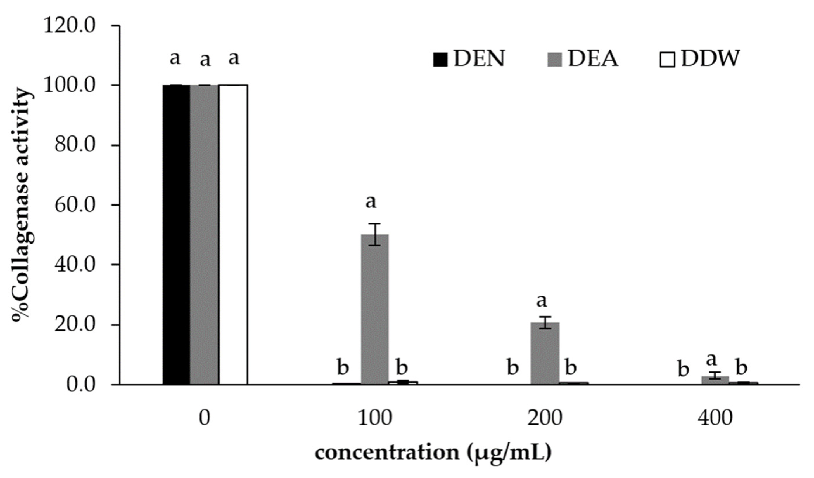
The collagenase enzyme can degrade collagen. As a result, collagen in the underskin or dermis layer is weakened by this enzyme and shows wrinkles because collagenase plays an important role in the skin’s aging process. Incubating DEA, DDW and DEN extracts at concentrations of 100, 200 and 400 µg/mL in the collagenase inhibition test gave collagenase activities of 50.1 ± 3.6, 20.6 ± 2.0 and 2.9 ± 1.1% for the DEA extract, while the DDW and DEN extracts recorded no activity at all the studied concentrations, as shown in Figure 2.
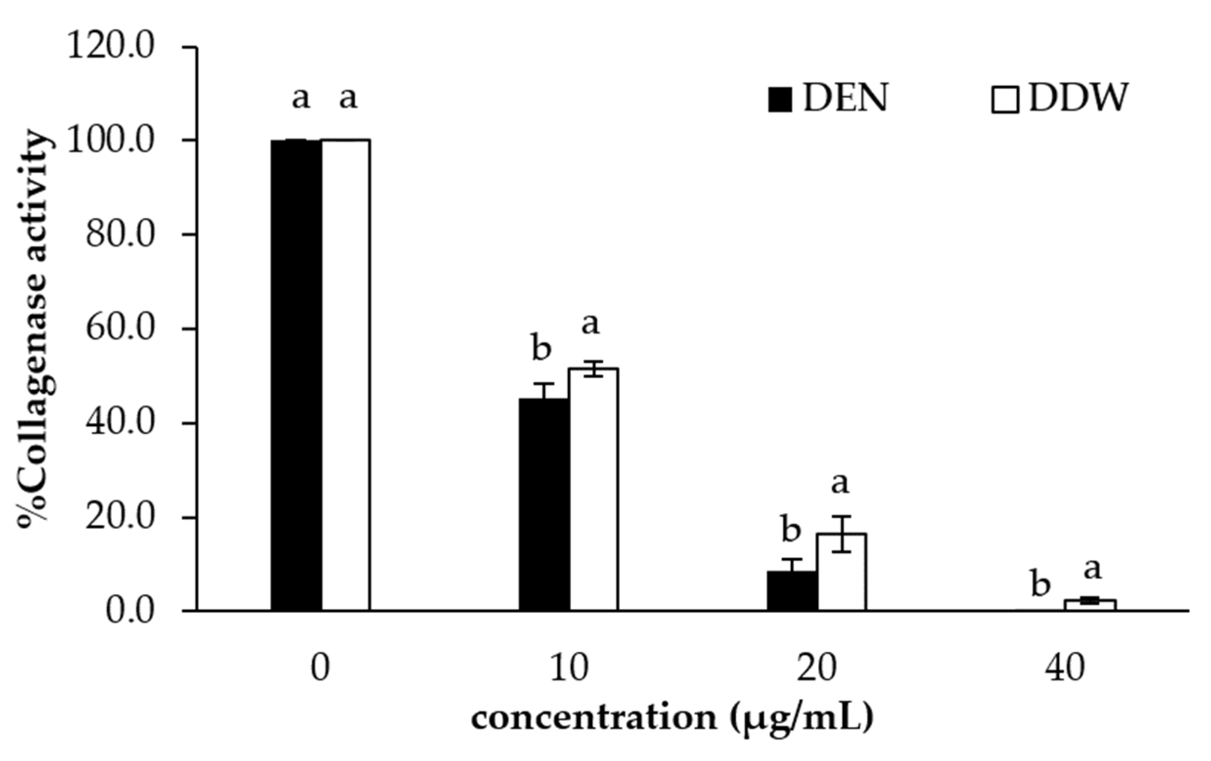
The results showed that the DDW and DEN extracts at concentrations of 100, 200 and 400 mg/mL inhibited the enzyme’s activity. Therefore, the extract concentrations were reduced to 10, 20 and 40 µg/mL, as shown in Figure 3. The DEN extract at 40 µg/mL showed the highest inhibition of collagenase activity, which was recorded at zero percent. Fourteen plant extracts in Thailand have been studied for collagenase inhibition, with only two species inhibiting collagenase enzyme, which were Ardisia elliptica (IC50 = 426.67 µg/mL) and Annona squamosal (IC50 = 426.67 µg/mL) [50]. According to research by Sin and Kim [51], flavonols have a stronger inhibitory impact on collagenase activity than flavones, isoflavones and flavanones. Therefore, it is thought that the flavonol group of chemicals, including quercetin and kaempferol, which may be found in A. occidentale, are what give this plant its strong anti-collagenase activity.
The cashew leaves extracted with ethanol contained many potent antioxidants, which inhibited the tyrosinase enzyme activity, which can cause dull skin with dark spots, and also inhibited the collagenase enzyme activity, which can lead to sagging skin and wrinkles. Therefore, the cashew leaves extracted with ethanol were suitable for further development as a skincare cosmetic product.

3.5. Stability Test of Facial Cream Products

The physical characteristics of the skin cream formulas were evaluated by testing at room temperature, 4 °C and 45 °C for 3 months and under alternating hot and cold conditions for six heating/cooling cycles. The products were kept at 45 °C for 24 h, alternating with cold conditions (4 °C) for 24 h for one cycle, and the pre- and post-test product changes were evaluated in terms of the texture, color, odor, stratification and acid–base (pH) characteristics of the product. The results showed that the formula was stable with satisfactory physical characteristics under the various storage conditions tested. The physical characteristics such as the basic formula remained the same. The color became darker but did not stratify, and the viscosity changed slightly, while the pH increased at 45 °C (Table 2). The heating/cooling cycle remained within the acceptable limits, and the smell remained the same, signifying that the product was stable.

4. Conclusions

The cashew leaves extracted with ethanol (DEN) showed substantial antioxidant activities and the highest inhibition of the crucial enzymes involved in skin aging such as tyrosinase and collagenase, with the potential for cosmeceutical product development for anti-aging, skin-whitening and antioxidant applications. The facial cream containing cashew leaf extracts was investigated for its stability and physical properties. The results revealed that the stability and physical properties of the facial skin cream were satisfactory under various storage conditions.

Author Contributions

Conceptualization, P.S.; methodology, P.S., L.K. and L.M.; validation, P.S., L.K. and L.M.; formal analysis, P.S.; investigation, P.S., L.K. and L.M.; writing—original draft preparation, P.S.; writing—review and editing, P.S. and L.M. All authors have read and agreed to the published version of the manuscript.

Funding

This research was funded by Phuket Rajabhat University, Thailand, grant number PKRU20/2560.

Institutional Review Board Statement

Not applicable.

Informed Consent Statement

Not applicable.

Data Availability Statement

Data are contained within the article.

Acknowledgments

The authors are grateful to the Faculty of Science and Technology, Phuket Rajabhat University and the Research and Innovation Center in Cosmetic Sciences and Natural products, Division of Cosmetic Sciences, School of Pharmaceutical Sciences, University of Phayao for their material support.

Conflicts of Interest

The authors declare no conflict of interest.

References

  1. Rabelo, M.C.; Fontes, C.P.M.L.; Rodrigues, S. Enzyme synthesis of oligosaccharides using cashew apple juice as substrate. Bioresour. Technol. 2009, 100, 5574–5580. [Google Scholar] [CrossRef] [PubMed]
  2. Zepka, L.Q.; Borsarelli, C.D.; da Silva, M.A.A.P.; Mercadante, A.Z. Thermal degradation kinetics of carotenoids in a cashew apple juice model and its impact on the system color. J. Agric. Food Chem. 2009, 57, 7841–7845. [Google Scholar] [CrossRef] [PubMed]
  3. Baptista, A.B.; Sarandy, M.M.; Gonçalves, R.V.; Novaes, R.D.; da Costa, C.G.; Leite, J.P.V.; do Carmo Gouveia Peluzio, M. Antioxidant and anti-inflammatory effects of Anacardium occidentale L. and Anacardium microcarpum D. extracts on the liver of IL-10 knockout mice. Evid.-based Complement. Altern. Med. 2020, 2020, 3054521. [Google Scholar] [CrossRef]
  4. Olajide, O.A.; Aderogba, M.A.; Fiebich, B.L. Mechanisms of anti-inflammatory property of Anacardium occidentale stem bark: Inhibition of NF-jB and MAPK signaling in the microglia. J. Ethnopharmacol. 2013, 145, 42–49. [Google Scholar] [CrossRef] [PubMed]
  5. Andarwulan, N.; Kurniasih, D.; Apriady, R.A.; Rahmat, H.; Roto, A.V.; Bolling, B.W. Polyphenols, carotenoids, and ascorbic acid in underutilized medicinal vegetables. J. Funct. Foods. 2012, 4, 339–347. [Google Scholar] [CrossRef]
  6. Kubo, I.; Nitoda, T.; Tocoli, F.E.; Green, I.R. Multifunctional cytotoxic agents from Anacardium occidentale. Phytother. Res. 2011, 25, 38–45. [Google Scholar] [CrossRef]
  7. Nugroho, A.E.; Malik, A.; Pramono, S. Total phenolic and flavonoid contents, and in vitro anti-hypertension activity of purified extract of Indonesian cashew leaves (Anacardium occidentale L.). Int. Food Res. J. 2013, 20, 299–305. [Google Scholar]
  8. Saxena, M.; Saxena, J.; Nema, R.; Singh, D.; Gupta, A. Phytochemistry of medical plants. J. Pharmacogn. Phytochem. 2013, 1, 169–182. [Google Scholar]
  9. Agidew, M.G. Phytochemical analysis of some selected traditional medicinal plants in Ethiopia. Bull. Natl. Res. Cent. 2022, 46, 87. [Google Scholar] [CrossRef]
  10. Shobana, G.; Keerthana, K.; John, N.A.A.; Felicita, E.S. In vitro antioxidant potentials of aqueous extract of Anacardium occidentale L. Int. J. Pharm. Pharm. Sci. 2016, 5, 1458–1467. [Google Scholar]
  11. Ojezele, M.O.; Agunbiade, S. Phytochemical constituents and medicinal properties of different extracts of Anacardium occidentale and Psidium guajava. Asian J. Biomed. Pharm. Sci. 2013, 3, 20–23. [Google Scholar]
  12. Desai, D.; Raorane, C.; Patil, S.; Gadgil, R.; Patkar, D. Anacardium occidentale: Fountain of phytochemicals; the qualitative profiling. World J. Pharm. Res. 2017, 6, 585–592. [Google Scholar] [CrossRef]
  13. Shukri, M.A.M.; Alan, C. Analysis of phenolics in Anacardium occidentale shoot extracts using a reversed-phase high performance liquid chromatography tandem mass spectrometry (RP-HPLC-MS). J. Trop. Agric. Food Sci. 2010, 38, 221–230. [Google Scholar]
  14. Holland, T.M.; Agarwal, P.; Wang, Y.; Leurgans, S.E.; Bennett, D.A.; Booth, S.L.; Morris, M.C. Dietary flavonols and risk of Alzheimer dementia. Neurology 2020, 94, e1749–e1756. [Google Scholar] [CrossRef]
  15. Costa, A.R.; AlmeiBezerra, J.W.; da Silva, T.G.; Pereira, P.S.; de Oliveira Borba, E.F.; Braga, A.L.; Fonseca, V.J.A.; de Menezes, S.A.; da Silva, F.S.H.; de Sousa Fernandes, P.A.; et al. Phytochemical profile and anti-Candida and cytotoxic potential of Anacardium occidentale L. (cashew tree). Biocatal. Agric. Biotechnol. 2021, 37, 102192. [Google Scholar] [CrossRef]
  16. Jennifer, C.; Stephie, C.M.; Abhishri, S.B.; Shalini, B.U. A review on skin whitening property of plant extracts. Int. J. Pharma Bio Sci. 2012, 3, 332–347. [Google Scholar]
  17. Oresajo, C.; Pillai, S.; Yatskayer, M.; Puccetti, G.; McDaniel, D.H. Antioxidants and skin aging: A review. Cosmet. Dermatol. 2009, 20, 563–570. [Google Scholar]
  18. Chang, T.S. An updated review of tyrosinase inhibitors. Int. J. Mol. Sci. 2009, 10, 2440–2475. [Google Scholar] [CrossRef] [Green Version]
  19. Loizzo, M.R.; Tundis, R.; Menichini, F. Natural and synthetic tyrosinase inhibitors as antibrowning agents: An update. Compr. Rev. Food Sci. Food Saf. 2012, 11, 378–398. [Google Scholar] [CrossRef]
  20. Ciganović, P.; Jakimiuk, K.; Tomczyk, M.; Končić, M.Z. Glycerolic licorice extracts as active cosmeceutical ingredients: Extraction optimization, chemical characterization, and biological activity. Antioxidants 2019, 8, 445. [Google Scholar] [CrossRef] [Green Version]
  21. Ola, A.R.B.; Metboki, G.; Lay, C.S.; Sugi, Y.; Rozari, P.D.; Darmakusuma, D.; Hakim, E.H. Single production of kojic acid by Aspergillus flavus and the revision of flufuran. Molecules 2019, 24, 4200. [Google Scholar] [CrossRef] [PubMed] [Green Version]
  22. Likhitwitayawuid, K. Oxyresveratrol: Sources, productions, biological activities, pharmacokinetics, and delivery systems. Molecules 2021, 26, 4212. [Google Scholar] [CrossRef] [PubMed]
  23. Phasha, V.; Senabe, J.; Ndzotoyi, P.; Okole, B.; Fouche, G.; Chuturgoon, A. Review on the use of kojic acid—A skin-lightening ingredient. Cosmetics 2022, 9, 64. [Google Scholar] [CrossRef]
  24. Thring, T.S.A.; Hili, P.; Naughton, D.P. Anti-collagenase, anti-elastase and anti-oxidant activities of extracts from 21 plants. BMC Complement. Altern. Med. 2009, 9, 1–11. [Google Scholar]
  25. Hussin, M.; Abdul Hamid, A.; Abas, F.; Ramli, N.S.; Jaafar, A.H.; Roowi, S.; Majid, N.A.; Pak Dek, M.S. NMR-based metabolomics profiling for radical scavenging and anti-aging properties of selected herbs. Molecules 2019, 24, 3208. [Google Scholar] [CrossRef] [PubMed] [Green Version]
  26. Jenkins, G. Molecular mechanisms of skin ageing. Mech. Ageing Dev. 2002, 123, 801–810. [Google Scholar] [CrossRef]
  27. Mungmai, L.; Preedalikit, W.; Aunsri, N.; Peerakam, N. Bioactivity test and GC–MS analysis of different solvent extracts from Perilla frutescens (Linn.) Britton and cosmetic product application for sensitive skin. Sci. Tech. Rmutt J. 2019, 9, 78–93. [Google Scholar]
  28. Chandler, S.F.; Dodds, J.H. The effect of phosphate nitrogen and sucrose on the production of phenolics and solasodine in callus cultures of Solanum laciniatum. Plant Cell Rep. 1983, 2, 205–208. [Google Scholar] [CrossRef]
  29. Ghasemi, K.; Ghasemi, Y.; Ali, E. Antioxidant activity, phenol and flavonoid contents of 13 citrus species peels and tissue. J. Pharm. Sci. 2009, 3, 277–281. [Google Scholar]
  30. Duan, X.; Jiang, Y.; Su, X.; Zhang, Z.; Shi, J. Antioxidant properties of anthocyanins extracted from litchi (Litchi chinenesis Sonn.) fruit pericarp tissues in relation to their role in the pericarp browning. Food Chem. 2007, 101, 1365–1371. [Google Scholar] [CrossRef]
  31. Oyaizu, M. Studies on product of browning reaction prepared from glucose amine. Jpn. J. Nutr. 1986, 44, 307–315. [Google Scholar] [CrossRef] [Green Version]
  32. Masuda, T.; Yamashita, D.; Takeda, Y.; Yonemori, S. Screening for tyrosinase inhibitors among extracts of seashore plants and identification of potent inhibitors from Garcinia subelliptica. Biosci. Biotechnol. Biochem. 2005, 69, 197–201. [Google Scholar] [CrossRef] [PubMed] [Green Version]
  33. Zinger, A.; Adir, O.; Alper, M.; Simon, A.; Poley, M.; Tzror, C.; Yaari, Z.; Krayem, M.; Kasten, S.; Nawy, G.; et al. Proteolytic nanoparticles replace a surgical blade by controllably remodeling the oral connective tissue. ACS Nano. 2018, 12, 1482–1490. [Google Scholar] [CrossRef] [PubMed]
  34. Leelapornpisid, P.; Mungmai, L.; Sirithunyalug, B.; Jiranusornkul, S.; Peerapornpisal, Y. A novel moisturizer extracted from freshwater macroalga [Rhizoclonium hieroglyphicum (C.Agardh) Kützing] for skin care cosmetic. Chiang Mai J. Sci. 2014, 41, 1195–1207. [Google Scholar]
  35. Do, Q.D.; Angkawijaya, A.E.; Tran-Nguyen, P.L.; Huong-Huynh, L.; Soetaredjo, F.E.; Ismadji, S.; Ju, Y.H. Effect of extraction solvent on total phenol content, total flavonoid content, and antioxidant activity of Limnophila aromatic. J. Food Drug Anal. 2014, 22, 296–302. [Google Scholar] [CrossRef] [Green Version]
  36. Dharamveer; Mishra, B.; Siddiqui, H.H. Pharmacognostical and phytochemical studies on Anacardium occidentale Linn. Leaves. Res. J. Pharm. Technol. 2013, 6, 75–79. [Google Scholar]
  37. Pin, K.Y.; Luqman Chuah, A.; Abdull Rashih, A.; Mazura, M.P.; Fadzureena, J.; Vimala, S.; Rasadah, M.A. Antioxidant and anti-inflammatory activities of extracts of betel leaves (Piper betle) from solvents with different polarities. J. Trop. For. Sci. 2010, 22, 448–455. [Google Scholar]
  38. Markom, M.; Hassan, M.; Wan Daud, W.R.; Singh, H.; Jahim, J.M. Evaluation of Piper betle on platelet activating factor (PAF) receptor binding activities. Malays. J. Sci. 2007, 26, 79–83. [Google Scholar]
  39. Boeing, J.S.; Barizão, É.O.; e Silva, B.C.; Montanher, P.F.; de Cinque Almeida, V.; Visentainer, J.V. Evaluation of solvent effect on the extraction of phenolic compounds and antioxidant capacities from the berries: Application of principal component analysis. Chem. Cent. J. 2014, 8, 48. [Google Scholar] [CrossRef] [Green Version]
  40. Hervert-Hernández, D.; García, O.P.; Rosado, J.L.; Goñi, I. The contribution of fruits and vegetables to dietary intake of poly-phenols and antioxidant capacity in a Mexican rural diet: Importance of fruit and vegetable variety. Int. Food Res. J. 2011, 44, 1182–1189. [Google Scholar] [CrossRef]
  41. Mungmai, L.; Preedalikit, W.; Pintha, K.; Tantipaiboonwong, P.; Aunsri, N. Collagenase and melanogenesis inhibitory effects of Perilla frutescens pomace extract and its efficacy in topical cosmetic formulations. Cosmetics 2020, 7, 69. [Google Scholar] [CrossRef]
  42. Tungmunnithum, D.; Thongboonyou, A.; Pholboon, A.; Yangsabai, A. Flavonoids and other phenolic compounds from me-dicinal plants for pharmaceutical and medical aspects: An overview. Medicines 2018, 5, 93. [Google Scholar] [CrossRef] [PubMed]
  43. Działo, M.; Mierziak, J.; Korzun, U.; Preisner, M.; Szopa, J.; Kulma, A. The potential of plant phenolics in prevention and therapy of skin disorders. Int. J. Mol. Sci. 2016, 17, 160. [Google Scholar] [CrossRef] [Green Version]
  44. Jaiswal, Y.S.; Tatke, P.A.; Gabhe, S.Y.; Ashok, V. Antioxidant activity of various extracts of leaves of Anacardium occidentale (cashew). Res. J. Pharm. Biol. Chem. Sci. 2010, 1, 112–119. [Google Scholar]
  45. Şöhretoğlu, D.; Sari, S.; Barut, B.; Özel, A. Tyrosinase inhibition by some flavonoids: Inhibitory activity, mechanism by in vitro and in silico studies. Bioorg. Chem. 2018, 81, 168–174. [Google Scholar] [CrossRef]
  46. Kishore, N.; Twilley, D.; van Staden, A.B.; Verma, P.; Singh, B.; Cardinali, G.; Kovacs, D.; Picardo, M.; Kumar, V.; Lall, N. Isolation of flavonoids and flavonoid glycosides from Myrsine africana and their inhibitory activities against mushroom ty-rosinase. J. Nat. Prod. 2018, 81, 49–56. [Google Scholar] [CrossRef]
  47. Kubo, I.; Kinst-Hori, I.; Yokokawa, Y. Tyrosinase inhibitors from Anacardium occidentale fruits. J. Nat. Prod. 1994, 57, 545–551. [Google Scholar] [CrossRef] [PubMed]
  48. Zhuang, J.X.; Hu, Y.H.; Yang, M.H.; Lui, F.J.; Qiu, L.; Zhou, X.W.; Chen, Q.X. Irreversible competitive inhibitory kinetics of cardol triene on mushroom tyrosinase. J. Agric. Food Chem. 2010, 58, 12993–12998. [Google Scholar] [CrossRef]
  49. Yu, X.P.; Su, W.C.; Wang, Q.; Zhuang, J.X.; Tong, R.Q.; Chen, Q.X.; Chen, Q.H. Inhibitory mechanism of cardanols on tyrosinase. Process Biochem. 2016, 51, 2230–2237. [Google Scholar] [CrossRef]
  50. Chatatikun, M. Chiabchalard, A. Thai plants with high antioxidant levels, free radical scavenging activity, anti-tyrosinase and anti-collagenase activity. BMC Complement. Altern. Med. 2017, 17, 487–495. [Google Scholar]
  51. Sin, B.Y.; Kim, H.P. Inhibition of collagenase by naturally-occurring flavonoids. Arch. Pharm. Res. 2005, 28, 1152–1155. [Google Scholar] [CrossRef] [PubMed]
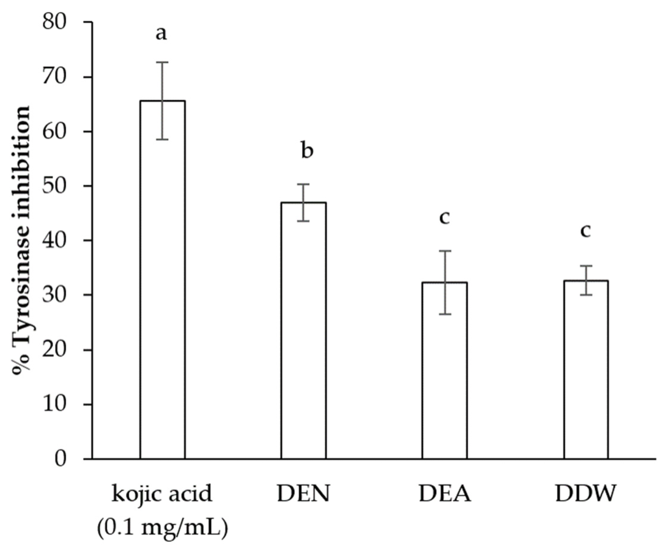
Figure 1. Tyrosinase inhibition of cashew leaf extract (0.1mg/mL) compared with the control (kojic acid). Values are reported as mean ± standard deviation. Different letters (a, b, c) indicate significant differences (p < 0.05).
Cosmetics 10 00017 g001
Figure 2. Comparison between cashew leaves extracted with ethanol (DEN), ethyl acetate (DEA) and water (DDW) at concentrations of 100, 200 and 400 µg/mL for collagenase activity. Values are reported as mean ± standard deviation. Different letters (a, b) indicate significant differences (p < 0.05).
Cosmetics 10 00017 g002
Figure 3. Comparison between cashew leaves extracted with ethanol (DEN), ethyl acetate (DEA) and water (DDW) at concentrations of 10, 20 and 40 µg/mL for collagenase activity. Values are reported as mean ± standard deviation. Different letters (a, b) indicate significant differences (p < 0.05).
Cosmetics 10 00017 g003
Table 1. Total phenolic content, total flavonoid content and antioxidant activities of cashew leaf extracts (mean ± standard deviation, n = 3).
Test MaterialTotal Phenolic Content (mg GAE/g Extract)Total Flavonoid Content (mg QE/g Extract)DPPH Assay
(mg GAE/g Extract)
Reducing Power Assay
(mg GAE/g Extract)
DEN41.39 ± 1.47 b7.63 ± 0.07 a152.04 ± 2.40 a37.90 ± 1.07 a
DEA0.91 ± 0.03 c0.32 ± 0.06 c31.43 ± 1.52 c0.53 ± 0.03 c
DDW111.00 ± 0.78 a3.88 ± 0.03 b144.21 ± 3.68 b35.59 ± 0.66 b
Different letters (a, b, c) in the same column indicate significant differences (p < 0.05).
Table 2. Stability testing results of facial cream containing cashew leaf extracts under various conditions: room temperature (RT), 4 °C and 45 °C for 3 months and six heating/cooling cycles (H/C cycles).
ParameterInitiationRT4 °C45 °CH/C Cycle
Viscosity (pas)1025 ± 3.061030.33 ± 4.161020.33 ± 13.581036.00 ± 5.571044.67 ± 10.26
Color Pale greenPale green Pale greenPale greenPale green
HomogeneityGoodGoodGoodGoodGood
Phase separationNoNoNoNoNo
Feel on skingood goodgoodgoodgood
pH6.016.126.226.356.56
Disclaimer/Publisher’s Note: The statements, opinions and data contained in all publications are solely those of the individual author(s) and contributor(s) and not of MDPI and/or the editor(s). MDPI and/or the editor(s) disclaim responsibility for any injury to people or property resulting from any ideas, methods, instructions or products referred to in the content.