Article

Amplification of Ultra-Trace DNA from Early Sheep Embryos Based on qPCR: Establishing a Gender Identification System

1
College of Life Science and Technology, Tarim University, Alar 843300, China
2
College of Animal Science and Technology, Shihezi University, Shihezi 832003, China
3
Key Laboratory of Tarim Animal Husbandry Science and Technology, Xinjiang Production & Construction Corps, Alar 843300, China
*
Author to whom correspondence should be addressed.
Biology 2025, 14(9), 1144; https://doi.org/10.3390/biology14091144
Submission received: 25 June 2025 / Revised: 19 August 2025 / Accepted: 25 August 2025 / Published: 29 August 2025
(This article belongs to the Special Issue The Biology of Animal Reproduction)

Simple Summary

Identifying the sex of early mammalian embryos is key for gender control. This study used ultra-trace amounts of sheep embryonic DNA templates and specific probes to amplify the SRY gene, establishing a time-saving, effective, simple, and reliable embryo sex identification method. The results showed that the accuracy of embryo sex identification was 100%. Combined with embryo transfer, this technology can accelerate breed selection and promote the development of embryo commercialization.

Abstract

The ability to identify and control the sex of embryos holds significant commercial implications for livestock production. Due to the high costs and instability of detection methods, it is necessary to establish a time-saving, effective, simple, and reliable method to identify the sex of mammalian embryos. This study designs primer probes based on the SRY gene sequence in the sex-determining region of the Y chromosome of sheep and combines qPCR to amplify both the SRY gene and the internal reference gene GAPDH as a control. By adjusting and optimizing the detection system with ultra-trace of genomic DNA (gDNA) from sheep blood and embryos, we sexed sheep embryos cultured in vitro. The results show that male embryos exhibit specific amplification bands for both the GAPDH and SRY genes, while female embryos only have amplification bands for the GAPDH gene. The sensitivity of the sheep embryo sex identification system established in this study can be as low as 5.64 pg of DNA, with an accuracy rate of 100% for embryo sex identification.

1. Introduction

In livestock production, many production traits are controlled by sex, and pre-selecting the sex of offspring guarantees improvement in the production efficiency and sustainability of the livestock industry. As important economic industries, dairy and poultry egg processing benefit from a higher female-to-male sex ratio when increasing output [1]. At the same time, selectively producing females is advantageous for optimizing the reproductive potential of parent stocks. Some human genetic diseases are transmitted through sex-related patterns [2,3]; therefore, developing technology to pre-select the sex of offspring is of great practical value for precise breeding management, promoting genetic improvement, and reducing the spread of sex-related diseases.
In recent years, technological innovation based on embryonic sex determination has accelerated, including single-cell sequencing, pre-implantation genetic diagnosis, non-invasive DNA testing, ultra-low temperature preservation, and embryo transfer [4,5,6]. Studies have shown that male and female mammalian embryos differ not only in chromosomal complement but also in transcriptional activity and epigenetic state. Sexual dimorphism in embryonic transcription affects carbohydrate and amino acid metabolism, with female embryos showing slower mitochondrial metabolism [7], providing new ideas for molecular markers combined with omics analysis for embryo sex identification. Herr et al. first used PCR methods for sex identification in bovine embryos [8]. Polisseni et al. optimized the time, accuracy, and stability of embryo sex identification, and this method has since been widely adopted [9]. However, current research predominantly focuses on the amplification efficiency of high-concentration genomic DNA (gDNA), while studies examining the amplification and detection of low-concentration gDNA, particularly at ultra-trace levels, remain relatively scarce.
The SRY gene is the main effective gene on the Y chromosome’s sex-determining region. It is composed of a single exon, encoding a protein structure domain with a high-mobility group (HMG) that binds to DNA and shows high sequence conservation [10]. The characteristics of the SRY-HMG make it suitable for developing a DNA sex determination test [11]. SRY is only expressed in males, so the absence of SRY gene amplification may result in false-positive outcomes. Glyceraldehyde 3-phosphate dehydrogenase (GAPDH) is an enzyme encoded by an autosomal gene, converting glucose into pyruvate in the glycolytic pathway [12]. Researchers have discovered that the GAPDH gene is highly conserved across mammals and is frequently employed as a classical reference gene in species such as cattle, sheep, horses, and chickens [13,14,15,16]. Therefore, amplifying the SRY and GAPDH genes, combined with fluorescence in situ hybridization (FISH), has been utilized for sex identification [17,18].
This study selects the SRY gene sequence and specific probes from rams to design primers and constructs a composite detection system by combining two sets of these primers. We aim to establish a method for identifying the sex of sheep embryos using ultra-trace embryonic DNA, thereby addressing the identified research gap. This avoids the potential false negatives that may occur if the SRY gene is not amplified in female embryos, improving the sensitivity and identification efficiency of the results.
We propose the following hypothesis: by optimizing qPCR amplification conditions, we can establish an efficient ultra-trace gDNA amplification system for sheep embryos, enabling accurate gene detection at minimal gDNA concentrations. Initially, we established an optimal identification system using sheep blood gDNA and evaluated its specificity at the lowest dilution factor through qPCR amplification. Ultimately, this method was employed to determine the sex of sheep embryos.

2. Materials and Methods

2.1. Ethics Statement

All animal experiments were conducted following the “Regulations and Guidelines for the Management of Experimental Animals” established by the Ministry of Science and Technology (Beijing, China, 2020 revision). This study was approved by the Institutional Animal Care and Use Committee of Tarim University, Xinjiang, China (protocol code DWBH20240814; approval date: 14 August 2024).

2.2. Experiment Location and Time

Experiments were conducted in Alar, China, Xinjiang (40°54′ N, 81°30′ E). Healthy sheep with an average age of 3 years were selected for the experiment. Adequate forage, management, and light intensity were ensured. The sheep were free to drink and supplemented with trace elements, and no other antioxidants were added to their forage.

2.3. Extraction of Blood gDNA

Venous blood samples from sheep (3 males and 3 females) were collected and preserved in anticoagulant tubes. Genomic DNA was extracted from the blood using a Transgen Biotech kit (Beijing, China; technical replicates n = 3). A nucleic acid detector (Nano-400A, Beijing, China) was used to measure the blood DNA concentration (males: 112.8 ± 3.2 ng/μL, females: 113.8 ± 2.9 ng/μL). The blood DNA was diluted in a gradient of 10, 100, 1000, 10,000, and 20,000 times with ddH2O, and the concentrations were measured and stored at −20 °C (males: 11.28 ± 0.21 ng/μL; females: 11.38 ± 0.18 ng/μL), (males: 1.128 ± 0.03 ng/μL; females: 1.138 ± 0.04 ng/μL), (males: 0.1128 ± 0.56 × 10⁻² ng/μL; females: 0.1138 ± 0.83 × 10⁻² ng/μL), (males: 0.01128 ± 0.74 × 10⁻³ ng/μL; females: 0.01138 ± 0.98 × 10⁻³ ng/μL), (males: 0.00564 ± 0.41 × 10⁻³ ng/μL; females: 0.005569 ± 0.46 × 10⁻³ ng/μL).

2.4. Extraction of Embryo gDNA

2.4.1. Embryo Production System

Sheep ovaries were collected from the Aksu slaughterhouse (Permit No. XJ-2023-092) and transported in sterile Phosphate-Buffered Saline (PBS; Saiweier Co., Ltd., Wuhan, China) supplemented with 200 IU/mL penicillin–streptomycin under controlled temperatures (38 ± 0.5 °C) within 2.5 ± 0.3 h.

2.4.2. Oocyte Maturation

Cumulus–oocyte complexes (COCs) were aspirated from 2 to 6 mm follicles using 18 G needles (Kangjian Medical, Taizhou, China, KJ-1820). The grading of COCs is based on their morphological characteristics. Grade A is defined by the presence of at least three layers of tightly arranged cumulus cells, showing no signs of degeneration or expansion. Grade B is characterized by fewer than three layers of cumulus cells, with a uniform oocyte cytoplasm. Grade C includes oocytes that are not surrounded by cumulus cells and may exhibit uneven granulation in the cytoplasm. Finally, Grade D is marked by significant signs of degeneration in the oocytes, including complete degeneration or the absence of cumulus cells. In this experiment, A/B grade COCs were selected for maturation, with 5% estrous sheep serum, 1 mM sodium pyruvate, 1.8 µg/µL heparin, 14 µM isoproterenol, and 2 mg/mL BSA (Solarbio Co., Ltd., Beijing, China) added to an SOF medium (Huankai Microbial, Guangzhou, China). This medium is manufactured by Sigma-Aldrich (St. Louis, MO, USA), a German company, and is identified by the product number M8776. Incubation parameters: 38.5 °C, 5% CO2, 100% humidity for 23 ± 0.5 h.

2.4.3. Sperm Preparation

Fresh sheep semen was collected and transported to the laboratory at a constant temperature of 38 °C. It was then diluted with Bicarbonate-Optimized buffer (BO buffer; Solarbio Co., Ltd., Beijing, China) at a 1:3 volume ratio and centrifuged at 800× g for 5 min. The semen sample was transferred to a 15 mL conical tube. The tube wall was carefully covered with 2 mL of capacitation medium (SP-TALP buffer; Huankai Microbial, Guangzhou, China) and incubated at 38.5 °C in a 5% CO2 environment for 45 min. Following incubation, we aspirated the upper 80% of the liquid, which contained the highly motile sperm [19].

2.4.4. Embryo Culture

The mature oocytes were washed three times with fertilization medium and subsequently transferred into 50 μL of pre-equilibrated fertilization medium, ensuring that a maximum of 15 COCs were added per drop. The upstream semen was added to the fertilization droplets (the sperm density was approximately 1 × 10⁶/mL). The sperm and oocytes were incubated in a CO2 incubator for 18 h (2% essential amino acids, 1.2 mM sodium pyruvate, NaHCO3 (Saiweier Co., Ltd., Wuhan, China), and 4% BSA were added to the SOF medium.

2.4.5. Embryo Biopsy Protocol

Morula-stage embryos were biopsied using an Eppendorf TransferMan® 4 micromanipulator mounted on an Olympus IX73 inverted microscope (Olympus Corporation, Tokyo, Japan). We utilized a holding pipette (Origio MXL3-100—outer diameter, 100 µm; inner diameter, 25 µm) to securely position the embryo, followed by a biopsy with a biopsy needle (Humagen MIC-20; inner diameter, 20 µm; Nikon Co., Ltd., Shenzhen, China).

2.4.6. gDNA Extraction

Biopsied cells (5–8 inner cells per embryo) were lysed in 56 °C lysis buffer containing the following: 0.5% SDS (Sigma, L3771; St. Louis, MO, USA), 200 μg/mL Proteinase K (ThermoFisher, EO0491; Waltham, MA, USA), and 10 mM Tris-HCl (pH 8.0). Thermal cycling: 95 °C for 10 min, ice quenching (2 min), centrifugation (2000× g, 10 min). The gDNA was diluted in a gradient of 1.5, 1.2, and 1 μL with ddH2O, and the concentrations were measured and stored at −20 °C: (males: 22.89 ± 1.32 pg/μL; females: 22.96 ± 1.28 pg/μL), (males: 11.42 ± 1.14 pg/μL; females: 11.48 ± 1.12 pg/μL), (males: 5.78 ± 0.65 pg/μL; females: 5.86 ± 0.71 pg/μL).

2.4.7. Cryopreservation

Another part of the morula-stage embryos was frozen using an embryo freezer (CL-5500, Guangzhou, China): the program started at 20 °C for 28 min (cooling rate 0.61 °C/min), −7 °C for 10 min (2.70 °C/min), −30 °C for 74 min (0.31 °C/min), −33 °C for 30 min (0.10 °C/min). Then, the sample was transferred to liquid nitrogen for storage.

2.5. Primer and Probe Design and Synthesis

The SRY and GAPDH genes sequences of the sheep Y chromosome sex-determining region (SRY, GenBank accession No: Z30265; GAPDH, GenBank accession No: NM-001190390) were designed using BLAST (Blastn, 2.14.0) and PREMIER (Premiere Pro 2020, 14.0) to create primers and probes (Table 1). Primers and probes were synthesized by New Bei Biological Technology Co., Ltd., Shanghai, China.

2.6. Blood qPCR Reaction System

This study utilized the Bio-Rad system (CFX96, Hercules, CA, USA) for qPCR experiments. SRY gene: 5 μL of 2 × S6Probe qPCR Mix, 0.2 μL each of forward and reverse primers (concentration 10 μmol/L), 0.1 μL of probe (5 μmol/L), 1 μL of template DNA (dilution concentrations at gradient 2.3), and ddH2O added to a final volume of 10 μL. GAPDH gene: 5 μL of 2×S6Universal SYBR qPCR Mix, 0.25 μL each of forward and reverse primers, 1 μL of template DNA, and ddH2O added to a final volume of 10 μL. qPCR reaction conditions: initial denaturation at 95 °C for 3 min, followed by 45 cycles of denaturation at 95 °C for 20 s, annealing at 60 °C for 20 s, and a final extension at 72 °C for 5 min, with the reaction products held at 4 °C.

2.7. Embryo Ultra-Trace qPCR Reaction System

SRY gene: gradient concentrations of gDNA at 1.5, 1.2, 1 μL, and 0.2 μL each of forward and reverse primers (dilution concentrations at gradient 2.4), 5 μL of 2 × S6Probe qPCR Mix, and ddH2O added to a final volume of 10 μL. GAPDH gene: 5 μL of 2 × S6Universal SYBR qPCR Mix, 0.25 μL each of forward and reverse primers (10 μmol/L), 1 μL of template DNA, and ddH2O added to a final volume of 10 μL. qPCR reaction conditions: initial denaturation at 95 °C for 3 min, followed by 45 cycles of denaturation at 95 °C for 20 s, annealing and extension at 60 °C for 20 s, and a final extension at 72 °C for 5 min, with the reaction products held at 4 °C.

2.8. Agarose Gel Electrophoresis

The qPCR amplification products (4 μL) were thoroughly mixed with the electrophoresis buffer (1 μL) and then added to the wells of a 2% agarose gel in sequence. Electrophoresis conditions: 150 V voltage, 130 mA current, 20 min. The results were observed using a gel imaging system. The amplification results for males showed two bands, while females had only one band.

2.9. Data Analysis

qPCR data was analyzed utilizing Bio-Rad’s CFX Manager (Version 3.1), while the real-time quantitative qPCR data were evaluated using the 2−ΔΔCt method. Furthermore, statistical analysis was performed using GraphPad Prism version 9.5.1.

3. Results

3.1. Establishing the Blood gDNA Ultra-Trace qPCR Amplification System

Sheep blood gDNA was diluted in a gradient of 10, 100, 1000, 10,000, and 20,000 times to obtain amplification curves, as shown in Figure 1A. All curves were stable, and the smaller the DNA concentration, the higher the ct (threshold cycle) value. The male sheep blood gDNA sample demonstrated amplification curves of the GAPDH and SRY genes, while the female sheep blood gDNA sample only demonstrated the amplification curve of the internal reference gene, GAPDH. The results were consistent with the experimental design.
The melting curve is demonstrated by amplifying sheep blood group DNA samples with gradient concentrations is shown in Figure 1B. The Tm values of the GAPDH and SRY genes were 86 °C and 83.5 °C, respectively. The melting curve was a single peak, and there was no primer dimer or non-specific amplification.

3.2. Verifying the Amplification Results for Blood gDNA Diluted 20,000 Times

The qPCR amplification curves of sheep blood gDNA diluted 20,000 times (♂5.64 pg/μL, ♀5.69 pg/μL) are shown in Figure 1C. Each sample was amplified in triplicate, and the detection accuracy of the SRY gene in the samples was 100%. The melting curve is shown in Figure 1D, and the results are completely consistent with the experimental design.

3.3. Serial Dilution of Blood gDNA for Electrophoresis Detection

The gradient concentration template amplification products of the blood gDNA were subjected to electrophoresis detection, and the results are shown in Figure 2. Males exhibited 162 bp SRY and 167 bp GAPDH gene amplification bands, while females only had a 167 bp GAPDH gene amplification band. The brightness of the amplification bands dimmed with decreasing concentrations of the amplified samples. The electrophoresis results confirmed the accuracy of the qPCR amplification results.

3.4. Gradient Concentration Amplification Results for Embryo gDNA

In this experiment, 1.5 μL, 1.2 μL, and 1 μL of gDNA solution were added to the qPCR reaction system. The results of the qPCR amplification are presented in Figure 3A. All six embryos amplified the GAPDH gene, and four embryos amplified the SRY gene, indicating they were male. Meanwhile, the optimal amplification curve was obtained when the volume of the gDNA solution was 1 μL.

3.5. Embryo Sex Identification

The experimental results concerning embryo sex identification show that ten representative embryos were randomly selected for analysis. The qPCR amplification results are illustrated in Figure 3B. All ten embryos amplified the GAPDH gene; six male embryos showed the SRY gene amplification curve, while four female embryos did not show the SRY gene amplification curve, with 100% identification accuracy.

4. Discussion

When combined with superovulation and embryo transfer techniques, sex control technology plays a crucial role in livestock production. As a primary method of sex control, embryo sex identification enables the production of offspring of a desired sex through embryo transfer, significantly contributing to improving production technologies and providing economic benefits [20]. This study established a qPCR amplification reaction system for ultra-trace gDNA in sheep’s blood and further adjusted the annealing temperature and cycle number to optimize the reaction system. The results show that the identification results are accurate and reliable under the following conditions: 20 μL of the amplification system, an annealing temperature of 60 °C, and 45 cycles. Nine sheep embryos were selected for gender identification using these methods, with a sensitivity of up to 5.64 pg and 100% accuracy. This method proved to be more sensitive and accurate than the findings of previous research [21,22,23].
Research shows that qPCR technology can quickly identify early embryos [24]. Mammalian Y chromosomes contain SRY-specific genes, which have high homology and high conservatism [25]. The sex identification protocol and primers used in this experiment are cheaper and more efficient than other methods described in the introduction, mainly for determining the sex of embryo biopsies before embryo transfer. The specific expression of SRY in male embryos is sufficient to determine their gender and has been used in different studies [26,27,28,29]. However, given the absence of the SRY gene in female embryos, it is not yet certain whether this indicates a positive result or PCR failure. Therefore, we designed SRY-specific probes, showing that the combined amplification of the housekeeping gene GAPDH can avoid the misinterpretation of false-negative results.
Ensuring reaction specificity with the optimal annealing temperature for specific primers is a critical step in improving the efficiency of qPCR amplification [30]. We designed temperature gradients and explored their effects multiple times, ultimately selecting an optimal annealing temperature of 60 °C. In addition, combining embryo gender identification with transplantation technology can provide greater benefits. This study conducted biopsies on embryos at the morula stage, employing micromanipulation instruments to extract 5–8 inner cells from each embryo. This procedure is widely used in preimplantation genetic diagnosis (PGD) and has been shown to be effective in obtaining genetic material for analysis [9,31].
Through blood tests, researchers such as María Sánchez and Rogers have found that the SRY gene product is only produced in ewes carrying male lambs, indicating that the detection time can only occur after the fetus has established contact with the mother [32,33]. Therefore, embryo gender identification is mainly used for embryos before implantation, requiring shorter cycles than those of natural delivery identification. The findings of this study indicate that biopsy does not significantly affect the quality or viability of embryos. The established ultra-trace gDNA amplification system, in conjunction with SRY-specific probes, greatly enhances the sensitivity of identification and will be significantly beneficial for research on early mammalian embryonic development characteristics and sex control technologies. Furthermore, future research should continue to optimize and consistently monitor biopsy techniques meticulously, enabling the safe application of this method for sex determination and other genetic analyses in early embryos.

5. Conclusions

This study established a rapid, sensitive, and accurate method for identifying the sex of sheep embryos, which can be further used to identify the sex and species of other mammalian biological samples. This method combines ultra-low-temperature freezing and embryo transfer techniques to obtain offspring of the desired gender.

Author Contributions

P.N. and D.F. conceived, drafted and approved this research article. Conceptualization: D.F.; data curation: P.N. and W.T.; formal analysis: F.H.; funding acquisition: D.F. and P.N.; methodology: D.F., P.N., Q.G., X.L. and X.W.; resources: P.N., F.H., J.W. and X.L.; visualization: P.N., X.W. and F.H.; roles/writing—original draft: P.N.; writing—review and editing: D.F., Q.G., W.T. and J.W. All authors have read and agreed to the published version of the manuscript.

Funding

This work was supported by the “Tianchi Talents” introduction program [grant numbers 2022CB021] and Tarim University President’s Fund Project [grant numbers TDZKBS202406].

Institutional Review Board Statement

All animal experiments were conducted in accordance with the “Regulations and Guidelines for the Management of Experimental Animals” established by the Ministry of Science and Technology (Beijing, China, 2020 revision). This study was approved by the Institutional Animal Care and Use Committee of Tarim University, Xinjiang, China (protocol code DWBH20240814; approval date: 14 August 2024).

Informed Consent Statement

Informed consent was obtained from all subjects involved in the study.

Data Availability Statement

The original contributions presented in the study are included in the article; further inquiries can be directed to the corresponding authors.

Conflicts of Interest

The authors declare that there are no competing interests associated with the manuscript.

References

  1. Xia, X.H.; Liang, N.; Ma, X.Y.; Lu, Q.M.; Wang, S.S.; Chang, Z.J. Inhibition of the NF-κB signaling pathway affects gonadal differentiation and leads to male bias in Paramisgurnus dabryanus. Theriogenology 2023, 207, 82–95. [Google Scholar] [CrossRef]
  2. Khalajzadeh, S.; Nejati-Javaremi, A.; Yeganeh, H. Effect of widespread and limited use of sexed semen on genetic progress and reproductive performance of dairy cows. Animal 2012, 6, 1398–1406. [Google Scholar] [CrossRef] [PubMed]
  3. Khramtsova, E.A.; Wilson, M.A.; Martin, J.; Winham, S.J.; He, K.Y.; Davis, L.K.; Stranger, B.E. Quality control and analytic best practices for testing genetic models of sex differences in large populations. Cell 2023, 186, 2044–2061. [Google Scholar] [CrossRef] [PubMed]
  4. Cusanovich, D.A.; Reddington, J.P.; Garfield, D.A.; Daza, R.M.; Aghamirzaie, D.; Marco-Ferreres, R.; Pliner, H.A.; Christiansen, L.; Qiu, X.; Steemers, F.J.; et al. The cis-regulatory dynamics of embryonic development at single-cell resolution. Nature 2018, 555, 538–542. [Google Scholar] [CrossRef] [PubMed]
  5. Yao, Y.; Yang, A.; Li, G.; Wu, H.; Deng, S.; Yang, H.; Ma, W.; Lv, D.; Fu, Y.; Ji, P.; et al. Melatonin promotes the development of sheep transgenic cloned embryos by protecting donor and recipient cells. Cell Cycle 2022, 21, 1360–1375. [Google Scholar] [CrossRef]
  6. Dvoran, M.; Nemcova, L.; Kalous, J. An Interplay between Epigenetics and Translation in Oocyte Maturation and Embryo Development: Assisted Reproduction Perspective. Biomedicines 2022, 10, 1689. [Google Scholar] [CrossRef]
  7. Fraire-Zamora, J.; Martínez, M.F.; Torra-Massana, M.; Zamora, M.J.; Vassena, R.; Miguel-Escalada, I.; Rodríguez, A.; Popovic, M. P-135 No difference in morphokinetic patterns between male and female preimplantation embryos. Hum. Reprod. 2023, 38. [Google Scholar] [CrossRef]
  8. Herr, C.; Matthaei, K.I.; Steel, T.; Reed, K.C. Rapid Y-chromosome-assay sexing of peripheral blood lymphocytes from bovinae of known phenotypic sex. Theriogenology 1990, 33, 246. [Google Scholar] [CrossRef]
  9. Polisseni, J.; Sá, W.F.; Guerra, M.O.; Machado, M.A.; Serapião, R.V.; Carvalho, B.C.; Camargo, L.S.; Peters, V.M. Post-biopsy bovine embryo viability and whole genome amplification in preimplantation genetic diagnosis. Fertil. Steril. 2010, 93, 783–788. [Google Scholar] [CrossRef]
  10. Christian, L.; Timothy, L.B.; Peter, K. Switching on sex: Transcriptional regulation of the testis-determining gene Sry. Development 2014, 141, 2195–2205. [Google Scholar] [CrossRef]
  11. Vincent, R.H.; Michael, C.; Anthony, A. The Molecular Action and Regulation of the Testis-Determining Factors, SRY (Sex-Determining Region on the Y Chromosome) and SOX9 [SRY-Related High-Mobility Group (HMG) Box 9]. Endocr. Rev. 2003, 24, 466–487. [Google Scholar] [CrossRef]
  12. Wei, H.; Movahedi, A.; Yang, J.; Zhang, Y.Y.; Liu, G.Y.; Zhu, S.; Yu, C.M.; Chen, Y.H.; Zhong, F.; Zhang, J. Characteristics and molecular identification of glyceraldehyde-3-phosphate dehydrogenases in poplar. Int. J. Biol. Macromol. 2022, 219, 185–198. [Google Scholar] [CrossRef] [PubMed]
  13. Rekawiecki, R.; Kowalik, M.K.; Kotwica, J. Validation of housekeeping genes for studying differential gene expression in the bovine myometrium. Acta Vet. Hung. 2013, 61, 505–516. [Google Scholar] [CrossRef] [PubMed]
  14. Sonowal, J.; Lal, P.C.; Dev, K.; Singh, R.; Barkathullah, N.; Akram, M.W.; Kumar, G.R.; Kant, A.R.; Kumar, D.; Saxena, S.; et al. Selection and validation of suitable reference gene for qPCR gene expression analysis in lamb testis cells under Sheep pox virus infection. Gene 2022, 831, 146561. [Google Scholar] [CrossRef] [PubMed]
  15. Azarpeykan, S.; Dittmer, K.E. Evaluation of housekeeping genes for quantitative gene expression analysis in the equine kidney. J. Equine Sci. 2016, 27, 165–168. [Google Scholar] [CrossRef]
  16. Mogilicherla, K.; Athe, R.P.; Chatterjee, R.N.; Bhattacharya, T.K. Identification of suitable reference genes for normalization of quantitative real-time PCR-based gene expression in chicken (Gallus gallus). Anim. Genet. 2022, 53, 881–887. [Google Scholar] [CrossRef] [PubMed]
  17. Lee, J.H.; Park, J.H.; Lee, S.H.; Park, C.S.; Jin, D.I. Sexing using single blastomere derived from IVF bovine embryos by fluorescence in situ hybridization (FISH). Theriogenology 2004, 62, 1452–1458. [Google Scholar] [CrossRef]
  18. Mishra, A.; Dhali, A.; Reddy, I.J.; Kolte, A.P. Sexing of pre-implantation ovine embryos through polymerase chain reaction-based amplification of GAPDH, SRY and AMEL genes. Reprod. Domest. Anim. 2020, 55, 885–892. [Google Scholar] [CrossRef]
  19. Nix, J.L.; Marrella, M.A.; Oliver, M.A.; Ealy, A.D.; Biase, F.H. Cleavage kinetics is a better indicator of embryonic developmental competency than brilliant cresyl blue staining of oocytes. Anim. Reprod. 2023, 248, 107174. [Google Scholar] [CrossRef]
  20. He, Q.F.; Huang, M.; Cao, X.Y.; Zhang, K.; Li, J.; Quan, F.S. Advancements in mammalian X and Y sperm differences and sex control technology. Zygote 2022, 30, 423–430. [Google Scholar] [CrossRef]
  21. Nix, J.L.; Schettini, G.; Biase, F.H. Sexing of cattle embryos using RNA-sequencing data or polymerase chain reaction based on a complete sequence of cattle chromosome Y. Front. Genet. 2023, 14, 1038291. [Google Scholar] [CrossRef] [PubMed]
  22. López-Gatius, F.; García-Ispierto, I. Sexing of Embryos at the Time of Twin Reduction: A Clinical Approach. Animals 2023, 13, 1326. [Google Scholar] [CrossRef] [PubMed]
  23. Cordeiro, C.D.; Gonceer, N.; Dorus, S.; Crill, J.E.; Moshayoff, V.; Lachman, A.; Moran, A.P.; Vilenchik, D.; Fedida-Metula, S. Fast, accurate, and cost-effective poultry sex genotyping using real-time polymerase chain reaction. Front. Vet. Sci. 2023, 10, 1196755. [Google Scholar] [CrossRef] [PubMed]
  24. Camargo, G.S.; Meira, C.D.; Bergfelt, D.R.; Oliveira, J.P.; Bromberger, C.R.; Barros, L.D.; Canesin, H.S.; Lopes, G.; Monteiro, G.A.; Ignácio, F.S. Equine blastocyst re-expansion rate, quality, and sex following embryonic collapse using needle perforation of blastocoel and subsequent PCR amplification of blastocoel DNA. J. Equine Vet. Sci. 2023, 118, 269. [Google Scholar] [CrossRef]
  25. Ridnik, M.; Abberbock, E.; Alipov, V.; Lhermann, S.Z.; Kaufman, S.; Lubman, M.; Poulat, F.; Gonen, N. Two redundant transcription factor binding sites in a single enhancer are essential for mammalian sex determination. Nucleic Acids Res. 2024, 52, 5514–5528. [Google Scholar] [CrossRef]
  26. Pathak, D.; Baksi, A.; Vasan, S.S.; Dighe, R.R. Molecular and Functional Characterization of Human Sex-Determining Region on the Y Chromosome Variants Using Protamine 1 Promoter. DNA Cell Biol. 2024, 43, 12–25. [Google Scholar] [CrossRef]
  27. Angove, M.L.; Turner, Z.; Conley, A.J.; McNabb, B.R.; Eenennaam, A.L. PSI-4 Investigation of a Naturally Occurring XX Male Bull. J. Anim. Sci. 2022, 100, 22–23. [Google Scholar] [CrossRef]
  28. Dujardin, É.; André, M.; Dewaele, A.; Mandon-Pepin, B.; Poulat, F.; Frambourg, A.; Thépot, D.; Jouneau, L.; Jolivet, G.; Pailhoux, É.; et al. DMRT1 is a testis-determining gene in rabbits and is also essential for female fertility. eLife 2023, 12, RP89284. [Google Scholar] [CrossRef]
  29. Li, N.; Zhou, J.; Zhang, W.Q.; Liu, W.J.; Wang, B.X.; She, H.B.; Mirbahar, A.A.; Li, S.F.; Zhang, Y.L.; Gao, W.J.; et al. A rapid method for assembly of single chromosome and identification of sex determination region based on single-chromosome sequencing. New Phytol. 2023, 240, 892–903. [Google Scholar] [CrossRef]
  30. Wu, L.R.; Chen, S.X.; Wu, Y.L.; Patel, A.A.; Zhang, D.Y. Multiplexed enrichment of rare DNA variants via sequence-selective and temperature-robust amplification. Nat. Biomed. Eng. 2017, 1, 714–723. [Google Scholar] [CrossRef]
  31. Coster, T.D.; Poucke, M.V.; Pascottini, O.B.; Angel-Velez, D.; Branden, E.V.; Peere, S.; Papas, M.; Gerits, I.; Govaere, J.; Peelman, L.; et al. Single closed-tube quantitative real-time PCR assay with dual-labelled probes for improved sex determination of equine embryos. Animal 2023, 17, 100952. [Google Scholar] [CrossRef]
  32. Rogers, F.; Rhoades, J.R.; Quail, L.K.; Rich, J.J. 113 Investigation of differences in pregnancy associated glycoproteins due to different fetal parameters (sex and number) using commercial tests in multiparous ewes. J. Anim. Sci. 2024, 102, 104–105. [Google Scholar] [CrossRef]
  33. Sánchez, J.M.; Gómez-Redondo, I.; Browne, J.A.; Planells, B.; Gutiérrez-Adán, A.; Lonergan, P. MicroRNAs in amniotic fluid and maternal blood plasma associated with sex determination and early gonad differentiation in cattle. Biol. Reprod. 2021, 105, 345–358. [Google Scholar] [CrossRef]
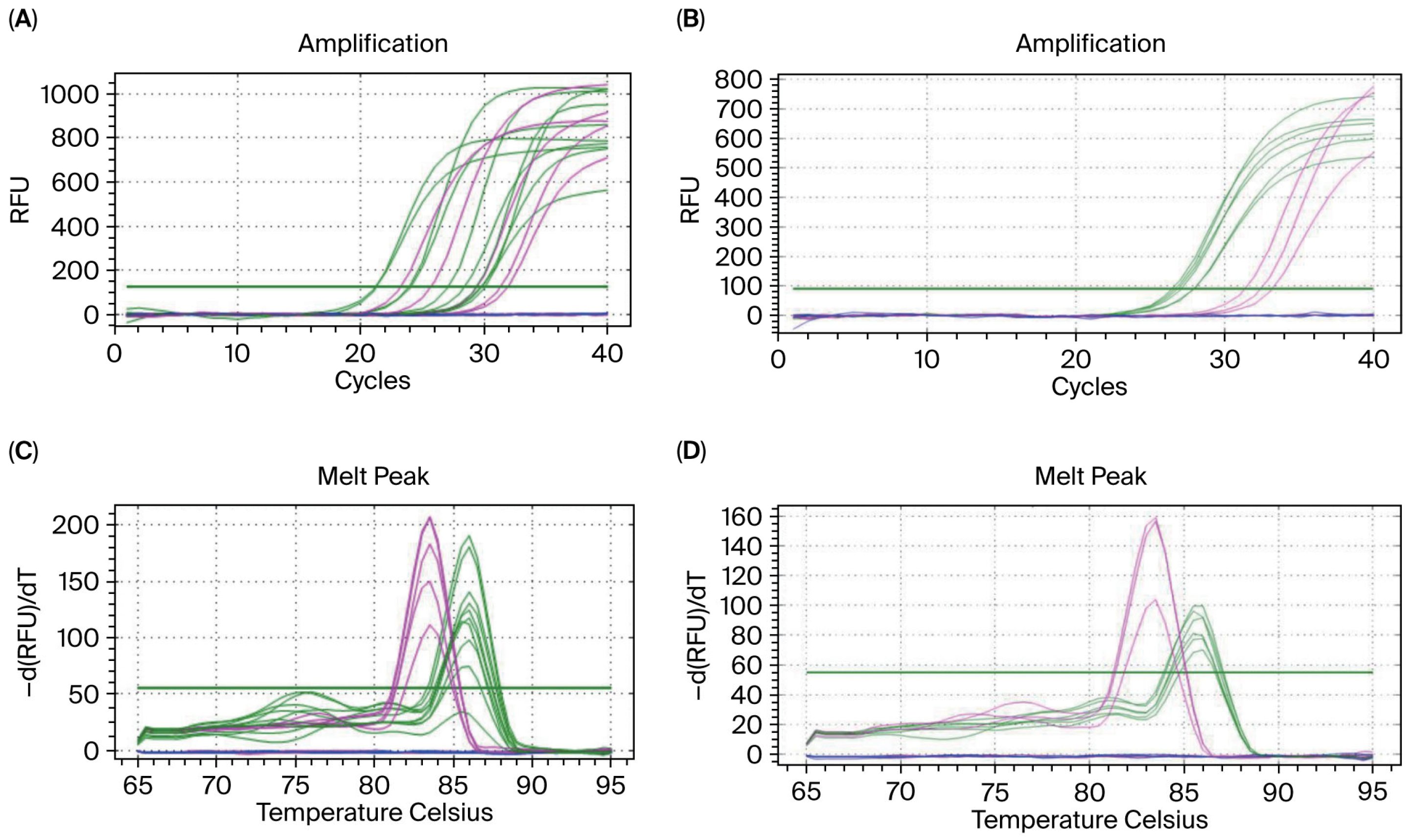
Figure 1. Establishment of sheep blood ultra-trace gDNA amplification system. Note: (A) Amplification curves at dilution factors of 10, 100, 1000, 10,000, and 20,000 times; (B) melting curves corresponding to the same dilution factors; (C) validation results for the qPCR amplification curve at the maximum dilution factor of 20,000 times; (D) validation results for the qPCR melting curve at the same maximum dilution factor. The green curve represents the GAPDH gene, the pink curve represents the SRY gene, and the blue curve indicates the blank control group. In addition, the Y-axis RFU represents the Relative Fluorescence Unit, while -d(RFU)/dT indicates the rate of change of the fluorescence signal concerning temperature.
Biology 14 01144 g001
Figure 2. Gradient-diluted gDNA detected through agarose gel electrophoresis. Note: The dilution multiples are 10/100/1000, and lane M is the DNA marker. The gDNA electrophoresis results for male sheep are as follows: lanes 1/2/3 are the GAPDH gene; lanes 5/6/7 are the SRY gene; the gDNA electrophoresis results for female sheep are as follows: lanes 9/10/11 are the GAPDH gene; lanes 13/14/15 are the SRY gene. Lanes 4/8/12/16 are the blank controls.
Biology 14 01144 g002
Figure 3. Sex identification based on sheep morula gDNA via qPCR amplification. Note: (A) qPCR amplification system for embryo gDNA gradient dilution (1.5, 1.2, 1 μL) is established; (B) amplification results for sex identification using ultra-trace of sheep embryo gDNA; the green curve represents the GAPDH gene, and the pink curve represents the SRY gene.
Biology 14 01144 g003
Table 1. Primer sequences for sex identification.
PrimersPrimer SequencesSize of Products (bp)Melting Temperature (°C)
GAPDH

SRY

SRY-Probe
Forward: GGTCCACATGGCCTCCAAG
Reverse: TCCATTTGTGAGTGTGTGGTCTT
Forward: CTATACACCGAGACAAATACCCG
Reverse: AATCGTCCCTGTATGTGAAGG
Forward: AM-AAGAGGCCACAGAAATCCCTTGCT-MGB (Minor Groove Binder)
167

148


65

60


Disclaimer/Publisher’s Note: The statements, opinions and data contained in all publications are solely those of the individual author(s) and contributor(s) and not of MDPI and/or the editor(s). MDPI and/or the editor(s) disclaim responsibility for any injury to people or property resulting from any ideas, methods, instructions or products referred to in the content.