Article

The Evolution of Molybdenum Dependent Nitrogenase in Cyanobacteria

by
Tomoaki Watanabe
1 and
Tokumasa Horiike
2,*
1
United Graduate School of Agricultural Science, Gifu University, Gifu 501-1193, Japan
2
Department of Bioresource Sciences, Shizuoka University, Shizuoka 422-8529, Japan
*
Author to whom correspondence should be addressed.
Biology 2021, 10(4), 329; https://doi.org/10.3390/biology10040329
Submission received: 31 March 2021 / Revised: 12 April 2021 / Accepted: 12 April 2021 / Published: 14 April 2021

Abstract

:

Simple Summary

Nitrogen fixation is the process by which nitrogen in the atmosphere is converted into ammonia and other nitrogen-containing organic compounds. It is carried out by a variety of bacteria, including Cyanobacteria. Previous studies have shown that several groups of Cyanobacteria have the ability to fix nitrogen; however, because these groups are scattered throughout the Cyanobacterial lineage, the evolutionary history of nitrogen fixation in these bacteria has not been clarified. In this study, we attempted to identify the origin of nitrogen fixation development in Cyanobacterium by focusing on molybdenum dependent nitrogenase, a major nitrogen fixing enzyme. We compared a phylogenetic tree from 179 species of Cyanobacteria to one generated from nitrogen fixation-related genes. We also compared the genomic locations of those genes. As a result, we found that nitrogen fixing genes were acquired in the Cyanobacterium common ancestor and subsequently lost in some lineages. The results demonstrate that inconsistencies between species phylogeny and organism characteristics can occur and be caused not only by horizontal gene transfer, but also by gene deletion.

Abstract

Nitrogen fixation plays a crucial role in the nitrogen cycle by helping to convert nitrogen into a form usable by other organisms. Bacteria capable of fixing nitrogen are found in six phyla including Cyanobacteria. Molybdenum dependent nitrogenase (nif) genes are thought to share a single origin as they have homologs in various phyla. However, diazotrophic bacteria have a mosaic distribution within the cyanobacterial lineage. Therefore, the aim of this study was to determine the cause of this mosaic distribution. We identified nif gene operon structures in the genomes of 85 of the 179 cyanobacterial strains for which whole genome sequences were available. Four nif operons were conserved in each diazotroph Cyanobacterium, although there were some gene translocations and insertions. Phylogenetic inference of these genes did not reveal horizontal gene transfer from outside the phylum Cyanobacteria. These results support the hypothesis that the mosaic distribution of diazotrophic bacteria in the cyanobacterial lineage is the result of the independent loss of nif genes inherited from common cyanobacterial ancestors in each lineage.

1. Introduction

Nitrogen fixation is the biochemical process by which the atmospheric nitrogen is converted to nitrogen-containing compounds including assimilation into organic compounds as part of the nitrogen cycle. Diazotrophic species are present in multiple phyla of bacteria and archaea [1]. Genes encoding nitrogen fixation enzymes are categorized into five groups (I–V) [2]. The genes related to nitrogen fixation such as nifH and nifD are conserved among species in different phyla of both bacteria and archaea [2,3]. Although some nif operons have been lost, duplicated, or horizontally transferred [4,5], many are conserved among these phyla [2]. These results indicate that nif genes originated from a common ancestor [6].
However, in the phylum Cyanobacteria, diazotrophic species are distributed in a mosaic pattern. Two explanations for this have been provided. One is that the last common ancestor of the Cyanobacteria did not have the ability to fix nitrogen, and some lineages independently obtained this ability by horizontal gene transfer from other phyla [7,8,9,10]. Alternatively, the last common ancestor may have been capable of nitrogen fixation, but this ability was subsequently lost in some lineages [11,12]. The former hypothesis is supported by evidence from a small number of species; however, diazotrophic species that diverged from the common ancestor of Cyanobacteria were not included in these analyses [7,8,9,10]. For example, Synechococcus sp. JA-3-3Ab, which can fix nitrogen and diverged earlier, was not included in these analyses [12]. Therefore, the former hypothesis is not well supported by the currently available data. In contrast, the studies that supported the latter possibility did not consider the possibility of horizontal gene transfer. As there are many horizontally transferred genes in Cyanobacterial genomes [13], this possibility cannot be ignored. Therefore, a phylogenetic analysis of each nif gene in Cyanobacteria was conducted to verify the potential of each hypothesis.
In the cyanobacterial genome, the nif genes form operon structures such as nifBSU, nifHDK, nifENXW, and nifVZT [14]. In this study, we identified the operon structures of the nif genes in each genome and performed a phylogenetic analysis of the proteins encoded by each operon structure to examine the reasons for the mosaic distribution of diazotrophic Cyanobacteria.

2. Materials and Methods

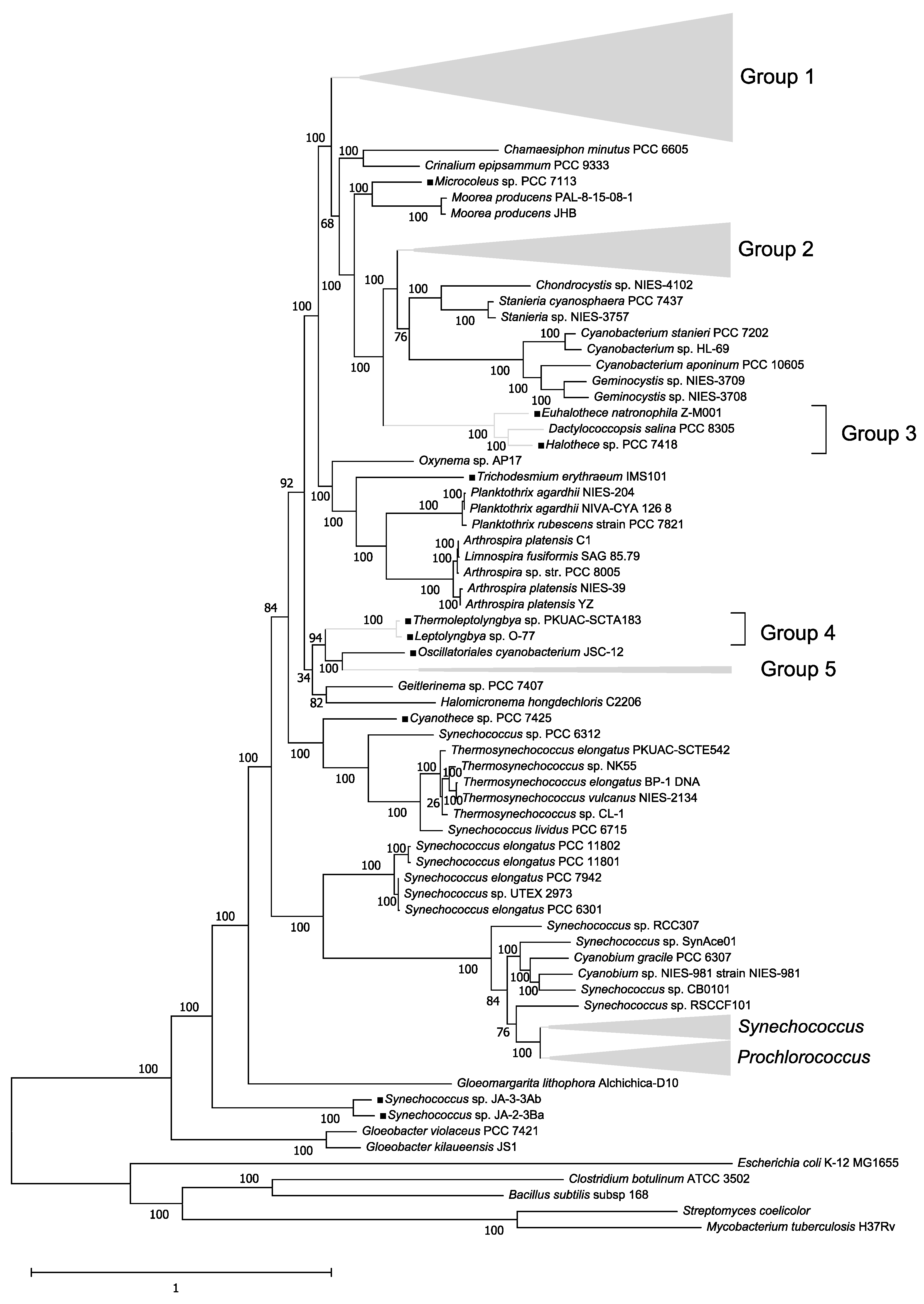
2.1. Construction of a Phylogenetic Tree of 179 Cyanobacteria Species

Genomic sequence data for 179 Cyanobacteria (Table S1) and five outgroups (Bacillus subtilis 168, Clostridium botulinum A str. ATCC 3502, Mycobacterium tuberculosis CDC1551, Streptomyces coelicolor A3 (2), and Escherichia coli K-12 MG1655) were obtained in the GenBank format from the National Center for Biotechnology Information database (ftp://ftp.ncbi.nlm.nih.gov/genomes/genbank/bacteria/, 13 September 2020). Of the 179 species, 85 belong to a diazotroph genus (Table S1). We detected 31 marker proteins for species phylogenetic analysis using AMPHORA2 [15]. Each species’ marker protein sequences were concatenated into one sequence. Multiple alignments of the concatenated sequences were performed using MAFFT version 7.271 [16]. Using the alignment data, a species phylogenetic tree was constructed based on the maximum likelihood method using RaxML version 8.29 [17]. Bootstrap tests were performed with the “-# autoMR” option. The optimal substitution model was estimated using RaxML.

2.2. Collection of nif Amino Acid Sequence Data

First, OrthoFinder [18] was used to generate ortholog datasets for all proteins. In these datasets, we detected proteins annotated with “nif’” and identified their orthologs. All proteins belonging to orthogroups [18] were considered nif candidates. For example, if a protein belonged to an orthogroup containing nifD, it was considered a nifD candidate. There was a nifD gene in Nostoc punctiforme PCC 73102 that was not registered by the coding sequence, we manually added information about its encoded protein. NifE and nifN genes are fused in some genomes (Anabaena variabilis ATCC 29413, Anabaena sp. YBS01, Calothrix sp. NIES-2098, Calothrix sp. NIES-2100, Trichormus variabilis 0441, Calothrix brevissima NIES-22, Chroococcidiopsis thermalis, Thermoleptolyngbya sp. PKUAC-SCTA183, and Trichodesmium erythraeum IMS101). The fused nifs are called nifEN.

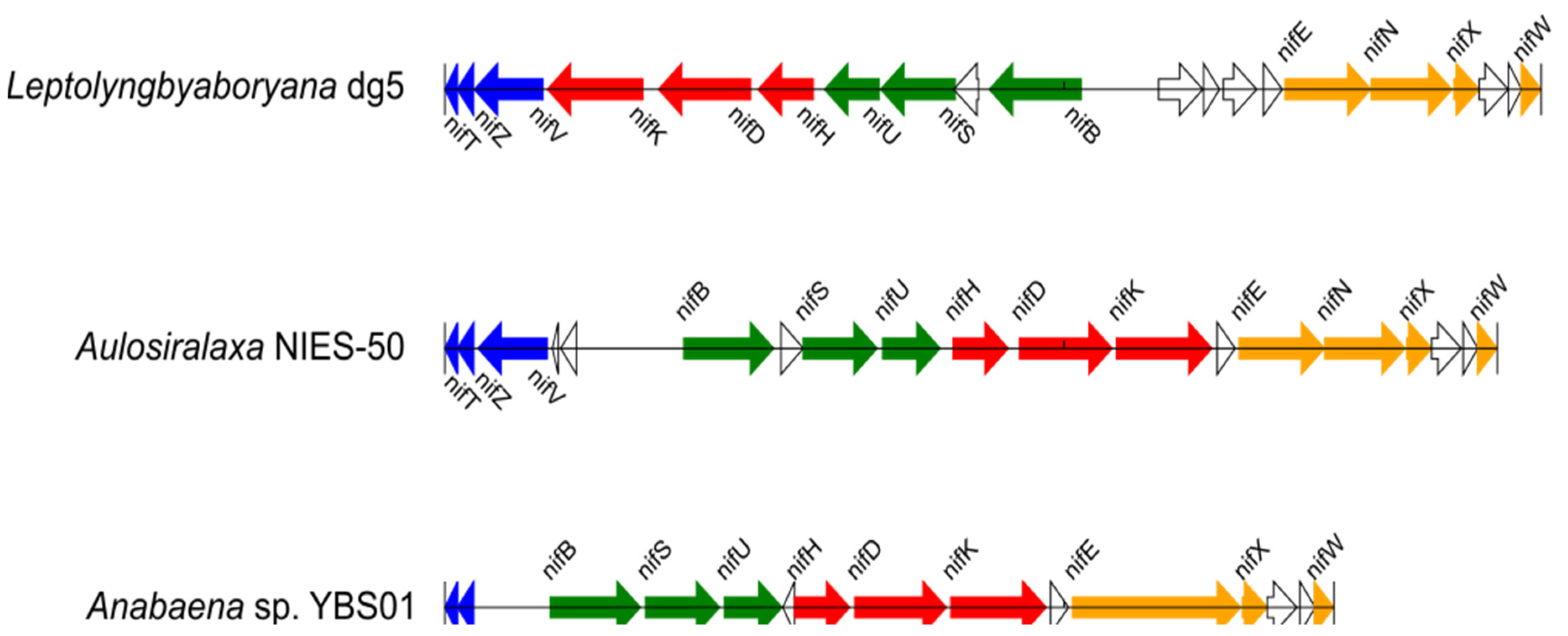
2.3. Detection of nif Gene Operon Structures

Location information for each nif gene in each genome was obtained from the GenBank file. Based on this information, nif genes with the potential to form known nif operons (nifBSU, nifENXW, nifHDK, nifVZT) were detected, and if there were <4 genes between neighboring nif genes, they were considered to be in the same operon. For example, if nifB, nifS, and nifU were in close proximity, they were considered to form one operon. In addition, if ≥4 genes existed between nif genes but a nif operon was present upstream and downstream of the nif gene, these genes were considered part of the same operon. In some genomes (for example, A. variabilis ATCC 29413 and Leptolyngbya boryana), the nif genes comprise of one large operon such as nifBSUHDKENXW [19,20]. However, in this analysis, this operon was treated as separated operons (nifBSU, nifHDK, and nifENXW) for comparison with other nif operons.

2.4. Selection of nif Genes from Multiple nif Genes in An Operon Structure

If there were multiple nif genes believed to be in the operon structure (for example, two nifB genes in an operon), we chose one coding sequence for each nif gene as follows. First, a phylogenetic tree was inferred using all nif proteins encoded by the nif genes in the operon by the maximum likelihood method using RaxML. These phylogenetic trees were compared to the species phylogenetic tree, and the nif whose phylogenetic position was more similar to the species phylogenetic tree was selected. The chosen nifs and the nif genes were used for later analyses.

2.5. Phylogenetic Analysis of Cyanobacterial nif Proteins

Proteins used in the phylogenetic analysis were nifB, nifD, nifE, nifH, nifK, nifN, nifS, nifT, nifU, nifV, nifW, nifX, and nifZ. Of the 179 cyanobacterial species, 82–85 species (depending on the type of nif) contained nifs that met the above criteria. Therefore, we performed phylogenetic analysis of the nifs in these species. Using the alignment data, phylogenetic trees were constructed in RaxML using the maximum likelihood method. Bootstrap tests were performed with the “-# autoMR” option. The optimal substitution model was estimated using RaxML. All other parameters were set to default values.

2.6. Discovery and Phylogenetic Analysis of nifs in Non-Diazotrophic Cyanobacteria

All ortholog sequences for nifs encoded by the nif genes comprising the operons estimated in Section 2.3 were obtained from non-diazotrophic species. Phylogenetic analysis was performed on the nif proteins of non-diazotrophs and diazotrophs as described in Section 2.5.

3. Results and Discussion

3.1. Phylogenetic Tree of 179 Cyanobacteria Species

A phylogenetic tree of the 179 concatenated Cyanobacteria genomes was constructed (Figure 1). Proteobacteria (E. coli), Actinobacteria (M. tuberculosis and S. coelicolor), and Firmicutes (B. subtilis and C. botulinum) species were used as outgroups. The first species to diverge from the common ancestor of Cyanobacteria belonged to the genus Gloeobacter. This genus consists of primitive Cyanobacteria in which a portion of the genes related to the thylakoid membranes and photochemical systems I and II are not present, and it has been previously suggested to be the first branch from the common ancestor of Cyanobacteria [21]. As many Cyanobacteria have synonyms, Cyanobacteria belonging to one genus may not be monophyletic; however, species belonging to the same genera formed roughly the same clusters.

3.2. Operon Structures of the nif Genes

Of the 179 Cyanobacteria, nif genes were identified in the genomes of the 85 species thought to be diazotrophs including those comprising the nifBSU, nifHDK, nifENXW, and nifVZT operons. The operon structures of representative cyanobacterial nif genes are shown in Figure 2. A map of nif genes comprising operon structures in the cyanobacterial genome is shown in Table S2. There were some translocations and insertions in places, but each operon was essentially conserved.
In nine Cyanobacteria (Anabaena sp. YBS01, A. variabilis ATCC 29413, A. laxa NIES-50, C. brevissima NIES-22, Calothrix sp. NIES-2098, Calothrix sp. NIES-2100, T. tenuis PCC 7101, T. variabilis 0441, and N. carneum NIES-2107), two nif operon sets were present at distant positions from each other in the genome. The nifVZT operon was more conserved than the other nif operons, and no other genes were present among the three nif genes. In rare cases, only two genes (nifV and nifZ, nifZ and nifT, or nifV and nifT) formed an operon; in these cases, the other nif gene was located at a distance (Table S2). Genes in the nifHDK operon encode a protein that plays a central role in nitrogen fixation, but the operon structure is not highly conserved (Table S2). Nevertheless, nifH, nifD, and nifK did not often exist on their own, and many species maintained an operon structure for at least two genes (either nifH and nifD or nifD and nifK). There were also several species in which the nifK gene was found upstream of nifENXW and was considered a part of the same operon. Several species were identified in which multiple genes were inserted between nifD and nifK, which would have presumably originally formed an operon. The nifB-fdxN-nifSU operon typically assumed its basic form; however, there were often a few genes between nifB and nifS including fdxN. nifS and nifU were always adjacent to each other, while nifB sometimes formed operons at a distance to nifSU or did not form operons. Cyanobacterium endosymbiont of Epithemia turgida isolate EtSB Lake Yunoko lacked a nifU gene; however, these contained a pseudogene for nifU downstream of nifS. This nifU gene was remotely located, did not form an operon, and had low sequence similarity, suggesting that it serves as an alternative to the original nifU gene. The nifENXW operon was relatively well conserved. Most Cyanobacteria formed an operon structure with nifE, nifN, and nifX adjacent to each other and nifW between genes of unknown function.

3.3. Phylogenetic Analysis of nif Proteins

We inferred phylogenetic trees for each nif encoded by a nif gene contained in an operon structure (Figure S1a–m). The common ancestor of Synechococcus sp. JA-3-3 Ab and Synechococcus sp. JA-2-3 Ba diverged first among the diazotrophic Cyanobacteria in the species phylogenetic tree (Figure 1), therefore, we used these species as the outgroup in each tree. If nifs are true orthologs, the tree shapes should be consistent. We divided the diazotrophic Cyanobacteria into five groups (note that this classification is not the traditional classification of Cyanobacteria, section I–V [22]) based on the species phylogenetic trees to compare phylogenetic trees easily (Table S1). However, the two species used as an outgroup and five diazotrophs with no related species (C. thermalis PCC 7203, Microcoleus sp. PCC 7113, T. erythraeum IMS 101, Cyanothece sp. PCC 7425, and Oscillatoriales cyanobacterium JSC-12) were not included in these groups. Groups I–V were all monophyletic (Figure 1). The following discussion is based on the positions of these groups within the phylogenetic trees.
As above-mentioned, nine Cyanobacteria had two sets of nif operons. A previous study reported the presence of two sets of nif operons (nif1 and nif2) in the genome of A. variabilis ATCC 29413 [23]. In this study, of the two sets of nifs, we considered the nif closely related to nif1 of A. variabilis ATCC 29413 on the nif phylogenetic tree to be nif1, and the nif closely related to nif2 to be nif2 (Figure 1). Nif1 genes have been inherited from the common ancestor of diazotrophic Cyanobacteria because the locations of nif1 on the nif phylogenetic tree are similar to those on the species phylogenetic tree. However, nif2 genes may have arisen before the common ancestor of group I diverged from the other groups by horizontal gene transfer. All Cyanobacteria with nif2 form heterocysts. It is possible that A. variabilis ATCC 29413 was able to fix nitrogen efficiently by using both the original nif1 and the nif2 coded by nif2 genes introduced by horizontal transfer [23].
The phylogenetic tree of nifB, encoded by the nifBSU operon, resulted in monophyly for all groups, whereas the nifS and nifU trees did not (Figure S1a–c). In the nifS tree, group III was contained within group II; and in the nifU tree, groups II and III were contained in group I. All three phylogenetic trees for the proteins encoded by the nifHDK operon were monophyletic (Figure S1d–f). In particular, the phylogenetic positions of the groups in the nifD and nifK trees were almost identical, suggesting that they evolved in the same way. The three phylogenetic trees for proteins encoded by the nifVZT operon were different (Figure S1g–i). The nifV tree was monophyletic for all groups. The nifZ phylogenetic tree included all groups except for group I in group I. In the nifT tree, group I’ was divided into two, one of which included group IV. Of the four phylogenetic trees for nif proteins encoded by the nifENXW operon, only the nifE tree was monophyletic for all groups (Figure S1j–m). The nifN tree included group III in group II, the nifX tree included group IV in group I’, and group I in the nifW tree was paraphyly.
The phylogenetic relationships within each group were similar for the nifBSU, nifHDK, nifVZT, and nifENXW operons, but the branching patterns between some groups were not consistent. The branches that differed among phylogenetic trees corresponded to branches with low bootstrap values. Such branches are thought to be unreliable. However, some nifs (nifX, nifW, nifZ, and nifT) had many internal branches with low bootstrap values, and these nifs showed larger differences in phylogenetic tree shape from other nifs coded by the same operon. The average amino acid length of nifX, nifW, nifZ, and nifT was 136.8, 106.4, 93.6, and 66.9, respectively. The short amino acid length of these nifs may have prevented accurate phylogenetic inference due to their small amount of information, resulting in many unreliable internal branches.
None of the nifs encoded by the operons had long external branches, suggesting that horizontal gene transfers from outside the phylum Cyanobacteria did not occur during the evolution of the Cyanobacterial nif genes. In contrast, a previous study has reported the horizontal transfer of nif genes from Deltaproteobacteria [24].

3.4. Nif Genes in Non-Diazotrophic Cyanobacteria

Orthologs of nif were detected in four non-diazotrophic Cyanobacteria (Table 1): nifS in Cyanobacterium aponinum PCC 10605 and Cyanobacterium stanieri PCC 7202, and nifV in Moorea producens PAL-8-15-08-1 and M. producens JHB. The nif genes encoding these proteins could have been derived from a common Cyanobacteria ancestor or through horizontal transfer from different phyla. If the former is true, the species may have originally been diazotrophs; however, after losing their nitrogen-fixing ability, their nif genes were gradually lost. However, some of these genes are thought to have functions other than nitrogen-fixing and were not lost. In this case, as the remaining nif genes are orthologous to those of closely related diazotrophs, the positions of the species on the nif phylogenetic trees would be expected to be the same or similar to their positions on the species phylogenetic tree. Conversely, if the latter is true, the nifs of non-diazotrophs and diazotrophs that are phylogenetically distant to each other could be closely related in the nif phylogenetic trees.
According to the phylogenetic tree, C. aponinum PCC 10605 and C. stanieri PCC 7202 were closely related, and both are non-diazotrophs that contain nifS genes (Figure 1). However, in the nifS tree, the internal branches leading to these two species diverged from near the roots of the phylum and were extremely long (Figure S2a). Therefore, the nifS genes of the ancestor of these species may have resulted from horizontal transfer from outside the phylum Cyanobacteria. In the species phylogenetic tree, M. producens PAL-8-15-08-1 and M. producens JHB were closely related (Figure 1). Conversely, in the nifV phylogenetic tree, the external branches connected to these species were very long (Figure S2b). Thus, the nifV genes in these species are likely to have been independently obtained through horizontal transfer from outside the phylum Cyanobacteria.

3.5. General Discussion

The phylogenetic trees of the nifs coded by the nif operon (Figure S1) did not have very long branches, like those leading to the nifs of non-diazotrophic Cyanobacteria (Figure S2). This suggests that after the common ancestor of diazotrophic Cyanobacteria acquired the first nif operon, none of the nif genes comprising the operon were acquired by horizontal transfer from outside the phylum Cyanobacteria. The fact that the nif operon is conserved also supports that horizontal gene transfer is restricted. As a result, it is conceivable that the nif genes of each lineage of diazotrophic Cyanobacteria were derived from the nif genes of the common ancestor of diazotrophic Cyanobacteria, and that independent gene loss may have occurred in each lineage of the non-diazotrophic Cyanobacteria.
The topologies of nif phylogenetic trees of each nif operon were inconsistent, but the approximate phylogenetic relationships within and between groups were similar. In addition, the bootstrap values tended to be very low in the internal branches, where the phylogenetic relationships among the groups differed within the same operon. Therefore, it is possible that some errors occurred in phylogenetic inference among some groups.
In this analysis, we used 179 cyanobacterial species including 85 diazotrophs. As the number of species and lineages used was increased from the previous studies [7,8,9,10,11,12,14], it is presumed that reliable results were obtained. However, as a sufficient number of nif genes could not be detected, certain diazotrophic cyanobacteria were excluded from the analysis and therefore, could not be considered in this study. In future, we expect to explore other nitrogenases such as vnf and anf in diazotrophs that were excluded from this study; this would allow for a comprehensive understanding of the evolution of the nitrogen-fixing capacity of Cyanobacteria.

4. Conclusions

Phylogenetic analysis of 179 cyanobacterial species with sequenced genomes showed that diazotrophs are distributed in a mosaic pattern. We estimated the operon structures of 85 diazotroph species and found that all were conserved; however, there were some differences among many species. Phylogenetic trees of species and individual nif proteins were generated and compared, and many differences were found. The relatively conserved group structure and preserved operon structure suggest that horizontal transfer of the genes comprising the operons is restricted. Therefore, differences in the shape of the phylogenetic trees are more likely due to low accuracy in phylogenetic inference than horizontal gene transfer among Cyanobacteria.
Two sets of nif operons were detected in nine diazotrophic Cyanobacteria. One operon set is “nif1 operon,” inherited from the common ancestor of diazotrophic Cyanobacteria with species divergence, and the other is “nif2 operon,” inherited by horizontal gene transfer from within the phylum Cyanobacteria.
By comparing the phylogenetic tree of each nif protein, species phylogenetic tree, and the inferred operon structure, we were able to test the possibility of horizontal gene transfer within and outside Cyanobacteria. This approach will allow us to understand the origin of not only the nitrogen-fixing capacity of Cyanobacteria, but also the other properties of its mosaic distribution in the phylogenetic tree. We concluded that the mosaic distribution of diazotrophic bacteria in the cyanobacterial lineage is the result of the independent loss of nif genes inherited from common cyanobacterial ancestors in each lineage.

Supplementary Materials

The following are available online at https://www.mdpi.com/article/10.3390/biology10040329/s1, Figure S1: Phylogenetic tree of 179 Cyanobacteria species. The phylogenetic tree was inferred using the concatenated sequences. Some of the phylogenetic relationships among species are compressed in Figure 1, but the complete phylogenetic tree is shown in Figure S1. Black squares indicate diazotrophic Cyanobacteria. Diazotrophic Cyanobacteria belonging to groups I–V are shown in brackets. Diazotrophic cyanobacteria with no closely related species were not included in the groups. Bootstrap values are shown at the internal branches (<100). Figure S2: Nif phylogenetic trees for diazotrophic Cyanobacteria. Phylogenetic trees of nifB (a), nifS (b), nifU (c), nifH (d), nifD (e), nifK (f), nifV (g), nifZ (h), nifT (i), nifE (j), nifN (k), nifX (l), and nifW (m) are shown. Diazotrophic Cyanobacteria belonging to groups I–V and I’ are shown in brackets. The nif1 are shown in black circles, whereas the nif2 are shown in black squares. Bootstrap values are shown at the internal branches (<100). Figure S3: Phylogenetic tree of nifV, which is coded by nif operon, and nifV, which is not. Diazotrophic Cyanobacteria belonging to groups I–V and I’ are shown in brackets. Black diamonds indicate the nifs that are not coded by the nif operons. Bootstrap values are shown at the internal branches (<100). Table S1: List of the Cyanobacteria species included in the analysis species with and without nitrogen fixation ability by “+”. Diazotrophic Cyanobacteria that have been grouped are indicated by their group numbers; those without groups are indicated by “None.” Table S2: Map of nif genes comprising the operon structure in the Cyanobacterial genome. The locations of all nif genes in each genome are shown. Each line indicates each genome. H, D, and K that are shown in red stand for nifH, nifD, and nifK, respectively. B, S, and U that are shown in green stand for nifB, nifS, and nifU, respectively. E, N, X, and W that are shown in brown stand for nif E, nif N, nif X, and nif W, respectively. V, Z, and T that are shown in blue stand for nifV, nifZ, and nifT, respectively. The numbers in white cells indicate the number of non-nif genes present.

Author Contributions

Conceptualization, T.W. and T.H.; Formal Analysis, T.W.; Methodology, T.W. and T.H.; Software, T.W.; Writing—Original Draft Preparation, T.W. and T.H.; Writing—Review and Editing, T.W. and T.H. All authors have read and agreed to the published version of the manuscript.

Funding

This research received no specific grant from any funding agency in the public, commercial, or not-for-profit sectors.

Institutional Review Board Statement

Not applicable.

Informed Consent Statement

Not applicable.

Acknowledgments

We thank Koichiro Awai and Rei Narikawa in the Department of Biological Science at Shizuoka University for their advice.

Conflicts of Interest

The authors declare no conflict of interest.

References

  1. Boyd, E.S.; Hamilton, T.L.; Peters, J.W. An alternative path for the evolution of biological nitrogen fixation. Front. Microbiol. 2011, 2, 205. [Google Scholar] [CrossRef] [PubMed] [Green Version]
  2. Raymond, J.; Siefert, J.L.; Staples, C.R.; Blankenship, R.E. The natural history of nitrogen fixation. Mol. Biol. Evol. 2004, 21, 541–554. [Google Scholar] [CrossRef] [Green Version]
  3. Staley, J.T.; Reysenbach, A.L. Biodiversity of Microbial Life: Foundation of Earth’s Biosphere; Wiley & Sons: Hoboken, NJ, USA, 2002; p. 592. [Google Scholar]
  4. Xiong, J.; Fischer, W.M.; Inoue, K.; Nakahara, M.; Bauer, C.E. Molecular evidence for the early evolution of photosynthesis. Science 2000, 289, 1724–1730. [Google Scholar] [CrossRef] [Green Version]
  5. Mus, F.; Colman, D.R.; Peters, J.W.; Boyd, E.S. Geobiological feedbacks, oxygen, and the evolution of nitrogenase. Free Radic. Biol. Med. 2019, 140, 250–259. [Google Scholar] [CrossRef]
  6. Boyd, E.S.; Peters, J.W. New insights into the evolutionary history of biological nitrogen fixation. Front. Microbiol. 2013, 4, 201. [Google Scholar] [CrossRef] [Green Version]
  7. Turner, S.; Huang, T.C.; Chaw, S.M. Molecular phylogeny of nitrogen-fixing unicellular cyanobacteria. Bot. Bull. Acad. Sin. 2001, 42, 181–186. [Google Scholar]
  8. Berman-Frank, I.; Lundgren, P.; Falkowski, P. Nitrogen fixation and photosynthetic oxygen evolution in cyanobacteria. Res. Microbiol. 2003, 154, 157–164. [Google Scholar] [CrossRef]
  9. Sánchez-Baracaldo, P.; Hayes, P.K.; Blank, C.E. Morphological and habitat evolution in the Cyanobacteria using a compartmentalization approach. Geobiology 2005, 3, 145–165. [Google Scholar] [CrossRef]
  10. Shi, T.; Falkowski, P.G. Genome evolution in cyanobacteria: The stable core and the variable shell. Proc. Natl. Acad. Sci. USA 2008, 105, 2510–2515. [Google Scholar] [CrossRef] [Green Version]
  11. Larsson, J.; Nylander, J.A.A.; Bergman, B. Genome fluctuations in cyanobacteria reflect evolutionary, developmental and adaptive traits. BMC Evol. Biol. 2011, 11, 187. [Google Scholar] [CrossRef] [Green Version]
  12. Latysheva, N.; Junker, V.L.; Palmer, W.J.; Codd, G.A.; Barker, D. The evolution of nitrogen fixation in cyanobacteria. Bioinformatics 2012, 28, 603–606. [Google Scholar] [CrossRef] [PubMed]
  13. Yerrapragada, S.; Siefert, J.L.; Fox, G.E. Horizontal gene transfer in cyanobacterial signature genes. Methods Mol. Biol. 2009, 532, 339–366. [Google Scholar]
  14. Esteves-Ferreira, A.A.; Cavalcanti, J.H.F.; Vaz, M.G.M.V.; Alvarenga, L.V.; Nunes-Nesi, A.; Araújo, W.L. Cyanobacterial nitrogenases: Phylogenetic diversity, regulation and functional predictions. Genet. Mol. Biol. 2017, 40, 261–275. [Google Scholar] [CrossRef] [Green Version]
  15. Wu, M.; Scott, A.J. Phylogenomic analysis of bacterial and archaeal sequences with AMPHORA2. Bioinformatics 2012, 28, 1033–1034. [Google Scholar] [CrossRef] [PubMed]
  16. Katoh, K.; Misawa, K.; Kuma, K.; Miyata, T. MAFFT: A novel method for rapid multiple sequence alignment based on fast Fourier transform. Nucleic Acids Res. 2002, 30, 3059–3066. [Google Scholar] [CrossRef] [Green Version]
  17. Stamatakis, A. RAxML version 8: A tool for phylogenetic analysis and post-analysis of large phylogenies. Bioinformatics 2014, 30, 1312–1313. [Google Scholar] [CrossRef]
  18. Emms, D.M.; Kelly, S. OrthoFinder: Solving fundamental biases in whole genome comparisons dramatically improves orthogroup inference accuracy. Genome Biol. 2015, 16, 157. [Google Scholar] [CrossRef] [Green Version]
  19. Thiel, T.; Lyons, E.M.; Erker, J.C. Characterization of genes for a second Mo-dependent nitrogenase in the cyanobacterium Anabaena variabilis. J. Bacteriol. 1997, 179, 5222–5225. [Google Scholar] [CrossRef] [PubMed] [Green Version]
  20. Tsujimoto, R.; Kamiya, N.; Fujita, Y. Transcriptional regulators ChlR and CnfR are essential for diazotrophic growth in nonheterocystous cyanobacteria. Proc. Natl. Acad. Sci. USA 2014, 111, 6762–6767. [Google Scholar] [CrossRef] [PubMed] [Green Version]
  21. Schirrmeister, B.E.; Antonelli, A.; Bagheri, H.C. The origin of multicellularity in cyanobacteria. BMC Evol. Biol. 2011, 11, 45. [Google Scholar] [CrossRef] [PubMed] [Green Version]
  22. Thiel, T.; Pratte, B.S. Regulation of three nitrogenase gene clusters in the Cyanobacterium Anabaena variabilis ATCC 29413. Life (Basel) 2014, 4, 944–967. [Google Scholar] [CrossRef] [PubMed] [Green Version]
  23. Rippka, R.; Deruelles, J.; Waterbury, J.B.; Herdman, M.; Stanier, R.Y. Generic Assignments, Strain Histories and Properties of Pure Cultures of Cyanobacteria Free. Microbiology 1979, 111, 1–61. [Google Scholar]
  24. Bolhuis, H.; Severin, I.; Confurius-Guns, V.; Wollenzien, U.I.; Stal, L.J. Horizontal transfer of the nitrogen fixation gene cluster in the cyanobacterium Microcoleus chthonoplastes. ISME J. 2010, 4, 121–130. [Google Scholar] [CrossRef] [PubMed]
Figure 1. Phylogenetic tree of 179 Cyanobacteria species. The phylogenetic tree was inferred using the concatenated sequences. Some of the phylogenetic relationships among species are compressed in Figure 1, but the complete phylogenetic tree is shown in Figure S1. Black squares indicate diazotrophic Cyanobacteria. Diazotrophic Cyanobacteria belonging to groups I–V are shown in brackets. Diazotrophic cyanobacteria with no closely related species were not included in the groups. Bootstrap values are shown at the internal branches (<100).
Biology 10 00329 g001
Figure 2. Operon structures of Cyanobacteria nif genes. The nif operon structures of Leptolyngbya boryana dg5, Aulosira laxa NIES-50, and Anabaena sp. YBS01 are shown. Blue, red, green, and orange arrows indicate the nifVZT, nifHDK, nifBSU, and nifENXW operons, respectively. White arrows indicate the genes unrelated to nitrogen fixation.
Biology 10 00329 g002
Table 1. Non-diazotrophic Cyanobacteria with homologs to nifs coded by nif operons of diazotrophs.
SpeciesNif
Moorea producens PAL-8-15-08-1nifV
Moorea producens JHBnifV
Cyanobacterium stanieri PCC 7202nifS
Cyanobacterium aponinum PCC 10605nifS
Publisher’s Note: MDPI stays neutral with regard to jurisdictional claims in published maps and institutional affiliations.

