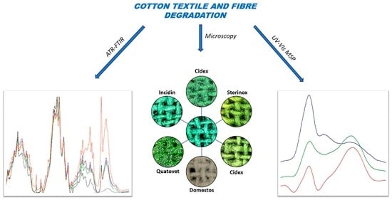
1. Introduction
Clothing textiles, including component fibers, may degrade under the influence of various factors occurring during their usage. As a result of the transformation of such materials under the influence of external factors, physicochemical changes occur in the color and structure of the textiles and the fibers they contain. External factors may include light, high temperatures, unfavorable environmental conditions (humidity, microorganisms, UV radiation), and chemicals used in the washing process (detergents, bleaches, enzymes, organic solvents) [
1,
2].
From a forensic perspective, degraded clothing textiles acquire unique physicochemical features. These features allow for the identification of degraded textile samples from other manufactured products of this type, making them more likely to be associated with a specific criminal event. In particular, degradation processes taking place in textiles and fibers under the influence of specific factors, such as thermal, biological, mechanical, and of a complex nature (e.g., gunshot), have been of interest for years. The effects of such processes can be observed by a forensic expert when analyzing textile materials secured in connection with many existing criminal events [
3,
4,
5,
6].
Most of the studies conducted so far have focused on cotton fibers. This fact is not surprising because the majority of clothing used by European consumers, thus constituting a main part of the evidential material examined in forensic laboratories, is made, at least to some extent, of cotton fibers [
7]. These fibers are relatively easy to defragment, e.g., under the conditions of a criminal event and the influence of frictional forces. The cotton fibers are staple fibers with a convoluted surface structure. Hence, the most common type of fiber microtraces are fragments of single cotton fibers, which have been subjected to identification and comparative tests for years [
8,
9,
10]. Comparative physiochemical examinations of cotton fibers revealed at the scene of the crime, on the victim’s clothing, or, for example, under the victim’s fingernails, with the cotton fibers present in the composition of clothing of known origin, e.g., belonging to the suspect, are very often carried out for forensic experts. The results of research can be very valuable evidence in all stages of criminal procedures in courts and before authorities conduct pre-trial proceedings [
10,
11].
The effects of specific chemicals on cotton textiles and fibers have been documented in the textile literature. For example, Schindler presented methods for identifying the main damage to cellulose fibers, pointing out that they are relatively resistant to alkalis but sensitive to acids [
2]. Moreover, the author indicates that cellulose fibers may be damaged under the influence of oxidizing factors.
Currently, fibers are identified using various microscopic and spectroscopic methods, following the principles of green chemistry and not using chemicals dangerous to the environment or the researcher’s health. [
9,
11]. Schindler describes the procedure for analyzing textile damage as well as a number of characteristic chemical reactions that allow determining its degree and type [
2]. This procedure includes the application of optical microscopy and IR spectroscopy, which are routinely used in forensic examinations of fibers. The author also indicates methods such as solubility and staining tests or thin-layer chromatography, which are rarely used due to their destructive nature. And although nowadays we strive to ensure that this type of risk occurs sporadically in people’s surroundings, a small share of harmful chemicals is present, e.g., in disinfection, sterilization, and DNA degradation agents, the use of which in everyday life has increased significantly in the era of the COVID-19 pandemic.
This is why the authors undertook the issue of recognizing the impact of this type of agent, with a complex composition that is not fully disclosed by the producers, on different colored cotton textiles and fibers. At the same time, the possibility of recognizing the type of agent used was assessed, among others, from a forensic point of view. An additional objective was to assess the use of routine microscopic and spectroscopic techniques to identify and compare such degraded material, i.e., textiles and single fibers [
11].
2. Materials and Methods
2.1. Original Material
The subject of the presented research consisted of samples of four textile materials made of 100% cotton, the basic characteristics of which are presented in
Table 1. The examined materials differed in product type (woven fabric, knitted fabric), structure (mass per unit area and thickness), and color and dye composition. These products came from one of the dyeing plants and were intended for sale (ready for use), but no knowledge about additives, temperature, or time intervals used in the dyeing processes was shared with the authors. This was not an isolated case because market goods are the subject of forensic examinations, and therefore it is not possible to precisely identify their components but to compare their characteristic features thoroughly.
2.2. Exposure of Original Material to Disinfectants, Sterilizers, and Degrading DNA Agents
Six widely available and commonly used agents for disinfection, sterilization, and DNA degradation were used to degrade cotton textiles. These agents had different commercial names, properties, and active ingredients. The agents are used in such areas of life as medicine, the food industry, households, or forensic examinations. The main characteristics of the chemical agents investigated in this study are presented in
Table 2.
The preparation of the material for experimental research consisted of the first stage in the separation of 7 pieces, with an area of about 10 mm × 10 mm, from each cotton fabric, marked as Sample 1–Sample 4. One set of materials was left untreated as original material. The remaining pieces were placed on a glass substrate and then treated with selected disinfectants, sterilizers, and DNA degrading agents, marked as Agent 1 and Agent 6. These agents (0.5 mL) were applied to the material sample with a Pasteur pipette so that it was wet throughout. The samples obtained in this way were left for 5–7 days in room conditions to completely dry.
2.3. Optical Microscopy Methods Used for Textile and Fiber Analysis
Observations of the research textiles (Sample 1–Sample 4), untreated and treated with all disinfectants, sterilizers, and DNA degrading agents (Agent 1–Agent 6) were realized with the use of a low-power M125 C stereomicroscope (Leica, Germany), with a magnification of 8× to 100×. The observed images were recorded by an MC 190 HD high-definition color microscope camera and an LAS-V4-12 system for the processing of the obtained data (both Leica, Germany). For detailed optical microscopy examination, experimental fabric samples were flattened by covering them with a microscope glass slide (Menzel-Glaser, Braunschweig, Germany).
Observations of fibers present in research material (Sample 1–Sample 4), coming from the untreated fabrics and those treated with selected agents (Agent 1–Agent 4, Agent 6) were conducted with the use of high-power, bright-field, polarized light and fluorescence microscopy DM2700 P (Leica, Germany) with a magnification of 100× to 400×. A 12 V/100 W halogen lamp in the Leica EL6000 external light source was used to excite the fluorescence. Leica fluorescent filter sets for UV (A—BP 340/380); UV and violet (D—BP 355/425); blue (I3—BP 450/490); and green (N2.1—BP 515/560) light were applied. Images were recorded using a DMC 4500 high-definition color microscope camera, and the LAS V4.13.0 system was used for the processing of the obtained data (both Leica, Germany).
For detailed bright-field microscopy examination, fiber samples were placed on microscope glass slides (Menzel-Glaser, Braunschweig, Germany) with a drop of a mounting medium—pure glycerine (Merck KGaA, Darmstadt, Germany)—and covered with coverslips (Menzel-Glaser, Braunschweig, Germany).
2.4. ATR-FTIR Analysis of Textiles
The infrared spectroscopy research was conducted for all research textiles (Sample 1–Sample 4), untreated and treated with all disinfectants, sterilizers, and DNA degrading agents (Agent 1–Agent 6) with the use of a Nicolet iS50 FTIR spectrometer (Thermo Fisher Scientific, Waltham, MA, USA), with a built-in, all-reflective diamond ATR. The spectra were recorded by collecting 32 scans ranging from 400 cm−1 to 4000 cm−1, with a spectral resolution of 4 cm−1. The spectra obtained during measurements were pre-processed directly in OMNIC Spectra Software (Thermo Fisher Scientific, Waltham, MA, USA) by doing background correction and smoothing. The fabric samples were tested directly by making three replicate measurements from different parts of the samples. Before each measurement, the crystal surface was washed with ethanol, and the background spectrum was collected.
2.5. UV–Vis Microspectrophotometry Analysis of Fibers
Microspectrophotometric measurements were performed for colored fibers extracted at random from the weft and the warp of different parts of Sample 2, i.e., the one with the strongest dyeing, untreated and treated with selected disinfectants, sterilizers, and DNA degrading agents (Agent 1–Agent 4 and Agent 6). The examined fibers were mounted between quartz microscope slides and cover slips (CRAIC Technologies, San Dimas, CA, USA), using pure glycerine (Merck KGaA, Darmstadt, Germany) as a mounting medium.
Absorbance spectra were obtained using a 20/20 PV™ microspectrophotometer (CRAIC Technologies, San Dimas, CA, USA) equipped with a Xenon lamp and an Ultrafluar 40× Zeiss objective. The spectra were recorded in the ultraviolet and visible ranges, between 200 and 800 nm. Measuring conditions were: aperture size—6.0 µm × 6.0 µm; scans to average—25; resolution value—1 nm. Reference data were taken exclusively from an area immediately adjacent to the mounted fiber; this was directly followed by measurements of the fiber sample. During the measurements, the same orientation of the cotton fibers relative to the position of the lamp was maintained to eliminate the phenomenon of dichroism. In order to avoid the bleaching of cotton fibers in UV light, very fast scanning, as well as closing off the path of the light source immediately after a single measurement, were used.
In total, ten measurements from five fibers, representing the particular fabric samples, were collected. All operations on spectra (averaging, comparison) were performed using CRAIC MSP Data Acquisition Software (CRAIC Technologies, San Dimas, CA, USA).
3. Results and Discussion
3.1. Low-Power Microscopy Analysis
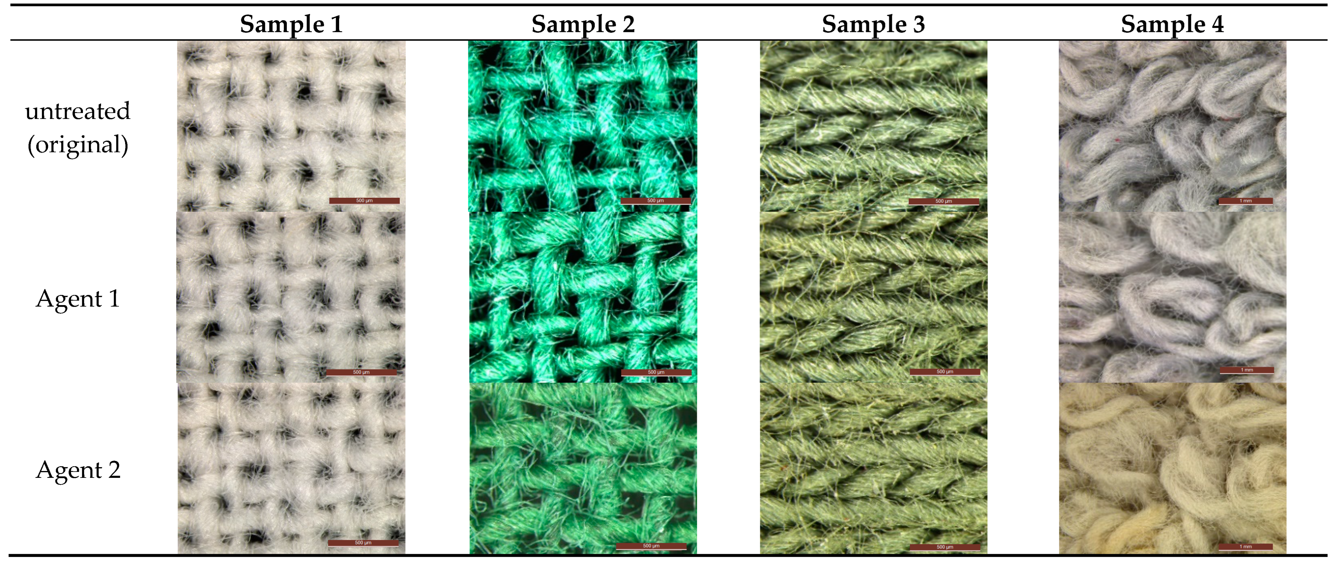
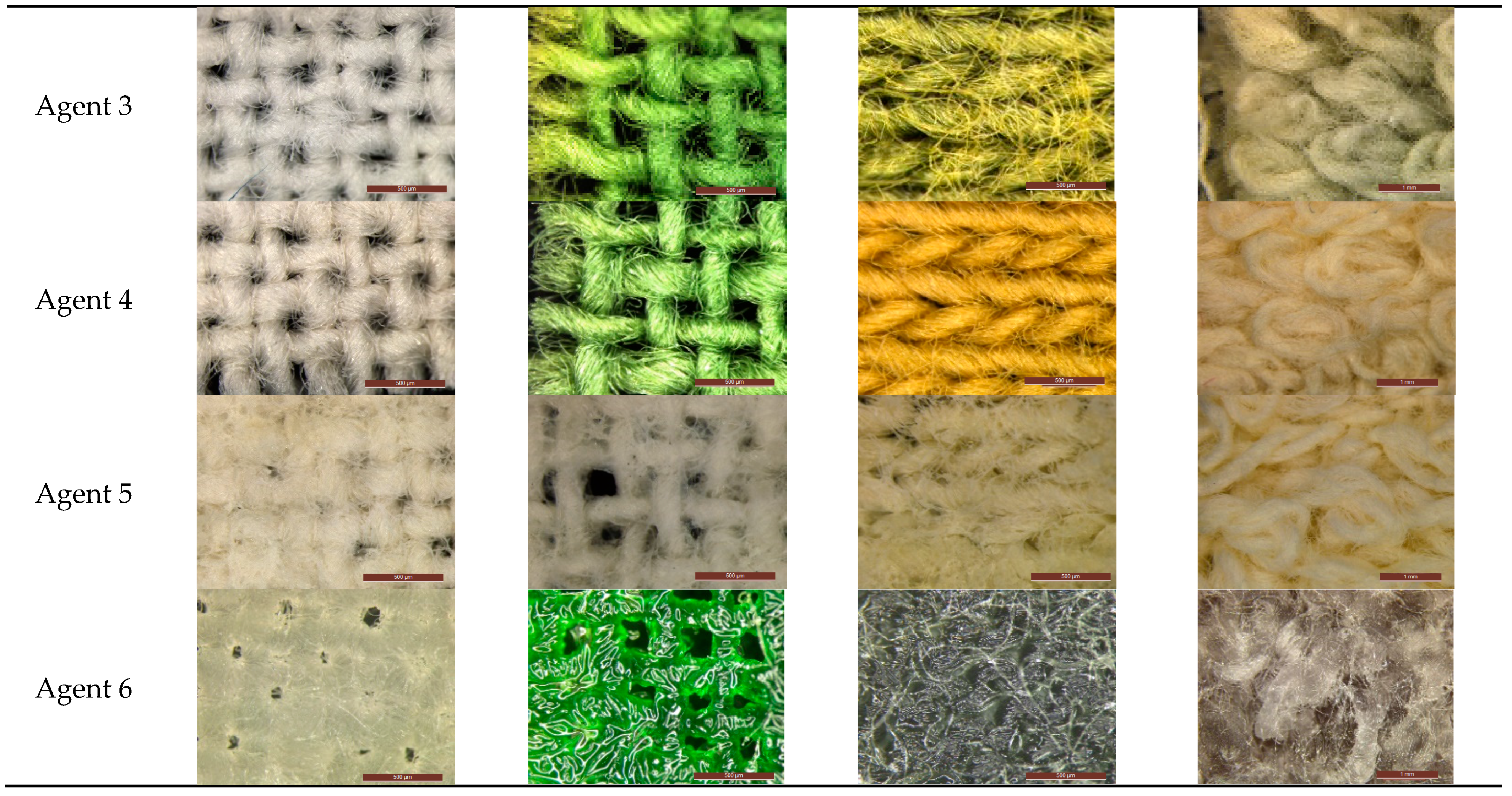
During the microscopic examination, samples of fabrics treated with selected agents were compared with those untreated. These studies were mainly of a preliminary and illustrative nature. However, on their basis, the effects of the changes taking place, characteristic of the agents used, were already observed.
For all samples treated with Agent 1 and Agent 2, no significant changes were observed. Only Sample 1 and Sample 4 turned slightly yellowish as a result of treatment with Agent 2 (
Figure 1). Agent 3 and Agent 4 led to a color change in three of the four examined samples, i.e., (Sample 2–Sample 4), while the structure of Sample 1 became more compact and stiff. Agent 5 had the most destructive effect on samples of cotton textiles, causing discoloration of colored materials, i.e., Sample 2–Sample 4, stiffening the structure of all samples and making them very brittle. This is not surprising because Agent 5 contained active chlorine released from sodium hypochlorite, which is a strong oxidant by nature [
18]. In addition, the presence of particles of a crystalline white substance on the surfaces of all these materials was confirmed. The use of Agent 6 also changed the color of individual samples (Sample 1–Sample 4), but because they did not dry completely, their structure became more dense and compact. This may be related to the chemical structure of quaternary ammonium salts, which provides unique properties of activation at the phase boundary and interaction with the surface [
19].
Based on the analysis of stereomicroscopic images obtained for individual samples of cotton fabrics (Sample 1–Sample 4), it was possible to assess the degree to which they have undergone a structural and color change as a result of exposure to selected disinfecting, sterilizing, and DNA degrading agents (
Figure 1). The effects observed in the case of the use of most agents, i.e., Agent 2–Agent 6, were so characteristic that on this basis it can be initially indicated which of the experimental agents, used for disinfection, sterilization, or DNA degradation, had an impact on cotton textiles.
3.2. High-Power Microscopy Analysis
In the case of comparative studies of cotton fibers contained within experimental cotton textiles, untreated and treated by disinfecting, sterilizing, and DNA-degrading agents, their color was one of the most important physicochemical properties subjected to verification. Initially, the color of the fibers was compared using bright-field microscopy, followed by fluorescence microscopy, with the odd use of different filters for epifluorescence imaging. The authors expected changes in the fluorescent properties of fibers, which were indeed observed for all cotton fibers treated with Agents 2–4 and Agent 6. In this case, however, these were mainly changes in color intensity.
Naturally, parallel changes in the morphology of the fibers themselves were sought. An observation of microscopic images of cotton fibers taken from treated experimental textiles did not reveal any evident, visible changes in the fiber structure compared to untreated ones. As indicated in
Section 2.3, samples degraded by Agent 5 were excluded from these studies. The fibers contained in them had significantly degraded, becoming more brittle and consequently torn and frayed, already at the time of preparation of the microscopic slides.
3.3. ATR-FTIR Analysis
In the course of infrared spectroscopy studies, samples of untreated cotton textiles (Sample 1–Sample 4) were first examined. The obtained spectra were compared with each other and with the analogous spectrum of cellulose, the main component of cotton. Literature data were used for the identification of bands in the infrared spectra of cellulose [
9,
20,
21,
22], and the most important of them are presented in
Table 3.
Each of the infrared spectra obtained for untreated cotton textiles (Sample 1–Sample 4) was consistent with adequate literature data and with each other.
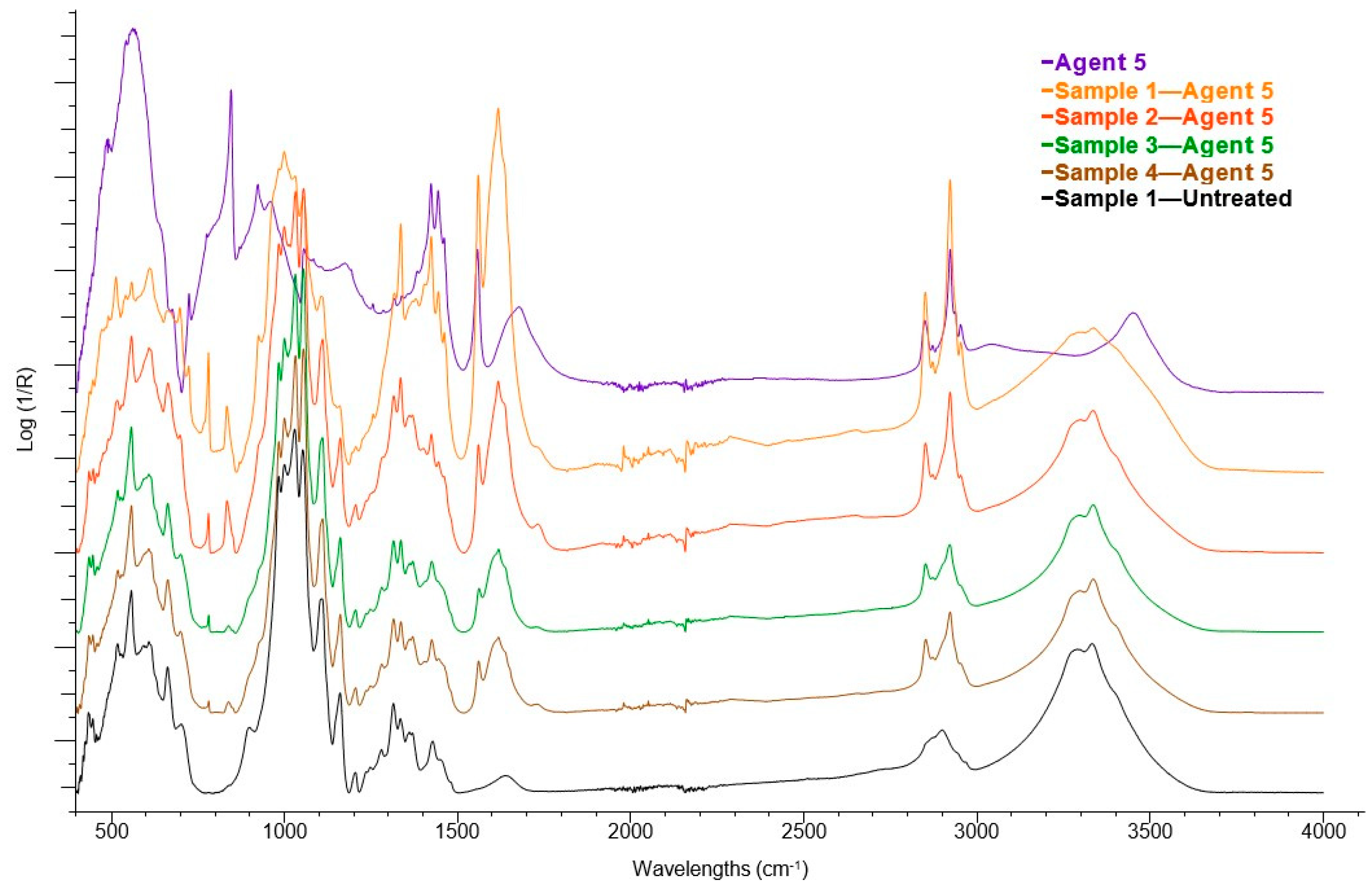
Then, samples exposed to disinfectants, sterilization, and DNA degrading Agents (Agent 1–Agent 6) were examined. The results show, that most ATR-FTIR spectra obtained for these samples and exactly treated by Agent 1–Agent 4 were analogous to those obtained for untreated textiles.
Additional bands were observed in spectra obtained from all samples (Sample 1–Sample 4) treated with Agent 5 and Agent 6. Both agents contained quaternary ammonium salts (Agent 5—hexadecyl trimethylammonium chloride; Agent 6—alkyl (C12-16) dimethylbenzyl ammonium chloride—
Table 2). And signals characteristic of these compounds (quaternary ammonium salts) appeared in the ATR-FTIR spectra of treated textiles (
Figure 2). Characteristic signals appeared in the spectral range 3000–2600 cm
−1 (asymmetric stretching vibrations and symmetrical stretching vibrations C-H bonds in the methylene group—green area in
Figure 2); 1600–1350 cm
−1 (deformation vibrations C-H bonds in the methyl and methylene group—blue area in
Figure 3); and 1000–700 cm
−1 (stretching vibrations C-C bonds—yellow area in
Figure 3).
Summarizing the results of the analysis carried out using the ATR-FTIR technique, it should be stated that this technique allowed for the determination of the type of basic component (biopolymer) of the cotton fibers. In some cases, ATR-FTIR may be helpful in the detection of textile degradation agents, in particular those with the addition of quaternary ammonium salts. However, based on the obtained research, it was not possible to identify the dyes of cotton textiles as well as detect the use of the rest of the disinfectants, sterilizants, and DNA-degrading agents used.
3.4. UV–Vis MSP Analysis
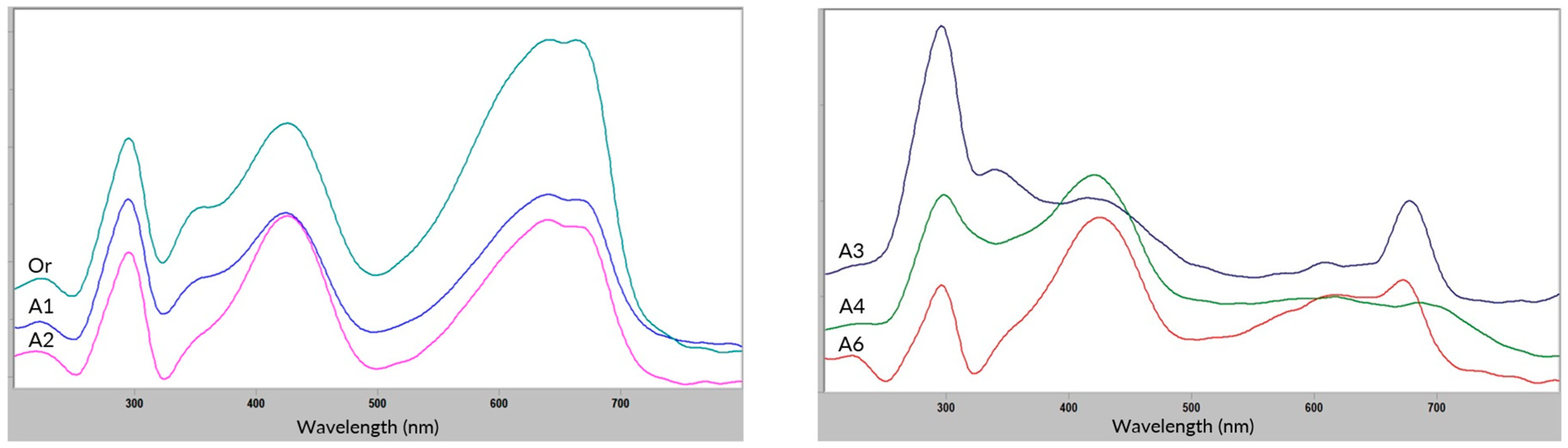
Since visible and UV microspectrophotometry is routinely used in forensic laboratories as an objective method of distinguishing colored single cotton fibers, it was selected to track the color change of the fibers included in Sample 2. It was the sample with the strong dyeing, treated by Agent 1–Agent 4, and Agent 6.
Figure 3 shows the comparison of the mean UV–Vis spectra of the original fibers from Sample 2 and those treated by Agent 1 and Agent 2. The results show that both of the applied agents had no major effect on the fibers or their coloration.
In the case of Sample 2 treated with Agent 3–Agent 4, and Agent 6 (
Figure 3), significant changes were observed in the area of dyed fibers, which theoretically could help to differentiate the agents used. However, in practice, this can be difficult given the high variability between cotton fibers within a single thread. The spectra of those fibers on the outside may show greater differences because they were more exposed to the agent’s impact in relation to the spectra of the fibers inside the thread or contained in the original sample.
4. Practical Aspects of the Examination—A Forensic Case Study
As an example of the practical use of the presented research, there was a forensic case study elaborated by the Institute of Forensic Research in Krakow, Poland, related to the event that occurred in one of the confectionery shops. The case was concerned with the exposure of a few-year-old boy to the immediate danger of loss of life or serious damage to health by leaving highly corrosive substances in the toilet available for customers. The boy entered the toilet alone and left it with deep burns to the skin of his hands and other parts of his body.
The opinion was developed mainly by experts in the field of forensic toxicology. However, because the evidence was, among others, children’s clothing, such as socks, trousers, a sweatshirt, and a sweater, it was advisable to examine the damaged clothing in terms of textile science. The aim of this research was to answer whether the action of the household chemistry products, secured by the process authority in the toilets of the confectionery shops and sent for testing, could have caused the observed damage to the evidence clothing. In order to reply to the question posed, examinations of damaged and undamaged fragments of the evidence clothing were carried out. Low-power microscopy studies were performed, followed by fiber identification and comparative examinations using high-power microscopy.
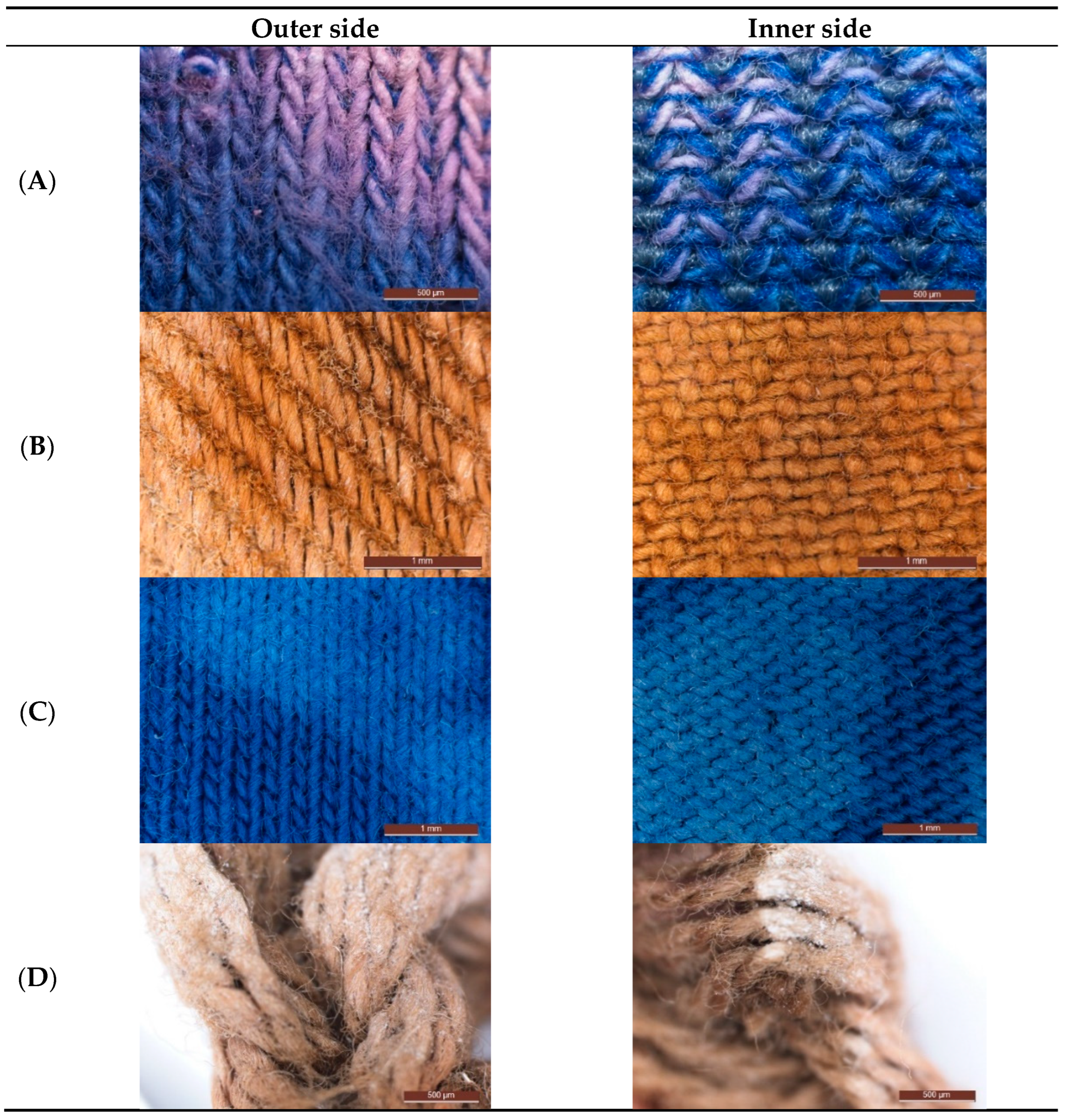
A pair of blue socks with pink discoloration was the first evidential material examined (
Figure 4A). After conducting a sequence of identification examinations of the fibers included in the analyzed clothing materials using bright-field and polarized light microscopy and FTIR spectroscopy, it was established that the knitted fabric of the sock was made of four types of fibers, i.e., cotton, polyamide, polyester, and elastic, of different colors, among others blue cotton fibers. As a result of the observation of the socks’ threads with the use of stereomicroscopy, it was found that only the blue cotton fibers included in them were discolored, i.e., they changed color to light pink.
The trousers were made of brown fabric, partially discolored, and there was a trace of a white substance on their surface (
Figure 4B). The fabric consisted of brown cotton fibers that were losing color while being damaged.
Then, it was established that the knitted fabric of the sweatshirt was discolored in a few places, especially on the sleeves (
Figure 4C). It consisted of white and blue cotton fibers, and the latter have lightened in places of damage.
The brown knitted sweater was discolored in several places, especially on the sleeves, and a trace of a white substance was visible on its surface (
Figure 4D). The knitted fabric consisted of light brown cotton fibers that were losing color while being damaged.
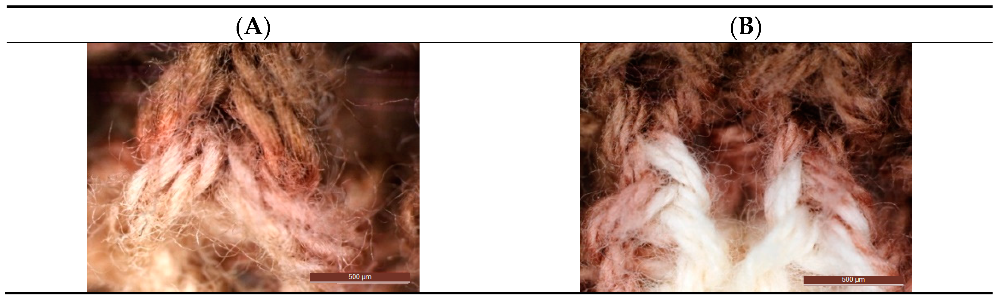
Then, a number of experiments were carried out, consisting of the impact of household chemicals sent for testing on small fragments of undamaged materials from which evidence clothing was made and observing the changes taking place. The experimentally obtained changes in the textiles and their fibers were compared with those observed in the evidence clothing using analogous microscopic techniques.
Among the numerous tested household chemicals, only in the case of analyzing the effects of two of them on fragments of undamaged fabrics from which evidence clothing was made were changes similar to those observed in the evidential materials noticed. For comparative tests, fragments of threads from damaged areas of evidence clothing and threads from damage caused by experiments with the use of these two preparations, i.e., Domestos citrus and Domestos pine, were taken. As a result of the observations, it was established that the direction of color changes in the places of original damage is consistent with the direction of color changes in places treated with these preparations, and the observed changes rather indicate the effect of the diluted preparation (
Figure 5A,B).
It was concluded that the damage to the textiles and fibers in the evidence clothing could therefore be caused by Domestos citrus and/or Domestos pine. Domestos preparations, and more precisely the sodium hypochlorite and hexadecyl trimethylammonium chloride they contain, had strong caustic properties until decomposition, chlorine release, conversion to sodium carbonate, and the associated decrease in pH. The white substance found on the seized clothing was identified by toxicologists as sodium carbonate.
In this way, it was confirmed that the injuries suffered by the boy could be caused by the action of specific preparations that had been carelessly left in the toilet for customers.
5. Conclusions
In this study, the authors evaluated the impact of popular agents used for disinfection, sterilization, and DNA degradation on cotton textiles in order to assess the possibility of identifying the type of agent used based on the changes observed in treated textiles and fibers. The obtained results of microscopic and spectroscopic examinations allowed us to observe changes in the physicochemical nature of samples coming from different cotton fabrics and their constituent fibers under the influence of particular agents of different chemical compositions and intended uses. Due to the lack of research on this topic, a comparison of the obtained results with the literature was not possible.
Differences in color, from a slight change to complete discoloration, and in the structure of the examined fabrics, which became more rigid, brittle, or, for example, compact, were already noticed with the use of various optical microscopy methods. Additional examination with the use of ATR FTIR allowed us to identify the presence in the exposed fabrics of residues of these agents that contained quaternary ammonium salts. For the rest of the samples, ATR-FTIR did not enable the identification of the agents used. This is most likely due to the type of active ingredient that could evaporate completely (alcohols), had a poor IR spectrum (inorganic compounds), or the characteristic bands were obscured by originating bands from cellulose. Bright-field microscopy made it possible to show, above all, changes or lack thereof in the fluorescence properties of single fibers present in the treated fabrics in relation to the control ones. With the use of UV–Vis microspectrophotometry, changes in colored fibers following the action of a specific agent on the examined fabrics were checked. As an application aspect of the conducted research, forensic case research was presented, in which the use of concrete disinfectants was recognized based on examinations of visual changes observed in cotton clothing.
The presented research concerned the visual assessment of textiles and fibers and the tracking of changes occurring in them using spectroscopic methods. The prospect of using alternative chromatographic methods and techniques for identification and comparative studies of such material seems very interesting. Therefore, the authors conducted extensive research on their development, and the identification of reactive dyes extracted from unchanged and degraded cotton textiles will be the subject of further study.


