Review

Recent Advances in Superhydrophobic and Antibacterial Coatings for Biomedical Materials

1
School of Mechanical Engineering, Dongguan University of Technology, Dongguan 523808, China
2
Mechanical and Electrical Engineering Institute, Zhengzhou University of Light Industry, Zhengzhou 450002, China
3
Zhengzhou Phray Technology Co., Ltd., Zhengzhou 450019, China
4
Prenatal Diagnosis Center of Dongguan Kanghua Hospital, Dongguan 523808, China
5
Guangdong HUST Industrial Technology Research Institute, Guangdong Provincial Key Laboratory of Digital Manufacturing Equipment, Dongguan 523808, China
*
Authors to whom correspondence should be addressed.
Coatings 2022, 12(10), 1469; https://doi.org/10.3390/coatings12101469
Submission received: 15 September 2022 / Revised: 29 September 2022 / Accepted: 30 September 2022 / Published: 5 October 2022

Abstract

:
In recent years, biomedical materials have been used in the response to the emergence of medical infections that pose a serious threat to the health and life of patients. The construction of superhydrophobic coatings and antimicrobial coatings is among the most effective strategies to address this type of medical derived infection. Firstly, this paper reviews the preparation methods of superhydrophobic surface coatings and their applications; summarizes the advantages and disadvantages of superhydrophobic surface preparation schemes based on the template method, spraying methods, etching methods, and their respective improvement measures; and focuses on the applications of superhydrophobic surfaces in self-cleaning and antibacterial coatings. Then, the action mechanisms of contact antibacterial coatings, anti-adhesion bacteriostatic coatings, anti-adhesion bactericidal coatings, and intelligent antibacterial coatings are introduced, and their respective characteristics, advantages, and disadvantages are summarized. The application potential of antimicrobial coatings in the field of biomedical materials is highlighted. Finally, the applications of superhydrophobic and antimicrobial coatings in medical devices are discussed in detail, the reasons for their current difficulties in commercial application are analyzed, and the future directions of superhydrophobic coatings and antimicrobial coatings are considered.

1. Introduction

As a kind of advanced multifunctional material, biomedical materials can be used to diagnose, cure, repair, or replace human tissues and organs or enhance their functions. Biomedical materials play a very important role in the lives and health of patients. In recent years, with the frequent use of biomedical materials, the problem of medical infections caused by pathogenic bacteria (e.g., Staphylococcus aureus, Escherichia coli, Candida albicans, etc.) adhering to the surface of medical devices or implantable biomaterials has become one of the greatest threats to patient health worldwide [1,2]. Bacterial infections are among the leading global causes of death among sick individuals [3,4,5,6]. Contamination of medical devices by harmful microorganisms during surgery or in the postoperative phase when the wound is not fully healed often leads to associated infections. In addition, bacteria can reach the surface of biomaterials through the spread of infection from other parts of the body, resulting in blood-borne implant infections [7]. Many medical biomaterials are at risk of bacterial infection. For example, dental implants have an infection rate of about 1% [8], the risk of urethral tube infection increases by 3%–7% per day [9], infection rates for vascular prostheses range from 0.5 to 5% [10], and the infection rate of plates and screws in trauma patients is even higher [11]. This type of infection is called “Biomaterial Centered Infection“ (BCI) [12].
To reduce morbidity and mortality due to bacterial infections, the preparation of antimicrobial and self-cleaning materials plays a vital role in health care. It also encourages scientific researchers to develop materials with antimicrobial activity to meet the practical needs of medical devices and public health products. Coatings can impart the desired surface functionality without affecting the overall performance of the material. The development of coatings with antimicrobial and self-cleaning properties has become a very active research field and is one of the most effective strategies to impart antimicrobial activity to medical devices and biological implants. Because certain microorganisms can survive on medical devices for more than 90 days [13], medical antimicrobial materials are required to have significant antimicrobial and antibacterial effects on pathogenic microorganisms, along with the ability to maintain antimicrobial properties for a longer period of time. In addition, for medical bio-implants, antimicrobial materials need to have good biocompatibility and self-cleaning ability.
Superhydrophobic coatings have unique non-wetting properties and are self–cleaning [14,15], corrosion-resistant [16], and resistant to bioadhesion [17]. They have extremely broad applications and prospects in the fields of apparel and textiles, biomedicine, daily necessities and packaging, transportation tools, and trace analysis [18,19]. Especially in recent years, superhydrophobic surfaces have been introduced into biological materials such as medical devices and artificial blood vessels to improve their anticoagulation and blood compatibility [20,21,22]. Antibacterial coating [23,24] refers to a kind of coating material that can inhibit the growth of bacteria on the surface or directly kill bacteria by surface modification. At present, the preparation of superhydrophobic and antibacterial coatings on the surfaces of medical devices and biological implants has become a hot topic in the fields of medicine and materials.
The rest of this paper is organized as follows: Section 2 outlines the hydrophobic mechanism of superhydrophobic coatings, the preparation methods, and the advantages and disadvantages of each of these preparation methods. Section 3 summarizes the bacteriostatic or bactericidal effects of antimicrobial coatings with the distinction of bactericidal mechanisms. Section 4 reviews the applications of superhydrophobic and antimicrobial coatings in the field of medical devices. Section 5 discusses the antimicrobial effects, persistence, and commercial applications of the coatings. Section 6 summarizes the conclusions and outlooks of this work.

2. Superhydrophobic Coatings

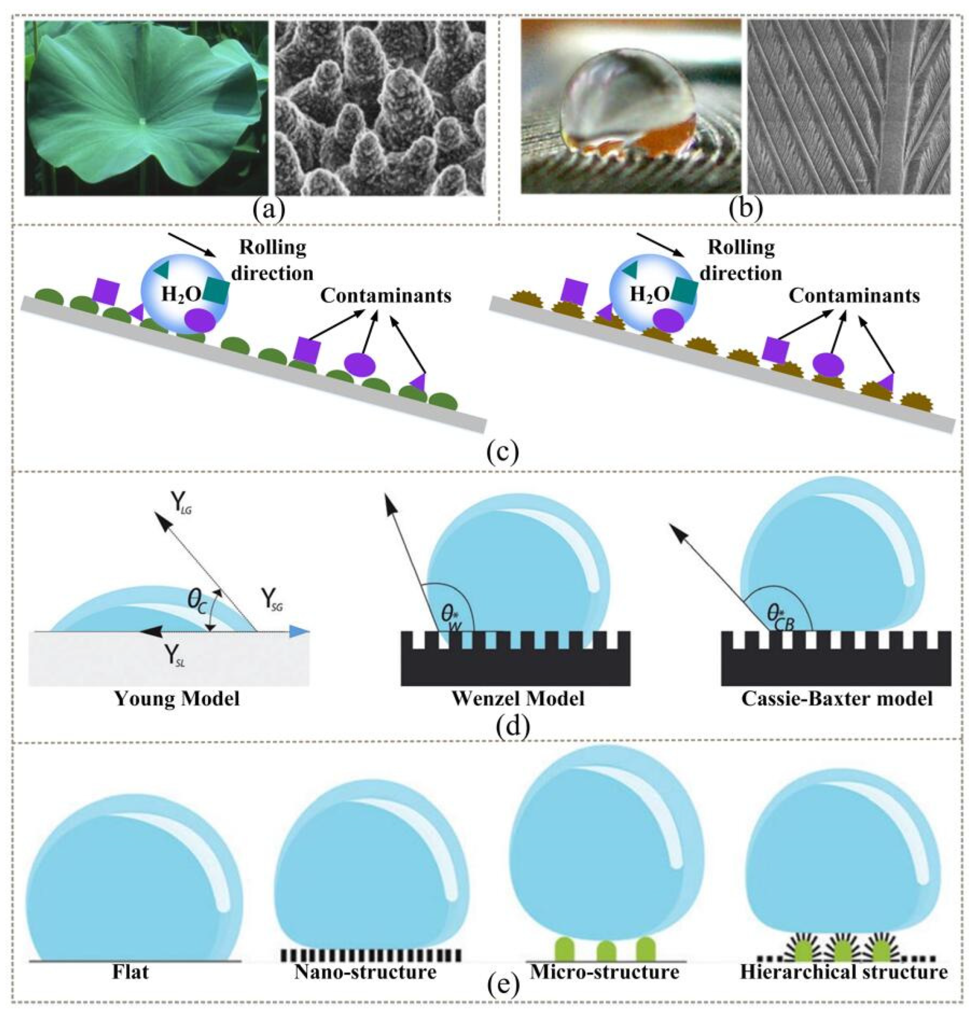
2.1. Background and Mechanism

Superhydrophobic surfaces have attracted a lot of attention for their excellent self-cleaning properties and great application potential. Lotus leaves are a common self-cleaning material observed in nature [25,26], and the surface of lotus leaves is also a typical natural superhydrophobic surface. Other plant and animal parts in nature, such as water strider legs, pigeon feathers, gecko feet, and butterfly wings [27,28,29,30] belong to the category of superhydrophobic surfaces. Figure 1a demonstrates the surface structure of lotus leaves and their surface SEM images. The surface structure of bird feathers and their surface SEM images are shown in Figure 1b. These plants and animals use their natural superhydrophobic surfaces to remove embedded dirt as an evolutionary survival strategy [31]. Superhydrophobic surfaces not only serve as a survival tool for plants and animals, but can also protect human life and health. Currently, the application of superhydrophobic surfaces in the biomedical field is of great interest. Surgical instruments in the clinic are prone to contamination by blood adhesion and bacterial adherence during surgery [32], and contaminated instruments can interfere with the surgeon’s vision and cause bacterial infections in patients, leading to medical malpractice. In contrast, blood-contacting medical devices treated with superhydrophobic coatings [33] can prevent blood adhesion and bacterial adherence, greatly safeguarding the life and health of patients.
Figure 1c describes the self-cleaning mechanism of superhydrophobic surfaces. For many medical and industrial applications, the mechanism of action of self-cleaning surfaces is fascinating [39,40]. Anti-adhesive and antibacterial coatings constructed from superhydrophobic functional surfaces can effectively prevent non-specific adhesion of biomolecules (such as proteins) on the material surface and effectively reduce the initial attachment of bacteria and inhibit the formation of bacterial biofilms, achieving antibacterial effects. Superhydrophobicity is elucidated by two physical principles: low surface energy, and rough structures on a microscopic scale. Surface chemistry and surface topography are the main factors that interfere with liquid-solid interface interactions. Surface energy affects the adhesion of substances to the interface, including fluids and microorganisms. Low surface energy reduces the adhesion work and, therefore, increases the hydrophobicity. Superhydrophobicity is usually achieved by a combination of surface structures, usually formed at the micro/nano scale in combination with compounds with low surface energy. In 1805, Thomas Young first introduced the concepts of contact angle (CA) and wettability and developed Young’s equation [41]. According to the Wenzel model and the Cassie–Baxter model, surface roughness plays a crucial role in wettability. Superhydrophobic surfaces are mainly characterized by a water CA of less than 150° and a water sliding angle (WSA) of less than 2° [42,43,44]. The theoretical basis for the surface wettability of superhydrophobic surfaces was developed from Young’s equation to the Wenzel model [45] and, finally, to the Cassie–Baxter model [46]; the schematic diagram of these three models is shown in Figure 1d.
The currently reported superhydrophobic models are all based on a modification of the Cassie–Baxter model. Bittoun et al. [47] theoretically studied the different types of surface morphologies. By varying the roughness scale, the Cassie–Baxter state was found to be more thermodynamically stable than the Wenzel state. In addition, they concluded that multiscale roughness increases the mechanical stability of the surface and favors superhydrophobicity. Zheng [48] suggested that by changing the surface topography from flat to structured, and by including a hierarchical organization, the water CA would be modified and superhydrophobicity could be achieved. Figure 1e displays the different structures, from planar to hierarchical. The lotus leaf proves this theory, and its superhydrophobicity is due to the presence of its micro/nanoscale features regardless of the hydrophobic coating on its surface.

2.2. Fabricating Methods

Superhydrophobic surfaces are achieved by preparing micro/nanostructures and then passivating them with low surface energy molecules. In recent years, many different micro/nanostructures inspired by nature have been prepared with superhydrophobic structural properties. For example, the dual–scale hierarchical structure of a lotus leaf [49], the micro/nanobrush dual structure [50], the multidimensional hierarchical structure of a butterfly wing [30], the re–entrant structure [51], and the flower–like structure [52]. The most commonly used preparation methods for superhydrophobic coatings are the template method [53], spraying methods (including powder spraying [54], chemical deposition [55], and chemical deposition [56]), and etching methods (including dry etching [57] and wet etching techniques [58]), and the flow of these preparation methods is shown in Figure 2. Other preparation methods, such as hydrothermal treatment [59], anodic oxidation [20], and microarc oxidation [60], also have their own characteristics and advantages. From the perspective of hydrophobic principles, these methods are mainly divided into two types: one is to change the roughness to obtain superhydrophobicity, while the other is to reduce the surface energy and form superhydrophobic surfaces via chemical modifications.

2.2.1. Template Method

The template method is a common method to obtain superhydrophobic surfaces. This method focuses on obtaining superhydrophobicity by replicating the rough structure on the surface of a template with low surface energy. The templates can be divided into “soft templates” and “hard templates” according to the materials used. The research of the template method is mainly divided into two approaches: In the first, the artificial substrate is formed by etching and other methods, and then a variety of required superhydrophobic structures are obtained by soft-template curing and hard-template imprinting. In the other, the “hard template” method is improved so that it can be applied to a variety of hard templates, and the template is in the superhydrophobic state with a contact angle higher than 150°. The substrates used are divided into two types: natural substrates and artificial substrates. Natural substrates include lotus leaves, petals, and other superhydrophobic texture structures, whereas artificial substrates include microstructures formed through etching and other methods.
The replication of natural substrates by the template method is the main way to achieve a superhydrophobic surface, from natural to artificial, and is an important means to study their biological superhydrophobic properties. Hong et al. [64] replicated the functional nanometer patterns of cicada wings by the “hard template method”, first by thermal embossing onto PVC polymer to form a mirror-image template, followed by deposition, activation, and photolithographic curing of the resin to replicate the cicada wing structure onto a glass plate. The surface contact angle obtained by this method was about 132°, which was higher than that of the PVC polymer at 86°. Sun et al. [53] used the soft template method to cast polydimethylsiloxane (PDMS) onto a lotus leaf and isolated it after curing a “mirror image template”. The PDMS was then used to cast the “mirror image template” again, and trimethylchlorosilane (TMCS) was used as an anti-adhesive between the two templates to replicate the lotus leaf structure on PDMS, which had a similar surface contact angle to the lotus leaf at 160°. On the micro/nano scales, the surface morphology of the replica and the natural lotus leaf was nearly identical, as shown in Figure 3a.
Laser etching is one of the methods used to form the substrate required for the template method. Toosi et al. [65] obtained the corresponding surface pattern by ablating the stainless steel surface with a femtosecond laser and, later, replicated the bilayer superhydrophobic surface by thermally embossing the pattern of this ablation on the HDPE surface. Figure 3b shows the SEM images of the superhydrophobic structure of HDPE using different structures (tripled roughness structure and cauliflower structure) as templates. Wang et al. [66] utilized a laser-etched aluminum alloy substrate and PDMS replication template method to create a superhydrophobic and blood-adhesive-resistant flexible tube. SEM images of the prepared flexible tube at different scales are shown in Figure 3c. This flexible tube can be used for medical transfusion devices to prevent contamination of medical devices.

2.2.2. Spraying Methods

The template method is mainly used to replicate the superhydrophobic structure on a low-surface-energy substrate, while spraying methods are used to spray or deposit a layer of particles with low surface energy on the surface of the substrate to give the surface a superhydrophobic effect. The spraying methods can be divided into solid spraying, liquid spraying, chemical deposition, etc. Since the mainstream liquid superhydrophobic coatings are usually volatile and irritating, harmful to the human body, and difficult to clean, the main direction of such coating methods uses the powder spraying, chemical deposition, and electrochemical deposition methods.
With the development and application of nanotechnology [67,68], powder spraying technology with nanoparticles as coatings provides an alternative solution to the environmental pollution problem caused by the solution spraying method. This method generally involves the electrostatic action of charged particles, which causes the powder to adhere to the substrate and form a superhydrophobic coating. Zhu et al. [36] prepared a PSU–CNTs–FEP nanocomposite coating by combining the advantages of superhydrophobic polysulfone and carbon nanotubes (CNTs) through an electrostatic coating method. The coating can be applied on the substrates of different materials, such as metal and glass, and can also be used for anti-corrosion of pipelines. In view of the pollution problem posed by the conventional spraying method, the development of green and non-polluting superhydrophobic coatings has also been one of the research directions in recent years. Shen et al. [69] prepared a superhydrophobic coating by designing and synthesizing a bio-based polymer material through the principles of green chemistry and spraying the design with the aid of CNT particles. The effects of CNT content on the coating, non-wetting, and morphology are shown in Figure 4a. The prepared surface was superhydrophobic (CA = 157°), with low adhesion (SA = 5°) and resistance to corrosion and contamination.
Electrochemical deposition is a technique in which ions undergo a redox reaction in the presence of an applied electric field to form a coating on an electrode. Electrochemical deposition can control the formation of surface roughness by adjusting the electrical parameters to make the prepared surface more uniform. However, electrodeposition cannot be applied to non-conductive materials, such as fibers, rubber, glass, etc. To solve the problem of weak adhesion of the coating to the substrate, Zou et al. [70] combined plasma electrolytic oxidation and electrodeposition techniques to obtain a composite structured superhydrophobic surface with strong adhesion, and the adhesive strength of the coating was evaluated by the scratch method, as shown in Figure 4b. The coating had good heat dissipation and corrosion protection and could resist 40 times of wear. Most of the existing electrodeposition methods need to go through two steps of roughness building and surface energy reduction, greatly increasing the complexity of the preparation process. One-step electrodeposition without low surface energy modification has become a hot research topic; Pan et al. [63] prepared a superhydrophobic surface via electrodeposition of a magnesium substrate in a mixture of cerium nitrate, stearic acid, and ethanol. The electrodeposition method requires only 1 min to produce a superhydrophobic surface. The CA of the prepared superhydrophobic surface was found to be 159.6° and could withstand a strong alkaline environment and 500 mm of sandpaper abrasion.
The chemical deposition method is used to add a coating on the surface of the substrate through the deposition of particles to give the substrate superhydrophobic properties. The chemical deposition method has the advantages of low cost, process reproducibility, and scalability [55]. However, the harmful nature of chemical substances and poor surface wear resistance are the fatal defects of the chemical deposition method. In order to reduce the toxicity of the reagents used, Zhang et al. [71] proposed a deep eutectic solvent containing Cr(III) on the surface of magnesium alloys, followed by surface modification with stearic acid, to create new chemically transformed chromate films with superhydrophobic and self-healing properties. The advantage of this solution is that the toxicity of Cr(III) is low and the pollution to the environment is relatively small. To improve the surface stability, Yu et al. [72] deposited hierarchical WO3@TiO2 nanosurfaces with superhydrophobicity through a liquid-phase deposition scheme using stainless steel as a substrate. The WO3@TiO2 surfaces had photocatalytic protection properties, so the surfaces could be used to protect metals from electrochemical corrosion. The superhydrophobic surface (CA = 162°) obtained by this method had long-term stability against corrosion. Transmission electron microscope (TEM) images of pristine WO3 and the WO3@TiO2 nanocomposite film are shown in Figure 4c.
Figure 4. Preparation effects of the spraying methods: (a) effects of the content of carbon nanotube materials on coatings, non-wettability, and morphology [69]; (b) scratching methods to evaluate the adhesion strength of the layers [70]; (c) TEM images of pristine WO3 and WO3@TiO2 nanocomposite film [72].
Coatings 12 01469 g004

2.2.3. Etching Methods

Spraying methods are used to apply or deposit a superhydrophobic particle coating on the substrate, while etching methods are used to increase the substrate’s surface roughness or reduce the surface energy directly through chemical modification or physical etching, forming a superhydrophobic surface for the purpose of reducing chemical activity and corrosion resistance. The most significant advantage of this method is the better stability and corrosion resistance of the resulting superhydrophobic surface, which can form a durable hydrophobic surface and avoid the problem of easy corrosion at the interface between the substrate and the coating.
Laser etching is the process of forming a superhydrophobic surface by changing the surface roughness using laser ablation of the surface. Pan et al. [63] prepared a superhydrophobic micro/nano cross-grooved surface for antibacterial purposes by modifying a stainless steel surface with picosecond laser technology, and the SEM images of the cross-grooved structure with magnifications of 200×, 1000×, and 50,000× are shown in Figure 5a. The optimal scanning pitch of the surface was 30 µm, and the surface CA reached 163° after three laser scans. The air layer formed on the surface was resistant to hard impacts and scratches and could be immersed in NaCl solution for 30 days and still be hydrophobic. The surface was also superhydrophobic underwater, inhibiting bacterial growth.
Chemical etching is a method of inducing random roughness on a surface by immersing the target surface in a corrosive/reactive chemical mixture to produce surface roughness [73], and this treatment is commonly used for metal and glass surfaces. Research in chemical etching in recent years has focused on improving the chemical properties of the prepared surface and minimizing the impact on the substrate strength. Jie [58] utilized a combination of chemical etching and thermal treatment to construct microstructures on a Cu surface and infiltrated them in an ethanol solution of stearic acid particles for surface modification to obtain superhydrophobic structures. The SEM images of the etched and superhydrophobic surfaces are shown in Figure 5b. Zhang et al. [74] prepared superhydrophobic surfaces via droplet etching with hydrochloric acid and chemical modification with perfluorooctanoic acid (PFOA) at 80 °C. This method is able to maintain the integrity of the aluminum material well while creating a rough structure on the surface. The SEM images of the etched and superhydrophobic surfaces are shown in Figure 5c. The prepared surfaces (CA = 156°, SA = 5°) had good thermal stability as well as anti-corrosion, self-cleaning, and anti-fouling abilities.
Figure 5. Preparation effects of etching methods: (a) SEM images of a superhydrophobic surface with cross-groove structures [63]; (b) SEM images of brass with a superhydrophobic surface [58]; (c) SEM images of the original surface, the etched surface, and the superhydrophobic surface [74].
Coatings 12 01469 g005

2.3. Summary

The advantages of the template method are that it can be used in large areas, the microstructures of different surface morphologies can be reproduced, and the operation is simple. However, the template method still has shortcomings, such as short service time, poor wear resistance, and susceptibility of the surface microstructure to damage. Of the spraying methods, the powder spraying technique is the most likely to replace liquid spraying and achieve mass production, but there are also problems of surface hydrophobicity and interface stability common to spraying methods. In addition, most of the particles used in the current spraying technology are colored particles, which make it difficult to spray onto glass surfaces with high transparency requirements. Chemical deposition and electrochemical deposition suffer from the environmental pollution of the chemicals used and the instability of the deposited interface. Most current chemical deposition techniques still require the deposition of low surface energy fluorides for modification. This leads to waste of raw materials, increases the cost of deposition, and makes it difficult to achieve mass production. Electrochemical deposition techniques, although more efficient, are only applicable to conductive substrates. The etching methods do not need to consider interfacial stability, but they may reduce the strength of the substrate during the etching process. The laser etching method can accurately obtain the desired surface but has the defects of high cost and long cooling time. The chemical etching method is simple and controllable, but the environmental pollution problem is difficult to solve. A summary of superhydrophobic surface preparation methods is shown in Table 1.
With the development of new materials and new technologies, superhydrophobic surface preparation methods have become more diverse and in-depth. The development of nanotechnology has meant that the spraying of solid particles is expected to replace liquid spraying methods; the development of biotechnology has led to the synthesis of many new biomaterials, which are expected to replace some toxic chemical reagents, realizing environmentally friendly superhydrophobic surfaces. The development of ultrafast laser technology can obtain more ideal surface parameters and greatly reduce the impact on substrate strength.

3. Antibacterial Coatings

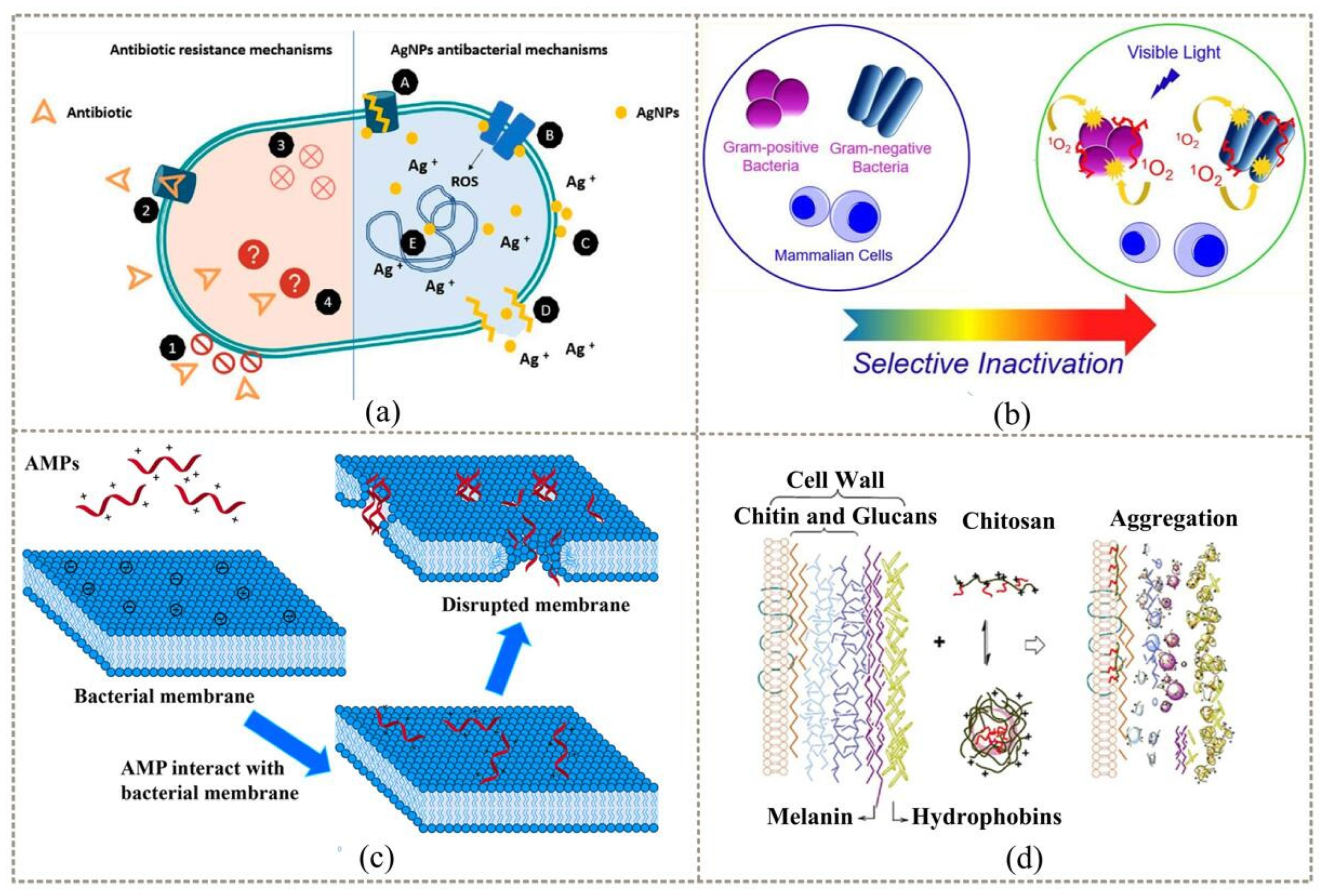
3.1. Background and Mechanism

In recent years, medical infections caused by bacterial growth on the surface of biomedical materials have become one of the hidden dangers that seriously threaten people’s lives and health, and the construction of antimicrobial coatings on the surface of materials is an important strategy to avoid such problems. The design and construction of surface coatings with antimicrobial function [75] is of great practical significance for the functional realization and application of functional materials, especially biomedical materials. Antimicrobial coatings [23,76] are a class of coating materials that are modified by the surface of the material to give them the function of inhibiting the growth of surface bacteria or directly killing the germs without changing the performance of the material itself. The current antimicrobial coatings are mainly classified into contact-type antimicrobial coatings [77], anti–adhesive antimicrobial coatings [78], and intelligent antimicrobial coatings [79] according to their mechanism of action. Therefore, they can be divided into inorganic antimicrobial coatings, represented by silver nanoparticles (Ag/NPs) [80,81,82], and organic antimicrobial coatings, represented by quaternary ammonium salts (QASs) [83,84,85]. In addition, certain organic coatings also use natural organic substances such as antimicrobial peptides (AMPs) and chitosan as antimicrobial agents [86,87]. The mechanisms of action of different antimicrobial agents are shown in Figure 6.
Contact-type antimicrobial coating [89] is the earliest type of antimicrobial coating studied. It works by immobilizing organic molecules with antimicrobial properties (such as QASs, AMPs, chitosan, etc.) directly onto the surface of the material. When bacteria come into contact with materials, surface antibacterial molecules use a chemical mechanism to kill the bacteria. Some inorganic antibacterial materials, such as Ag/NPs, gold nanoparticles (Au/NPs), and metal oxide nanoparticles are fixed on the surface of the material. Based on the chemical mechanism of metal ion dissolution, photocatalysis or photothermal effects, physical insertion destruction, and other physical mechanisms can also kill bacteria in contact with the material.
Anti-adhesive antimicrobial coatings can be divided into anti-adhesive antibacterial coatings and anti-adhesive bactericidal coatings [90]. Anti-adhesive antibacterial coatings are functional coatings constructed on the surface of materials that are simultaneously resistant to the adhesion of biomolecules such as bacteria, fungi, and proteins [79]. Antibacterial coating directly through the surface modification of materials changes the surface physicochemical properties of the materials (such as roughness, hydrophobicity, charge, etc.) so as to inhibit the adhesion of germs. Anti-adhesive germicidal coatings prevent bacterial, fungal, and protein biomolecular adhesion and continuously release germicidal ingredients, with the antibacterial and germicidal dual function of the coating generally being loaded directly into the anti-adhesive coating of various materials through physical chelation or chemical bonding of the antimicrobial agent. The antimicrobial agent is retained in the pores of the coating or in the polymer layer by physical adsorption and is released slowly or responsively to exert antimicrobial effects [86,87].
Intelligent antimicrobial coatings [91,92] achieve controllable antimicrobial processes, maintain “biological inertia” when there is no bacterial contact, can “activate” bactericidal function and release antimicrobial agents in the early stages of bacterial adhesion, and can achieve controlled release of antimicrobial agents by regulating intermolecular interactions and intelligent responsiveness. Intelligent antibacterial coating can effectively avoid the environmental pollution and health hazards caused by the irregular loss of toxic components such as heavy metals and antibiotics. In addition, intelligent antimicrobial coatings can quickly remove the dead bacteria and debris on the surface after killing bacteria through the intelligent responsiveness of the coating molecules (temperature, pH, light, magnetism, etc.) to maintain the long-term antibacterial function of the coating.

3.2. Fabrication Methods

There are various commonly used options available to immobilize antimicrobial coatings on material surface, such as dip-, spray-, and spin-coating methods [93], layer-by-layer self-assembly [94], and surface grafting [95]; the different immobilization processes are shown in Figure 7. The dip, spray, and spin-coating methods are the simpler techniques used to prepare polymer coatings. Layer-by-layer self-assembly is the alternating adsorption and deposition of differently charged substances by electrostatic forces to form composite multilayer films. This simple, reproducible, and flexible method is based on the sequential adsorption of different macromolecular components that are attracted to one another by hydrogen bonding, electrostatic interactions, van der Waals forces, and electron exchange. Surface grafting is an effective method to modulate the surface properties of substrates by grafting various polymers with specific functions. Although surface grafting is more laborious than simple physical adsorption, it allows better control of the chemical modification of the surface. Moreover, the covalent attachment of polymer chains to the material surface avoids delamination between them and ensures the long-term stability of the introduced chains.

3.2.1. Contact-Type Antibacterial Coatings

The construction of contact-type antimicrobial coatings is mainly achieved through direct contact of bacteria and other microorganisms with biocides fixed on the surface of the material and killed. Such coatings mostly use physical adsorption or chemical bonding to immobilize organic or inorganic biocides with strong antimicrobial properties on the surface of biomedical materials, and bacteria are rapidly killed by direct contact with the coating. QASs are the most widely used class of organic bactericides [96,97,98,99,100]. Guo et al. [101] pretreated glass-fiber membranes with plasma bombardment and further used chemical grafting to anchor QAS molecules on the surface of the glass-fiber membranes, and found that the antibacterial effect on the surface of the glass-fiber membranes was obvious after modification with QASs [102,103]; the preparation process is shown in Figure 8a. Wan et al. [104] synthesized QAS copolymers that, due to their better water solubility, allowed easier access to bacteria and exhibited excellent antibacterial properties. In addition, antimicrobial peptides [105,106,107] and chitosan [108,109,110] are also well-studied organic antimicrobial agents. Yazici et al. [111] designed a chimeric antimicrobial peptide with a bifunctional special structure. The chimeric structure of the antimicrobial peptide easily adhered to the surface of commonly used titanium implant materials at one end, and the exposed peptide molecules at the other end were effective against invading bacteria, especially against Streptococcus pyogenes, Staphylococcus epidermidis, and Escherichia coli.
Compared to organic molecules, inorganic nanomaterials are easier to design as functional coatings with rich microstructures, dynamics, and activity release. Therefore, metal nanomaterials are also highly preferred antimicrobial agents [112,113]. Among them, Ag/NPs are the most studied class of inorganic antimicrobial agents, and their antimicrobial mechanism is mainly based on the direct contact between silver ions released by oxidation and reactive oxygen species generated by contact with bacterial cell walls to destroy cell integrity; moreover, silver can bind to bacterial intracellular proteins, interfering with their normal function and killing bacteria [114,115]. Jung et al. [116] used ultrasound to treat starch–Ag/NPs antibacterial composite particles that could be coated on paper, which were prepared in one step by using a mixture of starch and silver nitrate, and not only had good antibacterial activity but was also biodegradable. Wu et al. [117] prepared a polyethylene imine (PEI)–Ag/NPs fluorinated caged polyhedral oligomeric sesquisiloxane solution based on a post-finishing dip-coating technique by sequentially dipping cotton fibers into a PEI–Ag/NPs fluorinated caged polyhedral solution, which had both excellent antibacterial properties and self-healing function. Superhydrophobic and wash-resistant cotton fibers with excellent antibacterial properties and self-healing functions were prepared, and the material has good application prospects in the field of medical textiles.
The use of organic and inorganic biocides in combination is also an effective strategy to construct efficient contact-type antimicrobial coatings [118,119,120,121,122,123,124]. Wang et al. [125] alternated negatively charged inorganic TNS and a positively charged natural biological antimicrobial agent (LSZ) via an electrostatic assembly technique and assembled them into a novel multilayer structured antimicrobial film, which exhibited a high inhibition rate against Micrococcus lysogenicus. The fabrication of the film is illustrated in Figure 8b. Rai et al. [126] covalently immobilized cecropin-melittin on nanoparticle-modified slides, and the composite coating was non-cytotoxic and killed pathogens by inducing bacterial inner and outer membrane penetration in the short term. The bactericidal rate of this coating was still higher than 90% after several test cycles, and its bactericidal mechanism is shown in Figure 8c.
Figure 8. Preparation process and bactericidal mechanism of contact-type antibacterial coatings: (a) preparation of a glass-fiber membrane modified by QASs [101]; (b) schematic illustration of the procedure for fabricating the TiO2 nanosheets (TNS)/lysozyme (LSZ) multilayer composite film [125]; (c) mechanism of action of immobilized CM against bacterial membranes [126].
Coatings 12 01469 g008

3.2.2. Anti-adhesion Antibacterial Coatings

Anti-adhesion and bacteriostatic coatings are generally constructed from superhydrophilic polymer brushes, hydrogels, or superhydrophobic functional surfaces, which can effectively prevent the non-specific adhesion of biomolecules such as proteins on the material surface [127] and inhibit the formation of bacterial biofilms, achieving antibacterial effects. Among them, superhydrophilic anti-pollution polymers such as polyethylene glycol (PEG) and its derivatives [128,129], amphoteric polymers [130,131], and poly(N-vinylpyrrolidone) [132] have been studied the most. The main focus has been on the construction of antimicrobial coatings in two forms: polymer brushes and hydrogels.
Jo et al. [133] covalently grafted PEG on the surface of a silanized oxide layer, and the coating exhibited excellent anti-adhesive properties to proteins. Wang et al. [134] utilized argon plasma to induce PEG grafting on the surface of membranes and membrane pores to prepare separation membranes with anti-adhesive properties. Ding et al. [135] grafted PEG on the surface of a medical catheter with an active polydopamine coating, and the coating was affected by the superhydrophilic component of PEG, which effectively inhibited the adhesion of Staphylococcus aureus and inhibited the formation of bacterial biofilms. The preparation process and antimicrobial effect of the coating are shown in Figure 9a. Hydrogel coatings are a class of extremely hydrophilic materials with a three-dimensional crosslinked network structure, which can swell rapidly in water and maintain a large volume of water in this swollen state without dissolving, and their high water retention gives them excellent anti-adhesive and antibacterial properties [135,136,137]. Zhao et al. [138] created a hybrid poly (N-hydroxyethyl acrylamide) salicylic acid gel coating with anti-adhesive and antibacterial properties and a (polyHEAA)-salicylic acid gel coating with anti-adhesive inhibition ability. The hydrogel surface showed good anti-adhesive ability to both proteins and bacteria, and the anti-adhesive effect of this gel coating on proteins is shown in Figure 9b.
The anti-adhesion bactericidal coating performs both bacterial inhibition and bactericidal functions. The construction strategy is to incorporate bactericidal components into the anti-adhesive coating via encapsulation, adsorption, or chemical bonding so that the composite functional coating can not only inhibit the adhesion of proteins or other biomolecules and bacteria on the material surface, but also kill bacteria by immobilizing bactericidal substances in direct contact with bacteria or releasing antimicrobial agents [139] to achieve long-term antimicrobial protection. According to their bactericidal characteristics, anti-adhesive bactericidal coatings can be divided into anti-adhesive contact bactericidal coatings and anti-adhesive release bactericidal coatings. Voo et al. [140] synthesized a tri-block polycarbonate polymer anti-adhesive contact bactericidal coating using PEG, antimicrobial cationic polycarbonate, and maleimide-functionalized polycarbonate, and achieved a 99.4% kill rate against Staphylococcus aureus. Paris et al. [141,142] constructed a contact bactericidal coating with an excellent killing effect on drug-resistant bacteria by pre-modifying hyaluronic acid on the surface of the material and immobilizing Streptococcus lactis peptides coupled with hyaluronic acid on the surface, which also avoided the accumulation of dead cells and maintained long-lasting antimicrobial properties. Nyström et al. [143] deposited antimicrobial-peptide-loaded poly(ethyl acrylate-co-methacrylic acid) microgels on the surface of glass substrates, and this composite coating released antimicrobial peptides with good anti-adhesive and bactericidal activity against Escherichia coli. The schematic diagram of the confocal laser scanning microscope imaging of Escherichia coli adhesion is shown in Figure 9c. Sadrearhami et al. [144] first modified a glass substrate with dopamine surface functionalization and then grafted PEG on the glass substrate via addition reaction and subsequently solidified carbon monoxide precursors, resulting in an efficient antimicrobial coating with anti-adhesive and carbon monoxide [145] release capabilities.
Figure 9. Bactericidal and bacteriostatic effects of anti-adhesion coatings: (a) preparation process and antibacterial effect of coating [135]; (b) anti-adhesion effect on proteins [138]; (c) confocal laser scanning microscope imaging and diagram of Escherichia coli adhesion [143].
Coatings 12 01469 g009

3.2.3. Intelligent Antibacterial Coatings

The development of intelligent antimicrobial coatings not only solves the problems of uncontrollable release of antimicrobial agents and residues of dead bacteria, but also greatly improves the validity of the functional antimicrobial coating [146,147]. The coatings can not only achieve excellent antibacterial purpose through the synergistic action of multiple antibacterial mechanisms, but also realize the controlled release of antimicrobial agents through physical and chemical intelligent stimulation responsiveness, reduce the pollution of antimicrobial agents to the environment, and remove dead bacteria in a timely manner to extend the antibacterial duration, making them the ideal antimicrobial coating materials at present.
The main mechanism of the coating is the physical triggering mechanism led by the temperature responsiveness. Figure 10a,b demonstrate the bactericidal mechanism of intelligent antimicrobial coatings based on temperature response. Yu et al. [148] first prepared an intelligent antibacterial coating based on a temperature-sensitive polymer and QAS. Above the low critical temperature, the molecular chain could promote the attachment of bacteria by dissolving and folding, and the QAS group was exposed to kill bacteria. Below the low critical temperature, the hydrophilic turnover of the molecular chain could quickly remove the dead bacteria attached to the surface of the material. Laloyaux et al. [149] prepared a temperature-responsive surface consisting of attached Xenopus antimicrobial peptides with grafted oligomeric (ethylene glycol) methacrylate. In response to external thermal stimulation, it could reversibly switch between antibacterial and bactericidal surface properties, being able to repel bacteria at physiological temperatures and kill bacteria at lower temperatures. At room temperature, the polymer chains on the surface stretched, effectively killing bacteria. Heating the polymer above 35 °C collapsed it, and PEG effectively repelled both attached and unattached bacteria on the surface. When the temperature dropped, the kill function was activated again.
In addition, intelligent antibacterial coatings based on photosensitive reactions are also widely used. Figure 10c demonstrates the bactericidal mechanism of an intelligent antimicrobial coating based on light response. Kim et al. [150] constructed a novel smart antibacterial coating based on surface photothermal interaction by tightly linking catechol-conjugated PVP and polyaniline through electrostatic interactions. The coating absorbed broadband near-infrared light, triggered surface photothermal conversion, and increased the temperature dramatically, causing the thermal decomposition of bacteria and killing 99.9% of Gram-positive and -negative bacteria within 3 min. Compared with the physical trigger mechanism above, the smart antibacterial coating under the chemical trigger mechanism was more flexible. It is well known that the pH of the surrounding environment decreases significantly (pH = 6.5) during bacterial infection. Figure 10d describes the bactericidal mechanism of the intelligent antimicrobial coating based on pH response. Wei et al. [151] prepared a smart antimicrobial surface with silicon nanowire (SiNW) arrays modified with poly(methacrylic acid) (PMAA). The ability of the SiNW–PMAA surface to bind lysozyme was very high when the pH was at acidic values; the adsorbed lysozyme was also removed by the SiNW–PMAA surface at pH = 7. The release of lysozyme molecules maintained the enzymatic activity and could be used as an antimicrobial agent to kill bacteria. After killing, dead bacteria and debris attached to the SiNW–PMAA surface could be removed by further increasing the pH to alkaline values, enabling simple surface switching by gradually changing the pH in the environment. Designing biological trigger mechanisms that are sensitive to biomolecules such as bacteria, fungi, and proteins in biological application environments will greatly improve the flexibility and specificity of smart antimicrobial coatings and is one of the important trends in the future development of smart antimicrobial coatings.
Figure 10. Bactericidal mechanisms of intelligent response antibacterial coatings: (a) based on temperature response [148]; (b) temperature change as the switch for antibacterial substances [149]; (c) based on NIR light response [150]; (d) based on pH response [151].
Coatings 12 01469 g010

3.3. Summary

At present, antibacterial coatings are mainly divided into the above four categories according to their different antibacterial action mechanisms. Their action mechanisms, construction methods, and disadvantages are shown in Table 2. The contact-type antibacterial coatings are safe and efficient, but the physical mechanism of killing bacteria mostly requires certain excitation conditions and consumes energy. In addition, the biggest problem with contact-type antibacterial coatings is that dead bacteria easily accumulate on the surface of the material, affecting the continuous development of antibacterial properties of the coating. Compared with the contact-type bactericidal coatings, the introduction of an anti-adhesion property can not only prevent the formation of bacterial biofilm, but also prevent the adhesion of biological macromolecules such as proteins, extending the antibacterial effectiveness of the coating. The emergence of intelligent antibacterial coatings not only solves the problem caused by the accumulation of dead bacteria on the material surface in contact-type antibacterial coatings, but also effectively solves the problems of the poor antibacterial effect of anti-adhesion antibacterial coatings and the uncontrollable release of their bactericidal components. The advantages and disadvantages of different antibacterial coatings are distinct. In practical application, appropriate coatings and construction methods should be selected according to the needs of specific application occasions. For biomaterials used in vivo in implantation environments, antibacterial coatings are required not only to have high resistance to biofilm formation, but also to be non-toxic and have excellent biocompatibility. In addition, implantable biomaterials often require high-durability and -stability antimicrobial coatings due to their long service life.

4. Applications

Biomedical materials are widely used and mainly divided into in vivo implanted medical materials and in vitro medical auxiliary materials, which can not only replace damaged organs and tissues, such as artificial heart valves, dentures, and blood vessels, but also improve and restore the functions of organs, such as contact lenses, pacemakers, etc. They can also be used for adjuvant therapy, such as vascular stents for interventional therapy, films for hemodialysis, drug carriers, controlled-release materials, etc. Studies have shown that with the emergence of drug-resistant bacteria, the iatrogenic infection rate of sutures, catheters, contact lenses, and other materials in clinical applications has been increasing in recent years [152], and the problems caused by iatrogenic infection are often more serious than the original disease [153]. For example, oral Streptococcus mutans is the leading cause of dental caries [154]; Staphylococcus aureus can cause osteomyelitis, peri-implantitis, and other infections [155]; Escherichia coli biofilms cause gastrointestinal infections [156]; Candida albicans cause mucosal lesions and skin lesions [157]; and various pathogenic bacteria can enter the blood and cause sepsis [158]. Constructing superhydrophobic or antibacterial coatings on the surface of biomedical materials is an effective way to solve this problem. In addition to their high antimicrobial properties, these coatings are often biologically inert, biocompatible, and self-cleaning [159].

4.1. Medical Implant Materials In Vivo

The use of implantable medical devices is an indispensable part of medical care. Due to the need for short-term or long-term existence in the body and frequent contact with the human body, the biological environment in the body is conducive to the colonization of microorganisms on the surface to form biofilms, leading to the infection of medical materials implanted in the body [160,161]. Due to the superhydrophobic surface of medical catheters, biological macromolecules such as proteins and platelets can easily adhere to the surface, thereby forming biofilms on the surface of catheters and causing related infections and thrombosis. Liu et al. [162] utilized poly(hexamethylene biguanide) hydrochloride–sodium stearate for surface coating to form an effective antibacterial coating, which could release bactericides to kill bacteria and prevent biofilm formation, especially since it had good killing performance against Staphylococcus aureus and Escherichia coli. In addition to the prominent problem of iatrogenic infection of catheters [163], the related infection rate of human implanted medical materials such as joint prostheses, heart valves, and cardiovascular stents during surgery is as high as 3% [164], seriously threatening the life and health of patients [165,166]. Studies have found that such infection problems can be solved by constructing hyaluronic acid–lysozyme composite anti-adhesion bactericidal coatings [167] and hydroxyapatite–zinc oxide synergistic contact antibacterial coatings [168] on biomedical alloys.
As an ophthalmic optical device implanted in the body or temporarily in contact with the body, contact lenses have become popular and are being used more and more widely. However, long-term wear can easily lead to clinical symptoms such as eye infections and dry eyes. The occurrence of these symptoms is related to the poor surface protein adhesion and antibacterial properties of contact lenses [169]. Direct deposition of Zn–CuO nanoparticles on the surface of contact lens materials results in a uniform and stable coating with excellent antibacterial properties [170]. It is also an effective method to directly coat poly(acrylic acid) (PAA) on the surface of the material and then immobilize the antimicrobial peptide by covalent coupling. The prepared contact lens modified with antimicrobial peptide on the surface not only had excellent optical indices, but also was non-irritating and non-toxic to the eyes, and had efficient anti-adhesion antibacterial properties and excellent biocompatibility [171]. With the increasing aging of society, the incidence of senile cataracts is also increasing, and lens replacement is a common procedure [172,173]. However, protein adhesion and bacterial infection on the surface of the intraocular lens not only seriously affect postoperative efficacy, but also easily lead to postoperative infection. Parra et al. [174] utilized quaternary ammonium salts and methacrylates containing benzothiazole groups as monomers to prepare intraocular lenses with a high refractive index and, subsequently, constructed a controllable-release fungicidal chlorhexidine on their surface. The antibacterial coating [175] greatly reduced the risk of postoperative infection. In addition, in the use of oral biomedical materials, the adhesion and growth of bacteria on the tooth interface is the main reason for the failure of dental composite restorations. Plaque biofilm inhibition and dental restoration have great potential with light-emitting diode (LED) light-cured Ag–ZnO antibacterial coatings [176].
For medical human implants, not only their antibacterial effects but also their biocompatibility should be considered. Superhydrophobic surfaces have antibacterial and biocompatible properties that make them suitable for biomedical implant applications. A superhydrophobic surface coating can prevent the rejection of biomedical implants in the human body. Supriadi et al. [177] developed a superhydrophobic material for dental implants using commercial stainless steel (17–4 PH), stearic acid, and chemical etching compounds (i.e., CuCl2 and HCl). The experimental results confirmed that the implants were less sticky to food and bacteria. The currently used vascular stents and valves are all made of metal materials that directly contact the blood, and the most important choices are titanium and stainless steel. Superhydrophobic titanium was prepared by the anodizing method [178]. Various titanium structures were used together with fluorine coatings in valves and stents to reduce platelet adhesion and thrombosis. The application of superhydrophobic surfaces as medical devices in preventing blood adhesion is shown in Figure 11. Ohko et al. [179] prepared a self-cleaning silicone medical catheter by coating the surface of a tethered-liquid perfluorocarbon (TLP) catheter with titanium dioxide. Titanium-dioxide-coated silicone catheters exhibit significant antimicrobial properties, especially against gram-negative Escherichia coli. Although the hydrophobicity of the coated catheter remains to be further investigated, its bactericidal effect and self-sterilizing properties provide new insights into the preparation of blood-compatible superhydrophobic coatings on medical devices. Silane treatment has been shown to be a hydrophobicity-enhancing strategy that not only reduces the surface free energy, but also helps to improve the long-term stability of the coating [180].
Antibacterial coatings and superhydrophobic coatings are effective methods to prevent implant infection caused by bacterial colonization. Although there are many theoretical studies on the construction of antibacterial and superhydrophobic coatings on the surface of implanted medical materials, due to the complex in vivo environment the theoretical study of effective coatings is often difficult to translate to efficacy in practical applications. In addition, for medical materials implanted in vivo, due to direct contact with human tissue and generally requiring long-term service, the biocompatibility, toxicity, durability, and stability of coatings are also issues to be considered [181].

4.2. Medical Auxiliary Materials In Vitro

Although in vitro medical auxiliary materials do not exist in the body for a long time, their dosage is generally huge. If the antibacterial properties of such materials are not paid attention to, they are likely to cause a large area of iatrogenic infection [182]. Surgical site infection is a frequent complication after surgery, and the antimicrobial properties of surgical sutures are particularly important. The development strategy of antibacterial surgical threads is mostly to form an antibacterial coating on the surface of surgical sutures. Bains et al. [183] loaded an antibacterial agent (benzimidazole-type dicationic liquid) as an iron-chelating agent on the dressing gauze, which gave the dressing good antibacterial effect, and its bactericidal effect is shown in Figure 12a. The dressing gauze could not only deform the bacterial cell wall or directly cause the bacterial cell wall to rupture, killing the bacteria, but also had no cytotoxicity. Hydrogel dressings have received extensive attention due to their good flexibility, elasticity, water swelling, and extracellular-matrix-like structure [183,184]. Mixing inorganic antibacterial agents into polysaccharide hydrogels can prepare antibacterial gels with rapid self-healing ability and good biocompatibility, which have good application prospects in medical dressings [185,186,187,188]. Wang et al. [189] used ethylene oxide–polylysine hydrogels to coat MnO2 nanosheets and self-assembled insulin micelles to prepare a multifunctional product with excellent antibacterial ability, good hemostatic effect, and that could reduce inflammation and promote cell proliferation. Gel dressings play an important role in promoting skin wound-healing in diabetic patients, and Figure 12b describes their antibacterial and hemostatic effects. Dialysis is a common form of rescue for acute or chronic kidney failure, where drugs or other toxins have built up in the body. There are many kinds of dialysis membrane materials, and the surfaces of untreated dialysis membrane materials can easily cause non-specific adhesion of proteins and other biomolecules and bacteria, resulting in dialysis failure or iatrogenic infection.
Zinc oxide nanoparticles have unique broad-spectrum antibacterial activity [190] and can be used to treat bacterial infections during peritoneal dialysis in patients with end-stage renal failure [191]. When performing gastric and colonoscopy testing, endoscopes not only need anti-fog properties, but also require antibacterial properties to prevent unnecessary hospital-derived infections. In response to this problem, an anti-fog, anti-adhesion, and antibacterial multifunctional coating can be constructed using cationic copolymers and hydrophilic copolymers [192]. The description of the anti-fog and antibacterial properties of the coating is shown in Figure 12c. At present, new cases of coronary pneumonia are still raging around the world. Medical ventilators are currently the most powerful tool for adjuvant treatment of pneumonia. However, studies have found that iatrogenic infection of human endotracheal tubes is closely related to the formation of bacterial biofilms on the surface of ventilators. To solve this problem, the anti-adhesion and antibacterial coating of a QAS-modified glycosylated brush can be constructed on the surface of the ventilator [193]. Materials designed for the construction of body implants must be biocompatible, i.e., matching the mechanical properties of the replaced tissue and not acting as cytotoxic, mutagenic, or immunogenic. Due to their good biocompatibility and self-cleaning properties, superhydrophobic coatings are not only suitable for application in biomedical implants, but also attract wide attention in the field of in vitro medical auxiliary materials. Superhydrophobic films are often used in plasma separators [194], hemorrhagic dressings and bandages [195], etc. Li et al. [196] designed a hemostatic gauze using immobilized carbon nanofibers (CNFs). CNF coatings were prepared by mixing CNFs with polytetrafluoroethylene (PTFE) powder or PDMS. The CNF–PTFE and CNF–PDMS composites were sprayed on cotton fabrics. These superhydrophobic surfaces were capable of withstanding large amounts of blood pressure, preventing blood loss. Furthermore, the superhydrophobic CNF-coated gauze exhibited lower bacterial adhesion, ascribed to its low surface energy and rough texture. These characteristics demonstrate the effectiveness of CNF as a hemostatic material.
Figure 12. Effects of antibacterial coatings as in vitro medical aid materials: (a) time–killing curves of MRSA, Staphylococcus aureus, and Escherichia coli at compound concentrations of 120, 80, and 60 μg/mL, respectively [183]; (b) antibacterial performance and hemostatic ability of FEM hydrogel [189]; (c) description of antifogging and antibacterial properties of PPQA/PHG blended coatings [192].
Coatings 12 01469 g012
Superhydrophobic biosensors fabricated in combination with superhydrophobic surfaces are a next-generation method in terms of accuracy, stability, less analyte required, and high surface-to-volume ratio. They can detect biomarkers more accurately, which is helpful for the early diagnosis of cancer. Lei et al. [197] designed a superhydrophobic material biosensor using a platinum-modified carbon-fiber mesh and immobilized glucose oxidase on it. Glucose oxidase as a catalyst and platinum-modified carbon-fiber mesh as a superhydrophobic surface played a significant role in promoting the detection, precision, and accuracy of the biosensor. Ninno et al. [198] fabricated a novel plasmonic biosensor that could detect specific protein biomarkers of Alzheimer’s disease. This biosensor could detect ferritin in small blood samples using the plasmonic effect together with the superhydrophobic surface [199]. Xu et al. [200] described the detection mechanism of superhydrophobic biosensors for prostate cancer biomarkers. Their sensor had good detection ability for micro ribonucleic acid in small samples, and Figure 13 demonstrates the design steps of this sensor. Superhydrophobic coatings are also being applied in places with a higher chance of infection, such as examination rooms in hospitals, frequently used thermometers, stethoscopes, and devices that often come into contact with multiple patients. They can also be used as antimicrobial coatings for operating rooms, bathroom surfaces, physicians’ boots, surgical gloves, and mobility aids [201]. Superhydrophobic coatings are also used in personal protective equipment (PPE) kits, face shields, and masks to combat the COVID-19 pandemic [202,203]. For example, protective masks prepared with a superhydrophobic coating have good self-cleaning and anti-fogging ability and are completely harmless to the human body [204].
Medical auxiliary materials are the most commonly used consumables in clinical practice, and most of them are disposable products. However, most of the materials are in direct contact with wounds during use. If they are not properly treated with antibacterial agents, they will cause serious infections and threaten the lives of patients. Compared with medical materials implanted in the body, medical auxiliary materials involve a wide variety of materials and shapes. There are now many materials and procedures that can be used to create coatings with antimicrobial effects. The only limitations are large-scale production costs, biocompatibility, and environmentally friendly coatings. The goal of future commercial development and application is to create a universal surface coating that is efficient, antibacterial, self-cleaning, and biocompatible.

5. Discussion

At present, the preparation process of most superhydrophobic materials is faced with many difficulties, such as harsh preparation conditions, complex production processes, long process times, unstable hydrophobic effects, etc., which is the reason for the difficulty of their large-scale application and transformation [205,206,207,208]. The advantages of the template method are that it can be used in large areas, the microstructure of different surface morphologies can be reproduced, and the operation is simple. However, the template method also has great defects. The biggest problem is that the superhydrophobic surface produced by this method cannot be used for a long time. Of the coating methods, powder spraying technology is the most likely to replace liquid spraying and achieve mass production. Because this method can be applied to all templates, its operation is simple, convenient, and fast. However, there are also problems with surface hydrophobicity and interface stability common to coatings methods. Deposition methods include chemical deposition and electrochemical deposition [209,210]. Compared with other methods, especially the electrochemical deposition method have a faster surface preparation rate and can obtain a superhydrophobic surface in a short time. However, these two methods have the problems of environmental pollution and interface instability. Although the stability of the interface is not considered in the etching methods, the strength of the substrate may be reduced during the etching process. Laser etching can accurately obtain the desired surface, but it is expensive and takes a long time to cool down [211,212]. Chemical etching is simple and controllable, but the problem of environmental pollution is difficult to solve. Table 3 describes the relative comparison of different preparation methods for superhydrophobic surfaces in terms of economic investment, stability, and environmental pollution.
In view of the various problems existing in traditional coating and etching methods, self-healing superhydrophobic coatings [213,214,215] inspired by the self-healing function of plants have become a new research direction. The coating methods have the advantage of achieving durability and wear resistance, giving the coating the ability to repair low surface energy compounds or restore damaged rough topography, thereby saving the need for redeposition or etching after the coating is damaged. In addition to the difficulty of preparation, the medical application of superhydrophobic coatings also faces the challenge of biosafety that is, to achieve superhydrophobicity while being harmless to the environment, safe to humans, and having good biocompatibility. The issue of coating reliability must also be a major concern, and it is essential to ensure that the material or coating has a sustained antibacterial effect. Although superhydrophobic materials can inhibit bacterial adhesion without causing tissue damage and bacterial drug resistance, they are still in the laboratory research stage in the medical field, and there is still a long way to go before their real clinical application.
It is effective to construct antibacterial coatings on the surface of biomedical materials via suitable surface modification methods [216,217,218]. At present, according to the function of antibacterial coatings, they are mainly divided into contact-type antibacterial coatings, anti-adhesion antibacterial/bactericidal coatings, and intelligent antibacterial coatings. Among them, intelligent antibacterial coatings can not only solve the problem of bacterial corpse adhesion and aggregation faced by contact-type antibacterial coatings, but can also achieve controlled release of bactericidal substances through physical and chemical excitation response mechanisms to avoid environmental hazards. Moreover, through the synergistic effect of different antibacterial methods, they can often achieve high antibacterial efficacy, which is important in the future development of antibacterial coatings. The classification of different antibacterial agents and their bactericidal mechanisms are shown in Figure 14. In practice, because of the long-term contact with human tissue, the performance of antibacterial coatings on the surface of biomedical materials implanted in the body is high. It must not only be highly effective, durable, and antibacterial, but also non-toxic and harmless, with excellent biocompatibility [219,220]. Due to the different types and quantities of medical auxiliary materials implanted in vivo and in vitro, the construction of antibacterial coatings often needs to choose different methods for surface modification according to the different physical and chemical properties of the actual material surface, greatly increasing the difficulty of industrial mass production of antibacterial functional biomedical materials.

6. Outlook

The preparation of superhydrophobic and antibacterial coatings on the surfaces of medical devices is an effective method to reduce hospital-acquired infections, and can effectively reduce medical costs and ensure the safety of patients’ lives. However, under the premise of ensuring that the coating has corresponding bacteriostatic and bactericidal effects, it is still difficult to realize the commercial application of superhydrophobic coatings and antibacterial coatings in the field of medical devices. Based on the literature review in this study, the future development directions of superhydrophobic coatings and antimicrobial coatings in the field of medical devices are as follows:
(1)
With the development of new materials and the emergence of new technologies, the preparation methods of superhydrophobic surfaces have become more diverse and in-depth. In light of the swift advancement of nanotechnology, the spraying of solid particles is expected to replace the liquid spraying method. The development of biotechnology has enabled the synthesis of many new biological materials, which are expected to replace some toxic chemical reagents, thereby realizing an environmentally friendly superhydrophobic surface. The emergence of these new technologies will further improve the biocompatibility of superhydrophobic coatings and expand the application of superhydrophobic surface coatings in the field of biomedical materials.
(2)
It is crucial to develop a durable and stable antibacterial coating with excellent broad-spectrum and high-efficiency antibacterial properties that is non-toxic and harmless, with no pollution, no drug resistance, and a universal surface antibacterial coating construction method. In the future, antibacterial coatings will inevitably further improve the antibacterial properties and functionality of biomedical materials, minimizing the risk of iatrogenic infection and reducing medical costs.
(3)
At present, the COVID-19 pandemic is still raging around the world. The development of superhydrophobic and antibacterial coatings is conducive to the development of reusable COVID-19 protective equipment and detection equipment, which will greatly reduce the cost of COVID-19 detection. For medical human implants, improving their biocompatibility under the premise of satisfying their long-term antibacterial properties is also the main development direction of superhydrophobic coatings and antibacterial coatings. In addition, for some medical devices that need to be worn outside the body for a long time, such as portable insulin pumps, because they need to inject drugs into the human body for a long time good antibacterial properties and biocompatibility are also required. The preparation of a superhydrophobic coating or antibacterial coating on the surface of the drug delivery needle is undoubtedly an effective solution.
(4)
Most applications of superhydrophobic coatings and antibacterial coatings in the field of medical devices are still in the laboratory stage, and it is difficult to achieve real large-scale applications. The main reason for this is that the preparation process is faced with harsh preparation conditions, a complex production process, long process time, and unstable effects. With the development of new materials and the emergence of new technologies, the preparation methods of superhydrophobic coatings and antibacterial coatings have become more diverse and in-depth, the surface effects prepared are more durable, and the mechanisms of action are more intelligent. It is becoming increasingly possible to realize the commercial application of superhydrophobic coatings and antibacterial coatings. In the future, superhydrophobic and antibacterial coatings will inevitably further improve the antibacterial properties and functionality of biomedical materials, minimizing the risk of iatrogenic infection, reducing medical costs, and benefiting society.

Author Contributions

Conceptualization, writing—first draft, L.W. and X.G.; literature review, W.M. and X.G.; formal analysis and data analysis, H.Z., K.L. and Y.L.; funding acquisition, Y.W., H.L., L.W. and X.G. contributed equally to this review and are co-first authors. In addition, Y.L. and W.M. contributed equally to this review and are co-corresponding authors. All authors have read and agreed to the published version of the manuscript.

Funding

This work was supported by the Innovation and Entrepreneurship Team Program of Zhengzhou Airport Economic Zone. In addition, this work was supported by the Local Innovative and Research Teams Project of Guangdong Pearl River Talents Program (2017BT01G167) and by the Henan Provincial Key Scientific and Technological Project (222102220011).

Institutional Review Board Statement

Not applicable.

Informed Consent Statement

Not applicable.

Conflicts of Interest

The authors declare no conflict of interest.

References

  1. Nicolle, L.E. Antimicrobial stewardship in long term care facilities: What is effective? Antimicrob. Resist. Infect. Control 2014, 3, 6. [Google Scholar] [CrossRef] [PubMed] [Green Version]
  2. Domenico, E.G.D.; Oliva, A.; Guembe, M. The current knowledge on the pathogenesis of tissue and medical device-related biofilm infections. Microorganisms 2022, 10, 1259. [Google Scholar] [CrossRef] [PubMed]
  3. Allegranzi, B.; Nejad, S.B.; Combescure, C.; Graafmans, W.; Attar, H.; Donaldson, L.; Pittet, D. Burden of endemic health-care-associated infection in developing countries: Systematic review and meta-analysis. Lancet 2011, 377, 228–241. [Google Scholar] [CrossRef]
  4. Peleg, A.Y. Hospital-acquired infections due to gram-negative bacteria. N. Engl. J. Med. 2010, 362, 1804–1813. [Google Scholar] [CrossRef]
  5. Hall-Stoodley, L.; Costerton, J.W.; Stoodley, P. Bacterial biofilms: From the natural environment to infectious diseases. Nat. Rev. Microbiol. 2004, 2, 95–108. [Google Scholar] [CrossRef]
  6. Zaborowska, M.; Welch, K.; Brånemark, R.; Khalilpour, P.; Engqvist, H.; Thomsen, P.; Trobos, M. Bacteria-material surface interactions: Methodological development for the assessment of implant surface induced antibacterial effects: Bacteria-material surface interactions. J. Biomed. Mater. Res. B Appl. Biomater. 2015, 103, 179–187. [Google Scholar] [CrossRef]
  7. Trampuz, A.; Zimmerli, W. Diagnosis and treatment of implant-associated septic arthritis and osteomyelitis. Curr. Infect. Dis. Rep. 2008, 10, 394–403. [Google Scholar] [CrossRef] [Green Version]
  8. Zhao, B.; Ren, Y.; Kuijer, R.; van der Mei, H.; Busscher, H.; Rochford, E. The implant infection paradox: Why do some succeed when others fail? opinion and discussion paper. Eur. Cells Mater. 2015, 29, 303–313. [Google Scholar] [CrossRef] [Green Version]
  9. Johnson, J.R.; Kuskowski, M.A.; Wilt, T.J. Systematic Review: Antimicrobial urinary catheters to prevent catheter-associated urinary tract infection in hospitalized patients. Ann. Intern. Med. 2006, 144, 116. [Google Scholar] [CrossRef] [Green Version]
  10. Hernández-Richter, T.; Schardey, H.M.; Wittmann, F.; Mayr, S.; Schmitt-Sody, M.; Blasenbreu, S.; Heiss, M.M.; Gabka, C.; Angele, M.K. Rifampin and triclosan but not silver is effective in preventing bacterial infection of vascular dacron graft material. Eur. J. Vasc. Endovasc. Surg. 2003, 26, 550–557. [Google Scholar] [CrossRef]
  11. Moroni, A.; Vannini, F.; Mosca, M.; Giannini, S. State of the art review: Techniques to avoid pin loosening and infection in external fixation. J. Orthop. Trauma 2002, 16, 189–195. [Google Scholar] [CrossRef]
  12. Gristina, A.G. Biomaterial-centered infection: Microbial adhesion versus tissue integration. Sci. New Ser. 1987, 237, 1588–1595. [Google Scholar] [CrossRef]
  13. Neely, A.N.; Maley, M.P. Survival of enterococci and staphylococci on hospital fabrics and plastic. J. Clin. Microbiol. 2000, 38, 724–726. [Google Scholar] [CrossRef] [Green Version]
  14. Latthe, S.S.; Sutar, R.S.; Kodag, V.S.; Bhosale, A.K.; Kumar, A.M.; Kumar Sadasivuni, K.; Xing, R.; Liu, S. Self-cleaning superhydrophobic coatings: Potential industrial applications. Prog. Org. Coat. 2019, 128, 52–58. [Google Scholar] [CrossRef]
  15. Sun, Y.; Guo, Z. Recent advances of bioinspired functional materials with specific wettability: From nature and beyond nature. Nanoscale Horiz. 2019, 4, 52–76. [Google Scholar] [CrossRef]
  16. Si, Y.; Dong, Z.; Jiang, L. Bioinspired designs of superhydrophobic and superhydrophilic materials. ACS Cent. Sci. 2018, 4, 1102–1112. [Google Scholar] [CrossRef]
  17. Esmeryan, K.D.; Avramova, I.A.; Castano, C.E.; Ivanova, I.A.; Mohammadi, R.; Radeva, E.I.; Stoyanova, D.S.; Vladkova, T.G. Early stage anti-bioadhesion behavior of superhydrophobic soot based coatings towards pseudomonas putida. Mater. Des. 2018, 160, 395–404. [Google Scholar] [CrossRef]
  18. Liu, K.; Tian, Y.; Jiang, L. Bio-inspired superoleophobic and smart materials: Design, fabrication, and application. Prog. Mater. Sci. 2013, 58, 503–564. [Google Scholar] [CrossRef]
  19. Mohamed, A.M.A.; Abdullah, A.M.; Younan, N.A. Corrosion behavior of superhydrophobic surfaces: A review. Arab. J. Chem. 2015, 8, 749–765. [Google Scholar] [CrossRef] [Green Version]
  20. Yang, Y.; Lai, Y.; Zhang, Q.; Wu, K.; Zhang, L.; Lin, C.; Tang, P. A novel electrochemical strategy for improving blood compatibility of titanium-based biomaterials. Colloids Surf. B Biointerfaces 2010, 79, 309–313. [Google Scholar] [CrossRef]
  21. Sun, T.; Tan, H.; Han, D.; Fu, Q.; Jiang, L. No platelet can adhere—largely improved blood compatibility on nanostructured superhydrophobic surfaces. Small 2005, 1, 959–963. [Google Scholar] [CrossRef] [PubMed]
  22. Jiang, J.Y.; Xu, J.L.; Liu, Z.H.; Deng, L.; Sun, B.; Liu, S.D.; Wang, L.; Liu, H.Y. Preparation, corrosion resistance and hemocompatibility of the superhydrophobic TiO2 coatings on biomedical Ti-6Al-4V Alloys. Appl. Surf. Sci. 2015, 347, 591–595. [Google Scholar] [CrossRef]
  23. Hoyos-Nogués, M.; Buxadera-Palomero, J.; Ginebra, M.-P.; Manero, J.M.; Gil, F.J.; Mas-Moruno, C. All-in-one trifunctional strategy: A cell adhesive, bacteriostatic and bactericidal coating for titanium implants. Colloids Surf. B Biointerfaces 2018, 169, 30–40. [Google Scholar] [CrossRef] [PubMed]
  24. Zhang, G.; Wu, Z.; Yang, Y.; Shi, J.; Lv, J.; Fang, Y.; Shen, Z.; Lv, Z.; Li, P.; Yao, X.; et al. A multifunctional antibacterial coating on bone implants for osteosarcoma therapy and enhanced osteointegration. Chem. Eng. J. 2022, 428, 131155. [Google Scholar] [CrossRef]
  25. Diamanti, M.V.; Luongo, N.; Massari, S.; Lupica Spagnolo, S.; Daniotti, B.; Pedeferri, M.P. Durability of self-cleaning cement-based materials. Constr. Build. Mater. 2021, 280, 122442. [Google Scholar] [CrossRef]
  26. Peng, S.; Bhushan, B. Mechanically durable superoleophobic aluminum surfaces with microstep and nanoreticula hierarchical structure for self-cleaning and anti-smudge properties. J. Colloid Interface Sci. 2016, 461, 273–284. [Google Scholar] [CrossRef]
  27. Qu, M.; Ma, X.; Hou, L.; Yuan, M.; He, J.; Xue, M.; Liu, X.; He, J. Fabrication of durable superamphiphobic materials on various substrates with wear-resistance and self-cleaning performance from kaolin. Appl. Surf. Sci. 2018, 456, 737–750. [Google Scholar] [CrossRef]
  28. Ragesh, P.; Anand Ganesh, V.; Nair, S.V.; Nair, A.S. A review on ‘self-cleaning and multifunctional materials’. J. Mater. Chem. A 2014, 2, 14773–14797. [Google Scholar] [CrossRef]
  29. Shang, Q.; Zhou, Y. Fabrication of transparent superhydrophobic porous silica coating for self-cleaning and anti-fogging. Ceram. Int. 2016, 42, 8706–8712. [Google Scholar] [CrossRef]
  30. Yanlong, S.; Wu, Y.; Xiaojuan, F.; Yongsheng, W.; Guoren, Y.; Shuping, J. Fabrication of superhydrophobic-superoleophilic copper mesh via thermal oxidation and its application in oil–water separation. Appl. Surf. Sci. 2016, 367, 493–499. [Google Scholar] [CrossRef]
  31. Hasan, J.; Roy, A.; Chatterjee, K.; Yarlagadda, P.K. Mimicking insect wings: The roadmap to bio-inspiration. ACS Biomater. Sci. Eng. 2019, 5, 3139–3160. [Google Scholar] [CrossRef]
  32. Tesler, A.B.; Kim, P.; Kolle, S.; Howell, C.; Ahanotu, O.; Aizenberg, J. Extremely durable biofouling-resistant metallic surfaces based on electrodeposited nanoporous tungstite films on steel. Nat. Commun. 2015, 6, 8649. [Google Scholar] [CrossRef] [Green Version]
  33. Yao, X.; Song, Y.; Jiang, L. Applications of bio-inspired special wettable surfaces. Adv. Mater. 2011, 23, 719–734. [Google Scholar] [CrossRef]
  34. Ensikat, H.J.; Ditsche-Kuru, P.; Neinhuis, C.; Barthlott, W. Superhydrophobicity in perfection: The outstanding properties of the lotus leaf. Beilstein J. Nanotechnol. 2011, 2, 152–161. [Google Scholar] [CrossRef] [Green Version]
  35. Bormashenko, E.; Bormashenko, Y.; Stein, T.; Whyman, G.; Bormashenko, E. Why do pigeon feathers repel water? hydrophobicity of pennae, Cassie–Baxter wetting hypothesis and Cassie–Wenzel capillarity-induced wetting transition. J. Colloid Interface Sci. 2007, 311, 212–216. [Google Scholar] [CrossRef]
  36. Zhu, Y.; Sun, F.; Qian, H.; Wang, H.; Mu, L.; Zhu, J. A biomimetic spherical cactus superhydrophobic coating with durable and multiple anti-corrosion effects. Chem. Eng. J. 2018, 338, 670–679. [Google Scholar] [CrossRef]
  37. Shen, D.; Ming, W.; Ren, X.; Xie, Z.; Liu, X. Progress in non-traditional processing for fabricating superhydrophobic surfaces. Micromachines 2021, 12, 1003. [Google Scholar] [CrossRef]
  38. Morán, G.; Méallet-Renault, R. Superhydrophobic surfaces toward prevention of biofilm- associated infections. In Bacterial Pathogenesis and Antibacterial Control; Kırmusaoğlu, S., Ed.; InTech: London, UK, 2018; ISBN 978-1-78923-160-1. [Google Scholar]
  39. Dalawai, S.P.; Saad Aly, M.A.; Latthe, S.S.; Xing, R.; Sutar, R.S.; Nagappan, S.; Ha, C.-S.; Kumar Sadasivuni, K.; Liu, S. Recent Advances in durability of superhydrophobic self-cleaning technology: A critical review. Prog. Org. Coat. 2020, 138, 105381. [Google Scholar] [CrossRef]
  40. Rasitha, T.P.; Thinaharan, C.; Vanithakumari, S.C.; Philip, J. A Simple approach for fabrication of superhydrophobic titanium surface with self-cleaning and bouncing properties. Colloids Surf. Physicochem. Eng. Asp. 2022, 636, 128110. [Google Scholar] [CrossRef]
  41. Wenzel, R.N. Surface roughness and contact angle. J. Phys. Chem. 1949, 53, 1466–1467. [Google Scholar] [CrossRef]
  42. Celik, N.; Torun, I.; Ruzi, M.; Esidir, A.; Onses, M.S. Fabrication of robust superhydrophobic surfaces by one-step spray coating: Evaporation driven self-assembly of wax and nanoparticles into hierarchical structures. Chem. Eng. J. 2020, 396, 125230. [Google Scholar] [CrossRef]
  43. Xia, M.; Yang, T.; Chen, S.; Yuan, G. Fabrication of superhydrophobic eucalyptus wood surface with self-cleaning performance in air and oil environment and high durability. Colloid Interface Sci. Commun. 2020, 36, 100264. [Google Scholar] [CrossRef]
  44. Yap, S.W.; Johari, N.; Mazlan, S.A.; Hassan, N.A. Mechanochemical durability and self-cleaning performance of zinc oxide-epoxy superhydrophobic coating prepared via a facile one-step approach. Ceram. Int. 2021, 47, 15825–15833. [Google Scholar] [CrossRef]
  45. Wenzel, R.N. Resistance of solid surfaces to wetting by water. Ind. Eng. Chem. 1936, 28, 988–994. [Google Scholar] [CrossRef]
  46. Khorasani, M.T.; Mirzadeh, H.; Kermani, Z. Wettability of porous polydimethylsiloxane surface: Morphology study. Appl. Surf. Sci. 2005, 242, 339–345. [Google Scholar] [CrossRef]
  47. Bittoun, E.; Marmur, A. The role of multiscale roughness in the lotus effect: Is it essential for super-hydrophobicity? Langmuir 2012, 28, 13933–13942. [Google Scholar] [CrossRef]
  48. Zheng, L. Superhydrophobicity from microstructured surface. Chin. Sci. Bull. 2004, 49, 1779. [Google Scholar] [CrossRef]
  49. Lima, A.C.; Mano, J.F. Micro/nano-structured superhydrophobic surfaces in the biomedical field: Part II: Applications overview. Nanomedicine 2015, 10, 271–297. [Google Scholar] [CrossRef]
  50. Liu, K.; Jiang, L. Bio-inspired design of multiscale structures for function integration. Nano Today 2011, 6, 155–175. [Google Scholar] [CrossRef]
  51. Chen, L.; Guo, Z.; Liu, W. Biomimetic multi-functional superamphiphobic FOTS-TiO2 particles beyond lotus leaf. ACS Appl. Mater. Interfaces 2016, 8, 27188–27198. [Google Scholar] [CrossRef]
  52. Wang, J.; Wang, H. Integrated device based on cauliflower-like nickel hydroxide particles–coated fabrics with inverse wettability for highly efficient oil/hot alkaline water separation. J. Colloid Interface Sci. 2019, 534, 228–238. [Google Scholar] [CrossRef] [PubMed]
  53. Sun, M.; Luo, C.; Xu, L.; Ji, H.; Ouyang, Q.; Yu, D.; Chen, Y. Artificial lotus leaf by nanocasting. Langmuir 2005, 21, 8978–8981. [Google Scholar] [CrossRef] [PubMed]
  54. Jordan, E.H.; Jiang, C.; Gell, M. The solution precursor plasma spray (spps) process: A review with energy considerations. J. Therm. Spray Technol. 2015, 24, 1153–1165. [Google Scholar] [CrossRef]
  55. Su, F.; Yao, K. Facile Fabrication of superhydrophobic surface with excellent mechanical abrasion and corrosion resistance on copper substrate by a novel method. ACS Appl. Mater. Interfaces 2014, 6, 8762–8770. [Google Scholar] [CrossRef]
  56. Liu, Y.; Li, S.; Zhang, J.; Liu, J.; Han, Z.; Ren, L. Corrosion inhibition of biomimetic super-hydrophobic electrodeposition coatings on copper substrate. Corros. Sci. 2015, 94, 190–196. [Google Scholar] [CrossRef]
  57. Long, J.; Zhong, M.; Zhang, H.; Fan, P. Superhydrophilicity to superhydrophobicity transition of picosecond laser microstructured aluminum in ambient air. J. Colloid Interface Sci. 2015, 441, 1–9. [Google Scholar] [CrossRef]
  58. Jie, H.; Xu, Q.; Wei, L.; Min, Y. Etching and heating treatment combined approach for superhydrophobic surface on brass substrates and the consequent corrosion resistance. Corros. Sci. 2016, 102, 251–258. [Google Scholar] [CrossRef]
  59. Xu, B.; Cai, Z. Fabrication of a superhydrophobic Zno nanorod array film on cotton fabrics via a wet chemical route and hydrophobic modification. Appl. Surf. Sci. 2008, 254, 5899–5904. [Google Scholar] [CrossRef]
  60. Egorkin, V.S.; Gnedenkov, S.V.; Sinebryukhov, S.L.; Vyaliy, I.E.; Gnedenkov, A.S.; Chizhikov, R.G. Increasing thickness and protective properties of peo-coatings on aluminum alloy. Surf. Coat. Technol. 2018, 334, 29–42. [Google Scholar] [CrossRef]
  61. Maghsoudi, K.; Momen, G.; Jafari, R.; Farzaneh, M. Direct replication of micro-nanostructures in the fabrication of superhydrophobic silicone rubber surfaces by compression molding. Appl. Surf. Sci. 2018, 458, 619–628. [Google Scholar] [CrossRef]
  62. Liu, Q.; Chen, D.; Kang, Z. One-step electrodeposition process to fabricate corrosion-resistant superhydrophobic surface on magnesium alloy. ACS Appl. Mater. Interfaces 2015, 7, 1859–1867. [Google Scholar] [CrossRef]
  63. Pan, Q.; Cao, Y.; Xue, W.; Zhu, D.; Liu, W. Picosecond laser-textured stainless steel superhydrophobic surface with an antibacterial adhesion property. Langmuir 2019, 35, 11414–11421. [Google Scholar] [CrossRef]
  64. Hong, S.-H.; Hwang, J.; Lee, H. Replication of cicada wing’s nano-patterns by hot embossing and uv nanoimprinting. Nanotechnology 2009, 20, 385303. [Google Scholar] [CrossRef]
  65. Toosi, S.F.; Moradi, S.; Ebrahimi, M.; Hatzikiriakos, S.G. Microfabrication of polymeric surfaces with extreme wettability using hot embossing. Appl. Surf. Sci. 2016, 378, 426–434. [Google Scholar] [CrossRef]
  66. Wang, J.; Wu, Y.; Zhang, D.; Li, L.; Wang, T.; Duan, S. Preparation of superhydrophobic flexible tubes with water and blood repellency based on template method. Colloids Surf. Physicochem. Eng. Asp. 2020, 587, 124331. [Google Scholar] [CrossRef]
  67. Ming, W.; Jiang, Z.; Luo, G.; Xu, Y.; He, W.; Xie, Z.; Shen, D.; Li, L. progress in transparent nano-ceramics and their potential applications. Nanomaterials 2022, 12, 1491. [Google Scholar] [CrossRef]
  68. Huang, H.; Xu, Y.; Luo, G.; Xie, Z.; Ming, W. Molecular dynamics study of laser interaction with nanoparticles in liquids and its potential application. Nanomaterials 2022, 12, 1524. [Google Scholar] [CrossRef]
  69. Shen, Y.; Wu, Z.; Tao, J.; Jia, Z.; Chen, H.; Liu, S.; Jiang, J.; Wang, Z. Spraying preparation of eco-friendly superhydrophobic coatings with ultralow water adhesion for effective anticorrosion and antipollution. ACS Appl. Mater. Interfaces 2020, 12, 25484–25493. [Google Scholar] [CrossRef]
  70. Zou, Y.; Wang, Y.; Xu, S.; Jin, T.; Wei, D.; Ouyang, J.; Jia, D.; Zhou, Y. Superhydrophobic double-layer coating for efficient heat dissipation and corrosion protection. Chem. Eng. J. 2019, 362, 638–649. [Google Scholar] [CrossRef]
  71. Zhang, J.; Gu, C.; Tong, Y.; Yan, W.; Tu, J. A smart superhydrophobic coating on az31b magnesium alloy with self-healing effect. Adv. Mater. Interfaces 2016, 3, 1500694. [Google Scholar] [CrossRef]
  72. Yu, S.Q.; Ling, Y.H.; Wang, R.G.; Zhang, J.; Qin, F.; Zhang, Z.J. Constructing superhydrophobic WO3@TiO2 nanoflake surface beyond amorphous alloy against electrochemical corrosion on iron steel. Appl. Surf. Sci. 2018, 436, 527–535. [Google Scholar] [CrossRef]
  73. Shen, P.; Uesawa, N.; Inasawa, S.; Yamaguchi, Y. Characterization of flowerlike silicon particles obtained from chemical etching: Visible fluorescence and superhydrophobicity. Langmuir 2010, 26, 13522–13527. [Google Scholar] [CrossRef]
  74. Zhang, X.; Zhao, J.; Mo, J.; Sun, R.; Li, Z.; Guo, Z. Fabrication of superhydrophobic aluminum surface by droplet etching and chemical modification. Colloids Surf. Physicochem. Eng. Asp. 2019, 567, 205–212. [Google Scholar] [CrossRef]
  75. Song, F.; Koo, H.; Ren, D. Effects of material properties on bacterial adhesion and biofilm formation. J. Dent. Res. 2015, 94, 1027–1034. [Google Scholar] [CrossRef]
  76. Cloutier, M.; Mantovani, D.; Rosei, F. Antibacterial coatings: Challenges, perspectives, and opportunities. Trends Biotechnol. 2015, 33, 637–652. [Google Scholar] [CrossRef]
  77. Godoy-Gallardo, M.; Wang, Z.; Shen, Y.; Manero, J.M.; Gil, F.J.; Rodriguez, D.; Haapasalo, M. Antibacterial coatings on titanium surfaces: A comparison study between in vitro single-species and multispecies biofilm. ACS Appl. Mater. Interfaces 2015, 7, 5992–6001. [Google Scholar] [CrossRef] [Green Version]
  78. Dhand, C.; Ong, C.Y.; Dwivedi, N.; Varadarajan, J.; Halleluyah Periayah, M.; Jianyang Lim, E.; Mayandi, V.; Goh, E.T.L.; Najjar, R.P.; Chan, L.W.; et al. Mussel-inspired durable antimicrobial contact lenses: The role of covalent and noncovalent attachment of antimicrobials. ACS Biomater. Sci. Eng. 2020, 6, 3162–3173. [Google Scholar] [CrossRef]
  79. Bazmi zeynabad, F.; Salehi, R.; Alizadeh, E.; Kafil, H.S.; Hassanzadeh, A.M.; Mahkam, M. PH-controlled multiple-drug delivery by a novel antibacterial nanocomposite for combination therapy. RSC Adv. 2015, 5, 105678–105691. [Google Scholar] [CrossRef]
  80. Bruna, T.; Maldonado-Bravo, F.; Jara, P.; Caro, N. Silver nanoparticles and their antibacterial applications. Int. J. Mol. Sci. 2021, 22, 7202. [Google Scholar] [CrossRef]
  81. Xu, Y.; Li, S.; Yue, X.; Lu, W. Review of silver nanoparticles (agnps)-cellulose antibacterial composites. BioResources 2017, 13, 2150–2170. [Google Scholar] [CrossRef]
  82. Patil, M.P.; Kim, G.-D. Eco-friendly approach for nanoparticles synthesis and mechanism behind antibacterial activity of silver and anticancer activity of gold nanoparticles. Appl. Microbiol. Biotechnol. 2017, 101, 79–92. [Google Scholar] [CrossRef] [PubMed]
  83. Huang, Y.; Pappas, H.C.; Zhang, L.; Wang, S.; Cai, R.; Tan, W.; Wang, S.; Whitten, D.G.; Schanze, K.S. Selective imaging and inactivation of bacteria over mammalian cells by imidazolium-substituted polythiophene. Chem. Mater. 2017, 29, 6389–6395. [Google Scholar] [CrossRef]
  84. Xu, Q.; He, P.; Wang, J.; Chen, H.; Lv, F.; Liu, L.; Wang, S.; Yoon, J. Antimicrobial activity of a conjugated polymer with cationic backbone. Dye. Pigment. 2019, 160, 519–523. [Google Scholar] [CrossRef]
  85. Wang, X.; Nakamoto, T.; Dulińska-Molak, I.; Kawazoe, N.; Chen, G. Regulating the stemness of mesenchymal stem cells by tuning micropattern features. J. Mater. Chem. B 2016, 4, 37–45. [Google Scholar] [CrossRef]
  86. Rabea, E.I.; Badawy, M.E.-T.; Stevens, C.V.; Smagghe, G.; Steurbaut, W. Chitosan as antimicrobial agent: Applications and mode of action. Biomacromolecules 2003, 4, 1457–1465. [Google Scholar] [CrossRef]
  87. Li, J.; Zhuang, S. Antibacterial activity of chitosan and its derivatives and their interaction mechanism with bacteria: Current state and perspectives. Eur. Polym. J. 2020, 138, 109984. [Google Scholar] [CrossRef]
  88. Sun, H.; Hong, Y.; Xi, Y.; Zou, Y.; Gao, J.; Du, J. Synthesis, self-assembly, and biomedical applications of antimicrobial peptide–polymer conjugates. Biomacromolecules 2018, 19, 1701–1720. [Google Scholar] [CrossRef]
  89. Li, Y.; Luo, B.; Guet, C.; Narasimalu, S.; Dong, Z. Preparation and formula analysis of anti-biofouling titania–polyurea spray coating with nano/micro-structure. Coatings 2019, 9, 560. [Google Scholar] [CrossRef] [Green Version]
  90. Ferreira, L.; Zumbuehl, A. Non-leaching surfaces capable of killing microorganisms on contact. J. Mater. Chem. 2009, 19, 7796. [Google Scholar] [CrossRef]
  91. Subramanyam, S.; Yurkovetsiky, A.; Hale, D.; Sawan, S.P. A Chemically intelligent antimicrobial coating for urologic devices. J. Endourol. 2000, 14, 43–48. [Google Scholar] [CrossRef]
  92. Wei, T.; Yu, Q.; Chen, H. Responsive and synergistic antibacterial coatings: Fighting against bacteria in a smart and effective way. Adv. Healthc. Mater. 2019, 8, 1801381. [Google Scholar] [CrossRef]
  93. Faure, B.; Salazar-Alvarez, G.; Ahniyaz, A.; Villaluenga, I.; Berriozabal, G.; De Miguel, Y.R.; Bergström, L. Dispersion and surface functionalization of oxide nanoparticles for transparent photocatalytic and uv-protecting coatings and sunscreens. Sci. Technol. Adv. Mater. 2013, 14, 023001. [Google Scholar] [CrossRef]
  94. Li, H.; Gao, C.; Tang, L.; Wang, C.; Chen, Q.; Zheng, Q.; Yang, S.; Sheng, S.; Zan, X. Lysozyme (Lys), tannic acid (ta), and graphene oxide (go) thin coating for antibacterial and enhanced osteogenesis. ACS Appl. Bio Mater. 2020, 3, 673–684. [Google Scholar] [CrossRef] [Green Version]
  95. Morgese, G.; Benetti, E.M. Polyoxazoline biointerfaces by surface grafting. Eur. Polym. J. 2017, 88, 470–485. [Google Scholar] [CrossRef]
  96. Chien, H.-W.; Chen, Y.-Y.; Chen, Y.-L.; Cheng, C.-H.; Lin, J.-C. Studies of PET nonwovens modified by novel antimicrobials configured with both n -halamine and dual quaternary ammonium with different alkyl chain length. RSC Adv. 2019, 9, 7257–7265. [Google Scholar] [CrossRef] [Green Version]
  97. Zhao, C.; Wang, X.; Yu, L.; Wu, L.; Hao, X.; Liu, Q.; Lin, L.; Huang, Z.; Ruan, Z.; Weng, S.; et al. Quaternized carbon quantum dots with broad-spectrum antibacterial activity for the treatment of wounds infected with mixed bacteria. Acta Biomater. 2022, 138, 528–544. [Google Scholar] [CrossRef]
  98. Xie, D.; Weng, Y.; Guo, X.; Zhao, J.; Gregory, R.L.; Zheng, C. Preparation and evaluation of a novel glass-ionomer cement with antibacterial functions. Dent. Mater. 2011, 27, 487–496. [Google Scholar] [CrossRef]
  99. Ye, X.; Qin, X.; Yan, X.; Guo, J.; Huang, L.; Chen, D.; Wu, T.; Shi, Q.; Tan, S.; Cai, X. π–π conjugations improve the long-term antibacterial properties of graphene oxide/quaternary ammonium salt nanocomposites. Chem. Eng. J. 2016, 304, 873–881. [Google Scholar] [CrossRef]
  100. Wang, C.-H.; Liu, W.-S.; Sun, J.-F.; Hou, G.-G.; Chen, Q.; Cong, W.; Zhao, F. Non-toxic o-quaternized chitosan materials with better water solubility and antimicrobial function. Int. J. Biol. Macromol. 2016, 84, 418–427. [Google Scholar] [CrossRef]
  101. Guo, M.; Meng, F.; Li, G.; Luo, J.; Ma, Y.; Xia, X. Effective antibacterial glass fiber membrane prepared by plasma-enhanced chemical grafting. ACS Omega 2019, 4, 16591–16596. [Google Scholar] [CrossRef]
  102. Kang, C.K.; Kim, S.S.; Kim, S.; Lee, J.; Lee, J.-H.; Roh, C.; Lee, J. Antibacterial cotton fibers treated with silver nanoparticles and quaternary ammonium salts. Carbohydr. Polym. 2016, 151, 1012–1018. [Google Scholar] [CrossRef]
  103. Chu, X.; Zhang, P.; Wang, Y.; Sun, B.; Liu, Y.; Zhang, Q.; Feng, W.; Li, Z.; Li, K.; Zhou, N.; et al. Near-infrared carbon dot-based platform for bioimaging and photothermal/photodynamic/quaternary ammonium triple synergistic sterilization triggered by single nir light source. Carbon 2021, 176, 126–138. [Google Scholar] [CrossRef]
  104. Wan, X.; Zhang, Y.; Deng, Y.; Zhang, Q.; Li, J.; Wang, K.; Li, J.; Tan, H.; Fu, Q. Effects of interaction between a polycation and a nonionic polymer on their cross-assembly into mixed micelles. Soft Matter 2015, 11, 4197–4207. [Google Scholar] [CrossRef]
  105. Savini, F.; Loffredo, M.R.; Troiano, C.; Bobone, S.; Malanovic, N.; Eichmann, T.O.; Caprio, L.; Canale, V.C.; Park, Y.; Mangoni, M.L.; et al. Binding of an antimicrobial peptide to bacterial cells: Interaction with different species, strains and cellular components. Biochim. Biophys. Acta (BBA)-Biomembr. 2020, 1862, 183291. [Google Scholar] [CrossRef]
  106. Etienne, O.; Picart, C.; Taddei, C.; Haikel, Y.; Dimarcq, J.L.; Schaaf, P.; Voegel, J.C.; Ogier, J.A.; Egles, C. Multilayer polyelectrolyte films functionalized by insertion of defensin: A new approach to protection of implants from bacterial colonization. Antimicrob. Agents Chemother. 2004, 48, 3662–3669. [Google Scholar] [CrossRef] [Green Version]
  107. Alves, D.; Olívia Pereira, M. Mini-review: Antimicrobial peptides and enzymes as promising candidates to functionalize biomaterial surfaces. Biofouling 2014, 30, 483–499. [Google Scholar] [CrossRef]
  108. Yu, D.; Feng, J.; You, H.; Zhou, S.; Bai, Y.; He, J.; Cao, H.; Che, Q.; Guo, J.; Su, Z. The microstructure, antibacterial and antitumor activities of chitosan oligosaccharides and derivatives. Mar. Drugs 2022, 20, 69. [Google Scholar] [CrossRef] [PubMed]
  109. Li, J.; Fu, J.; Tian, X.; Hua, T.; Poon, T.; Koo, M.; Chan, W. Characteristics of chitosan fiber and their effects towards improvement of antibacterial activity. Carbohydr. Polym. 2022, 280, 119031. [Google Scholar] [CrossRef] [PubMed]
  110. Dai, X.; Li, S.; Li, S.; Ke, K.; Pang, J.; Wu, C.; Yan, Z. High antibacterial activity of chitosan films with covalent organic frameworks immobilized silver nanoparticles. Int. J. Biol. Macromol. 2022, 202, 407–417. [Google Scholar] [CrossRef] [PubMed]
  111. Yazici, H.; O’Neill, M.B.; Kacar, T.; Wilson, B.R.; Oren, E.E.; Sarikaya, M.; Tamerler, C. Engineered chimeric peptides as antimicrobial surface coating agents toward infection-free implants. ACS Appl. Mater. Interfaces 2016, 8, 5070–5081. [Google Scholar] [CrossRef] [PubMed]
  112. Rigo, S.; Cai, C.; Gunkel-Grabole, G.; Maurizi, L.; Zhang, X.; Xu, J.; Palivan, C.G. Nanoscience-based strategies to engineer antimicrobial surfaces. Adv. Sci. 2018, 5, 1700892. [Google Scholar] [CrossRef]
  113. Wang, L.; Hu, C.; Shao, L. The Antimicrobial activity of nanoparticles: Present situation and prospects for the future. Int. J. Nanomed. 2017, 12, 1227–1249. [Google Scholar] [CrossRef] [Green Version]
  114. Arciola, C.R.; Campoccia, D.; Speziale, P.; Montanaro, L.; Costerton, J.W. Biofilm formation in staphylococcus implant infections. a review of molecular mechanisms and implications for biofilm-resistant materials. Biomaterials 2012, 33, 5967–5982. [Google Scholar] [CrossRef]
  115. Dibrov, P.; Dzioba, J.; Gosink, K.K.; Häse, C.C. Chemiosmotic mechanism of antimicrobial activity of Ag+ in vibrio cholerae. Antimicrob. Agents Chemother. 2002, 46, 2668–2670. [Google Scholar] [CrossRef] [Green Version]
  116. Jung, J.; Raghavendra, G.M.; Kim, D.; Seo, J. One-step synthesis of starch-silver nanoparticle solution and its application to antibacterial paper coating. Int. J. Biol. Macromol. 2018, 107, 2285–2290. [Google Scholar] [CrossRef]
  117. Wu, M.; Ma, B.; Pan, T.; Chen, S.; Sun, J. Silver-nanoparticle-colored cotton fabrics with tunable colors and durable antibacterial and self-healing superhydrophobic properties. Adv. Funct. Mater. 2016, 26, 569–576. [Google Scholar] [CrossRef]
  118. Song, J.; Kong, H.; Jang, J. Bacterial Adhesion inhibition of the quaternary ammonium functionalized silica nanoparticles. Colloids Surf. B Biointerfaces 2011, 82, 651–656. [Google Scholar] [CrossRef]
  119. Elena, P.; Miri, K. Formation of contact active antimicrobial surfaces by covalent grafting of quaternary ammonium compounds. Colloids Surf. B Biointerfaces 2018, 169, 195–205. [Google Scholar] [CrossRef]
  120. Simões, D.; Miguel, S.P.; Ribeiro, M.P.; Coutinho, P.; Mendonça, A.G.; Correia, I.J. Recent advances on antimicrobial wound dressing: A review. Eur. J. Pharm. Biopharm. 2018, 127, 130–141. [Google Scholar] [CrossRef]
  121. Qiao, Z.; Yao, Y.; Song, S.; Yin, M.; Luo, J. Silver nanoparticles with ph induced surface charge switchable properties for antibacterial and antibiofilm applications. J. Mater. Chem. B 2019, 7, 830–840. [Google Scholar] [CrossRef]
  122. Pejman, M.; Firouzjaei, M.D.; Aktij, S.A.; Das, P.; Zolghadr, E.; Jafarian, H.; Shamsabadi, A.A.; Elliott, M.; Esfahani, M.R.; Sangermano, M.; et al. Improved antifouling and antibacterial properties of forward osmosis membranes through surface modification with zwitterions and silver-based metal organic frameworks. J. Membr. Sci. 2020, 611, 118352. [Google Scholar] [CrossRef]
  123. Li, D.; Gong, Y.; Chen, X.; Zhang, B.; Zhang, H.; Jin, P.; Li, H. Room-temperature deposition of hydroxyapatite/antibiotic composite coatings by vacuum cold spraying for antibacterial applications. Surf. Coat. Technol. 2017, 330, 87–91. [Google Scholar] [CrossRef]
  124. Xu, Q.; Li, X.; Jin, Y.; Sun, L.; Ding, X.; Liang, L.; Wang, L.; Nan, K.; Ji, J.; Chen, H.; et al. Bacterial self-defense antibiotics release from organic–inorganic hybrid multilayer films for long-term anti-adhesion and biofilm inhibition properties. Nanoscale 2017, 9, 19245–19254. [Google Scholar] [CrossRef]
  125. Wang, Y.; Zhang, D. Hetero-nanostructured film of titania nanosheets and lysozyme: Fabrication and synergistic antibacterial properties. Surf. Coat. Technol. 2012, 210, 71–77. [Google Scholar] [CrossRef]
  126. Rai, A.; Pinto, S.; Evangelista, M.B.; Gil, H.; Kallip, S.; Ferreira, M.G.S.; Ferreira, L. High-density antimicrobial peptide coating with broad activity and low cytotoxicity against human cells. Acta Biomater. 2016, 33, 64–77. [Google Scholar] [CrossRef]
  127. Fang, Y.; Gonuguntla, S.; Soh, S. Universal nature-inspired coatings for preparing noncharging surfaces. ACS Appl. Mater. Interfaces 2017, 9, 32220–32226. [Google Scholar] [CrossRef]
  128. Shang, D.; Sun, X.; Shen, X.; Hang, J.; Jin, L.; Shi, L. Effects of PEG-TMS on the stability and antifouling performances of hydrocarbon-modified amphiphilic xerogel coatings. Prog. Org. Coat. 2018, 121, 142–150. [Google Scholar] [CrossRef]
  129. Zhou, Z.; Calabrese, D.R.; Taylor, W.; Finlay, J.A.; Callow, M.E.; Callow, J.A.; Fischer, D.; Kramer, E.J.; Ober, C.K. Amphiphilic Triblock Copolymers with PEGylated Hydrocarbon Structures as environmentally friendly marine antifouling and fouling-release coatings. Biofouling 2014, 30, 589–604. [Google Scholar] [CrossRef]
  130. Hoque, J.; Ghosh, S.; Paramanandham, K.; Haldar, J. Charge-switchable polymeric coating kills bacteria and prevents biofilm formation in vivo. ACS Appl. Mater. Interfaces 2019, 11, 39150–39162. [Google Scholar] [CrossRef] [PubMed]
  131. Chu, X.; Zhang, M.; Zhou, N.; Wu, F.; Sun, B.; Shen, J. Synthesis and characterization of a novel antibacterial material containing poly(sulfobetaine) using reverse atom transfer radical polymerization. RSC Adv. 2018, 8, 33000–33009. [Google Scholar] [CrossRef] [PubMed]
  132. Ramesh, K.; Gundampati, R.K.; Singh, S.; Mitra, K.; Shukla, A.; Jagannadham, M.V.; Chattopadhyay, D.; Misra, N.; Ray, B. self-assembly, doxorubicin-loading and antibacterial activity of well-defined ABA-type Amphiphilic poly(N-Vinylpyrrolidone)-b-poly(D, L-Lactide)-b-poly(N-Vinyl Pyrrolidone) Triblock Copolymers. RSC Adv. 2016, 6, 25864–25876. [Google Scholar] [CrossRef]
  133. Jo, S.; Park, K. Surface modification using silanated poly(ethylene glycol)s. Biomaterials 2000, 21, 605–616. [Google Scholar] [CrossRef]
  134. Wang, P.; Tan, K.L.; Kang, E.T.; Neoh, K.G. Antifouling Poly(Vinylidene Fluoride) Microporous membranes prepared via plasma-induced surface grafting of poly(ethylene glycol). J. Adhes. Sci. Technol. 2002, 16, 111–127. [Google Scholar] [CrossRef]
  135. Ding, X.; Yang, C.; Lim, T.P.; Hsu, L.Y.; Engler, A.C.; Hedrick, J.L.; Yang, Y.-Y. Antibacterial and antifouling catheter coatings using surface grafted PEG-bcationic polycarbonate diblock copolymers. Biomaterials 2012, 33, 6593–6603. [Google Scholar] [CrossRef]
  136. Zhang, Z.; Chen, S.; Jiang, S. Dual-functional biomimetic materials: Nonfouling poly(Carboxybetaine) with active functional groups for protein immobilization. Biomacromolecules 2006, 7, 3311–3315. [Google Scholar] [CrossRef]
  137. Zhang, J.; Shen, B.; Chen, L.; Chen, L.; Mo, J.; Feng, J. Antibacterial and antifouling hybrid ionic–covalent hydrogels with tunable mechanical properties. ACS Appl. Mater. Interfaces 2019, 11, 31594–31604. [Google Scholar] [CrossRef]
  138. Zhao, C.; Li, X.; Li, L.; Cheng, G.; Gong, X.; Zheng, J. Dual Functionality of antimicrobial and antifouling of poly(N-Hydroxyethylacrylamide)/salicylate hydrogels. Langmuir 2013, 29, 1517–1524. [Google Scholar] [CrossRef]
  139. Castelletto, V.; Edwards-Gayle, C.J.C.; Hamley, I.W.; Barrett, G.; Seitsonen, J.; Ruokolainen, J. Peptide-stabilized emulsions and gels from an arginine-rich surfactant-like peptide with antimicrobial activity. ACS Appl. Mater. Interfaces 2019, 11, 9893–9903. [Google Scholar] [CrossRef]
  140. Voo, Z.X.; Khan, M.; Narayanan, K.; Seah, D.; Hedrick, J.L.; Yang, Y.Y. Antimicrobial/antifouling polycarbonate coatings: Role of block copolymer architecture. Macromolecules 2015, 48, 1055–1064. [Google Scholar] [CrossRef]
  141. Paris, J.-B.; Seyer, D.; Jouenne, T.; Thébault, P. Various methods to combine hyaluronic acid and antimicrobial peptides coatings and evaluation of their antibacterial behaviour. Int. J. Biol. Macromol. 2019, 139, 468–474. [Google Scholar] [CrossRef]
  142. Paris, J.-B.; Seyer, D.; Jouenne, T.; Thébault, P. Elaboration of antibacterial plastic surfaces by a combination of antiadhesive and biocidal coatings of natural products. Colloids Surf. B Biointerfaces 2017, 156, 186–193. [Google Scholar] [CrossRef]
  143. Nyström, L.; Strömstedt, A.A.; Schmidtchen, A.; Malmsten, M. Peptide-loaded microgels as antimicrobial and anti-inflammatory surface coatings. Biomacromolecules 2018, 19, 3456–3466. [Google Scholar] [CrossRef]
  144. Sadrearhami, Z.; Shafiee, F.N.; Ho, K.K.K.; Kumar, N.; Krasowska, M.; Blencowe, A.; Wong, E.H.H.; Boyer, C. Antibiofilm nitric oxide-releasing polydopamine coatings. ACS Appl. Mater. Interfaces 2019, 11, 7320–7329. [Google Scholar] [CrossRef]
  145. Duong, H.T.T.; Jung, K.; Kutty, S.K.; Agustina, S.; Adnan, N.N.M.; Basuki, J.S.; Kumar, N.; Davis, T.P.; Barraud, N.; Boyer, C. Nanoparticle (star polymer) delivery of nitric oxide effectively negates Pseudomonas aeruginosa biofilm formation. Biomacromolecules 2014, 15, 2583–2589. [Google Scholar] [CrossRef]
  146. Hao, X.; Chen, S.; Qin, D.; Zhang, M.; Li, W.; Fan, J.; Wang, C.; Dong, M.; Zhang, J.; Cheng, F.; et al. Antifouling and antibacterial behaviors of capsaicin-based pH responsive smart coatings in marine environments. Mater. Sci. Eng. C 2020, 108, 110361. [Google Scholar] [CrossRef]
  147. Wu, J.; Zhao, S.; Xu, S.; Pang, X.; Cai, G.; Wang, J. Acidity-triggered charge-reversible multilayers for construction of adaptive surfaces with switchable bactericidal and bacteria-repelling functions. J. Mater. Chem. B 2018, 6, 7462–7470. [Google Scholar] [CrossRef]
  148. Yu, Q.; Cho, J.; Shivapooja, P.; Ista, L.K.; López, G.P. Nanopatterned smart polymer surfaces for controlled attachment, killing, and release of bacteria. ACS Appl. Mater. Interfaces 2013, 5, 9295–9304. [Google Scholar] [CrossRef]
  149. He, M.; Wang, Q.; Zhang, J.; Zhao, W.; Zhao, C. Substrate-independent Ag-nanoparticle-loaded hydrogel Coating with regenerable bactericidal and thermoresponsive antibacterial properties. ACS Appl. Mater. Interfaces 2017, 9, 44782–44791. [Google Scholar] [CrossRef]
  150. Kim, S.H.; Kang, E.B.; Jeong, C.J.; Sharker, S.M.; In, I.; Park, S.Y. Light controllable surface coating for effective photothermal killing of bacteria. ACS Appl. Mater. Interfaces 2015, 7, 15600–15606. [Google Scholar] [CrossRef]
  151. Wei, T.; Tang, Z.; Yu, Q.; Chen, H. Smart antibacterial surfaces with switchable bacteria-killing and bacteria-releasing capabilities. ACS Appl. Mater. Interfaces 2017, 9, 37511–37523. [Google Scholar] [CrossRef]
  152. Mutters, N.T.; Günther, F.; Heininger, A.; Frank, U. Device-related infections in long-term healthcare facilities: The challenge of prevention. Future Microbiol. 2014, 9, 487–495. [Google Scholar] [CrossRef]
  153. Ganewatta, M.S.; Miller, K.P.; Singleton, S.P.; Mehrpouya-Bahrami, P.; Chen, Y.P.; Yan, Y.; Nagarkatti, M.; Nagarkatti, P.; Decho, A.W.; Tang, C. Antibacterial and biofilm-disrupting coatings from resin acid-derived materials. Biomacromolecules 2015, 16, 3336–3344. [Google Scholar] [CrossRef] [PubMed]
  154. Bowen, W.H.; Koo, H. Biology of Streptococcus mutans-derived glucosyltransferases: Role in extracellular matrix formation of Cariogenic Biofilms. Caries Res. 2011, 45, 69–86. [Google Scholar] [CrossRef] [PubMed]
  155. Archer, N.K.; Mazaitis, M.J.; Costerton, J.W.; Leid, J.G.; Powers, M.E.; Shirtliff, M.E. Staphylococcus aureus biofilms: Properties, regulation, and roles in human disease. Virulence 2011, 2, 445–459. [Google Scholar] [CrossRef] [PubMed] [Green Version]
  156. Khan, S.; Zaidi, S.; Alouffi, A.S.; Hassan, I.; Imran, A.; Khan, R.A. Computational proteome-wide study for the prediction of Escherichia coli Protein targeting in host cell organelles and their implication in development of colon cancer. ACS Omega 2020, 5, 7254–7261. [Google Scholar] [CrossRef] [PubMed] [Green Version]
  157. Finkel, J.S.; Mitchell, A.P. Genetic control of candida albicans biofilm development. Nat. Rev. Microbiol. 2011, 9, 109–118. [Google Scholar] [CrossRef] [PubMed]
  158. Hoque, J.; Konai, M.M.; Gonuguntla, S.; Manjunath, G.B.; Samaddar, S.; Yarlagadda, V.; Haldar, J. Membrane active small molecules show selective broad spectrum antibacterial activity with no detectable resistance and eradicate biofilms. J. Med. Chem. 2015, 58, 5486–5500. [Google Scholar] [CrossRef]
  159. Xu, X.; Chen, Y.; Tan, Q.; Chen, Z.; Li, Y.; Wu, W.; Wang, X.; Liu, Y. Construction of multilayer films with bactericidal and long-term antitumor drug release properties as a non-vascular stent coating for therapy in obstruction. J. Mater. Chem. B 2019, 7, 4963–4972. [Google Scholar] [CrossRef]
  160. Francolini, I.; Vuotto, C.; Piozzi, A.; Donelli, G. Antifouling and antimicrobial biomaterials: An overview. APMIS 2017, 125, 392–417. [Google Scholar] [CrossRef] [Green Version]
  161. Veerachamy, S.; Yarlagadda, T.; Manivasagam, G.; Yarlagadda, P.K. Bacterial adherence and biofilm formation on medical implants: A review. Proc. Inst. Mech. Eng. Part H J. Eng. Med. 2014, 228, 1083–1099. [Google Scholar] [CrossRef] [Green Version]
  162. Liu, P.; Li, X.; Zhang, H.; Li, W.; Li, S.; Ren, Y.; Shi, H.; Li, X. PH-responsive spiropyran-based copolymers and their application in monitoring and antibacterial Coatings. Prog. Org. Coat. 2021, 156, 106259. [Google Scholar] [CrossRef]
  163. Vaterrodt, A.; Thallinger, B.; Daumann, K.; Koch, D.; Guebitz, G.M.; Ulbricht, M. Antifouling and antibacterial multifunctional polyzwitterion/enzyme coating on silicone catheter material prepared by electrostatic Layer-by-Layer assembly. Langmuir 2016, 32, 1347–1359. [Google Scholar] [CrossRef]
  164. Ullah, I.; Siddiqui, M.A.; Liu, H.; Kolawole, S.K.; Zhang, J.; Zhang, S.; Ren, L.; Yang, K. Mechanical, Biological, and antibacterial characteristics of plasma-sprayed (Sr,Zn) substituted hydroxyapatite coating. ACS Biomater. Sci. Eng. 2020, 6, 1355–1366. [Google Scholar] [CrossRef]
  165. Xia, C.; Cai, D.; Tan, J.; Li, K.; Qiao, Y.; Liu, X. Synergistic effects of N/Cu dual ions Implantation on stimulating antibacterial ability and angiogenic activity of titanium. ACS Biomater. Sci. Eng. 2018, 4, 3185–3193. [Google Scholar] [CrossRef]
  166. Chouirfa, H.; Bouloussa, H.; Migonney, V.; Falentin-Daudré, C. Review of titanium surface modification techniques and coatings for antibacterial applications. Acta Biomater. 2019, 83, 37–54. [Google Scholar] [CrossRef]
  167. Agarwal, S.; Riffault, M.; Hoey, D.; Duffy, B.; Curtin, J.; Jaiswal, S. Biomimetic hyaluronic acid-Lysozyme composite coating on AZ31 Mg alloy with combined antibacterial and osteoinductive activities. ACS Biomater. Sci. Eng. 2017, 3, 3244–3253. [Google Scholar] [CrossRef]
  168. Geuli, O.; Lewinstein, I.; Mandler, D. Composition-tailoring of ZnO-hydroxyapatite nanocomposite as bioactive and antibacterial coating. ACS Appl. Nano Mater. 2019, 2, 2946–2957. [Google Scholar] [CrossRef]
  169. Xiao, A.; Dhand, C.; Ming, L.C.; Beuerman, R.W.; Lakshminarayanan, R. Strategies to design antimicrobial contact lenses and contact lens cases. J. Mater. Chem. B 2018, 6, 2171–2186. [Google Scholar] [CrossRef]
  170. Tuby, R.; Gutfreund, S.; Perelshtein, I.; Mircus, G.; Ehrenberg, M.; Mimouni, M.; Gedanken, A.; Bahar, I. Fabrication of a stable and efficient antibacterial nanocoating of Zn-CuO on contact lenses. ChemNanoMat 2016, 2, 547–551. [Google Scholar] [CrossRef]
  171. Dutta, D.; Kamphuis, B.; Ozcelik, B.; Thissen, H.; Pinarbasi, R.; Kumar, N.; Willcox, M.D.P. Development of silicone hydrogel antimicrobial contact lenses with mel4 peptide coating. Optom. Vis. Sci. 2018, 95, 937–946. [Google Scholar] [CrossRef]
  172. Topete, A.; Pinto, C.A.; Barroso, H.; Saraiva, J.A.; Barahona, I.; Saramago, B.; Serro, A.P. High hydrostatic pressure as sterilization method for drug-loaded intraocular lenses. ACS Biomater. Sci. Eng. 2020, 6, 4051–4061. [Google Scholar] [CrossRef]
  173. Li, X.; Zhao, Y.; Wang, K.; Wang, L.; Yang, X.; Zhu, S. Cyclodextrin-containing hydrogels as an intraocular lens for sustained drug release. PLoS ONE 2017, 12, e0189778. [Google Scholar] [CrossRef] [Green Version]
  174. Parra, F.; Vázquez, B.; Benito, L.; Barcenilla, J.; San Román, J. Foldable antibacterial acrylic intraocular lenses of high refractive index. Biomacromolecules 2009, 10, 3055–3061. [Google Scholar] [CrossRef]
  175. Pérez-Köhler, B.; Fernández-Gutiérrez, M.; Pascual, G.; García-Moreno, F.; San Román, J.; Bellón, J.M. In vitro assessment of an antibacterial quaternary Ammonium-Based polymer loaded with chlorhexidine for the coating of polypropylene prosthetic meshes. Hernia 2016, 20, 869–878. [Google Scholar] [CrossRef]
  176. Wang, S.; Huang, Q.; Liu, X.; Li, Z.; Yang, H.; Lu, Z. Rapid antibiofilm effect of Ag/ZnO nanocomposites assisted by dental LED curing Light against facultative anaerobic oral pathogen streptococcus mutans. ACS Biomater. Sci. Eng. 2019, 5, 2030–2040. [Google Scholar] [CrossRef]
  177. Supriadi, S.; Suharno, B.; Widjaya, T.; Ayuningtyas, S.T.; Baek, E.R. Development of superhydrophobic material SS 17-4 PH for bracket orthodontic application by metal injection molding. IOP Conf. Ser. Mater. Sci. Eng. 2018, 299, 012096. [Google Scholar] [CrossRef]
  178. Jokinen, V.; Kankuri, E.; Hoshian, S.; Franssila, S.; Ras, R.H.A. Superhydrophobic blood-repellent surfaces. Adv. Mater. 2018, 30, 1705104. [Google Scholar] [CrossRef] [PubMed] [Green Version]
  179. Ohko, Y.; Utsumi, Y.; Niwa, C.; Tatsuma, T.; Kobayakawa, K.; Satoh, Y.; Kubota, Y.; Fujishima, A. Self-sterilizing and self-cleaning of silicone catheters coated with TiO2 photocatalyst thin films: A preclinical work. J. Biomed. Mater. Res. 2001, 58, 97–101. [Google Scholar] [CrossRef]
  180. Zimmermann, J.; Artus, G.R.J.; Seeger, S. Superhydrophobic silicone nanofilament coatings. J. Adhes. Sci. Technol. 2008, 22, 251–263. [Google Scholar] [CrossRef] [Green Version]
  181. Xue, H.; Zhao, Z.; Chen, S.; Du, H.; Chen, R.; Brash, J.L.; Chen, H. Antibacterial coatings based on microgels containing quaternary ammonium Ions: Modification with polymeric sugars for improved cytocompatibility. Colloid Interface Sci. Commun. 2020, 37, 100268. [Google Scholar] [CrossRef]
  182. Manna, J.; Begum, G.; Kumar, K.P.; Misra, S.; Rana, R.K. Enabling antibacterial coating via bioinspired mineralization of nanostructured ZnO on fabrics under mild conditions. ACS Appl. Mater. Interfaces 2013, 5, 4457–4463. [Google Scholar] [CrossRef]
  183. Bains, D.; Singh, G.; Kaur, N.; Singh, N. Development of biological self-cleaning wound-dressing gauze for the treatment of bacterial Infection. ACS Sustain. Chem. Eng. 2019, 7, 969–978. [Google Scholar] [CrossRef]
  184. Li, P.; Poon, Y.F.; Li, W.; Zhu, H.-Y.; Yeap, S.H.; Cao, Y.; Qi, X.; Zhou, C.; Lamrani, M.; Beuerman, R.W.; et al. A polycationic antimicrobial and biocompatible hydrogel with microbe membrane suctioning ability. Nat. Mater. 2011, 10, 149–156. [Google Scholar] [CrossRef]
  185. Zhou, L.; Lei, D.; Wang, Q.; Luo, X.; Chen, Y. Biocompatible polyphosphorylcholine hydrogels with inherent antibacterial and nonfouling behavior effectively promote skin wound healing. ACS Appl. Bio Mater. 2020, 3, 5357–5366. [Google Scholar] [CrossRef]
  186. Chen, S.; Tang, F.; Tang, L.; Li, L. Synthesis of Cu-nanoparticle hydrogel with self-Healing and photothermal properties. ACS Appl. Mater. Interfaces 2017, 9, 20895–20903. [Google Scholar] [CrossRef]
  187. Agnihotri, S.; Mukherji, S.; Mukherji, S. Antimicrobial chitosan–PVA hydrogel as a nanoreactor and Immobilizing matrix for silver nanoparticles. Appl. Nanosci. 2012, 2, 179–188. [Google Scholar] [CrossRef] [Green Version]
  188. Kumar, P.T.S.; Lakshmanan, V.-K.; Biswas, R.; Nair, S.V.; Jayakumar, R. Synthesis and biological evaluation of chitin hydrogel/nano ZnO composite bandage as antibacterial wound dressing. J. Biomed. Nanotechnol. 2012, 8, 891–900. [Google Scholar] [CrossRef]
  189. Wang, S.; Zheng, H.; Zhou, L.; Cheng, F.; Liu, Z.; Zhang, H.; Wang, L.; Zhang, Q. Nanoenzyme-reinforced injectable hydrogel for healing diabetic wounds Infected with multidrug resistant bacteria. Nano Lett. 2020, 20, 5149–5158. [Google Scholar] [CrossRef]
  190. Alves, M.M.; Bouchami, O.; Tavares, A.; Córdoba, L.; Santos, C.F.; Miragaia, M.; de Fátima Montemor, M. New Insights into antibiofilm effect of a nanosized ZnO coating against the pathogenic methicillin resistant Staphylococcus aureus. ACS Appl. Mater. Interfaces 2017, 9, 28157–28167. [Google Scholar] [CrossRef]
  191. Pranjali, P.; Meher, M.K.; Raj, R.; Prasad, N.; Poluri, K.M.; Kumar, D.; Guleria, A. Physicochemical and antibacterial properties of PEGylated zinc oxide nanoparticles dispersed in peritoneal dialysis fluid. ACS Omega 2019, 4, 19255–19264. [Google Scholar] [CrossRef]
  192. Bai, S.; Li, X.; Zhao, Y.; Ren, L.; Yuan, X. Antifogging/antibacterial coatings constructed by N-Hydroxyethylacrylamide and quaternary ammonium-containing copolymers. ACS Appl. Mater. Interfaces 2020, 12, 12305–12316. [Google Scholar] [CrossRef]
  193. Lee, H.-S.; Yee, M.Q.; Eckmann, Y.Y.; Hickok, N.J.; Eckmann, D.M.; Composto, R.J. Reversible swelling of chitosan and quaternary ammonium modified chitosan brush layers: Effects of pH and counter anion size and functionality. J. Mater. Chem. 2012, 22, 19605. [Google Scholar] [CrossRef]
  194. Liu, C.; Liao, S.-C.; Song, J.; Mauk, M.G.; Li, X.; Wu, G.; Ge, D.; Greenberg, R.M.; Yang, S.; Bau, H.H. A high-efficiency superhydrophobic plasma separator. Lab. Chip 2016, 16, 553–560. [Google Scholar] [CrossRef] [Green Version]
  195. Liu, J.; Ye, L.; Sun, Y.; Hu, M.; Chen, F.; Wegner, S.; Mailänder, V.; Steffen, W.; Kappl, M.; Butt, H. Elastic superhydrophobic and photocatalytic active films used as blood repellent dressing. Adv. Mater. 2020, 32, 1908008. [Google Scholar] [CrossRef]
  196. Li, Z.; Milionis, A.; Zheng, Y.; Yee, M.; Codispoti, L.; Tan, F.; Poulikakos, D.; Yap, C.H. Superhydrophobic hemostatic nanofiber composites for fast clotting and minimal adhesion. Nat. Commun. 2019, 10, 5562. [Google Scholar] [CrossRef] [Green Version]
  197. Lei, Y.; Sun, R.; Zhang, X.; Feng, X.; Jiang, L. Oxygen-rich enzyme biosensor based on superhydrophobic electrode. Adv. Mater. 2016, 28, 1477–1481. [Google Scholar] [CrossRef] [PubMed]
  198. Ninno, A.D.; Ciasca, G.; Gerardino, A.; Calandrini, E.; Papi, M.; Spirito, M.D.; Nucara, A.; Ortolani, M.; Businaro, L.; Baldassarre, L. An integrated superhydrophobic-plasmonic biosensor for mid-Infrared protein detection at the femtomole level. Phys. Chem. Chem. Phys. 2015, 17, 21337–21342. [Google Scholar] [CrossRef] [PubMed]
  199. Khanmohammadi Chenab, K.; Sohrabi, B.; Rahmanzadeh, A. Superhydrophobicity: Advanced biological and biomedical applications. Biomater. Sci. 2019, 7, 3110–3137. [Google Scholar] [CrossRef] [PubMed]
  200. Xu, T.; Song, Y.; Gao, W.; Wu, T.; Xu, L.-P.; Zhang, X.; Wang, S. Superwettable electrochemical biosensor toward detection of cancer biomarkers. ACS Sens. 2018, 3, 72–78. [Google Scholar] [CrossRef] [Green Version]
  201. Self-Cleaning Materials for Doctors and Hospitals. Available online: https://www.dr-hempel-network.com/medtec-medical-technology/self-cleaning-materials/ (accessed on 19 August 2022).
  202. Meguid, S.A.; Elzaabalawy, A. Potential of combating transmission of COVID-19 using novel self-cleaning superhydrophobic surfaces: Part I—Protection Strategies against Fomites. Int. J. Mech. Mater. Des. 2020, 16, 423–431. [Google Scholar] [CrossRef]
  203. Zhong, H.; Zhu, Z.; Lin, J.; Cheung, C.F.; Lu, V.L.; Yan, F.; Chan, C.-Y.; Li, G. Reusable and recyclable graphene masks with outstanding superhydrophobic and photothermal performances. ACS Nano 2020, 14, 6213–6221. [Google Scholar] [CrossRef]
  204. Lai, Y.; Tang, Y.; Gong, J.; Gong, D.; Chi, L.; Lin, C.; Chen, Z. Transparent superhydrophobic/superhydrophilic TiO2-Based coatings for self-cleaning and anti-fogging. J. Mater. Chem. 2012, 22, 7420. [Google Scholar] [CrossRef]
  205. Simpson, J.T.; Hunter, S.R.; Aytug, T. Superhydrophobic Materials and Coatings: A Review. Rep. Prog. Phys. 2015, 78, 086501. [Google Scholar] [CrossRef]
  206. Hooda, A.; Goyat, M.S.; Pandey, J.K.; Kumar, A.; Gupta, R. A review on fundamentals, constraints and fabrication techniques of superhydrophobic coatings. Prog. Org. Coat. 2020, 142, 105557. [Google Scholar] [CrossRef]
  207. Ruzi, M.; Celik, N.; Onses, M.S. Superhydrophobic coatings for food packaging applications: A Review. Food Packag. Shelf Life 2022, 32, 100823. [Google Scholar] [CrossRef]
  208. Razavi, S.M.R.; Oh, J.; Haasch, R.T.; Kim, K.; Masoomi, M.; Bagheri, R.; Slauch, J.M.; Miljkovic, N. Environment-friendly antibiofouling superhydrophobic coatings. ACS Sustain. Chem. Eng. 2019, 7, 14509–14520. [Google Scholar] [CrossRef]
  209. Ming, W.; Xie, Z.; Ma, J.; Du, J.; Zhang, G.; Cao, C.; Zhang, Y. Critical review on sustainable techniques in electrical discharge machining. J. Manuf. Process. 2021, 72, 375–399. [Google Scholar] [CrossRef]
  210. Ming, W.; Zhang, S.; Zhang, G.; Du, J.; Ma, J.; He, W.; Cao, C.; Liu, K. Progress in modeling of electrical discharge machining process. Int. J. Heat Mass Transf. 2022, 187, 122563. [Google Scholar] [CrossRef]
  211. Zhang, Z.; Liu, D.; Zhang, Y.; Xue, T.; Huang, Y.; Zhang, G. Fabrication and droplet impact performance of superhydrophobic Ti6Al4V surface by laser induced plasma micro-machining. Appl. Surf. Sci. 2022, 605, 154661. [Google Scholar] [CrossRef]
  212. Zhang, Z.; Zhang, Y.; Liu, D.; Zhang, Y.; Zhao, J.; Zhang, G. Bubble behavior and its effect on surface integrity in laser-induced plasma micro-machining silicon wafer. J. Manuf. Sci. Eng. 2022, 144, 091008. [Google Scholar] [CrossRef]
  213. Chen, K.; Zhou, S.; Yang, S.; Wu, L. Fabrication of all-water-based Self-repairing superhydrophobic coatings Based on uv-responsive microcapsules. Adv. Funct. Mater. 2015, 25, 1035–1041. [Google Scholar] [CrossRef]
  214. Li, B.; Zhang, J. Polysiloxane/Multiwalled carbon nanotubes nanocomposites and their applications as ultrastable, healable and superhydrophobic coatings. Carbon 2015, 93, 648–658. [Google Scholar] [CrossRef]
  215. Li, Y.; Li, L.; Sun, J. Bioinspired self-Healing superhydrophobic coatings. Angew. Chem. Int. Ed. 2010, 49, 6129–6133. [Google Scholar] [CrossRef]
  216. Montali, A. Antibacterial coating systems. Injury 2006, 37, S81–S86. [Google Scholar] [CrossRef]
  217. Pesode, P.A.; Barve, S.B. Recent advances on the antibacterial coating on titanium implant by micro-arc oxidation process. Mater. Today Proc. 2021, 47, 5652–5662. [Google Scholar] [CrossRef]
  218. Balagna, C.; Perero, S.; Ferraris, S.; Miola, M.; Fucale, G.; Manfredotti, C.; Battiato, A.; Santella, D.; Vernè, E.; Vittone, E.; et al. antibacterial coating on polymer for space application. Mater. Chem. Phys. 2012, 135, 714–722. [Google Scholar] [CrossRef] [Green Version]
  219. Rivero, P.J.; Urrutia, A.; Goicoechea, J.; Zamarreño, C.R.; Arregui, F.J.; Matías, I.R. An antibacterial coating based on a Polymer/Sol-Gel hybrid matrix loaded with silver nanoparticles. Nanoscale Res. Lett. 2011, 6, 305. [Google Scholar] [CrossRef] [PubMed] [Green Version]
  220. Romanò, C.L.; Scarponi, S.; Gallazzi, E.; Romanò, D.; Drago, L. Antibacterial coating of implants in orthopaedics and trauma: A classification proposal in an evolving panorama. J. Orthop. Surg. 2015, 10, 157. [Google Scholar] [CrossRef]
Figure 1. Typical hydrophobic mechanisms of superhydrophobic surfaces in nature: (a) SEM images of lotus leaf surfaces [34]; (b) SEM images of bird feather surfaces [35]; (c) self-cleaning mechanism of superhydrophobic surface [36]; (d) liquid droplet on solid surfaces, representing the Young, Wenzel, and Cassie–Baxter models [37]; (e) different types of surface roughness and wettability [38].
Coatings 12 01469 g001
Figure 2. Preparation methods and principles of superhydrophobic surfaces: (a) template method [61]; (b) spraying method [62]; (c) etching method [63].
Coatings 12 01469 g002
Figure 3. Preparation effects of the template method: (a) SEM images of the natural lotus leaf and its positive replica [53]; (b) SEM images of superoleophobic rough fibrous structures of high–density polyethylene (HDPE) [65]; (c) SEM images of the flexible tube [66].
Coatings 12 01469 g003
Figure 6. Bactericidal mechanisms of the most common antibacterial substances in different types of antibacterial coatings: (a) inorganic antibacterial coating [80]; (b) organic polymer antibacterial coating [83]; (c) natural material antibacterial coating (peptides) [88]; (d) natural material antibacterial coating (chitosan) [87].
Coatings 12 01469 g006
Figure 7. Different methods of attaching antibacterial coatings to material surfaces: (a) infiltrate, spray, and rotate [93]; (b) layer-by-layer self-assembly [94]; (c) surface grafting [95].
Coatings 12 01469 g007
Figure 11. Coatings utilized as blood-repellent surfaces for medical devices [178].
Coatings 12 01469 g011
Figure 13. Design of ultra-wettable electrochemical microchips for biosensing [200].
Coatings 12 01469 g013
Figure 14. Features and mechanisms of commonly used antimicrobial substances.
Coatings 12 01469 g014
Table 1. Summary of preparation methods for superhydrophobic surfaces.
MethodProcessAdvantagesDisadvantagesSubstrates
Template
method
Replicate rough microstructures on low-surface-energy template surfacesTime–saving, low cost, good reproducibilityHard to endure, poor abrasion resistancePolymer, glass
Powder
spraying
Spray a solid coating on the surface of an easily corroded substrateWide application range, convenient and fast, easy to manipulateUnstable interface, uneven coating surface, poor abrasion resistanceGlass, polymer, metal, wood
Electro-
chemical
deposition
External electric field; a redox reaction occurs in the plating layer and is formed on an electrodeTime–saving, low cost, mass production, easy to controlEnvironmental pollution, poor adhesion strength and abrasion resistanceGlass, polymer, metal, wood
Chemical
deposition
A coating or film is formed by a reaction between the substrate and a solution or gas containing a metal elementTime-saving, good reproducibilityEnvironmental pollution, difficult to control, poor adhesion strengthConductor (metal)
Laser
etching
Ablation on the surface by a laser to change the rough surface structureCorrosion resistance, good stability, uniform surfaceHigh cost, long processing time, difficult to widely useMetal, glass, silicon
Chemical
etching
Roughness caused by immersion of the target surface in chemical mixture or gas discharge produces roughnessLow cost, easy to control, corrosion resistanceLimited in application, air pollution, poor strengthMetal, glass
Table 2. Comparison of four kinds of antibacterial coatings.
TypeMechanismConstructionCharacteristics
Contact-type
antibacterial coating
Directly killing bacteria or interfering with their normal reproduction by destroying the integrity of the cell membraneCovalent bonding by chemical reaction, surface depositionAntibacterial with high efficiency; some components may be toxic
Anti-adhesion bacteriostatic coatingInhibition of bacteria by enhancing physical and energy barriers prevents bacteria from adhering to material surfacesSurface-initiated graft polymerization, crosslinkingAntibacterial with low efficiency; most components are non-toxic
Anti-adhesion bactericidal coatingAnti-adhesion, both bacterial killing and inhibitionSurface-initiated graft polymerization, crosslinking, embeddingAntibacterial with high efficiency; some components may be toxic
Intelligent
antibacterial coating
Environment responsive as a “switch” to release the antibacterial component for killing bacteria and eliminating their bodiesSurface-initiated graft polymerization, embeddingAntibacterial with high efficiency; most components are non-toxic
Table 3. Relative comparisons of different methods for fabricating superhydrophobic surfaces.
MethodsEvaluation Indicators
InvestmentStabilityPollutionOperation RequirementsEfficiency
Template methodLowLowLowLowHigh
Powder sprayingLowLowLowLowMedium
Electrochemical depositionLowMediumHighHighHigh
Chemical depositionMediumMediumHighHighHigh
Laser etchingHighHighLowLowLow
Chemical etchingMediumHighHighLowMedium
Publisher’s Note: MDPI stays neutral with regard to jurisdictional claims in published maps and institutional affiliations.

