
An Ancient Egyptian Multilayered Polychrome Wooden Sculpture Belonging to the Museo Egizio of Torino: Characterization of Painting Materials and Design of Cleaning Processes by Means of Highly Retentive Hydrogels

 , , , ,
  , and
, and
Abstract
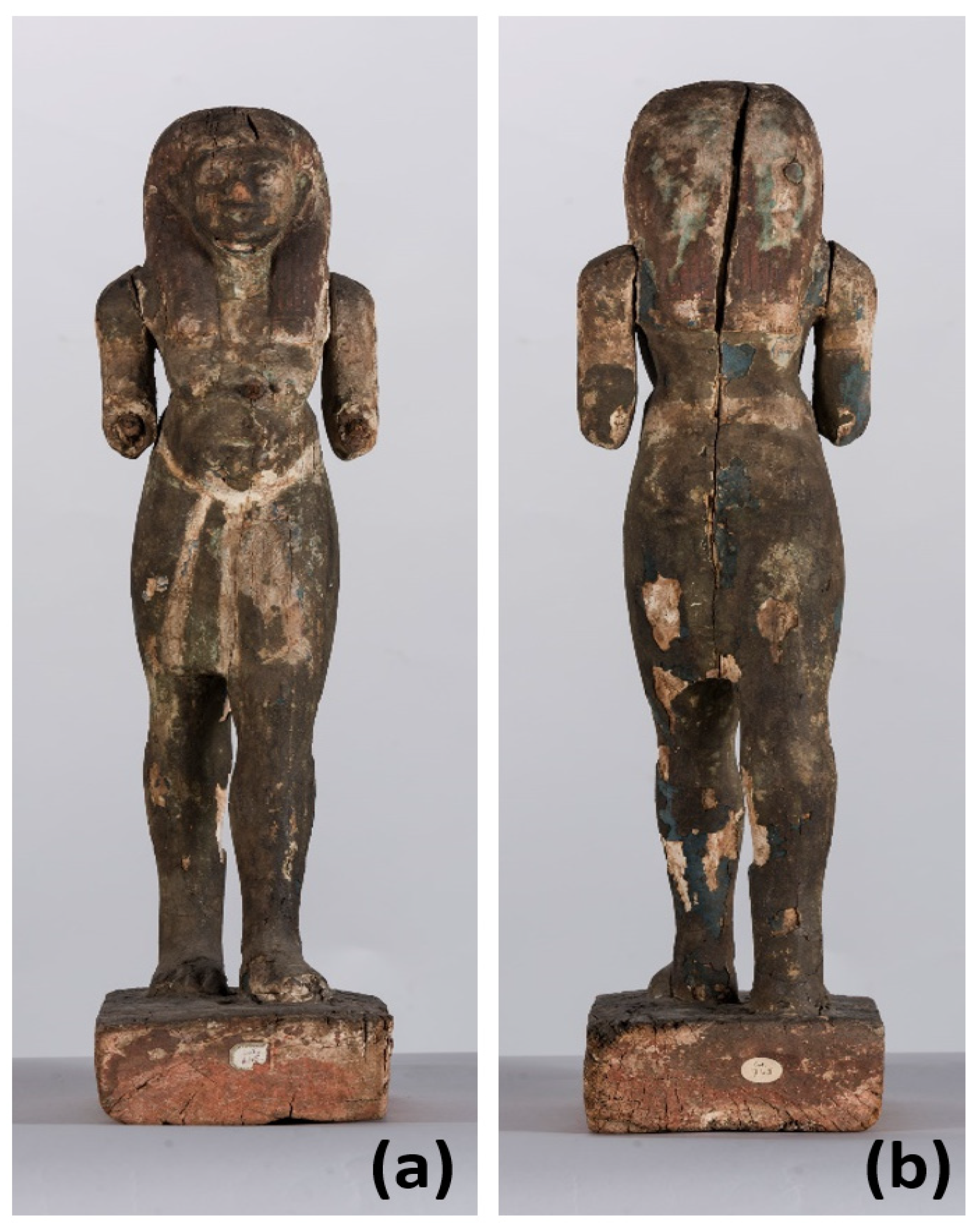
:1. Introduction
2. Materials and Methods
2.1. Painting Materials Analysis
2.2. Mockups for Cleaning Test
2.2.1. Mockups
2.2.2. Selected Cleaning Materials
2.2.3. Assessment of Cleaning Results
3. Results and Discussion
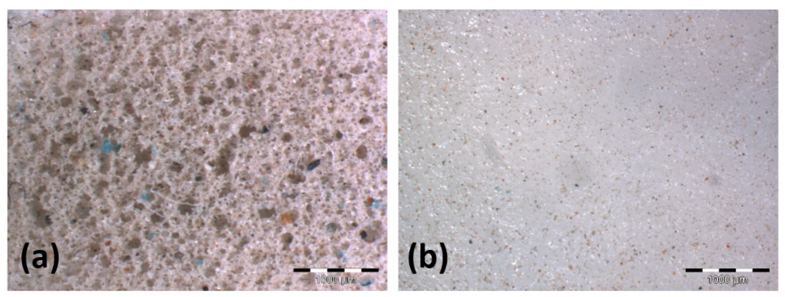
3.1. Painting Materials Characterization
3.1.1. Blue and Green Pigments
3.1.2. The brown Pigment Layer
3.1.3. Other Pigments
3.2. Cleaning Tests on Mockups
3.3. Cleaning Treatment of the Egyptian Statuette
4. Conclusions
Supplementary Materials
Author Contributions
Funding
Acknowledgments
Conflicts of Interest
References
- Baines, J. Fecundity Figures. Egyptian Personification and the Iconology of a Genre; Aris & Phillips Ltd.: Warminster, UK, 1985; pp. 317–318. ISBN 0-85668-087-7. [Google Scholar]
- Mottica, R. Scultura Lignea in Terra d’Egitto: Analisi Valutativa di una Statuetta del Dio Nilo Conservata al Museo Egizio di Torino. Bachelor’s Thesis, Università di Torino, Torino, Italy, 2006. [Google Scholar]
- Santoro, S. Il Colore nell’Antico Egitto: Inquadramento e Riflessioni Tecnico-Artistiche e Simboliche sui Colori di Statuetta del dio Nilo, Integrate da Indagini Archeometriche al SEM-EDS. Bachelor’s Thesis, Università di Torino, Torino, Italy, 2006. [Google Scholar]
- Serrapede, M. I Colori del Dio Nilo: Caratterizzazione delle Stesure Cromatiche di una Statuetta Lignea del Dio per Mezzo di XRF, PIXE e SEM-EDS. Bachelor’s Thesis, Università di Torino, Torino, Italy, 2007. [Google Scholar]
- Manfredda, N. Conservation Problems of an Artefact with Double Layers of Polychromy: A Wooden Sculpture of New Kingdom from the Museo Egizio of Torino. Master’s Thesis, Università di Torino in agreement with Centro Conservazione e Restauro la Venaria Reale, Torino, Italy, 2019. [Google Scholar]
- Fabretti, A.; Rossi, F.; Lanzone, R.V. Regio Museo di Torino. Antichità Egizie; Stamperia Reale: Torino, Italy, 1882; Volume 1, p. 58. [Google Scholar]
- Daudin-Schotte, M.; Bisschoff, M.; Joosten, I.; van Keulen, H.; van den Berg, K.J. Dry cleaning approaches for unvarnished paint surfaces. In New Insights into the Cleaning of Paintings: Proceedings from the Cleaning 2010 International Conference, Universidad Politécnica de Valencia and Museum Conservation Institute; Mecklenburg, M.F., Charola, E., Koestler, R.J., Eds.; Smithsonian Contributions to Museum Conservation; Smithsonian Institution Scholarly Press: Washington, DC, USA, 2013; pp. 209–219. [Google Scholar]
- Baglioni, M.; Poggi, G.; Chelazzi, D.; Baglioni, P. Advanced materials in cultural heritage conservation. Molecules 2021, 26, 3967. [Google Scholar] [CrossRef]
- Domingues, J.A.L.; Bonelli, N.; Giorgi, R.; Fratini, E.; Gorel, F.; Baglioni, P. Innovative hydrogels based on semi-interpenetrating p(HEMA)/PVP networks for the cleaning of water-sensitive cultural heritage artifacts. Langmuir 2013, 29, 2746–2755. [Google Scholar] [CrossRef] [PubMed]
- Bonelli, N.; Montis, C.; Mirabile, A.; Bertia, D.; Baglioni, P. Restoration of paper artworks with microemulsions confined in hydrogels for safe and efficient removal of adhesive tapes. Proc. Natl. Acad. Sci. USA 2018, 115, 5932–5937. [Google Scholar] [CrossRef] [Green Version]
- Cardaba, I.; Poggi, G.; Baglioni, M.; Chelazzi, D.; Maguregui, I.; Giorgi, R. Assessment of aqueous cleaning of acrylic paints using innovative cryogels. Microchem. J. 2020, 152, 104311. [Google Scholar] [CrossRef]
- Bonelli, N.; Poggi, G.; Chelazzi, D.; Giorgi, R.; Baglioni, P. Poly(vinyl alcohol)/poly(vinyl pyrrolidone) hydrogels for the cleaning of art. J. Colloid Interface Sci. 2019, 536, 339–348. [Google Scholar] [CrossRef] [PubMed]
- Bartoletti, A.; Barker, R.; Chelazzi, D.; Bonelli, N.; Baglioni, P.; Lee, J.; Angelova, L.V.; Ormsby, B. Reviving WHAAM! a comparative evaluation of cleaning systems for the conservation treatment of Roy Lichtenstein’s iconic painting. Herit. Sci. 2020, 8, 9. [Google Scholar] [CrossRef]
- Mastrangelo, R.; Chelazzi, D.; Poggi, G.; Fratini, E.; Pensabene Buemi, L.; Petruzzelis, M.L.; Baglioni, P. Twin-chain polymer hydrogels based on poly(vinyl alcohol) as new advanced tool for the cleaning of modern and contemporary art. Proc. Natl. Acad. Sci. USA 2020, 117, 7011–7020. [Google Scholar] [CrossRef] [Green Version]
- Vigorelli, L.; Re, A.; Guidorzi, L.; Cavaleri, T.; Buscaglia, P.; Nervo, M.; Del Vesco, P.; Borla, M.; Grassini, S.; Lo Giudice, A. Multi-analytical approach for the study of an ancient Egyptian wooden statuette from the collection of Museo Egizio of Torino. Acta Imeko 2021, in press. [Google Scholar]
- Petrakakis, K.; Dietrich, H. MINSORT: A program for the processing and archivation of microprobe analysis of silicate and oxide minerals. Neues Jb. Miner. Abh. 1985, 8, 379–384. [Google Scholar]
- Lo Giudice, A.; Re, A.; Angelici, D.; Corsi, J.; Gariani, G.; Zangirolami, M.; Ziraldo, E. Ion microbeam analysis in cultural heritage: Application to lapis lazuli and ancient coins. Acta Imeko 2017, 6, 76–81. [Google Scholar] [CrossRef] [Green Version]
- Oleari, C. Misurare il Colore, 2nd ed.; Hoepli: Milano, Italy, 2008; pp. 222–224. ISBN 8-820-34126-3. [Google Scholar]
- Piquette, K.E. Reflectance transformation imaging (RTI) and Ancient Egyptian material culture. Damqatum CEHAO Newsl. 2011, 7, 16–20. [Google Scholar]
- Serotta, A. An investigation of tool marks on Ancient Egyptian hard stone sculpture: Preliminary report. In Metropolitan Museum Studies in Art, Science, and Technology; Centeno, S.A., Kennedy, N.W., Manuels, M., Schorsch, D., Stone, R.E., Sun, Z.J., Wypyski, M.T., Eds.; Metropolitan Museum of Art: New York, NY, USA, 2014; Volume 2, pp. 197–201. ISBN 0-300-20439-6. [Google Scholar]
- Wolbers, R. Cleaning Painted Surfaces: Aqueous Methods; Archetype Publications: London, UK, 2000; ISBN 978-1-873132-36-4. [Google Scholar]
- Hatton, G.D.; Shortland, A.J.; Tite, M.S. The production technology of Egyptian blue and green frits from second millennium BC Egypt and Mesopotamia. J. Archaeol. Sci. 2008, 35, 1591–1604. [Google Scholar] [CrossRef]
- Scott, D.A. A review of ancient Egyptian pigments and cosmetics. Stud. Conserv 2016, 61, 185–202. [Google Scholar] [CrossRef]
- Pagès-Camagna, S.; Colinart, S. The Egyptian green pigment: Its manufacturing process and links to Egyptian blue. Archaeometry 2003, 45, 637–658. [Google Scholar] [CrossRef]
- Asperger, A.; Engewald, W.; Fabian, G. Advances in the analysis of natural waxes provided by thermally assisted hydrolysis and methylation (THM) in combination with GC/MS. J. Anal. Appl. Pyrolysis 1999, 52, 51–63. [Google Scholar] [CrossRef]
- Asperger, A.; Engewald, W.; Fabian, G. Thermally assisted hydrolysis and methylation—A simple and rapid online derivatization method for the gas chromatographic analysis of natural waxes. J. Anal. Appl. Pyrolysis 2001, 61, 91–109. [Google Scholar] [CrossRef]
- Girod, A.; Weyermann, C. Lipid composition of fingermark residue and donor classification using GC/MS. Forensic Sci. Int. 2014, 238, 68–82. [Google Scholar] [CrossRef] [PubMed] [Green Version]
- Riedo, C.; Scalarone, D.; Chiantore, O. Advances in identification of plant gums in cultural heritage by thermally assisted hydrolysis and methylation. Anal. Bioanal. Chem. 2010, 396, 1559–1569. [Google Scholar] [CrossRef] [PubMed]
- Riedo, C.; Scalarone, D.; Chiantore, O. Multivariate analysis of pyrolysis-GC/MS data for identification of polysaccharide binding media. Anal. Methods 2013, 5, 4060–4067. [Google Scholar] [CrossRef] [Green Version]
- Daniels, V.; Stacey, R.; Middleton, A. The blackening of paint containing Egyptian blue. Stud. Conserv. 2004, 49, 217–230. [Google Scholar] [CrossRef]
- Challinor, J.M. Characterisation of wood by pyrolysis derivatisation-gas chromatography/mass spectrometry. J. Anal. Appl. Pyrolysis 1995, 35, 93–107. [Google Scholar] [CrossRef]
- Buckley, S.A.; Evershed, R.P. Organic chemistry of embalming agents in Pharaonic and Graeco-Roman mummies. Nature 2001, 413, 837–841. [Google Scholar] [CrossRef] [PubMed]
- Fabbri, D.; Chiavari, G.; Galle, G.C. Characterization of soil humin by pyrolysis(/methylation)-gas chromatography/mass spectrometry: Structural relationships with humic acids. J. Anal. Appl. Pyrolysis 1996, 37, 161–172. [Google Scholar] [CrossRef]
- Kaal, J.; Nierop, K.G.J.; Kraal, P.; Preston, C.M. A first step towards identification of tannin-derived black carbon: Conventional pyrolysis (Py-GC-MS) and thermally assisted hydrolysis and methylation (THM-GC-MS) of charred condensed tannins. Org. Geochem. 2012, 47, 99–108. [Google Scholar] [CrossRef]
- Chiavari, G.; Montalbani, S.; Prati, S.; Keheyan, Y.; Baroni, S. Application of analytical pyrolysis for the characterisation of old inks. J. Anal. Appl. Pyrolysis 2007, 80, 400–405. [Google Scholar] [CrossRef]
- Guidotti, C.V. Micas in metamorphic rocks. Rev. Mineral. 1984, 13, 357–467. [Google Scholar]
- Massone, H.J.; Schreyer, W. Phengite geobarometry based on the limiting assemblage with K-feldspar, phlogopite and quartz. Contrib. Mineral. Petrol. 1987, 96, 212–224. [Google Scholar] [CrossRef]
- De Putter, T.; Karlshausen, C. Les Pierres Utilisées dans la Sculpture et L’architecture de l’Ègypte Pharaonique: Guide Pratique Illustré; Connaissance de l’Égypte Ancienne: Bruxelles, Belgium, 1992; ISBN 2-87268-003-9. [Google Scholar]
- Klitzsch, E.; Harms, J.C.; Lejal-Nicol, A.; List, F.K. Major subdivisions and depositional environments of Nubia strata, Southwestern Egypt. Am. Assoc. Pet. Geol. Bull. 1979, 63, 967–974. [Google Scholar] [CrossRef]
- Finger, F.; Dörr, W.; Gerdes, A.; Gharib, M.; Dawoud, M. U-Pb zircon ages and geochemical data for the Monumental Granite and other granitoid rocks from Aswan, Egypt: Implications for the geological evolution of the western margin of the Arabian Nubian Shield. Mineral. Petrol. 2008, 93, 153–183. [Google Scholar] [CrossRef]
- Borghi, A.; Vaggelli, G.; D’Amicone, E.; Fiora, L.; Maschali, O.; Shalaby, B.; Vigna, L. Bekhen stone artifacts in the Egyptian Antiquity Museum of Turin (Italy): A mineropetrographic study. In Proceedings of the 2nd International Conference on the Geology of the Tethys, Cairo University, Cairo, Egypt, 19–22 March 2007. [Google Scholar]
- Lee, L.; Quirke, S. Painting materials. In Ancient Egyptian Materials and Technology; Nicholson, P.T., Shaw, I., Eds.; Cambridge University Press: Cambridge, UK, 2000; pp. 104–120. ISBN 0-521-12098-5. [Google Scholar]


| Model Sample | Pigments of Paint Layer 1 | Layering of Materials 1 |
|---|---|---|
| A | None | Preparation layer |
| B | Egyptian blue (KP.10060) | Preparation layer-KP.10060 |
| C | Egyptian green (KP.10064) | Preparation layer-KP.10064 |
| D | Yellow ochre (KP.40301) + | Preparation layer-KP.40301-Z.C0008 |
| Red ochre (Z.C0008) | ||
| E | Yellow ochre (KP.40301) + | Preparation layer-KP.40301-S.7060030 (lines)-Brown |
| Black (S.7060030) + Brown | ||
| F | Red ochre (Z.C0008) + | Preparation layer-Z.C0008-S.7060030 (stripes)-Brown |
| Black (S.7060030) + Brown | ||
| G | Egyptian green (KP.10064) + | Preparation layer-KP.10064-S.7060030 (lines)-Brown |
| Black (S.7060030) + Brown |
| Materials Tested on Each Sample | Objective | Test Name | Test Description |
|---|---|---|---|
| PG6 | Tuning the length of gel sheet’s application | 2a | 180 s |
| 2b | 150 s | ||
| 2c | 120 s + 60 s | ||
| 3a | 90 s + 90 s | ||
| 3b | 60 s + 60 s | ||
| 3c | 30 s | ||
| PG6 PG5 Gum | Comparing the effect of gel gums applied on wet and dry surfaces | 4a | PG6 (90 s + 90 s) + PG5 Gum on a still wet surface |
| 4b | PG6 (90 s + 90 s) + | ||
| PG5 Gum on a dried surface | |||
| PG6 PG5 Gum PU sponge (DJ) 1 | Comparing the best result obtained with hydrogels with the traditional dry cleaning method | 1a | PG6 (90 s + 90 s) + PG5 Gum on a dried surface |
| 1b | Mechanical removal. Sponges previously washed in demineralized water | ||
| PG6 PG5 Gum PU sponge (DJ) 1 | Evaluating the boost in efficacy by combining the two methods | 5a | PU sponge (DJ) 1 + PG6 (90 s) + PG5 Gum on a dried surface |
| 5b | PU sponge (DJ) 1 + PG6 (120 s) | ||
| 5c | PU sponge (DJ) 1 + PG5 Gum on a dried surface |
| Peak n. | Retention Time [min] | Assignment |
|---|---|---|
| 1 | 5.95 | 1,2,3-Trimethoxypropane |
| 2 | 8.08 | 2-Butendioic acid dimethyl ester |
| 3 | 8.27 | 2-Butandioic acid dimethyl ester |
| 4 | 9.32 | Benzoic acid methyl ester |
| 5 | 11.71 | 2-Methoxybutendioic acid dimethyl ester |
| 6 | 12.15 | Permethylated 3-deoxypentenoic acid methyl ester |
| 7 | 12.40 | Permethylated 3-deoxypentenoic acid methyl ester |
| 8 | 13.16 | Permethylated 3,6-deoxyhexenoic acid methyl ester |
| 9 | 13.32 | 1,2,4-Trimethoxybenzene |
| 10 | 13.40 | Permethylated 3,6-deoxyhexenoic acid methyl ester |
| 11 | 13.42 | 4-Methoxybenzoic acid methyl ester |
| 12 | 13.87 | 1,2,3-Propaentricarboxylic acid trimethyl ester |
| 13 | 14.25 | Octanedioic acid dimethyl ester |
| 14 | 14.69 | Permethylated 3-deoxyhexenoic acid methyl ester |
| 15 | 15.17 | Permethylated 3-deoxyhexenoic acid methyl ester |
| 16 | 15.49 | Nonanedioic acid dimethyl ester |
| 17 | 16.08 | 3,4-Dimethoxybenzoic acid methyl ester |
| 18 | 16.39 | 2,3,4,6-Tetra-O-methyl-D-gluconic acid δ-lactone |
| 19 | 17.51 | Tetradecanoic acid methyl ester |
| 20 | 17.56 | 3,4,5-Trimethoxybenzoic acid methyl ester |
| 21 | 18.67 | 1,2,3-Benzentricarboxylic acid trimethyl ester |
| 22 | 18.86 | 1,2,4-Benzentricarboxylic acid trimethyl ester |
| 23 | 19.39 | 9-Hexadecenoic acid methyl ester |
| 24 | 19.62 | Hexadecanoic acid methyl ester |
| 25 | 21.30 | 9-Octadecenoic acid methyl ester |
| 26 | 21.54 | Octadecanoic acid methyl ester |
| 27 | 23.27 | Eicosanoic acid methyl ester |
| 28 | 24.89 | Docosanoic acid methyl ester |
| 29 | 25.65 | 21-Methyldocosanoic acid methyl ester |
| 30 | 26.15 | Heptacosane |
| 31 | 26.40 | Tetracosanoic acid methyl ester |
| 32 | 26.86 | Octacosane |
| 33 | 27.09 | Squalene |
| 34 | 27.59 | Nonacosane |
| 35 | 27.86 | Hexacosanoic acid methyl ester |
| 36 | 28.38 | Tricontane |
| 37 | 29.36 | 3-Methoxycholest-5-ene |
| 38 | 29.65 | Octacosanoic acid methyl ester |
Publisher’s Note: MDPI stays neutral with regard to jurisdictional claims in published maps and institutional affiliations. |
© 2021 by the authors. Licensee MDPI, Basel, Switzerland. This article is an open access article distributed under the terms and conditions of the Creative Commons Attribution (CC BY) license (https://creativecommons.org/licenses/by/4.0/).
Share and Cite
Manfredda, N.; Buscaglia, P.; Gallo, P.; Borla, M.; Aicardi, S.; Poggi, G.; Baglioni, P.; Nervo, M.; Scalarone, D.; Borghi, A.; et al. An Ancient Egyptian Multilayered Polychrome Wooden Sculpture Belonging to the Museo Egizio of Torino: Characterization of Painting Materials and Design of Cleaning Processes by Means of Highly Retentive Hydrogels. Coatings 2021, 11, 1335. https://doi.org/10.3390/coatings11111335
Manfredda N, Buscaglia P, Gallo P, Borla M, Aicardi S, Poggi G, Baglioni P, Nervo M, Scalarone D, Borghi A, et al. An Ancient Egyptian Multilayered Polychrome Wooden Sculpture Belonging to the Museo Egizio of Torino: Characterization of Painting Materials and Design of Cleaning Processes by Means of Highly Retentive Hydrogels. Coatings. 2021; 11(11):1335. https://doi.org/10.3390/coatings11111335
Chicago/Turabian StyleManfredda, Nicole, Paola Buscaglia, Paolo Gallo, Matilde Borla, Sara Aicardi, Giovanna Poggi, Piero Baglioni, Marco Nervo, Dominique Scalarone, Alessandro Borghi, and et al. 2021. "An Ancient Egyptian Multilayered Polychrome Wooden Sculpture Belonging to the Museo Egizio of Torino: Characterization of Painting Materials and Design of Cleaning Processes by Means of Highly Retentive Hydrogels" Coatings 11, no. 11: 1335. https://doi.org/10.3390/coatings11111335