
The Life of a Painting as Traced by Technical Analysis: Original Materials and Posthumous Alterations in Édouard Manet’s Woman in Striped Dress

 ,
,
Abstract
:
1. Introduction
2. Materials and Methods
2.1. IR Reflectography and Transmittography
2.2. X-Radiography
2.3. Point XRF
2.4. MA-XRF
2.5. FTIR
2.6. Raman
2.7. SEM/EDS
2.8. EBSD
2.9. Py-GC/MS
2.10. Preparation of Cross Sections
3. Results and Discussion
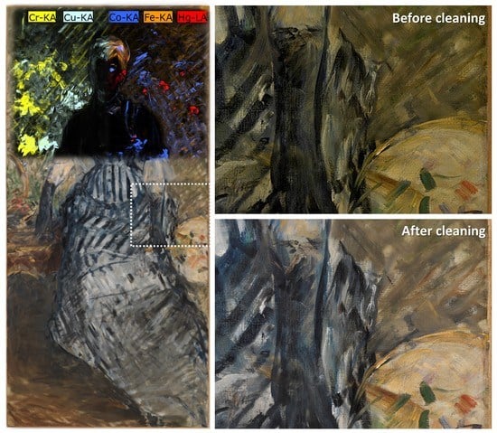
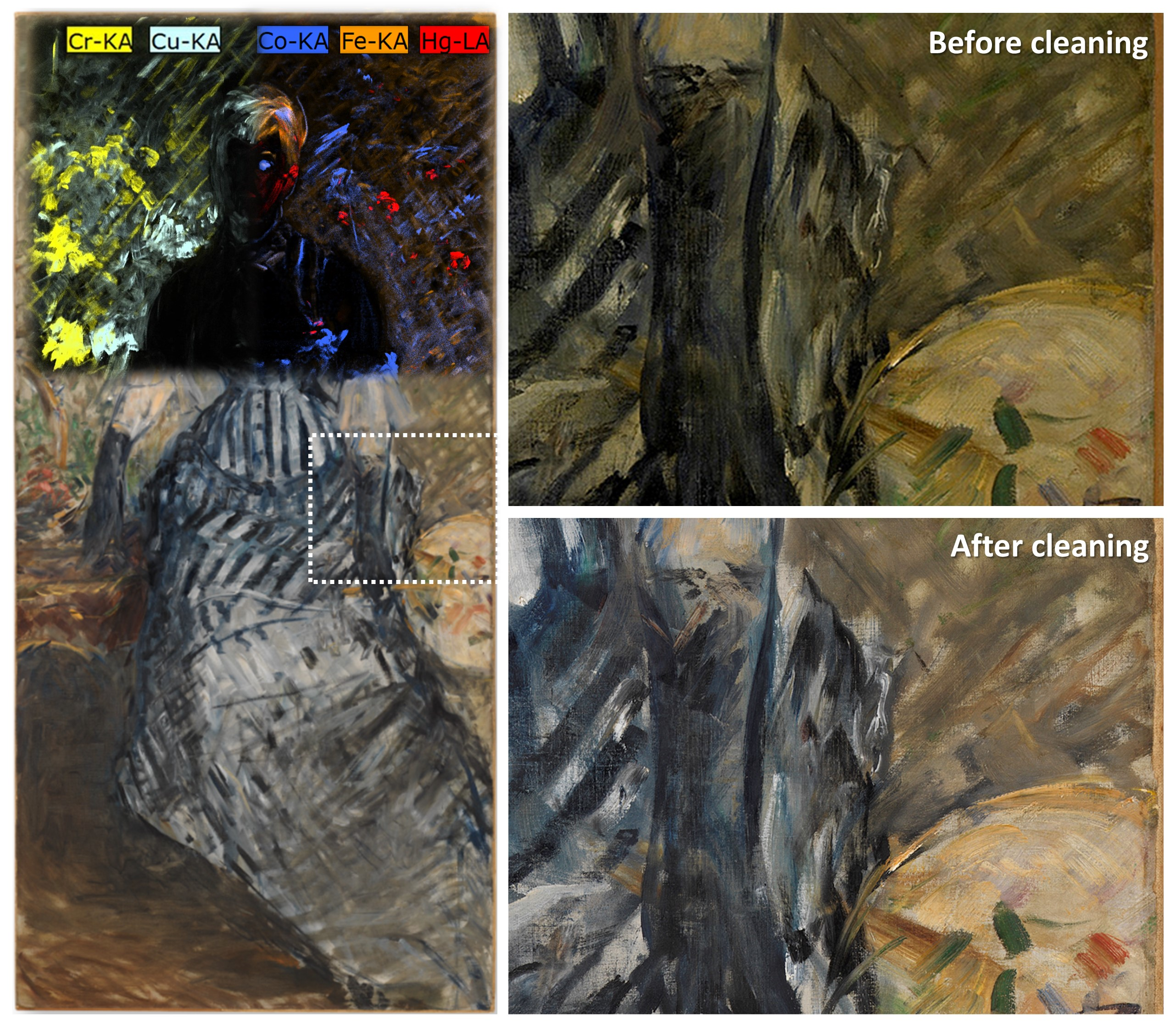
3.1. Canvas Support and Ground Preparation
3.2. Paint Layers
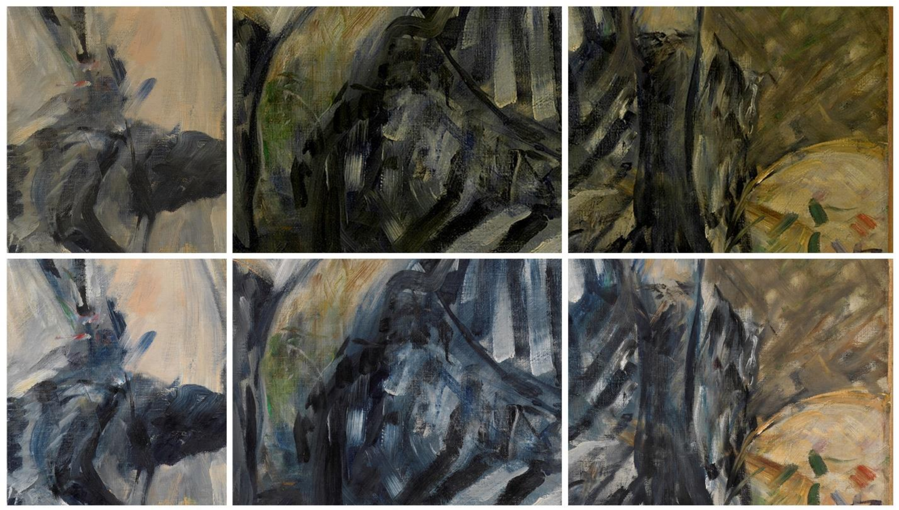
3.3. Varnish
4. Conclusions
Supplementary Materials
Author Contributions
Funding
Institutional Review Board Statement
Informed Consent Statement
Data Availability Statement
Acknowledgments
Conflicts of Interest
References
- McMillan, G.; Pozzi, F. Édouard Manet, Woman in Striped Dress, Materials and Process. In Thannhauser Collection: French Modernism at the Guggenheim; Fontanella, M., Ed.; Guggenheim Museum Publications: New York, NY, USA, 2018; pp. 108–110, 298. [Google Scholar]
- Greene, V.; McMillan, G. Revealing Édouard Manet’s Woman in Striped Dress. In Thannhauser Collection: French Modernism at the Guggenheim; Fontanella, M., Ed.; Guggenheim Museum Publications: New York, NY, USA, 2018; pp. 111–115, 298–300. [Google Scholar]
- Proust, A. Édouard Manet (Souvenirs). La Rev. Blanche 1897, 12, 132–133. [Google Scholar]
- Hanson, A.C. Manet and the Modern Tradition; Yale University Press: New Haven, CT, USA, 1977; p. 160. [Google Scholar]
- Stuckey, C.F. Manet Revised: Whodunit? Art Am. 1983, 71, 158–177, 239–241. [Google Scholar]
- Bomford, D.; Roy, A. Manet’s “The Waitress”: An Investigation into its Origin and Development. Natl. Gallery Tech. Bull. 1983, 7, 3–19. [Google Scholar]
- McMillan, G. Édouard Manet, Before the Mirror, Materials and Process. In Thannhauser Collection: French Modernism at the Guggenheim; Fontanella, M., Ed.; Guggenheim Museum Publications: New York, NY, USA, 2018; pp. 104–106. [Google Scholar]
- Groom, G.; Westerby, G. (Eds.) Manet Paintings and Works on Paper at the Art Institute of Chicago. 2017. Available online: https://publications.artic.edu/manet/reader/manetart/ (accessed on 5 August 2021).
- Amato, S.R.; Burnstock, A.; Cross, M.; Janssens, K.; Rosi, F.; Cartechini, L.; Fontana, R.; Fovo, A.D.; Paolantoni, M.; Grazia, C.; et al. Interpreting Technical Evidence from Spectral Imaging of Paintings by Édouard Manet in the Courtauld Gallery. X-Ray Spectrom. 2019, 48, 282–292. [Google Scholar] [CrossRef]
- Eastaugh, N.; Walsh, V.; Chaplin, T.; Siddall, R. Pigment Compendium. A Dictionary and Optical Microscopy of Historic Pigments; Butterworth-Heinemann: Oxford, UK, 2004. [Google Scholar]
- van den Berg, K.J.; Boon, J.J.; Pastorova, I.; Spetter, L.F. Mass Spectrometric Methodology for the Analysis of Highly Oxidized Diterpenoid Acids in Old Master Paintings. J. Mass Spectrom. 2000, 35, 512–533. [Google Scholar] [CrossRef]
- Mills, J.; White, R. Organic Chemistry of Museum Objects, 2nd ed.; Butterworth-Heinemann: Oxford, UK, 1994. [Google Scholar]
- White, R.; Kirby, J. A Survey of Nineteenth- and Early Twentieth-Century Varnish Compositions Found on a Selection of Paintings in the National Gallery Collection. Natl. Gallery Tech. Bull. 2001, 22, 64–84. [Google Scholar]
- Duret, T. Histoire d’Édouard Manet et de Son Oeuvre; H. Floury: Paris, France, 1902. [Google Scholar]


| Samples | Analytical Techniques | Ground Preparation | Pigments, Fillers, and Extenders in Paint Layers | Varnish |
|---|---|---|---|---|
| (S2) Scraping of uppermost varnish; background, left of center, top quadrant | FTIR, Py-GC/MS | --- | --- | Diterpenoid natural resin belonging to the Pinaceae family, linseed oil with possible addition of driers |
| (S3) Cross section of ground; proper right edge, tape removed | Optical microscopy, SEM/EDS | Single-layer ground (20–50 μm): coarse lead white, with a few particles of calcite, barite, feldspar, and carbon-based black | --- | One layer (6–20 μm), on top of ground preparation |
| (S4) Cross section of green paint; foliage, proper right edge, farther into painting | Optical microscopy, SEM/EDS | Single-layer ground (50 μm): coarse lead white, with a few particles of calcite | Top: Calcite, gypsum, Fe oxide/oxy-hydroxide Bottom: Emerald green, ultramarine blue, cadmium yellow, Naples yellow, vermilion, Cr-based green (possibly viridian), lead white, Fe oxide/oxy-hydroxide, organic lake on Al substrate | Two layers (bottom 10–20 μm, top 5–10 μm), on top of paint layers |
| (S5) Scraping of green paint; foliage, proper right, top quadrant | FTIR, Raman | --- | Viridian, emerald green, lead white (hydrocerussite) | Natural resin and oil (varnish and/or binding medium) |
| (S6) Scraping of green paint; foliage, right of center, top quadrant | FTIR, Raman | --- | Viridian, ultramarine blue, chrome yellow, lead white (hydrocerussite), barite | Natural resin and oil (varnish and/or binding medium) |
| (S7) Scraping of green paint; foliage, left of center, top quadrant | FTIR, Raman | --- | Viridian, ultramarine blue, vermilion, red and yellow ochers, chrome yellow, lead white (cerussite), kaolinite | Natural resin and oil (varnish and/or binding medium) |
| (S8) Scraping of green paint; foliage, left of center, top quadrant | FTIR, Raman | --- | Viridian, ultramarine blue, chrome yellow, lead white (hydrocerussite), barite | Natural resin and oil (varnish and/or binding medium) |
| (S9) Cross section of brown paint; foliage, proper right edge, tape removed | Optical microscopy, SEM/EDS, EBSD | Single-layer ground (60–70 μm): coarse lead white, with a few particles of calcite and quartz | Top: Lead white, cobalt blue, ultramarine blue, emerald green, malachite, cadmium yellow, Fe oxide/oxy-hydroxide Bottom: Lead white, cadmium yellow, cobalt blue, ultramarine blue, Cr-based green (possibly viridian), emerald green, Fe oxide/oxy-hydroxide, red lake on Al substrate, Naples yellow, malachite, quartz | One layer (3–6 μm), on top of paint layers |
| (S10) Cross section of brown paint; foliage, proper right edge, farther into painting. Broken into two fragments, 10a and 10b | Optical microscopy, SEM/EDS | Single-layer ground (30 μm): coarse lead white, with a few particles of calcite and gypsum | Top: Calcite, gypsum, Fe oxide/oxy-hydroxide Bottom: Lead white, cadmium yellow, chrome yellow, cobalt blue, ultramarine blue, Cr-based green (possibly viridian), emerald green, Fe oxide/oxy-hydroxide, red lake on Al substrate, vermilion, Naples yellow, Cu-based green (possibly malachite), bone or ivory black, cerulean blue, quartz | Two layers (bottom 6–25 μm, top 3–6 μm), on top of paint layers |
| (S11) Scraping of green paint; foliage, proper right, top quadrant | FTIR, Raman | --- | Viridian, ultramarine blue, vermilion, chrome yellow, lead white (hydrocerussite), barite | Natural resin and oil (varnish and/or binding medium) |
| (S12) Cross section of pink-orange paint; basket of flowers, near proper right edge | Optical microscopy, SEM/EDS | Single-layer ground (partial): coarse lead white, with a few particles of calcite | Top: Lead white, barite, vermilion, chrome yellow, red lakes on Al and S substrates, emerald green, gypsumBottom: Red ocher, lead white, ultramarine blue, cobalt blue | Three layers: two in between paint layers (bottom 2–5 μm, top 4–5 μm); one at top of stratigraphy (8–10 μm) |
| (S13) Cross section of green paint; foliage, proper right, top quadrant | Optical microscopy, SEM/EDS | Single-layer ground (10–40 μm): coarse lead white, with a few particles of calcite, barite, silicates, and iron-containing earths | Top: Cr-based green (possibly viridian), ultramarine blue, chrome yellow, lead white, barite, Fe oxide/oxy-hydroxide, vermilion, calcite Bottom: Emerald green, lead white, Fe oxide/oxy-hydroxide, ultramarine blue, vermilion, Naples yellow, zinc yellow, bone or ivory black, calcite, barite | Two layers: one in between paint layers (5–15 μm); one on top of paint layers (20–40 μm) |
| (S14) Cross section of green paint; foliage, proper left, top quadrant | Optical microscopy, SEM/EDS | --- | Cr-based green (possibly viridian), chrome yellow, lead white, barite, red ocher, calcite | None observed |
| (S15) Scraping of blue paint; dress, near proper left edge, bottom quadrant | Raman | --- | Ultramarine blue, carbon-based black | --- |
| (S16) Scraping of blue paint; dress, center of picture | Raman | --- | Ultramarine blue | --- |
Publisher’s Note: MDPI stays neutral with regard to jurisdictional claims in published maps and institutional affiliations. |
© 2021 by the authors. Licensee MDPI, Basel, Switzerland. This article is an open access article distributed under the terms and conditions of the Creative Commons Attribution (CC BY) license (https://creativecommons.org/licenses/by/4.0/).
Share and Cite
Pozzi, F.; Centeno, S.A.; Caro, F.; McMillan, G.; Stringari, L.; Greene, V. The Life of a Painting as Traced by Technical Analysis: Original Materials and Posthumous Alterations in Édouard Manet’s Woman in Striped Dress. Coatings 2021, 11, 1334. https://doi.org/10.3390/coatings11111334
Pozzi F, Centeno SA, Caro F, McMillan G, Stringari L, Greene V. The Life of a Painting as Traced by Technical Analysis: Original Materials and Posthumous Alterations in Édouard Manet’s Woman in Striped Dress. Coatings. 2021; 11(11):1334. https://doi.org/10.3390/coatings11111334
Chicago/Turabian StylePozzi, Federica, Silvia A. Centeno, Federico Caro, Gillian McMillan, Lena Stringari, and Vivien Greene. 2021. "The Life of a Painting as Traced by Technical Analysis: Original Materials and Posthumous Alterations in Édouard Manet’s Woman in Striped Dress" Coatings 11, no. 11: 1334. https://doi.org/10.3390/coatings11111334
APA StylePozzi, F., Centeno, S. A., Caro, F., McMillan, G., Stringari, L., & Greene, V. (2021). The Life of a Painting as Traced by Technical Analysis: Original Materials and Posthumous Alterations in Édouard Manet’s Woman in Striped Dress. Coatings, 11(11), 1334. https://doi.org/10.3390/coatings11111334