Review

In Silico Modeling as a Perspective in Developing Potential Vaccine Candidates and Therapeutics for COVID-19

1
Institute of Chemical Industries Researches, National Research Centre, Dokki, Cairo 12622, Egypt
2
Department of Pharmaceutical Chemistry, Faculty of Pharmaceutical Sciences and Pharmaceutical Industries, Future University in Egypt (FUE), Cairo 11835, Egypt
3
Department of Surgery, Hamad Medical Corporation (HMC), Doha 3050, Qatar
4
Department of Chemical and Product Safety, German Federal Institute for Risk Assessment (BfR), Max-Dohrn-Strasse 8-10, 10589 Berlin, Germany
5
GALAB Laboratories GmbH, BioPharma Division, 21029 Hamburg, Germany
*
Author to whom correspondence should be addressed.
Coatings 2021, 11(11), 1273; https://doi.org/10.3390/coatings11111273
Submission received: 28 July 2021 / Revised: 29 September 2021 / Accepted: 12 October 2021 / Published: 20 October 2021
(This article belongs to the Special Issue Antiviral Coating as Sustainable Measure to Fight Viral Pandemic)

Abstract

:
The potential of computational models to identify new therapeutics and repurpose existing drugs has gained significance in recent times. The current ‘COVID-19’ pandemic caused by the new SARS CoV2 virus has affected over 200 million people and caused over 4 million deaths. The enormity and the consequences of this viral infection have fueled the research community to identify drugs or vaccines through a relatively expeditious process. The availability of high-throughput datasets has cultivated new strategies for drug development and can provide the foundation towards effective therapy options. Molecular modeling methods using structure-based or computer-aided virtual screening can potentially be employed as research guides to identify novel antiviral agents. This review focuses on in-silico modeling of the potential therapeutic candidates against SARS CoVs, in addition to strategies for vaccine design. Here, we particularly focus on the recently published SARS CoV main protease (Mpro) active site, the RNA-dependent RNA polymerase (RdRp) of SARS CoV2, and the spike S-protein as potential targets for vaccine development. This review can offer future perspectives for further research and the development of COVID-19 therapies via the design of new drug candidates and multi-epitopic vaccines and through the repurposing of either approved drugs or drugs under clinical trial.

1. Introduction

The identification of effective therapeutics for COVID-19 has been an important focus of research across the world ever since its emergence. Interestingly, with two years of effort in this domain, many approaches have been taken to recognize and develop potential candidates against this virus, with some being more efficient than others. This fast-paced pursuit for the identification of therapeutics has brought in-silico and computational modeling into the limelight, mainly driven by the inefficiency of experimental high-throughput screening (HTS) for COVID-19 drug discovery [1]. Computational modeling holds the key to this bottleneck by providing a more efficient solution to expedite drug discovery or repurposing for the COVID-19 pandemic by evaluating the biological interactions of drugs with viral molecular components based on existing datasets [2,3,4]. There are two types of computational approaches towards identifying therapeutics: target-based and disease-based. The former generates information on the drug–target interaction, while the latter utilizes existing databases to determine new indications of existing drugs via comparisons with disease characteristics [5,6].
Another popular mode of drug discovery is virtual screening (VS), an approach that combines initial computing followed by experimental targeting of specific biomolecules, such as DNA, protein, RNA, or lipid molecules, for the purpose of inhibiting or activating them [7]. This facilitates the rapid identification of viral proteins, with their structures determined using experimental and computational methods. Unlike conventional vaccine discovery, this approach provides a cost- and time-efficient route towards the identification of potential drug candidates for several microbial pathogens. Reverse vaccinology (RV), an approach that uses the genome to design vaccines, has revolutionized the field, as bacteria no longer need to be cultured to identify vaccine targets [8]. As a consequence, all putative target protein antigens can be identified, instead of being restricted to those isolated from bacterial cultures. Considering these benefits, researchers are focusing on developing RV prediction algorithms. Currently, there are no licensed drugs suitable for such viral infections. Consequently, drug repurposing has become the best course of action in this scenario, involving the effective adaptation of existing antiviral agents for alternative treatments. Several FDA-approved drug candidates that have been previously applied for SARS CoV1 and Middle Eastern Respiratory Syndrome (MERS) management are currently being investigated for SARS CoV2 therapy. Drug repurposing plays a specific role in drug discovery by identifying new therapeutic uses for already studied drugs [9,10,11]. Globally, more than 500 clinical trials of repurposed drugs have been registered and a further 150 have been initiated [5]. The current research studies are mainly focused on the utilization of antiviral drugs and repurposing strategies, as well as vaccine development [6,12,13,14,15,16,17]. The WHO roadmap [18] provides the necessary guidelines and support for regulation, ethics, and platform utilization to ensure the efficient development of vaccines and treatments.
Most coronaviruses share the same genetic features. As a result of their large genome size (about 27–32 kilobases), they are classified as RNA viruses with positive sense. Because they have overlapping ORF1a and ORF1b polyproteins, their genomes are not segmented. Polyproteins, as shown in Figure 1, arecomposed of 16 non-structural proteins (NSPs) and structural proteins in the form of spike (S), envelope (E), membrane (M), and nucleoproteins (N) [19]. After infection of host cells, different CoVs have been shown to undergo genome recombination [20]. This recombination contributes to their evolution into new types, which could include new intermediate hosts. As a result, CoVs have strong adaptation ability and the ability to be involved in a wide range of species; however, the identification of these viruses’ spike glycoproteins is crucial for vaccine development. In addition, the use of in-silico studies to design new antiviral drug candidates is beneficial [21,22]. This is why the targeting of SARS CoV2 spike (S) proteins, polyproteins, or other viral proteins for vaccine and therapeutic development is becoming increasingly essential approach [23].
Alternatively, natural plant-based bioactive compounds have been proven to possess antiviral properties that combat infection at various stages of the viral replication cycle, including virus entry inhibition into the host cells [24,25,26,27]. In addition to therapeutic identification, delivery is a crucial part of drug discovery. Nano-carriers hold huge potential in drug delivery and can be easily adapted for vaccine development or antiviral delivery targeting specific peptides of CoV protein [28,29,30]. Many datasets on the structure–activity relationships and toxicity of nano delivery systems are available as a consequence of the booming popularity of this field, which can be adapted for the current pandemic as well [31,32,33,34].
Despite these efforts from interdisciplinary groups of researchers, no effective antiviral treatment is available for the virus. While alternate strategies including stem cells, immunotherapy, and plant-based therapeutics, the need to ensure the safety of clinical trials means an ideal solution has not been found for the current scenario; hence, there is a huge scope for exploiting the wide spectrum of existing antivirals to target SARS CoV2 virus. Herein, this review gives insights on the molecular modeling studies of (1) SARS CoV1, (2) the new SARS CoV2, and (3) vaccine studies. This review aims to provide a road map for future researchers that can be used in identifying target-based antiviral molecules by employing computational models. We further hope to establish the significance of drug repositioning or repurposing as a means to combat any future viral infections. Further, this review can also be used as the basis for the future development of coronavirus therapeutics.

2. Diverse Studies on Molecular Targets of SARS CoV

Molecular modeling data on the structure and mechanism of host entry and replication of SARS CoV revealed several strategic sites that could be targets for developing the therapeutic inhibitors. The trimeric spike (S) glycoprotein (Figure 1) is an essential target for which many peptide inhibitors have been developed. During viral host interaction, this glycoprotein is subjected to further cleavage by host proteases into S1 subunit and S2 subunit. The S1 subunit consists of B200 amino acids and is concerned with receptor binding, while S2 subunit was identified to contain heptad repeat (HR) regions [35]. Detailed structural studies have displayed that the receptor-binding domain (RBD) consists of two subdomains: the core and external sub-domains. As S1 subunit involves the first set of interaction sequence between the virus and the host, they have become the main target for many inhibitors. The special binding with its Cys-145 sulfur atom has become the key element for the mechanism of SARS CoV inhibition.

2.1. Molecular Targeting of the Main Protease of SARS CoV (Mpro)

As viral proteases play a crucial part in protein maturation and clearance, they are widely employed as antiviral drug targets. For instance, aziridine- and oxirane-2-carboxylate derivatives were tested for their in-vitro inhibitory activity against SARS CoV Mpro. The significant inhibitory activity was contributed specifically by the Gly-Gly-containing peptide (1), Figure 2. This was attributed to the close proximity of the reactive center of the most active compound (1) with the sulfur atom of Cys-145 of the SI pocket [36].
Similarly, another study worked on developing two series of non-peptidyl inhibitors to target the chymotrypsin-like cysteine proteinases (SARS CoV Mpro). Series 1 was designed to possess a furan ring attached to a benzene derivative moiety, while series 2 possesses different six-membered cyclic aromatic systems attached to a central ester functionality. The results from molecular docking studies on these inhibitors indicated the importance of the 3-chloropyridine moiety for the inhibitory activity as it clusters in the S1 pocket. These studies also indicated the necessity of some functional groups such as chloromethyl ketones, epoxides, Michael acceptors, and azides to have the capability to covalently attach with the sulfur atom in Cys145, thereby inactivating the enzymatic activity of SARS CoV Mpro. Derivatization of the furan ring can maximize the interactions with S2 and S4 pockets, since they allow the insertion of bulky groups, meaning tighter interactions will virtually prevent the peptidyl substrates from entry [37].

2.2. Molecular Targeting of Chemotrypsin-Like Protease of SARS CoV (3CLpro)

Upon exploring the potential of plant-based protease inhibitors, it was identified that quercetin-3-b-galactoside showed inhibition of SARS CoV protease. Further structure–activity relationship studies employing molecular docking and SPR/FRET bioassays indicated that absence of the 7-hydroxy group of quercetin and acetoxylation of the sugar moiety resulted in loss of inhibition, while replacement of the galactose moiety with alternative sugars failed to show any effect on the inhibition potency [38].
Other chymotrypsin-like protease inhibitors were synthesized as two groups; one P4- substitution with oxazole and the other as a P4- substitution with a peptide. Enzymatic inhibition was performed together with an antiviral assay, showing that the peptide-like compounds had more significant enzymatic inhibition effects than their peers of P4-oxazole compounds. Despite the similar antiviral activity levels of the two groups, peptide substitution was not preferred due to the poor transcellular diffusion. The most active inhibitor was the P4-Boc-Ser-incorporated inhibitor (2), Figure 2. The X-ray structure determination of inhibitor (2) in a complex with SARS CoV 3CLpro indicated that the molecular binding between this inhibitor and the enzyme caused a covalent bond between the inhibitor and sulfur atom of Cys-145. P1 lactam NH was also found to form a hydrogen bond with His-163 imidazole ring, with a hydrophobic interaction with the Leu-27 side chain. The P3-carbonyl group formed a hydrogen bond with the NH of Glu-166. The P1–ethyl ester carbonyl group was very close to oxygen in Thr-26, while the P4-Ser side chain displayed hydrogen bonding with Glu-166 [39].
5-Chloropyridine ester-derived series were also designed and synthesized to target the chemotrypsin-like protease of SARS CoV. From this series, inhibitor (3), Figure 2, showed an antiviral EC50 equal to 6.9 M and IC50 value equal to 30 nM. It was concluded that the 5-chloropyridinyl ester specifically at position 4 of the indole ring is crucial for potency.
Molecular docking studies using GOLD 3.221 were performed to provide the possible binding modes for inhibitor (3) with two different crystal structures of SARS CoV 3CLpro (PDB ID: 2HOB and PDB ID: 2V6N). It was noted that the carbonyl carbon of the inhibitor and the sulfur atom of Cys-145 were restricted to the range of 2.5 to 3.5Å for one crystal structure. This orientation stacked the chloropyridyl group in the S1 pocket, with chloro group pointing towards the surface. The distance between the chloropyridinyl nitrogen and the nitrogen atom of the imidazole ring of His-163 was identified to be 2.4Å. Three essential hydrogen bonds with Cys-145(NH), Ser-144(NH), and Gly-143(NH) were identified to form a strong network that embedded the inhibitor into the active site and stabilized the carbonyl of the ester for nucleophilic attack by Cys-145. The indole group of the inhibitor was also found near the hydrophobic S2 pocket, whereby the indole nitrogen showed significant interactions with the imidazole group of His-41. As a result of His-41 flipping and the ability to p stack with an aromatic moiety in the second crystal structure, the inhibitor revealed an indolyl group displaced towards the S1 pocket and stacked with His-41’s shifted imidazole ring, locking the orientation [40]. The 2-(benzylthio)-6-oxo-4-phenyl-1,6-dihydropyrimidine derivatives were synthesized to inhibit SARS CoV 3CL protease, which was proven via in vitro protease inhibition assays. The compounds were also proven to be non-cytotoxic via MTT cytotoxicity assays. One compound of the series inhibitor, 4-(4-chlorophenyl)-2-((4-nitrobenzyl)thio)-6-oxo-1,6-dihydropyrimidine-5-carbonitrile (4), Figure 2, showed promising inhibitory activity against SARS CoV 3CLpro with an IC50 of 6.1 M.
To gainmore insight into the mechanistic action of this compound with the enzyme, a molecular docking study was carried out. The nitro group at C-4 was essential for this activity because of the perfect insertion of the nitro phenyl group into S1 pocket, whereby one oxygen atom of the nitro group made a hydrogen bond with Cys-145 and the other oxygen atom was very close to Gly-143, with a distance of 2.3Å. On the other hand, the chlorophenyl moiety was embedded in the S2 pocket forming hydrophobic interactions with both Met-49 and Gln-189. On comparison of the results of compound (4) with the rest of the compounds, it was concluded that the compounds that did not possess a nitro group in the aryl ring showed minimum activity. Additionally, the presence of a moderate electron-withdrawing group such as chloro functionality in the phenyl ring showed better inhibitory activity than for compounds possessing electron-donating groups such as methyl or methoxy functionalities [41].

2.3. Miscellaneous Targets of SARS CoV

In addition to the above mentioned studies, the molecular dynamics (MD) simulation technique was employed to investigate sixteen antiviral drugs taken from the NCI database against the structure and dynamics of SARS CoV. Among them, four compounds with trade names, namely Nevirapine, Glycovir, Virazole, and Calanolide A, were found to interact well with the active site of the SARS CoV proteinase displaying binding free energy in the range from −10.58 to −5.40 kcal/mol [42].
In vitro studies on aurintricarboxylic acid (ATA) revealed its selective inhibition of SARS CoV replication inside host cells. The mode of action was determined by molecular docking, where it was found that ATA acted on the S1 protein through binding to the incorporated catalytic domain, similar to the RNA-dependent RNA polymerase (RdRp) binding to Ca2+-activated neutral protease (m-calpain) and the protein tyrosine phosphatase (PTP) and then inhibiting its function [43].
Among the 1255 amino acids of the SARS CoV spike protein, receptor binding and membrane fusion sequences exit. The spike protein is further characterized by conserved N-terminal and C-terminal domains, with the former being responsible for membrane fusion. As such, therapeutic compounds that are capable of effective binding to this terminal can inhibit the fusion process, preventing viral entry into host cells. A solid-phase FMOC technique was used to synthesize antisense (negative) DNA peptides that interact selectively with their sense peptide. It was found that hydrophobic elements such as Lys/Arg methylene and Tyr/phenyl groups could engage with sense peptide hydrophobic side chains, while hydrophilic groups such as the amino group interacted with sense hydrophilic backbone elements. As a result, a dodecapeptide with high affinity to the sense peptide could be a successful inhibitor of SARS CoV [44].
Other studies presented aryl diketoacids (ADKs) as SARS CoV-selective inhibitors that showed action (IC50 = 5.4–13.6 M) against the unwinding activity of DNA duplex of the SARS CoV NTPase/helicase. Dihydroroxychromone derivatives (5), Figure 2, were synthesized, whereby an arylmethyl moiety was attached to one end and a catechol moiety was attached to the opposite end. The inhibitory activity levels of these derivatives were measured and noticeable increases in inhibition were shown as long as there were two spatially separated substituents. Based on the previous results, a pharmacophore model was built, indicating two different binding sites at the target enzyme; hence, the three key elements of ADKs essential for binding were a diketo acid core, a free catechol unit, and a hydrophobic aryl methyl substituent [45], as shown in Figure 2.

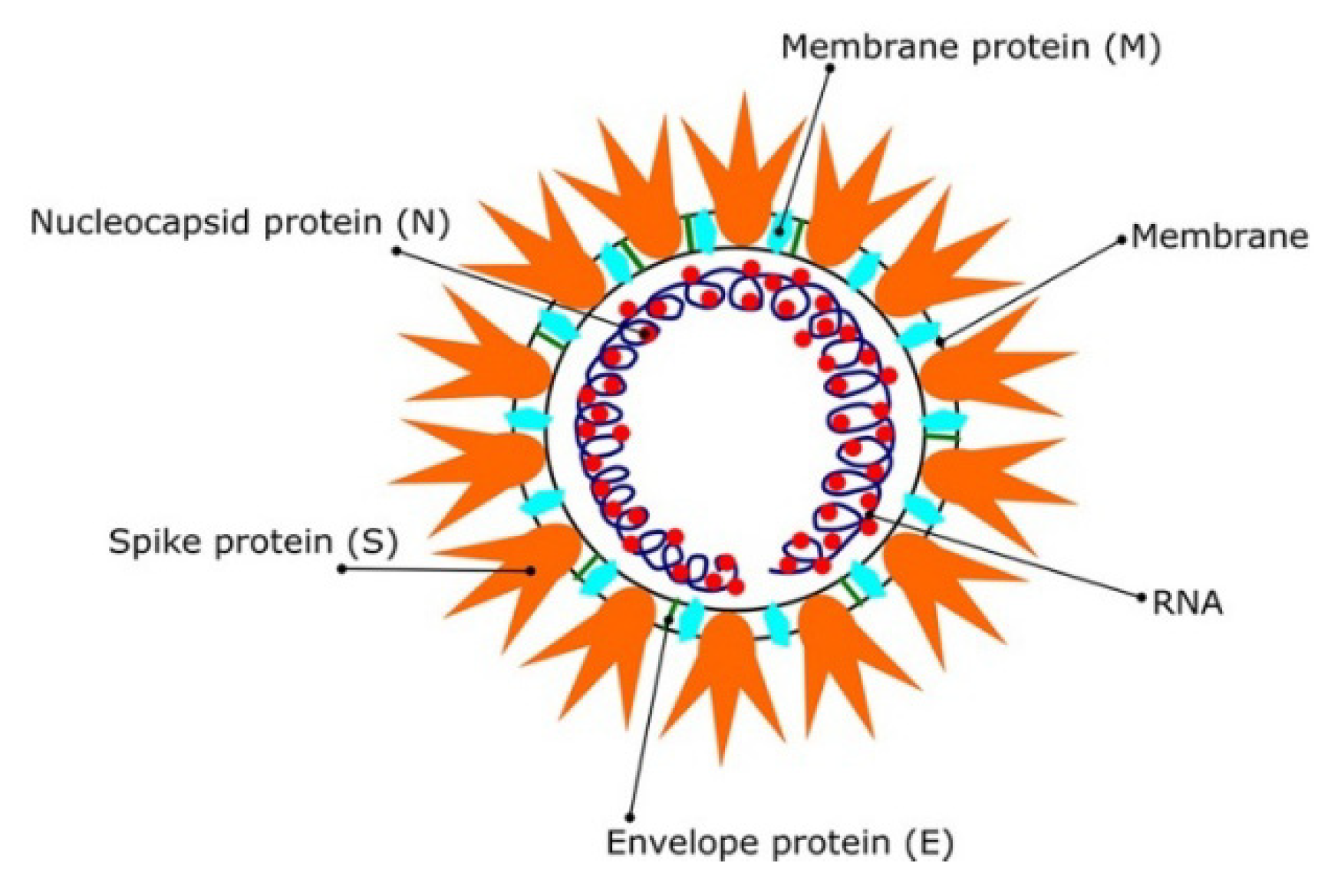
3. Molecular Modeling Studies for the SARS CoV-2Pandemic

In order to effectively repurpose SARS-CoV drugs for SARS CoV2, the extent of sequence similarities between them was investigated and found to be 79% [46,47]. The RNA sequence, including the untranslated region (UTR), open reading frames (ORF), and non-structural proteins (NSP) of the SARS CoV2 virion structure, is shown in Figure 3. The viral RNA is enveloped by nucleocapsid N protein. The viral entry to the host cell starts by interaction of the virial S protein to the angiotensin-converting enzyme 2 (ACE-2) in the host cell [48,49]. As such, both ACE2 and S proteins are considered to be major drug targets [50]. The virus replication isdependent on the host cell resources used. Both replication and transcription are enhanced by NSP [46,47]. RdRp, also known as nsp12, catalyzes the synthesis of viral RNA; therefore, targeting of RdRp has also been considered as an effective approach for COVID-19 drug development [51].
Proteases have also been shown to play a crucial role in viral replication [52]. As demonstrated in Figure 3, ORF1a encodes a (chymotrypsin-like cysteine protease, 3CLPro) main protease in coronaviruses [51,53].The SARS CoV2 RNA sequence encodes Mpro, which is embedded in the nsp5 region. Mpro is a homodimer that is split into two protomers with three distinct domains. Domains I and II have an antiparallel structure, although domain III has α-helices that are connected in parallel with domain II from one side to the other by a loop region. Domains I and II contain the primary Mpro binding site, which contains His41 and Cys145 in its catalytic activity [54]. As a virus replicates and transcribes, Mpro plays a critical role. Many polypeptides are cleaved, including RdRp and RNA processing domains. As a result, Mpro becomes an important target for the therapy of SARS CoV2 [55]. For antiviral medications against SARS, including SARS CoV2 and other coronavirus infections [51], it is widely believed that the key targets are the S protein, ACE-2, TMPRSS2, 3CLpro, RdRp, and PLpro (papain-like protease).

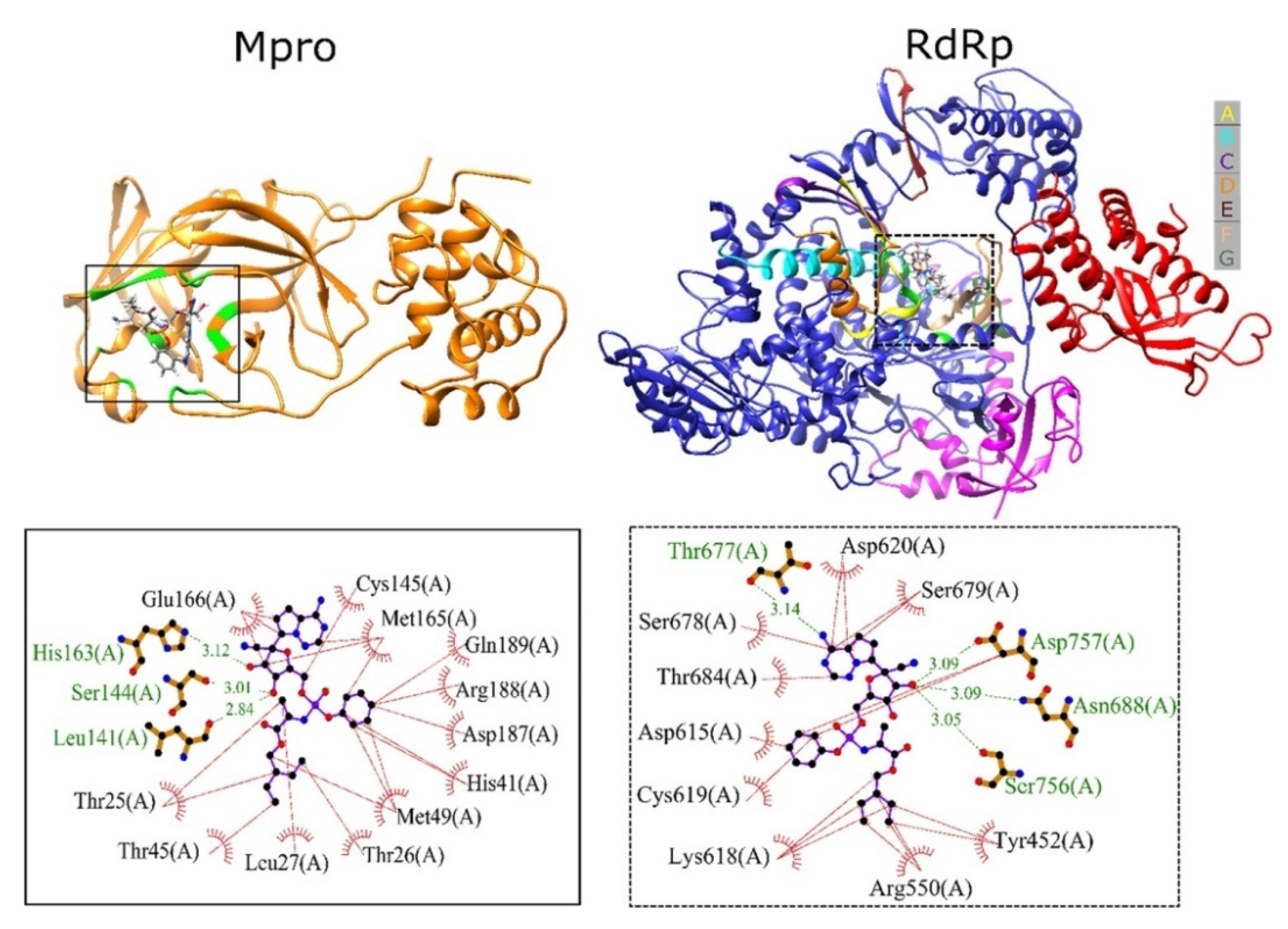
3.1. Molecular Modeling Studies on Drugs Acting on SARS CoV2 Mpro

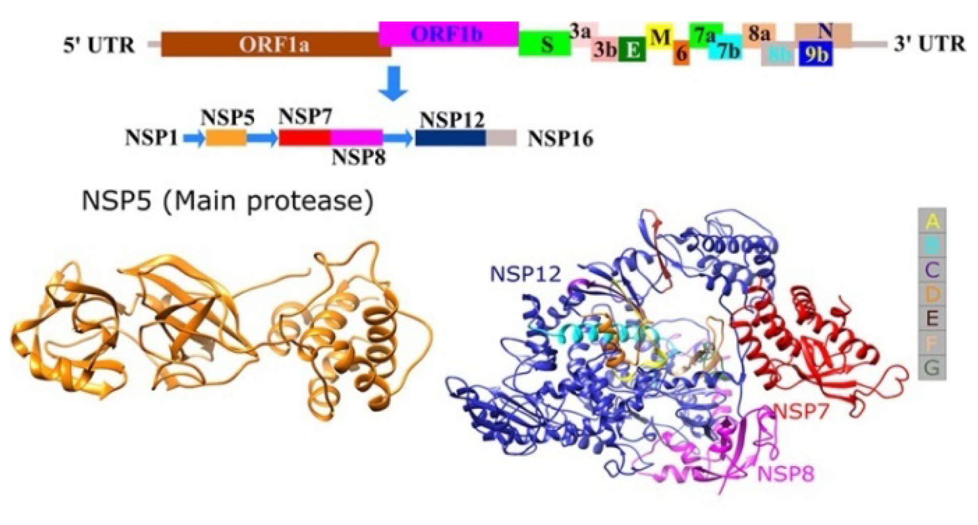
3.1.1. Sequence Analysis and Protease Homology Modeling of SARS CoV2 and SARS CoV

Non-structural protein 3 (NSP3) is considered the largest protein within the genomes of coronaviruses, with different domain functions for active replication. The 3C-like protease (3CLpro) and papain-like protease (PLpro) enzymes are essentials for formation of structural or non-structural protein components, whichare further required for the replication or packaging of the new-generation viruses.
The main protease of SARS virus (EC 3.4.22.69) is considered the key enzyme for polyprotein processing and the main target for SARS CoV antiviral drugs. Due to the high homology of this main protease between SARS CoV and SARS CoV2, these antiviral drugs targeting this protease can be repositioned for the current COVID-19 pandemic [35]. Compounds targeting SARS CoV 3CLpro have been determined from different sources, either from laboratory synthetic methods or from natural products, in addition to using virtual screening. Arun et al. hypothesized the homology of some SARS CoV proteins in the ORF1ab with non-structural protein 3 (nsp3) SARS CoVs. Binding site determination in these proteins for known antiviral ligands could be helpful. An overall bioinformatics analysis is encouraged, with sequence alignment and multiple sequence comparisons of the whole-genome sequences of SARS CoV2 and homology analysis of both the spike protein and polyproteins. In addition, a binding site analysis for possible antiviral drugs targets is presented in [20].
Comparing SARS coronavirus NSP12 linkages to cofactors NSP7 and NSP8, researchers found that the polyprotein sequence isolate SARS CoV2 HKU-SZ-001 2020 had a 98.94 percent identity. The results showed residues 3268–3573 in frame 2, with 306 amino acids, of the Wuhan-Hu-1 SARS CoV2 isolate (GenBank Accession Number MN908947.3) were 96% identical to the SARS CoVs 3C-like peptidase when modelled with template 2a5i from the PDB database. Because of this, it has the ability to attach to an aza-peptide epioxide (APE), which inhibits SARS CoV peptidase irreversibly. According to the docking profiles of nine distinct ligand conformers using Autodock Vina, the docking affinity was 7.7 kcal/mol. The 831 SARS CoV2 genomes compromised this site. The papain-like protease–deubiquitinase can limit SARS CoV replication when complexed with the ligand GRL0617, according to a genome model of residues 1568–1882 in frame 2, with 315 amino acids. These ligands are clearly in high demand as antivirals for SARS CoV2 [20].

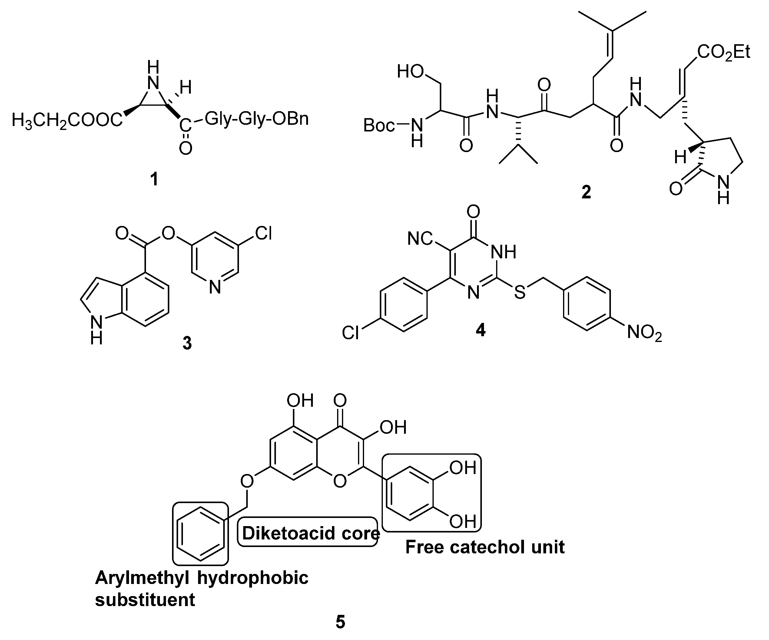
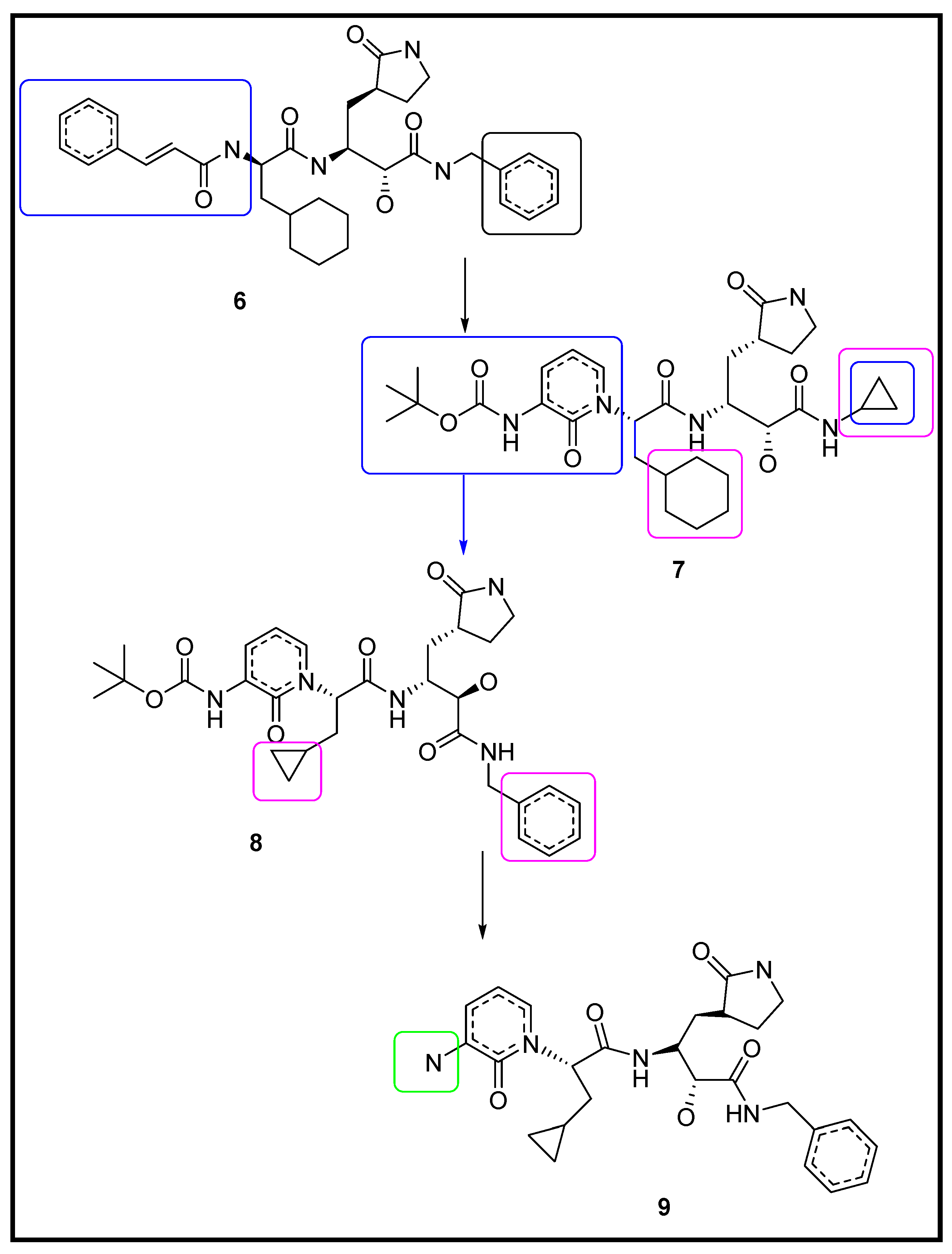
3.1.2. Designing the Improved Drugs for COVID-19: Targeting SARS CoV2 Main Protease Mpro

Using Zhang et al.’s [52] crystal structure analysis of SARS-main CoV2’s proteases (Mpro, 3CLpro), the unliganded SARS CoV2 Mpro structure and its complex, together with a beta-ketoamide inhibitor, were reported and studied in order to increase its plasma half-life through the incorporation of P3-P2 amide bonds into the pyridone ring. It was demonstrated that the crystal structure of Mpro could be used as a basis for designing more potent inhibitors to protease, with a beneficial tropism to the lung that is easily administered through inhalation [56]. The previously designed best inhibitor compound (6) was modified in order to increase its plasma half-life, reduce binding to plasma proteins, and increase solubility. In this study, the authors hid the amide bond in a pyridone ring to prevent it from cleavage by the known cellular proteases, thereby increasing its plasma half-life. New compounds (79) were prepared by Mengist et al., which are presented in Figure 4. The tert-butyloxycarbonyl (Boc) group of compound (8) was removed, producing the inactive form (9). This insures that the Boc group is necessary for cellular membrane crossing and the advantage of the hydrophobic moiety’s presence [56].
Similarly, Hilgenfeld’s group worked on the pharmacokinetics of compounds (7) and (8).The ADME levels of both compounds were similar, showing 90% binding to human plasma proteins. Compound (7) showed relatively less clearance than that of compound (7) and good tropism to the lungs, in addition to being well-tolerated through inhalation by mice. The designed α-ketoamide inhibitors showed improved inhibition efficiency against recombinant SARS CoV2 Mpro and can be used to suppress disease progression. These studies [56] have laid the ground work on the path towards identifying potent anticoronaviral drugs by employing molecular docking and molecular dynamic studies. By effective exploitation of these advanced methods, more insights on the chemistry involved in effective antiviral development can be derived; however, more structural and functional studies are required to improve the efficacy of new therapies to face coronavirus pandemics.

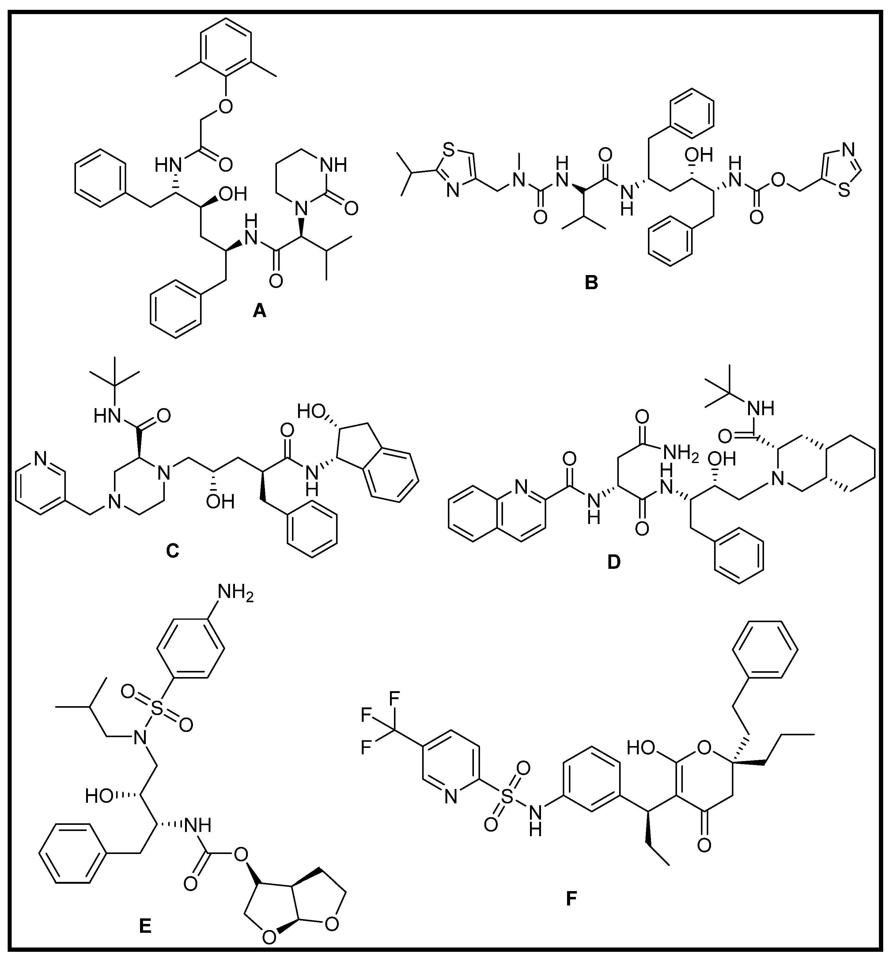
3.1.3. Repurposing of FDA-Approved Antiviral Drugs: Targeting the SARS CoV2 Main Protease Mpro

Six HIV protease inhibitors named ritonavir, lopinavir, indinavir, tipranavir, darunavir, and saquinavir have been targeted as clinical anti-SARS drugs against the new coronavirus pneumonia COVID-19, as shown in Figure 5 [57].
A molecular dynamics study indicated three distinct domains for both 3CLpro-1 and 3CLpro-2 (Figure 6); however, molecular docking results showed that the 3CLpro of SARS CoV2 (PDB ID: 6LU7) for all studied drugs had higher binding affinities compared to SARS CoV1 (PDB ID: 1UJ1) [57].
High docking scores were observed for both the indinavir and darunavir complexes. For the molecular interactions between these two protease inhibitors and SARS CoV’ 3CLpro, the binding free energy calculations (MMPBSA) were carried out. With the docked complexes, it was discovered that the binding pockets of 3CLpro-2 were significantly more closed and that indinavir and darunavir can bind deeper to the pocket with 19 contact residues. There were only 11 and 17 contact residues in complexes of 3CLpro-1–indinavir and 3CLpro-1–darunavir, respectively, indicating that the binding pocket for 3CLpro-1 is more openly visible. Additional residues increase the affinity of 3CLpro-2 for the inhibitors, explaining why the binding energy values of 3CLpro-2–inhibitor complexes were lower than those of 3CLpro-1. In its interactions with 3CLpro-2 and 3, darunavir creates five and three hydrogen bonds with 3CLpro-1. Indinavir, on the other hand, forms no hydrogen bonds with either of the two 3CLpro molecules. This means that darunavir may have a more stable enzyme inhibitor combination, making it more suitable for COVID-19 treatment [57].
In parallel, Lin et al. [58] studied the effects of an anti-HIV drug called Kaletra. This drug is composed of two protease inhibitors named ritonavir and lopinavir. As both competitive and non-competitive inhibitors exhibit strong binding to the enzyme, the binding ability levels of ritonavir and lopinavir in Kaletra were evaluated against the proteases in order to estimate their inhibitory effects. It was observed that the binding between CEP_C30 and ritonavir was most suitable based onboth libdock scores and intermolecular interactions. Compared to CEP_C30, both ritonavir and lopinavir seem not to be suitable for binding to PLVP; therefore, it was suggested that the therapeutic effect of Kaletra on Wuhan pneumonia and other coronavirus diseases might be due to the inhibitory effect of ritonavir on CEP_C30. This suggests that further studies should be focused on finding the catalytic mechanism of CEP_C30 and the role of ritonavir [58].
Another approach was taken using a molecule transformer–drug target interaction MT-DTI deep learning-based model, which predicts binding affinities based on chemical sequences (SMILES) and amino acid sequences (FASTA) of the target protein, without knowing their structural information. This could be helpful for determining drugs targeted for uncharacterized protein structures rather than using the traditional three-dimensional (3D) structure-based docking strategy [59]. MT-DTI was used to predict the binding affinities of 3410 FDA-approved medications against 3C-like proteinase (PDB ID: 6LU7), 30-to-50 exonuclease, endoRNAse, and 20-Oribose methyltransferase of SARS CoV2, which were then compared to values calculated by AutoDock Vina (3D structure-based docking algorithm). As a result, atazanavir (Kd 94.94 nM) was found to have the highest potency against SARS CoV2 3C-like proteinase. This was followed by remdesivir, efavirenz, ritonavir, and other antiviral drugs with predicted affinity levels of Kd > 100 nM, while none of the antiviral protease inhibitors had an affinity Kd < 1000 nM [59].

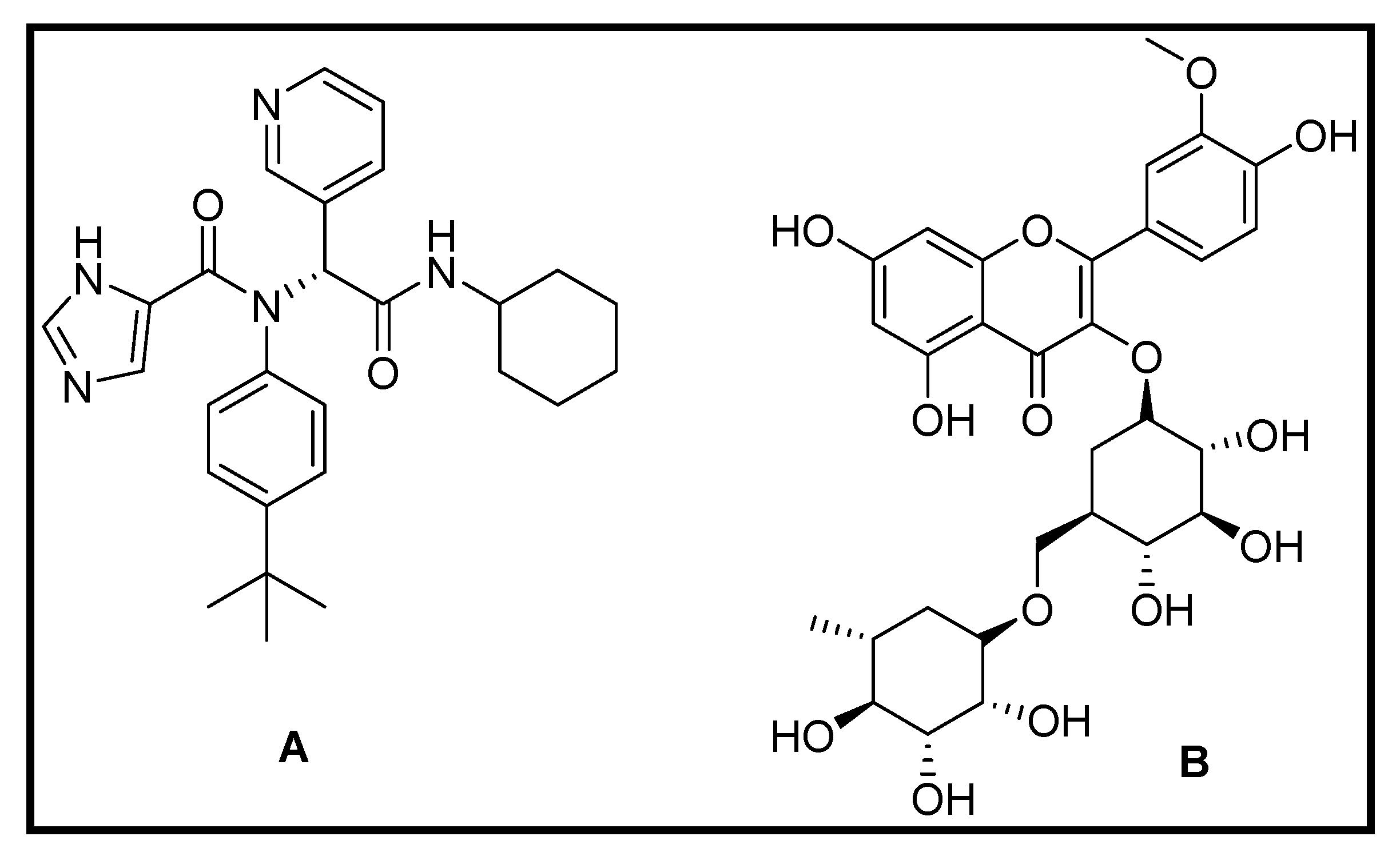
3.1.4. Repurposing of Natural Compound Drugs: Targeting the SARS CoV2 Main Protease Mpro

Several natural compounds, such as flavonoids in medicinal plants, have been known for their antiviral activity. The obtained binding energies to the protein, PDB ID: 6LU7, showed that nelfinavir and lopinavir (−10.72, −9.41 kcal/mol, respectively) are potential treatment options thatcan be used as standards for comparison. On the other hand, kaempferol, quercetin, luteolin-7-glucoside, demethoxycurcumin, naringenin, apigenin-7-glucoside, oleuropein, curcumin, catechin, and epicatechin-gallate might have the best potential, acting as COVID-19 Mpro inhibitors [60]; however, further studies are essential to investigate their medicinal use. In silico studies revealed that the compounds share the same pharmacophore as nelfinavir, as shown in Figure 7, due to the presence of a high number of phenolic compounds in the most abundant medicinal plants in nature [60].
Hydrogen bonds with 6LU7 amino acids are included in the docking analysis results. In the COVID-19 Mpro active site, the binding energy results are determined by the number of H-bonds that interact with the amino acids. As a result, the compound’s high affinity is highly dependent on the type and amount of bonding with the protein’s active site. When nelfinavir is combined with 6LU7, it forms a number of chemical bonds, including hydrogen bonds and hydrophobic bonds. As with nelfinavir, kaempferol, quercetin, and luteolin-7-glucoside form a number of chemical bonds. There is a higher affinity for the kaempferol bonds compared to the other compounds [60].
The flavanoid narcissoside showed promising results against the novel coronavirus COVID-19 via computational molecular docking screening using Molegro Virtual Docker (MVD) software with 30°A-grid resolution. Narcissoside showed a perfect fit at the active site of protein PDB ID: 6W63 as compared to the standard inhibitor (R)-N-(4-(tert-butyl)phenyl)-N-(2-(cyclohexylamino)-2-oxo-1-(pyridin-3-yl)ethyl)-1H-imidazole-5-carboxamide (standard compound (10), Figure 8), as recognized from the 6W63 protein inhibitor complex in the protein data bank [50]. This showed that the binding interactions only differed in three amino acid interactions. Narcissoside showed interactions with Leu 167, Pro 52, and Pro 168 and, while compound (10) showed interactions with His 172, Leu 27, and Thr 26 [62]. The calculated energy levels as assessed via two scoring functions named the Moldock score and Rerank score showed better results for narcissoside. It was determined that the affinity of narcissoside exceeds that of standard (10). Narcissoside also fits perfectly at the 6W63 active site via thirteen hydrogen bonds with nine amino acids in the cavity, compared to only four hydrogen bonds with amino acids in the case of the standard (10) [62].

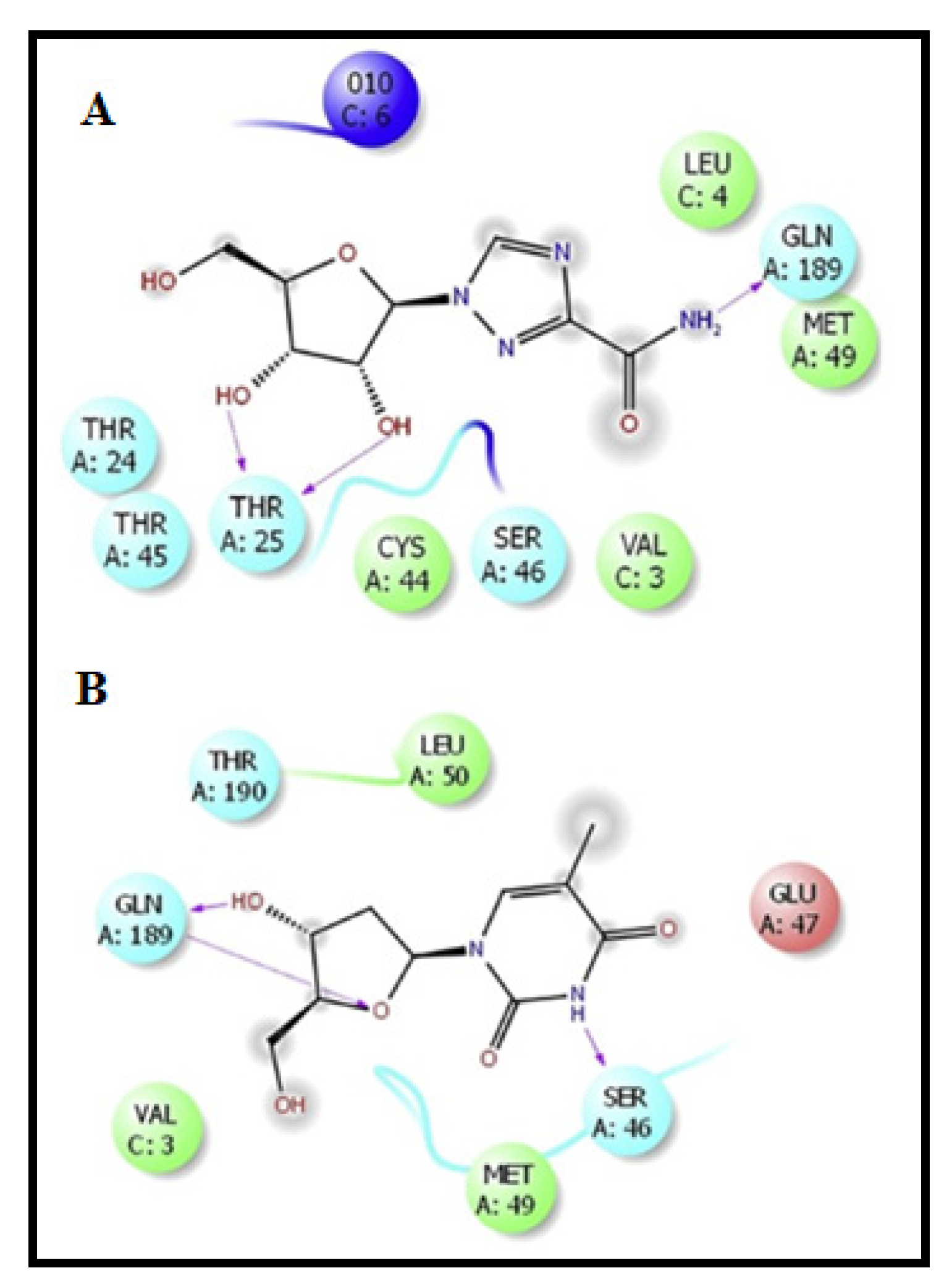
3.1.5. Virtual Screening Repurposing Studies: Targeting the SARS CoV2 Main Protease Mpro

A virtual screening study was presented, targeting the first resolved COVID-19 main protease crystal structure, PDB ID: 6LU7. Pairwise and multiple sequence comparisons of COVID-19 Mpro to SARS Mpro and MERS CoV Mpro revealed little difference in the residues number between COVID-19 and SARS CoV Mpros. MERS CoV Mpro showed low conservation with large differences in numbers compared to COVID-19 Mpro. This was related to phylogenetic relations being close to SARS showing COVID-19 Mpro, with 96.08% identity, compared to distant relations to MERS CoV with 51.61% identity [63]. Then, virtual screening of FDA-approved drugs was performed, with the results showing that the top 20 resulting drugs included the first two antivirals, followed by two antituberculous agents, two vitamins, and one anticancer, as well as other miscellaneous drugs. The results were relative to curcumin as an approved Mpro inhibitor that is used in SARS treatment [63].
Both ribavirin and telbivudine antivirals were ranked at the second and third positions, showing two-fold superiority regarding the docking scores as compared to curcumin, while the fourth and sixth positions went to vitamin B12 and nicotinamide, respectively. The main binding interactions were both hydrogen bonding and hydrophobic interactions, as shown in Figure 9. Based on the results, the repurposing of the antivirals ribavirin and telbivudine and of nicotinamide and vitamin B12 is recommended [63].
Another virtual and high-throughput screening method involving fluorescence resonance energy transfer was also used for a library of about 10,000 compounds targeting the main protease (Mpro) of SARS CoV2. Seven compounds were considered as primary hits, including the approved drugs disulfiram and carmofur, as well as preclinical or clinicaltrial candidate’s ebselen, shikonin, tideglusib, PX-12, and TDZD-8. A mechanism-based inhibitor (N3) was determined via computer-aided drug design and the crystal structure complex of Mpro SARS CoV2 with this compound was assessed [53]. A comparison of the binding modes between the structures with either SARS CoV2 Mpro–N3 or SARS CoV Mpro–N1 was performed. The IC50 values of the seven compounds were in range of 0.67 to 21.4 μM. Ebselen showed the best Mpro inhibition activity, withIC50 = 0.67 μM. It also showed antiviral activity in cell-based assays. The in vitro enzymatic inhibition results using quantitative real-time RT–PCR (qRT–PCR) showed that ebselen and N3 had the best antiviral activity at 10 μM concentration of Vero cells of SARS CoV2. TDZD-8 is claimed to be a non-specific Mpro inhibitor, which is why it was not considered for further investigation [53].
A structure-based drug design screening of more than 2000 FDA-approved drugs against the main protease enzyme (Mpro) crystal structure of COVID-19 in complex with the inhibitory peptide N3 PDB ID: 6LU7 was performed with docking simulations using the molecular operating environment (MOE) technique. The preliminary pharmacophores necessary to bind the COVID-19 virus via the Mpro substrate binding pocket were studied [64].
The results showed the top 11 hits based on binding affinity and S scores. The interacting amino acids and binding mode results revealed the significance of hydrophobic interactions together with the hydrogen bond network of His163, His164, Glu166, Gln189, and Thr190 for optimum fitting to the main proteases. Additionally, the presence of a terminal of either the sulphonic acid or carboxylic acid group can act as a bio-isosteric group to the phosphate moieties in antiviral drugs [65]. This includes certainantiviral drugs—mainly darunavir, nelfinavirand, and saquinavir. The hypercholesterolemia drug rosuvastatin is considered one of the most promising hits; however, further in vitro and in vivo testing should be carried out [64].
In silico studies of the main COVID-19 proteases were performed in order to determine the molecular interactions of drugs with certain therapeutic indications, including azithromycin, baricitinib, and hydroxychloroquine, as well asother similarly structured drugs such as chloroquine, quinacrine, and ruxolitinib, in the SARS CoV2 main protease (M-pro) PDB ID: 6LU7. The results showed that all inhibitors bound to the domain III enzyme site of the SARS CoV2 main protease [66]. The six tested inhibitors did not show any significant distances when compared to N3 complexed in the SARS CoV2 main protease (Mpro). Azithromycin, baricitinib, quinacrine, and ruxolitinib did not show any interactions with the key amino acid residues in the co-crystallized inhibitor bond site, located in domains I and II. All the inhibitors showed affinity and interactions with domain III [66]. This study suggests the beneficial combination of baricitinib and quinacrine with azithromycin; however, these simulation results are considered an initial step in the development and repurposing of antiviral molecules. Further studies showed death and heart arrhythmia of hospitalized patients being treated by the antimalarial drugs chloroquine and hydroxychloroquine [67,68].

3.2. Molecular Modeling Studies on Drugs Acting on RNA-Dependent RNA Polymerase of SARS CoV2

Repurposing of FDA-Approved Antiviral Drugs: Targeting RdRp

Remidesivir is being prescribed in many patients for COVID-19 management. It has shown in vitro activity against old SARS CoV and also MERS CoV viruses by acting as an RdRp inhibitor [51].Molecular docking studies, steered molecular dynamics (SMD) simulations, and umbrella sampling were performed to determine the interactions between remdesivir and both Mpro (PDB ID: 6LU7) and RdRp (PDB ID: 7BTF). The SMD results showed that remdesivir can efficiently bind to both targets, which might explain its effectiveness in treatment [51].
Remdesivir’s binding energies with Mpro and RNA polymerase were 7.9 kcal/mol and 6.5 kcal/mol, respectively. Remdesivir showed better affinity for Mpro than that for RdRp. Further, remdesivir showed an EC50 of 0.77 M for SARS CoV2 in in vitro studies [50]. The experimental binding energy, gexpbind (ΔGexp), may be calculated using the formula RTln(EC50), where R is the gas constant (R = 1.987 × 10⁻³ kcal/mol and T = 300 K) and EC50 is measured in M. This gives an estimate of −8.4 kcal/mol for ΔGexpbind, which is in line with the docking result for Mpro. This suggests that remdesivir’s binding to Mpro is greater than its known approved target, RdRp. Only the crude docking method produced this result, whereas the more accurate SMD and umbrella sampling produced the opposite findings. The binding site for remdesivir in Mpro is located between domains I and II of the protein. There were 3 hydrogen bonds (HBs; His163, Ser144, and Leu141) and 12 non-bond (Glu166, Cys145, Met165, Gln189, Arg188, Asp187, His41, Met49, Thr26, Leu27, Thr45, and Thr25) interactions between remdesivir and Mpro. As illustrated in Figure 9, non-bonded residues influence the interaction between remdesivir and Mpro, showing similarity with N3 in binding to His41 and Cys145 of the Mpro active site.
Remdesivir can affect the activity of nsp12 in RNA polymerase because the binding site is much closer to the nsp12 active site. Regarding Nsp12, there were four hydrogen bonds (HBs), namely Asp757, Asn688, and Thr677, as well aseight non-bond sites (NBS), namely Ser79, Tyr452, Ser620, Ser678, Cys618, Lys618, Thr684, Asp615, and Arg550. Figure 10 illustrates the docking simulation for RdRp–remdesivir, which was similar to Mpro, showing that there are more non-bond contacts involved than HBs [51]. In Figure 10, remdesivir is shown as a stick; Mpro is orange; and nsp12, nsp7, and nsp8 are blue, red, and magenta, respectively, in the upper panel. The binding site residues of the target are highlighted in green. The nsp12 active site is depicted in the seven motifs A–G. Meanwhile, in the bottom panel, the boxed areas are rendered in two-dimensional charts (2D). Green and red lines indicate HBs and non-bonded contacts, respectively. Chain A is denoted by the letter A in parentheses. When compared to RdRp’s three chains, Mpro has a single chain and nsp12 is designated as chain A.
The SARS CoV NSP12 RdRp homology model was also studied (PDB ID: 6NUR). It was determined that remdesivir had a reasonable binding mode, with a free energy of −8.28 ± 0.65 kcal/mol, which is higher than that of the natural ligand ATP (−4.14 ± 0.89 kcal/mol) in terms of the relative binding free energy. There is some evidence that remdesivir can replace ATP in blocking the RdRp binding site. Residues D618, S549, and R555 play key roles in remdesivir’s binding affinity; thus, it was identified that remdesivir can act as a SARS CoV2 RNA chain terminator [69].
Molecular dynamics and docking techniques were used to study the binding of remdesivir to SARS CoV2 RdRP (PDB ID: 6NUR). In addition, remdesivir-bound RdRP simulations showed that the template entry site was blocked.The conformational and thermodynamic parameters corroborated the experimental data and confirmed the involvement of arginine, serine, and aspartate residues in the peptide structure. The catalytic site consisting of SER 759, ASP 760, and ASP 761 (SDD) formed strong contacts with remdesivir [70].

3.3. Molecular Modeling Studies on Drugs Acting on Endoribonuclease

Virtual Screening Repurposing Studies: Targeting Endoribonuclease

NendoU (NSP15) (PDB ID: 6VWW) protein endoribonuclease was targeted with 8548 ligands. Glide-SP and Glide-XP showed good affinity for four drugs that could be repurposed for COVID-19 treatment. The FDA-approved drug DB00876 (Eprosartan) and investigational drugs DB15063 (Inarigivir soproxil), DB12307 (Foretinib), and DB01813 (an experimental drug) [71] are among them [72]; however, SARS CoV2 NendoU enzymes have different catalytic properties and substrate specificity from SARS CoV, H CoV-229E, and MERS CoV enzymes. Virtual screening of 8548 ligands targeting protein endoribonuclease NendoU (NSP15) (PDB ID: 6VWW) was performed. Four drugs were suggested to be repurposed for COVID-19 treatment, showing good affinity using Glide-SP and Glide-XP. These drugs were DB00876 (Eprosartan) approved by the FDA, as well as the investigational drugs DB15063 (Inarigivir soproxil), DB12307 (Foretinib), and DB01813 (an experimental drug) [71]. The results showed great similarity between SARS CoV2 NendoU and other virus strains, such as SARS CoV, H CoV 229E, and MERS CoV, but it has distinct catalytic characteristics and substrate selectivity. As a result of the SARS-(NSP15) CoV2endoribonuclease’s known structure (PDB ID: 6VWW), Krishnan et al. [72] performed a structural-based drug design investigation and found that NSP15 (COVID-19) shares a high degree of sequence similarity with SARS CoV2, MERS CoV, and HCoV-229E (human coronavirus).
Molecular docking procedures including two steps, standard precision (SP) and extra precision (XP), and in silico screening were performed to identify inhibitors of this enzyme through induced fit docking and screening of 3978 compounds with potential antiviral activity from an enamine database. Eight compounds showed promising docking scores in addition to good docking energy results [61]. All of the compounds interacted with Lys290, an important catalytic residue involved in nucleoside hydrolysis in SARS and MERS NSP15. Additionally, the interaction of5 out of 8 compounds with an essential residue for hydrolysis, His235, as well asSer294 and Tyr343 hydroxyl groups, confirmed the hydrogen bonding with ligands. Regarding MERS (NSP15), the importance of the Tyr343 (Y343A) residue for ribonuclease activity has been demonstrated by mutational studies. All compounds are located in the active site, suggesting docking without unusual steric problems [72]. The docking results revealed that the interaction is aligned with the expectations for good interactions of the inhibitor at the active site. The tested compounds could be useful in inhibiting the NSP15 endoribonuclease activity and arresting virus replication. It is worth noting that these compounds have already been synthesized and can be considered for further studies against the recent COVID-19 pandemic [72].

4. In Silico Modeling in Vaccine Development: A SARS CoV-2 Case Study

While antiviral identification and development help in managing viral infections, vaccine development will offer herd immunity in the longer run [73,74,75]. Currently, there are more than 20 vaccines being tested in clinical trials. The World Health Organization (WHO) is regularly updating its list of the developed vaccines [76]. COVID-19 vaccines have been the subject of several studies [9,10,11,12]. As a result of previous outbreaks of SARS CoV and MERS CoV, global industry leaders and researchers have attempted to develop a suitable vaccine [77,78]. The vaccine development approaches for SARS CoV2 include live attenuated virus [79,80,81,82], protein-based vaccines [83,84,85,86,87,88,89,90], viral vectors [79,91,92,93,94], DNA vaccines [95,96,97], mRNA vaccines [98,99,100], and other self-assembling vaccines [101]. The S protein/gene is the most preferred target site in SARS vaccine development, meaning the same strategy could potentially be useful in developing a SARS CoV2 vaccine [102,103].

4.1. Molecular Modeling of the Designed Multi-Epitopic Vaccines

Informatics tools are as convenient and cost-effective as in silico studies [104,105] and could help in designing a multi-epitope vaccine for COVID-19 treatments [106,107,108,109,110,111,112,113]. The potential peptide epitopes (antigenic and immunogenic) can help in facilitating the design of vaccines. The candidate vaccine constructs consist of distant epitopes. These epitopes are computed using B cells, cytotoxic T lymphocytes (CTL), and helper T lymphocytes (HTL) based on SARS CoV2 proteins [114,115]. The candidate vaccines are designed by fusing these epitopes with linkers [116,117,118,119]. These linkers help to stabilize the protein structure by producing a flexible conformation, protein folding, and functional domain separation. The binding affinity of the potential candidate vaccines with immune receptors was studied using the molecular docking approach.

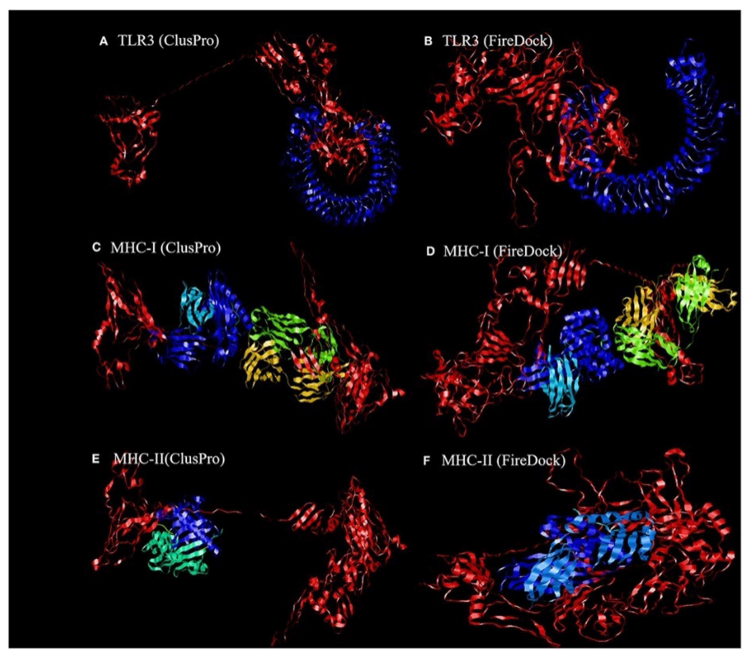
4.1.1. Molecular Docking of the Construct SARS CoV2 Vaccine with the Related Antigenic Recognition Receptors (TLR-3, MHC-I, and MHC-II)

Dong et al. [119] revealed the binding affinity between the candidate vaccines and antigenic recognition receptors of toll-like receptor-3 (TLR-3, 2A0Z) and the major histo compatibility complex (MHC-I, 4WUU), and MHC-II, 3C5J) present on the immune cell surfaces [120], which were evaluated using molecular docking techniques. The ClusPro server, https://cluspro.bu.edu/login.php?redir/queue.php (accessed on 20 October 2021), was used toperform the docking analysis. This server computed 44 models based on the electrostatic interaction and the desolvated energy. The server determined a total of twenty-nine model complexes each for TLR3, MHC-I, and MHC-II and the COVID-19 vaccine. The lowest binding energy scores of −1156.2, −1346.8, and −1309 kcal/mol were chosen and are represented in Figure 11. The PatchDock server (https://bioinfo3d.cs.tau.ac.il/PatchDock/ (accessed on 20 October 2021)) was used for further docking evaluation to confirm the binding affinity between the vaccine construct and the suggested receptors [121]. The top 10 complexes were then refined using the FireDock algorithm [121], which predicts the optimal complex with the help of energy functions. The best model complex of TLR3, MHC-I, and MHC-II receptors and the vaccine construct interaction (red color) showed global energy results, attractive van der Waals energy (VdW) results, repulsivevan der Waals (VdW) energy results, and atomic contact energy values (Figure 11, Table 1), verifying the stability of the docked complex.

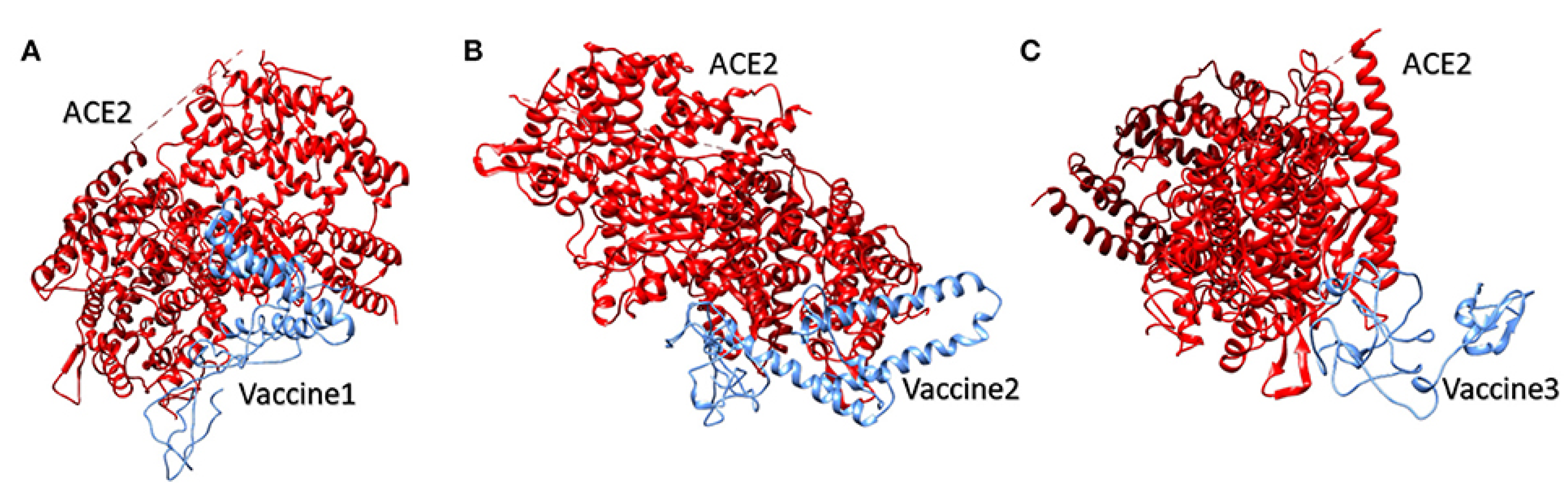
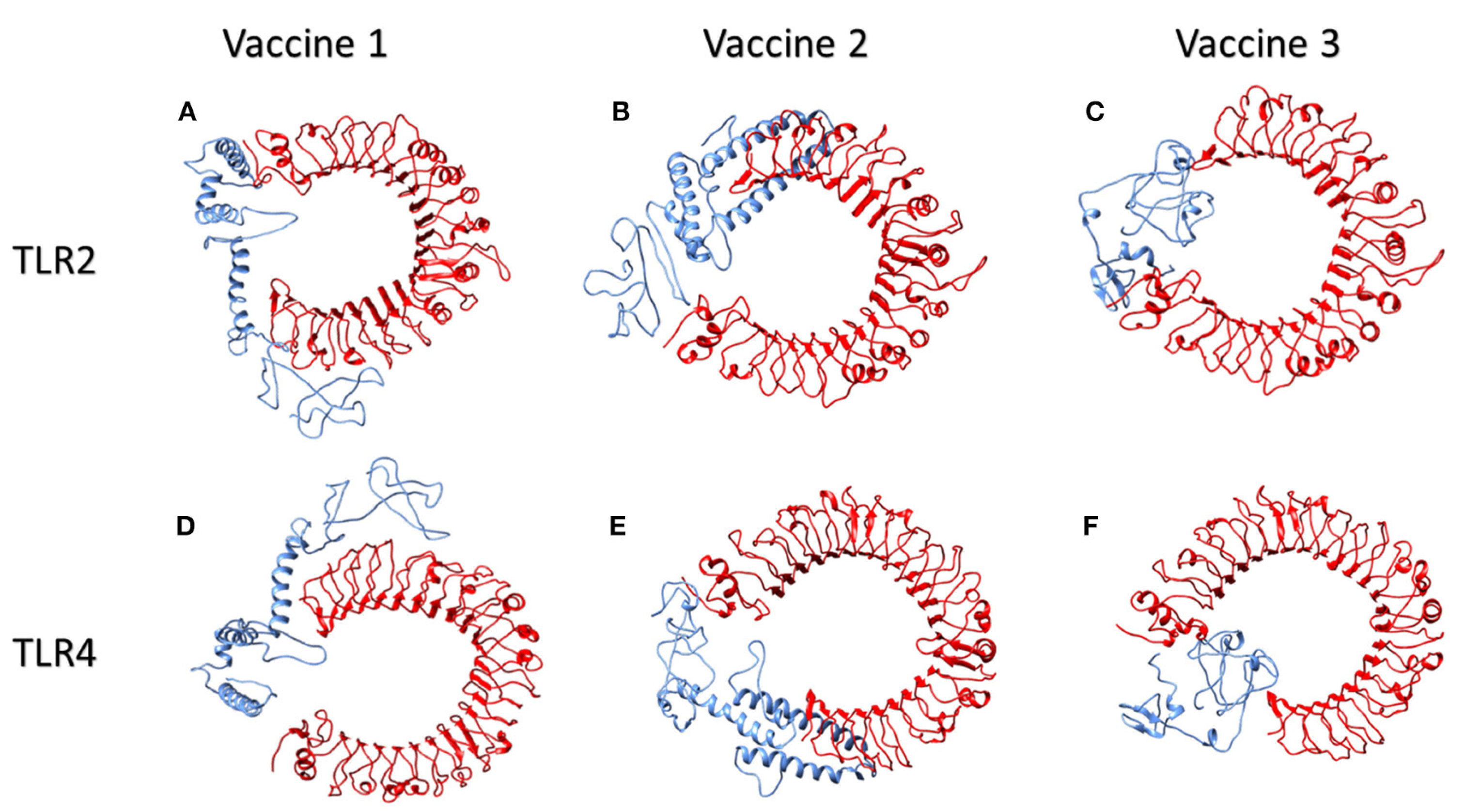
4.1.2. Molecular Docking of the SARS CoV2 Vaccine Construct with the Antigenic Recognition Receptors (ACE-2, TLR2, TLR4, HLA Alleles)

The candidate vaccines were designed and then tested with different epitopes [122]. The construct vaccines, which have high antigenicity and are expected to produce high antibody titers, were combined with multi-epitope vaccines in order to enhance the immune response [123,124]. Three different construct vaccine constructs were used, with one comprising the top-scoring CD4 and CD8 epitopes lying in the S1 domain and the second one formed by taking two epitopes from the S1 domain and two from the S2 domain of the spike SARS CoV 2 protein, which represents the MHC-I and MHC-II binders. The last vaccine was created by combining a B-cell epitope with the second vaccine using a different adjuvant. The candidate vaccine constructs were also docked to different type of receptors, such as the ACE-2 receptor using PDB ID: 3sci, TLR2 with PDB ID: 2Z7X, TLR4 with PDB ID: 4G8A, B-cell receptor (BCR) CD79 using PDB ID: 3KG5 and HLA super family alleles; HLA A*02 01 using PDB ID: 4U6Y, the other HLA B*51 01 using PDB ID: 4MJI, and also class II HLA-DRB1*1402 using PDB ID: 6ATF [122], representing broad-spectrum peptide-binding repertoires.

Molecular Docking of the Candidate Vaccines with ACE-2 Receptors

Vaccine 1 was docked with the ACE-2 receptor using the HADDOCK server (Guru Interface and refinement interface) [125]. The server clustered 36 structures into seven different clusters. Vaccine 2 was also docked with the ACE-2 receptor, while the HADDOCK server clustered 18 structures into three clusters. While working on vaccine 3 and the ACE-2 receptor, the server clustered 22 structures into five clusters. The top cluster score, Z-score, and water-refined models are represented in Figure 12. The latter (as shown in Figure 12) represents docking interactions of the h-ACE-2 protein complex (red) with the proposed multi-epitopic vaccines 1, 2, and 3 (blue), which were performed using the HADDOCKserver. The static interaction parameters of ACE-2 with the three construct vaccines 1,2, and 3 are displayed in Table 2.

Molecular Docking of Vaccines with the Potential TLR2 and TLR4 Receptors

Toll-like receptors (TLR) TLR2 and TLR4, which are found on the cell surfaces, are activated by viral glycoproteins. They detect structural and non-structural proteins of the virus, suggesting the need to study cytokine release and inflammation. These TLR agonists have been shown to enhance the immune system’s response and viral clearance [126]. TLR2 was docked with the three constructed vaccines. The interaction between vaccine 1 and TLR2 revealed 40 structures in six clusters. Similarly, the server clustered 80 structures into 12 clusters for the interaction of vaccine 2 and TLR2. Likewise, the HADDOCK server clustered 136 structures into 10 clusters; the top-scoring model, z- score, and the total water-refined model results from the interactions of TLR2 with vaccines 1, 2, and 3 are depicted in Table 3. The three candidate vaccines also interacted with TLR4.In the case of the interaction of vaccine 1 and TLR4, the server clustered 157 structures into 13 clusters. Similarly, vaccine 2 interacted with the TLR4 receptor, clustering 47 structures into nine clusters. Likewise, the HADDOCK clustered 93 structures into eight clusters for the interaction between vaccine3 and TLR4. The top HADDOCK cluster scores with Z-values and the water-refined models are presented in Table 3. The HADDOCK refinement interface was then used to refine the top cluster models. The 20 obtained structures were clustered into one single cluster. The final cluster consisted entirely of water-refined models. The observed statistics in the interaction of vaccines 1, 2, and 3 from their refined clusters were also studied [122] and the complexes are shown in Figure 13 and Table 3. Figure 13 illustrates the interactions of human TLR2 and TLR4 proteins (red) with the candidate multi-epitopic COVID-19 vaccines 1, 2, and 3 (blue).

Molecular Modeling Interactions of the Suggested Vaccines with HLA Alleles

Studies showed that the docking analysis interaction of the proposed vaccine 1 and the HLA A allele [122] with the water-refined models was59.0 percent. The HADDOCK server clustered 118 structures into 17 separate clusters. Likewise, the server clustered 97 structures into 17 clusters, depicting 48.5 percent of the water-refined models in the case of vaccine 2 and the HLA A allele. Similarly, the HADDOCK server clustered 187 structures into three clusters for vaccine 3, representing 93.5 percent of the water-refined models. The best HADDOCK results with z-scores are represented in Table 4. Here, 115 structures were clustered into 15 separate clusters using HADDOCK server for the interaction between vaccine 1 and the HLA B allele, representing a total of 57.5 percent of the water-refined models. Additionally, HADDOCK clustered 84 structures into nine clusters for the interaction of vaccine2 and the HLA B allele, depicting 42 percent of the water-refined models. The HADDOCK server aggregated 168 likely structures into 10 clusters representing 84 percent of the water-refined models for vaccine 3. The top-scoring cluster and the static parameters are depicted in Table 4. Moreover, when vaccine 1 was docked into the HLA DRB1 allele, the HADDOCK server showed 67 probable structures clustered into different clusters, accounting for 33.5 percent of the water-refined models. Likewise, the HADDOCK server clustered 64 structures into 11 different clusters for vaccine 2 and the HLA DRB1 allele, representing32 percent of the water-refined models. HADDOCK clustered 93 probable structures into 13 different clusters for vaccine 3, representing 46.5 percent of the water-refined models. The scoring parameters are shown in Table 4. The HADDOCK refinement interface refined and then clustered the top cluster model from 20 structures into one single cluster, representing 100 percent of the water-refined models. The observed statistics for the interactions of vaccines 1, 2, and 3 with their respective refined clusters were also studied [122]. Finally, the vaccine mRNA was improved using the Java Codon Adaptation Tool in order to ensure that the designed vaccines were effective in a specific expression system.

5. Conclusions and Outlook

Scientists worldwide are collaborating to face the COVID-19 pandemic so as to expedite drug development using computer-aided drug design, including the use of modeling studies for therapeutic drug andvaccine development. The current review summarizes the existing studies in this field, thereby offering an overview that could help ingenerating further findings, including the conversion of virtual screening results into clinically applied therapies using in vitro and in vivo techniques against COVID-19. This can be achieved either through screening for new drug candidates or repurposing of already approved drugs. Moreover, this review also establishes that in silico studies are an essential tool that can facilitate the design of multi-epitopic vaccines. In addition to computational methods, specific antibodies can be used as drugs to reduce COVID-19 symptoms and outcomes through the interactions of specific antibodies with SARS CoV2 proteins [127]. Additionally, recent studies have been performed on the diagnosis and detection or determination of SARS CoV2 [128]. SARS CoV2 is an RNA virus, with high mutating capability, making the process of drug discovery highly challenging. Any strategy that could effectively determine the sequences of all new possible variants of this virus could present insights in the application of DNA or RNA enzymes for biomedical applications [129]. Further, there is a requisite for the development of tandem therapeutics owing to the multitude of viral infections. In addition, lung defense to infections in inhalation toxicology can benefit from in vitro air–liquid interface (ALI) cell culture and exposure models, which were recently reviewed elsewhere [130] The experimental investigation of multifaceted particulate matter from the environment, cigarette smoke, and e-cigarette liquid vapes has made inhalation toxicology a major research topic concerning the adverse effects of these items on lung tissue, similarly to SARS CoV2 [131]. Due to the time-consuming development process involved in conventional drug discovery, drug repurposing seems to be a promising approach against sudden viral outbreaks. In particular, drug repositioning by exploiting the domains of computational models could greatly speed up the process. Drugs could be personalized for COVID 19 using advanced microfluidic devices and lung-on-chip (LOC) technologies, which have demonstrated huge potential against SARS CoV2. The unconventional technologies in the pipeline enabling drugs to cross different biological barriers, particularly regarding opportunistic infection of the brain, could lead to rapid antiviral solutions. In addition, the integration of bioinformatic, pharmaceutical, and toxicological studies is essential to address any bottlenecks related to the safety and dosage of repurposed drugs. In comparison to the conventional WHO procedures established for less urgent health risks, the time required to receive vaccines is greatly dependent on the regulatory authorities and the flexibility needed to expedite processing and vaccine approval. As reported, some drugs and candidates vaccine have been determined based on the previous SARS virus, which after development have been shown to be effective for COVID-19.

Author Contributions

Conceptualization, R.F.B., I.M.F. and A.V.S.; data curation, R.F.B., U.K., and A.V.S.; writing—original draft preparation, R.F.B., I.M.F. and A.A.M.; writing—review and editing, R.F.B., A.V.S., and V.C.; graphic design and visualization, R.F.B., A.A.M. and A.V.S.; supervision, R.F.B., and A.V.S.; project administration, R.F.B.; funding acquisition, R.F.B., A.V.S. & U.K. All authors have read and agreed to the published version of the manuscript.

Funding

This research received no external funding.

Institutional Review Board Statement

Not Applicable.

Informed Consent Statement

Not Applicable.

Data Availability Statement

Not Applicable.

Acknowledgments

The authors gratefully acknowledge the National Research Center, Cairo, Egypt, and Future University in Egypt, Cairo, Egypt, for their support.

Conflicts of Interest

The authors declare that there are no conflicts of interest.

References

  1. Singh, A.V.; Gemmati, D.; Vats, T.; Singh, A.; Zamboni, P. High throughput array technologies: Expanding applications from clinics to applied research. Front. Nanosci. Nanotechnol. 2019, 5, 1–2. [Google Scholar] [CrossRef] [Green Version]
  2. Singh, A.V.; Maharjan, R.-S.; Kanase, A.; Siewert, K.; Rosenkranz, D.; Singh, R.; Laux, P.; Luch, A. Machine-learning-based approach to decode the influence of nanomaterial properties on their interaction with cells. ACS Appl. Mater. Interf. 2021, 13, 1943–1955. [Google Scholar] [CrossRef] [PubMed]
  3. Muratov, E.N.; Amaro, R.; Andrade, C.H.; Brown, N.; Ekins, S.; Fourches, D.; Isayev, O.; Kozakov, D.; Medina-Franco, J.L.; Merz, K.M.; et al. A critical overview of computational approaches employed for COVID-19 drug discovery. Chem. Soc. Rev. 2021, 50, 9121–9152. [Google Scholar] [CrossRef]
  4. Abdellatiif, M.H.; Ali, A.; Ali, A.; Hussien, M.A. Computational studies by molecular docking of some antiviral drugs with COVID-19 receptors are an approach to medication for COVID-19. Open Chem. 2021, 19, 245–264. [Google Scholar] [CrossRef]
  5. Laws, M.; Surani, Y.M.; Hasan, M.; Chen, Y.; Jin, P.; Al-Adhami, T.; Chowdhury, M.; Imran, A.; Psaltis, I.; Jamshidi, S.; et al. Current trends and future approaches in small-molecule therapeutics for COVID-19. Curr. Med. Chem. 2018, 13, e0209034. [Google Scholar] [CrossRef]
  6. Kumar, R.; Harilal, S.; Al-Sehemi, A.G.; Mathew, G.E.; Carradori, S.; Mathew, B. The chronicle of COVID-19 and possible strategies to curb the pandemic. Curr. Med. Chem. 2020, 28, 2852–2886. [Google Scholar] [CrossRef] [PubMed]
  7. Ganser, L.R.; Lee, J.; Rangadurai, A.; Merriman, D.K.; Kelly, M.L.; Kansal, A.D.; Sathyamoorthy, B.; Al-Hashimi, H.M. High-performance virtual screening by targeting a high-resolution RNA dynamic ensemble. Nat. Struct. Mol. Biol. 2018, 25, 425–434. [Google Scholar] [CrossRef] [PubMed] [Green Version]
  8. Bullock, J.; Luccioni, A.; Pham, K.H.; Lam, C.S.N.; Luengo-Oroz, M. Mapping the landscape of Artificial Intelligence applications against COVID-19. J. Artif. Intell. Res. 2020, 69, 807–845. [Google Scholar] [CrossRef]
  9. Al-Karmalawy, A.A.; Dahab, M.A.; Metwaly, A.M.; Elhady, S.S.; Elkaeed, E.B.; Eissa, I.H.; Darwish, K.M. molecular docking and dynamics simulation revealed the potential inhibitory activity of ACEIs against SARS-CoV-2 targeting the hACE2 receptor. Front. Chem. 2021, 9, 661230. [Google Scholar] [CrossRef] [PubMed]
  10. Ng, Y.L.; Salim, K.C.; Chu, J.J.H. Drug repurposing for COVID-19: Approaches, challenges and promising candidates. Pharmacol. Ther. 2021, 228, 107930–107944. [Google Scholar] [CrossRef]
  11. Kifle, Z.D.; Ayele, A.G.; Enyew, E.F. Drug repurposing approach, potential drugs, and novel drug targets for covid-19 treatment. J. Environ. Public Health 2021, 2021, 6631721. [Google Scholar] [CrossRef]
  12. Boopathi, S.; Poma, A.B.; Kolandaivel, P. Novel 2019 coronavirus structure, mechanism of action, antiviral drug promises and rule out against its treatment. J. Biomol. Struct. Dyn. 2020, 29, 1–10. [Google Scholar] [CrossRef] [Green Version]
  13. Sayed, A.M.; Khattab, A.R.; AboulMagd, A.M.; Hassan, H.M.; Rateb, M.E.; Zaid, H.; Abdelmohsen, U.R. Nature as a treasure trove of potential anti-SARS-CoV drug leads: A structural/mechanistic rationale. RSC Adv. 2020, 10, 19790–19802. [Google Scholar] [CrossRef]
  14. Corey, B.L.; Mascola, J.R.; Fauci, A.S.; Collins, F.S. A strategic approach to COVID-19 vaccine R&D. Science 2020, 368, 948–950. [Google Scholar]
  15. Graham, B.S. Rapid COVID-19 vaccine development. Science 2020, 368, 945–946. [Google Scholar] [CrossRef] [PubMed]
  16. Mullard, A. World Report COVID-19 vaccine development pipeline gears up. Lancet 2020, 395, 1751–1752. [Google Scholar] [CrossRef]
  17. Callaway, E. The race for coronavirus vaccines: A graphical guide. Nature 2020, 580, 576–577. [Google Scholar] [CrossRef]
  18. WHO. A Coordinated Global Research Roadmap—2019 Novel Coronavirus; World Health Organization: Geneva, Switzerland, 2020; pp. 1–96. Available online: https://www.who.int/blueprint/priority-diseases/key-action/Roadmap-version-FINAL-for-WEB.pdf?ua=1 (accessed on 7 October 2020).
  19. Forni, D.; Cagliani, R.; Clerici, M.; Sironi, M. Molecular evolution of human coronavirus genomes. Trends Microbiol. 2017, 25, 35–48. [Google Scholar] [CrossRef] [PubMed] [Green Version]
  20. Luo, C.M.; Wang, N.; Yang, X.L.; Liu, H.Z.; Zhang, W.; Li, B.; Hu, B.; Peng, C.; Geng, Q.B.; Zhu, G.J.; et al. Discovery of novel bat coronaviruses in South China that use the same receptor as Middle East respiratory syndrome coronavirus. J. Virol. 2018, 92, 00116–00118. [Google Scholar] [CrossRef] [Green Version]
  21. Singh, A.V.; Chandrasekar, V.; Janapareddy, P.; Mathews, D.E.; Laux, P.; Luch, A.; Yang, Y.; Garcia-Canibano, B.; Balakrishnan, S.; Abinahed, J.; et al. Emerging application of nanorobotics and artificial intelligence to cross the bbb: Advances in design, controlled maneuvering, and targeting of the barriers. ACS Chem. Neurosci. 2021, 12, 1835–1853. [Google Scholar] [CrossRef] [PubMed]
  22. Singh, A.V.; Jahnke, A.V.; Wang, S.; Xiao, Y.; Alapan, Y.; Kharratian, S.; Onbasli, M.C.; Kozielski, K.; David, H.; Richter, G.; et al. Anisotropic gold nanostructures: Optimization via in silico modeling for hyperthermia. ACS Appl. Nano Mater. 2018, 1, 6205–6216. [Google Scholar] [CrossRef]
  23. Shanker, A.; Bhanu, D.; Alluri, A.; Gupta, S. Whole genome sequence analysis and homology modelling of a 3c like peptidase and a non-structural protein 3 of the sars-cov-2 shows protein ligand interaction with an aza-peptide and a noncovalent lead inhibitor with possible antiviral properties. New J. Chem. 2020, 44, 9202–9212. [Google Scholar] [CrossRef]
  24. Sharma, V.; Sharma, A.; Bharate, S.B. Natural products in mitigation of SARS CoV infections. Curr. Med. Chem. 2020, 28, 4454–4483. [Google Scholar] [CrossRef] [PubMed]
  25. Pandey, A.T.; Pandey, I.; Zamboni, P.; Gemmati, D.; Kanase, A.; Singh, A.V.; Singh, M.P. Traditional herbal remedies with a multifunctional therapeutic approach as an implication in covid-19 associated co-infections. Coatings 2020, 10, 761. [Google Scholar] [CrossRef]
  26. Ansari, M.H.D.; Lavhale, S.; Kalunke, R.M.; Srivastava, P.L.; Pandit, V.; Gade, S.; Yadav, S.; Laux, P.; Luch, A.; Gemmati, D.; et al. Recent advances in plant nanobionics and nanobiosensors for toxicology applications. Curr. Nanosci. 2020, 16, 27–41. [Google Scholar] [CrossRef]
  27. Pandeya, A.T.; Pandeyb, I.; Hachenberger, Y.; Krause, B.-C.; Haidar, R.; Laux, P.; Luch, A.; Singh, M.P.; Singh, A.V. Emerging paradigm against global antimicrobial resistance via bioprospecting of mushroom into novel nanotherapeutics development. Trends Food Sci. Technol. 2020, 106, 333–344. [Google Scholar] [CrossRef]
  28. Shen, Y.; Hao, T.; Ou, S.; Hu, C.; Chen, L. Applications and perspectives of nanomaterials in novel vaccine development. Med. Chem. Comm. 2018, 9, 226–238. [Google Scholar] [CrossRef]
  29. Gregory, A.E.; Titball, R.; Williamson, D. Vaccine delivery using nanoparticles. Front. Cell. Infect. Microbiol. 2013, 3, 13. [Google Scholar] [CrossRef] [Green Version]
  30. Chang, C.K.; Lo, S.C.; Wang, Y.S.; Hou, M.H. Recent insights into the development of therapeutics against coronavirus diseases by targeting N protein. Drug Discov. 2016, 21, 562–572. [Google Scholar] [CrossRef]
  31. Singh, A.V.; Ansari, M.H.D.; Rosenkranz, D.; Maharjan, R.S.; Kriegel, F.L.; Gandhi, K.; Kanase, A.; Singh, R.; Laux, P.; Luch, A. Artificial intelligence and machine learning in computational nanotoxicology: Unlocking and empowering nanomedicine. Adv. Healthc. Mater. 2020, 9, 1901862. [Google Scholar] [CrossRef]
  32. Singh, A.V.; Rosenkranz, D.; Ansari, M.H.D.; Singh, R.; Kanase, A.; Singh, S.P.; Johnston, B.; Tentschert, J.; Laux, P.; Luch, A. Artificial intelligence and machine learning empower advanced biomedical material design to toxicity prediction. Adv. Intell. Syst. 2020, 2, 2000084. [Google Scholar] [CrossRef]
  33. Panyam, J.; Labhasetwar, V. Biodegradable nanoparticles for drug and gene delivery to cells and tissue. Adv. Drug Deliv. Rev. 2003, 55, 329–347. [Google Scholar] [CrossRef]
  34. Sainz, V.; Conniot, J.; Matos, A.I.; Peres, C.; Zupanǒiǒ, E.; Moura, L.; Silva, L.C.; Florindo, H.F.; Gaspar, R.S. Regulatory aspects on nanomedicines. Biochem. Biophys. Res. Commun. 2015, 468, 504–510. [Google Scholar] [CrossRef] [PubMed]
  35. Yuan, Y.; Cao, D.; Zhang, Y.; Ma, J.; Qi, J.; Wang, Q.; Lu, G.; Wu, Y.; Yan, J.; Shi, Y.; et al. Cryo-EM structures of MERS-CoV and SARS-CoV spike glycoproteins reveal the dynamic receptor binding domains. Nat. Comm. 2017, 8, 1–9. [Google Scholar] [CrossRef] [PubMed]
  36. Martina, E.; Stiefl, N.; Degel, B.; Schulz, F.; Breuning, A.; Schiller, M.; Vicik, R.; Baumann, K.; Ziebuhr, J.; Schirmeister, T. Screening of electrophilic compounds yields an aziridinyl peptide as new active-site directed SARS-CoV main protease inhibitor. Bioorg. Med. Chem. Lett. 2005, 15, 5365–5369. [Google Scholar] [CrossRef]
  37. Niu, C.; Yin, J.; Zhang, J.; Vederas, J.C.; James, M.N. Molecular docking identifies the binding of 3-chloropyridine moieties specifically to the S1 pocket of SARS-CoV Mpro. Bioorg. Med. Chem. 2008, 16, 293–302. [Google Scholar] [CrossRef]
  38. Chen, L.; Li, J.; Luo, C.; Liu, H.; Xu, W.; Chen, G.; Liew, O.W.; Zhu, W.; Puah, C.M.; Shen, X.; et al. Binding interaction of quercetin-3-β-galactoside and its synthetic derivatives with SARS-CoV 3CLpro: Structure–activity relationship studies reveal salient pharmacophore features. Bioorg. Med. Chem. 2006, 14, 8295–8306. [Google Scholar] [CrossRef]
  39. Ghosh, A.K.; Xi, K.; Grum-Tokars, V.; Xu, X.; Ratia, K.; Fu, W.; Houser, K.V.; Baker, S.C.; Johnson, M.E.; Mesecar, A.D. Structure-based design, synthesis, and biological evaluation of peptidomimetic SARS-CoV 3CLpro inhibitors. Bioorg. Med. Chem. Lett. 2007, 17, 5876–5880. [Google Scholar] [CrossRef]
  40. Ghosh, A.K.; Gong, G.; Grum-Tokars, V.; Mulhearn, D.C.; Baker, S.C.; Coughlin, M.; Prabhakar, B.S.; Sleeman, K.; Johnson, M.E.; Mesecar, A.D. Design, synthesis and antiviral efficacy of a series of potent chloropyridyl ester-derived SARS-CoV 3CLpro inhibitors. Bioorg. Med. Chem. Lett. 2008, 18, 5684–5688. [Google Scholar] [CrossRef]
  41. Ramajayam, R.; Tan, K.P.; Liu, H.G.; Liang, P.H. Synthesis, docking studies, and evaluation of pyrimidines as inhibitors of SARS-CoV 3CL protease. Bioorg. Med. Chem. Lett. 2010, 20, 3569–3572. [Google Scholar] [CrossRef]
  42. Lee, V.S.; Wittayanarakul, K.; Remsungnen, T.; Parasuk, V.; Sompornpisut, P.; Chantratita, W.; Sangma, C.; Vannarat, S.; Srichaikul, P.; Hannongbua, S.; et al. Structure and dynamics of SARS coronavirus proteinase: The primary key to the designing and screening for anti-SARS drugs. Sci. Asia 2003, 29, 181–188. [Google Scholar]
  43. Yap, Y.; Zhang, X.; Andonov, A.; He, R. Structural analysis of inhibition mechanisms of aurintricarboxylic acid on SARS-CoV polymerase and other proteins. Comput. Biol. Chem. 2005, 29, 212–219. [Google Scholar] [CrossRef]
  44. Huang, Y.; Zhao, R.; Luo, J.; Xiong, S.; Shangguan, D.; Zhang, H.; Liu, G.H.; Chen, Y. Design, synthesis and screening of antisense peptide based combinatorial peptide libraries towards an aromatic region of SARS-CoV. J. Mol. Recognit. 2008, 2, 122–131. [Google Scholar] [CrossRef]
  45. Lee, C.; Lee, J.M.; Lee, N.R.; Kim, D.E.; Jeong, Y.J.; Chong, Y. Investigation of the pharmacophore space of Severe Acute Respiratory Syndrome coronavirus (SARS-CoV) NTPase/helicase by dihydroxychromone derivatives. Bioorg. Med. Chem. Lett. 2009, 19, 4538–4541. [Google Scholar] [CrossRef] [PubMed]
  46. Gorbalenya, A.E.; Baker, S.C.; Baric, R.S.; de Groot, R.J.; Drosten, C.; Gulyaeva, A.A.; Haagmans, B.L.; Lauber, C.; Leontovich, A.M.; Neuman, B.W.; et al. Coronaviridae Study Group of the International Committee on Taxonomy of viruses. The species severe acute respiratory syndromerelated coronavirus: Classifying 2019-nCoV and naming it SARS-CoV2. Nat. Microbiol. 2020, 5, 536–544. [Google Scholar]
  47. Zhou, P.; Yang, X.L.; Wang, X.-G.; Hu, B.; Zhang, L.; Zhang, W.; Si, H.R.; Zhu, Y.; Li, B.; Huang, C.L.; et al. A pneumonia outbreak associated with a new coronavirus of probable bat origin. Nature 2020, 579, 270–273. [Google Scholar] [CrossRef] [PubMed] [Green Version]
  48. Kuba, K.; Imai, Y.; Rao, S.; Gao, H.; Guo, F.; Guan, B.; Huan, Y.; Yang, P.; Zhang, Y.; Deng, W.; et al. A crucial role of angiotensin converting enzyme 2 (ACE2) in SARS coronavirus−induced lung injury. Nat. Med. 2005, 11, 875–879. [Google Scholar] [CrossRef] [PubMed]
  49. Nguyen, H.L.; Lan, P.D.; Thai, N.Q.; Nissley, D.A.; O’Brien, E.P.; Li, M.S. Does SARS-CoV-2 bind to human ACE2 more strongly than does SARS-CoV? J. Phys. Chem. B 2020, 124, 7336–7347. [Google Scholar] [CrossRef] [PubMed]
  50. Zumla, A.; Chan, J.F.W.; Azhar, E.I.; Hui, D.S.C.; Yuen, K.-Y. Coronaviruses—Drug discovery and therapeutic options. Nat. Rev. Drug Discov. 2016, 15, 327–347. [Google Scholar] [CrossRef] [Green Version]
  51. Nguyen, H.L.; Thai, N.Q.; Truong, D.T.; Li, M.S. Remdesivir strongly binds to both RNA-dependent RNA polymerase and main protease of SARS-CoV-2: Evidence from molecular simulations. J. Phys. Chem. B 2020, 124, 11337–11348. [Google Scholar] [CrossRef]
  52. Zhang, L.; Lin, D.; Sun, X.; Curth, U.; Drosten, C.; Sauerhering, L.; Becker, S.; Rox, K.; Hilgenfeld, R. Crystal structure of SARS-CoV-2 main protease provides a basis for design of improved α-ketoamide inhibitors. Science 2020, 368, 409–412. [Google Scholar] [CrossRef] [Green Version]
  53. Jin, Z.; Du, X.; Xu, Y.; Deng, Y.; Liu, M.; Zhao, Y.; Zhang, B.; Li, X.; Zhang, L.; Peng, C.; et al. Structure of M pro from SARS-CoV-2 and discovery of its inhibitors. Nature 2020, 582, 289–313. [Google Scholar] [CrossRef] [Green Version]
  54. Anand, K.; Palm, G.J.; Mesters, J.R.; Siddell, S.G.; Ziebuhr, J.; Hilgenfeld, R. Structure of coronavirus main proteinase reveals combination of a chymotrypsin fold with an extra α-helical domain. EMBO J. 2002, 21, 3213–3224. [Google Scholar] [CrossRef]
  55. Naik, V.R.; Munikumar, M.; Ramakrishna, U.; Srujana, M.; Goudar, G.; Naresh, P.; Kumar, B.N.; Hemalatha, R. Remdesivir (GS-5734) as a therapeutic option of 2019-nCOV main protease−in silico approach. J. Biomol. Struct. Dyn. 2020, 39, 4701–4714. [Google Scholar] [CrossRef]
  56. Mengist, H.M.; Fan, X.; Jin, T. Designing of improved drugs for COVID-19: Crystal structure of SARS-CoV-2 main protease M pro. Signal Transduct. Target Ther. 2020, 5, 1–2. [Google Scholar] [CrossRef]
  57. Sang, P.; Tian, S.H.; Meng, Z.H.; Yang, L.Q. Anti-HIV drug repurposing against SARS-CoV-2. RSC Adv. 2020, 10, 15775–15783. [Google Scholar] [CrossRef] [Green Version]
  58. Lin, S.; Shen, R.; Guo, X. Molecular modeling evaluation of the binding abilities of ritonavir and lopinavir to Wuhan pneumonia coronavirus proteases. Biorxiv 2020. [Google Scholar] [CrossRef]
  59. Beck, B.R.; Shin, B.; Choi, Y.; Park, S.; Kang, K. Predicting commercially available antiviral drugs that may act on the novel coronavirus (SARS-CoV-2) through a drug-target interaction deep learning model. Comput. Struct. Biotechnol. 2020, 18, 784–790. [Google Scholar] [CrossRef] [PubMed]
  60. Khaerunnisa, S.; Kurniawan, H.; Awaluddin, R.; Suhartati, S.; Soetjipto, S. Potential inhibitor of COVID-19 main protease (Mpro) from several medicinal plant compounds by molecular docking study. Preprints 2020, 20944, 1–4. [Google Scholar]
  61. Ravichandran, S.; Singh, N.; Donnelly, D.; Migliore, M.; Johnson, P.; Fishwick, C.; Luke, B.T.; Martin, B.; Maudsley, S.; Fugmann, S.D.; et al. Pharmacophore model of the quercetin binding site of the SIRT6 protein. J. Mol. Graph. 2014, 49, 38–46. [Google Scholar] [CrossRef] [PubMed] [Green Version]
  62. Dubey, K.; Dubey, R. Computation screening of narcissoside a glycosyloxyflavone for potential novel coronavirus 2019 (COVID-19) inhibitor. Biomed. J. 2020, 43, 363–367. [Google Scholar] [CrossRef]
  63. Kandeel, M.; Al-Nazawi, M. Virtual screening and repurposing of FDA approved drugs against COVID-19 main protease. Life Sci. 2020, 251, 117627. [Google Scholar] [CrossRef]
  64. Farag, A.; Wang, P.; Ahmed, M.; Sadek, H. Identification of FDA approved drugs targeting COVID-19 virus by structure-based drug repositioning. ChemRxiv. 2020. [Google Scholar] [CrossRef] [Green Version]
  65. Wang, F.; Chen, C.; Tan, W.; Yang, K.; Yang, H. Structure of main protease from human coronavirus NL63: Insights for wide spectrum anti-coronavirus drug design. Sci. Rep. 2016, 6, 22677. [Google Scholar] [CrossRef] [Green Version]
  66. Marinho, E.M.; de Andrade Neto, J.B.; Silva, J.; da Silva, C.R.; Cavalcanti, B.C.; Marinho, E.S.; Júnior, H.V. Virtual screening based on molecular docking of possible inhibitors of Covid-19 main protease. Microb. Pathog. 2020, 148, 104365. [Google Scholar] [CrossRef]
  67. Mahase, E. Hydroxychloroquine for covid-19: The end of the line? BMJ 2020, 369, 2378. [Google Scholar] [CrossRef] [PubMed]
  68. Funnell, S.G.; Dowling, W.E.; Muñoz-Fontela, C.; Gsell, P.S.; Ingber, D.E.; Hamilton, G.A.; Delang, L.; Rocha-Pereira, J.; Kaptein, S.; Dallmeier, K.H.; et al. Emerging preclinical evidence does not support broad use of hydroxychloroquine in COVID-19 patients. Nat. Commun. 2020, 11, 1–4. [Google Scholar] [CrossRef] [PubMed]
  69. Zhang, L.; Zhou, R. Binding mechanism of remdesivir to SARS-CoV-2 RNA dependent RNA polymerase. Preprints 2020, 124, 6955–6962. [Google Scholar]
  70. Koulgi, S.; Jani, V.; Uppuladinne, M.V.; Sonavane, U.; Joshi, R. Remdesivir-bound and ligand-free simulations reveal the probable mechanism of inhibiting the RNA dependent RNA polymerase of severe acute respiratory syndrome coronavirus 2. RSC Adv. 2020, 10, 26792–26803. [Google Scholar] [CrossRef]
  71. Joshi, G.; Poduri, R. Virtual screening enabled selection of antiviral agents against Covid-19 disease targeting coronavirus endoribonuclease NendoU: Plausible mechanistic interventions in the treatment of new virus strain. Chemrxiv 2020. [Google Scholar] [CrossRef]
  72. Krishnan, D.A.; Sangeetha, G.; Vajravijayan, S.; Nandhagopal, N.; Gunasekaran, K. Structure-based drug designing towards the identification of potential anti-viral for COVID-19 by targeting endoribonuclease NSP15. IMU 2020, 20, 100392. [Google Scholar] [CrossRef] [PubMed]
  73. Lurie, N.; Saville, M.; Hatchett, R.; Halton, J. Developing Covid-19 Vaccines at pandemic Speed. N. Engl. J. Med. 2020, 382, 1969–1973. [Google Scholar] [CrossRef] [PubMed]
  74. Gavi. The COVID-19 Vaccine Race. 2020. Available online: https://www.gavi.org/vaccineswork/covid-19-vaccine-race (accessed on 10 October 2020).
  75. Le, T.T.; Andreadakis, Z.; Kumar, A.; Román, R.G.; Tollefsen, S.; Saville, M.; Mayhew, S. The COVID-19 vaccine development landscape. Nat. Rev. Drug Discov. 2020, 19, 305–306. [Google Scholar] [CrossRef] [PubMed]
  76. WHO Draft Landscape of Covid-19 Candidate Vaccines. 2020. Available online: https://www.who.int/publications/m/item/draft-landscape-of-covid-19-candidate-vaccines?utm_source=Human+Vaccines+Project+COVID+Report&utm_campaign=089a88336d-EMAIL_CAMPAIGN_2018_08_21_06_59_COPY_01&utm_medium=email&utm_term=0_89e0163bb8-089a88336d-180996954 (accessed on 10 October 2020).
  77. Reed, S.G.; Orr, M.T.; Fox, C.B. Key roles of adjuvants in modern vaccines. Nat. Med. 2013, 19, 1597–1608. [Google Scholar] [CrossRef] [PubMed]
  78. Luo, F.; Liao, F.; Wang, H.; Tang, H.; Yang, Z.; Hou, W. Evaluation of antibody-dependent enhancement of SARS-CoV infection in rhesus macaques immunized with an inactivated SARS-CoV vaccine. Virol. Sin. 2018, 33, 201–204. [Google Scholar] [CrossRef] [Green Version]
  79. Funk, C.D.; Laferrière, C.; Ardakani, A. A Snapshot of the Global Race for Vaccines Targeting SARS-CoV-2 and the COVID-19 Pandemic. Front. Pharmacol. 2020, 11, 937. [Google Scholar] [CrossRef]
  80. Wang, N.; Shang, J.; Jiang, S.; Du, L. Subunit vaccines against emerging pathogenic human coronaviruses. Front. Microbiol. 2020, 11, 298. [Google Scholar] [CrossRef]
  81. Yong, C.Y.; Ong, H.K.; Yeap, S.K.; Ho, K.L.; Tan, W.S. Recent advances in the vaccine development against Middle East respiratory syndrome-coronavirus. Front. Microbiol. 2019, 10, 1781. [Google Scholar] [CrossRef] [Green Version]
  82. Shang, W.; Yang, Y.; Rao, Y.; Rao, X. The outbreak of SARS-CoV-2 pneumonia calls for viral vaccines. NPJ Vaccines 2020, 5, 18. [Google Scholar] [CrossRef] [Green Version]
  83. Anon. COVID-19 Treatment and Vaccine Tracker. Available online: https://airtable.com/shrSAi6t5WFwqo3GM/tblEzPQS5fnc0FHYR/viweyymxOAtNvo7yH?blocks=bipZFzhJ7wHPv7x9z (accessed on 30 October 2020).
  84. Anon. Draft Landscape of COVID-19 Candidate Vaccines. Available online: https://www.who.int/publications/m/item/draft-landscape-of-covid-19-candidate-vaccines (accessed on 30 October 2020).
  85. Anon. HKU Joins Global Partnership to Develop COVID-19 Vaccine. Available online: https://fightcovid19.hku.hk/hku-state-key-laboratory-for-emerging-infectious-diseases-joins-global-effort-to-develop-covid-19-vaccine/ (accessed on 11 December 2020).
  86. Voltron Therapeutics, Inc. Enters into Sponsored Research Agreement with The Vaccine & Immunotherapy Center at the Massachusetts General Hospital to Develop Potential COVID-19 Vaccine. Available online: https://www.prnewswire.com/news-releases/voltron-therapeutics-inc-enters-into-sponsored-research-agreement-with-the-vaccine--immunotherapy-center-at-the-massachusetts-general-hospital-to-develop-potential-covid-19-vaccine-301034225.html (accessed on 24 December 2020).
  87. Zhang, N.; Tang, J.; Lu, L.; Jiang, S.; Du, L. Receptor-binding domain-based subunit vaccines against MERS-CoV. Virus Res. 2015, 202, 151–159. [Google Scholar] [CrossRef]
  88. Lee, N.H.; Lee, J.A.; Park, S.Y.; Song, C.S.; Choi, I.S.; Lee, J.B. A review of vaccine development and research for industry animals in Korea. Clin. Exp. Vaccine Res. 2012, 1, 18–34. [Google Scholar] [CrossRef] [Green Version]
  89. Cao, Y.; Zhu, X.; Hossen, M.N.; Kakar, P.; Zhao, Y.; Chen, X. Augmentation of vaccine-induced humoral and cellular immunity by a physical radiofrequency adjuvant. Nat. Commun. 2018, 9, 3695. [Google Scholar] [CrossRef]
  90. Tu, Y.-F.; Chien, C.-S.; Yarmishyn, A.A.; Lin, Y.-Y.; Luo, Y.-H.; Lin, Y.-Y.; Lai, W.-Y.; Yang, D.-M.; Chou, S.-J.; Yang, Y.-P.; et al. A Review of SARS-CoV-2 and the Ongoing Clinical Trials. Int. J. Mol. Sci. 2020, 21, 2657. [Google Scholar] [CrossRef] [Green Version]
  91. Kim, E.; Erdos, G.; Huang, S.; Kenniston, T.W.; Balmert, S.C.; Carey, C.D.; Raj, V.S.; Epperly, M.W.; Klimstr, W.B.; Haagmans, B.L.; et al. Microneedle array delivered recombinant coronavirus vaccines: Immunogenicity and rapid translational development. EBioMedicine 2020, 55, 102743. [Google Scholar] [CrossRef]
  92. Arora, K.; Rastogi, R.; Arora, N.M.; Parashar, D.; Paliwal, J.; Naqvi, A.; Srivastava, A.; Singh, S.K.; Kalyanaraman, S.; Potdar, S.; et al. Multi-Antigenic Virus-like Particle of SARS-CoV-2 produced in Saccharomyces cerevisiae as a vaccine candidate. bioRxiv 2020, preprint. [Google Scholar] [CrossRef]
  93. Thanh, L.T.; Zacharias, A.; Arun, K.; G’omez, R.R.; Stig, T.; Melanie, S.; Stephen, M. The COVID-19 vaccine development landscape. Nat. Rev. Drug Discov. 2020, 19, 305–306. [Google Scholar] [CrossRef]
  94. Anon. Countries where COVID-19 Has Spread. Available online: https://www.worldometers.info/coronavirus/countries-where-coronavirus-has-spread (accessed on 24 December 2020).
  95. Anon. UW–Madison, FluGen, Bharat Biotech to Develop CoroFlu, a Coronavirus Vaccine. 2020. Available online: https://www.businesswire.com/news/home/20200402005666/en/UW%E2%80%93Madison-FluGen-Bharat-Biotech-develop-CoroFlu-coronavirus (accessed on 24 December 2020).
  96. Lauring, A.S.; Jones, J.O.; Andino, R. Rationalizing the development of live attenuated virus vaccines. Nat Biotechnol. 2010, 28, 573–579. [Google Scholar] [CrossRef]
  97. Yang, Z.Y.; Kong, W.P.; Huang, Y.; Roberts, A.; Murphy, B.R.; Subbarao, K.; Nabel, G.J. A DNA vaccine induces SARS coronavirus neutralization and protective immunity in mice. Nature 2004, 428, 561–564. [Google Scholar] [CrossRef] [Green Version]
  98. Modjarrad, K.; Roberts, C.C.; Mills, K.T.; Castellano, A.R.; Paolino, K.; Muthumani, K.; Reuschel, E.L.; Robb, M.L.; Racine, T.; Oh, M.; et al. Safety and immunogenicity of an anti-Middle East respiratory syndrome coronavirus DNA vaccine: A phase 1, open-label, single-arm, dose-escalation trial. Lancet Infect. Dis. 2019, 19, 1013–1022. [Google Scholar] [CrossRef] [Green Version]
  99. Ura, T.; Okuda, K.; Shimada, M. Developments in viral vector-based vaccines. Vaccines 2014, 624–641. [Google Scholar] [CrossRef] [PubMed] [Green Version]
  100. Mulligan, M.J.; Lyke, K.E.; Kitchin, N.; Absalon, J.; Gurtman, A.; Lockhart, S.; Neuzil, K.; Raabe, V.; Bailey, R.; Swanson, K.A.; et al. Phase 1/2 study to describe the safety and immunogenicity of a COVID-19 RNA vaccine candidate (BNT162b1) in adults 18 to 55 years of age: Interim report. MedRxiv 2020, preprint. [Google Scholar] [CrossRef]
  101. Pardi, N.; Hogan, M.J.; Porter, F.W.; Weissman, D. mRNA vaccines—A new era in vaccinology. Nat. Rev. Drug Discov. 2018, 17, 261–279. [Google Scholar] [CrossRef] [PubMed] [Green Version]
  102. Hobernik, D.; Bros, M. DNA vaccines-how far from clinical use? Int. J. Mol. Sci. 2018, 19, 3605. [Google Scholar] [CrossRef] [PubMed] [Green Version]
  103. Tomar, N.; De, R.K. Immunoinformatics: A brief review. Methods Mol. Biol. 2014, 1184, 23–55. [Google Scholar] [PubMed]
  104. Oli, A.N.; Obialor, W.O.; Ifeanyichukwu, M.O.; Odimegwu, D.C.; Okoyeh, J.N.; Emechebe, G.O.; Adejumo, S.A.; Ibeanu, G.C. Immunoinformatics and vaccine development: An overview. Immunotargets Ther. 2020, 26, 13–30. [Google Scholar] [CrossRef] [Green Version]
  105. Lia, L.; Suna, T.; He, Y.; Lia, W.; Fana, Y.; Zhanga, J. Epitope-based peptide vaccines predicted against novel coronavirus disease caused by SARS-CoV-2. Virus Res. 2020, 288, 198082. [Google Scholar]
  106. Bency, J.; Helen, M. Novel epitope based peptides for vaccine against SARS-CoV-2 virus: Immunoinformatics with docking approach. Int. J. Res. Med. Sci. 2020, 8, 2385–2394. [Google Scholar]
  107. Qamar, M.T.; Rehman, A.; Tusleem, K.; Ashfaq, U.A.; Qasim, M.; Zhu, X.; Fatima, I.; Shahid, F.; Chen, L.-L. Designing of a next generation multiepitope based vaccine (MEV) against SARS-COV-2: Immunoinformatics and in silico approaches. PLoS ONE 2020, 15, e0244176. [Google Scholar]
  108. Sanami, S.; Zandi, M.; Pourhossein, B.; Mobini, G.-R.; Safaei, M.; Abed, A.; Arvejeh, P.M.; Chermahini, F.A.; Alizadeh, M. Design of a multi-epitope vaccine against SARS-CoV-2 using immunoinformatics approach. Int. J. Biol. Macromol. 2020, 164, 871–883. [Google Scholar] [CrossRef]
  109. Jyotisha Singh, S.; Qureshi, I.A. Multi-epitope vaccine against SARS-CoV-2 applying immunoinformatics and molecular dynamics simulation approaches. J. Biomol. Struct. Dyn. 2021. [Google Scholar] [CrossRef]
  110. Chen, H.-Z.; Tang, L.-L.; Yu, X.-L.; Zhou, J.; Chang, Y.-F.; Wu, X. Bioinformatics analysis of epitope-based vaccine design against the novel SARS-CoV-2. Infect. Dis. Poverty 2020, 9, 88. [Google Scholar] [CrossRef]
  111. Sirohi, P.R.; Gupta, J.; Somvanshi, P.; Prajapati, V.K.; Grover, A. Multiple epitope-based vaccine prediction againstSARS-CoV-2 spike glycoprotein. J. Biomol. Struct. Dyn. 2020. [Google Scholar] [CrossRef] [PubMed]
  112. Dar, H.A.; Waheed, Y.; Najmi, M.H.; Ismail, S.; Hetta, H.F.; Ali, A.; Muhammad, K. Multiepitope subunit vaccine design against COVID-19 based on the spike protein of SARS-CoV-2: An in silico analysis. J. Immunol. Res. 2020, 2020. [Google Scholar] [CrossRef] [PubMed]
  113. Baruah, V.; Bose, S. Immunoinformatics-aided identification of T cell and B cell epitopes in the surface glycoprotein of 2019-now. J. Med. Virol. 2020, 2, 5. [Google Scholar]
  114. Abraham, P.K.; Srihansa, T.; Krupanidhi, S.; Vijaya, S.A.; Venkateswarulu, T.C. Design of multi-epitope vaccine candidate against SARS-CoV-2: An in-silico study. J. Biomol. Struct. Dyn. 2020, 1, 1–9. [Google Scholar] [CrossRef] [PubMed]
  115. Jespersen, M.C.; Peters, B.; Nielsen, M.; Marcatili, P. BepiPred-2.0: Improving sequence-based B-cell epitope prediction using conformational epitopes. Nucleic Acids Res. 2017, 45, W24–W29. [Google Scholar] [CrossRef] [PubMed] [Green Version]
  116. Pandey, R.K.; Ojha, R.; Aathmanathan, V.S.; Krishnan, M.; Prajapati, V.K. Immunoinformatics approaches to design a novel multiepitope subunit vaccine against HIV infection. Vaccine 2018, 36, 2262–2272. [Google Scholar] [CrossRef]
  117. Nezafat, N.; Ghasemi, Y.; Javadi, G.; Khoshnoud, M.J.; Omidinia, E. A novel multiepitope peptide vaccine against cancer: An in silico approach. Biology 2014, 349, 121–134. [Google Scholar]
  118. Barh, D.; Misra, A.N.; Kumar, A.; Azevedo, V. A novel strategy of epitope design in Neisseria gonorrhoeae. Bioinformation 2010, 5, 77–82. [Google Scholar] [CrossRef]
  119. Dong, R.; Chu, Z.; Yu, F.; Zha, Y. Contriving multi-epitope subunit of vaccine for covid-19: Immunoinformatics approaches. Front. Immunol. 2020, 11, 1784. [Google Scholar] [CrossRef]
  120. Vajda, S.; Yueh, C.; Beglov, D.; Bohnuud, T.; Mottarella, S.E.; Xia, B.; Hall, D.R.; Kozakov, D. New additions to the ClusPro server motivated by CAPRI. Proteins 2017, 85, 435–444. [Google Scholar] [CrossRef] [PubMed] [Green Version]
  121. Schneidman-Duhovny, D.; Inbar, Y.; Nussinov, R.; Wolfson, H.J. PatchDock and SymmDock: Servers for rigid and symmetric docking. Nucleic Acids Res. 2005, 33, W363–W367. [Google Scholar] [CrossRef] [PubMed] [Green Version]
  122. Naz, A.; Shahid, F.; Butt, T.T.; Awan, F.M.; Ali, A.; Malik, A. Designing multi-epitope vaccines to combat emerging coronavirus disease 2019 (COVID-19) by employing immuno-informatics approach. Front. Immunol. 2020, 11, 1663. [Google Scholar] [CrossRef]
  123. Guo, J.; Yang, Y.; Xiao, W.; Sun, W.; Yu, H.; Du, L.; Lustigman, S.; Jiang, S.; Koua, Z.; Zhou, Y. A truncated fragment of Ov-ASP-1 consisting of the core pathogenesis-related-1 (PR-1) domain maintains adjuvanticity as the full-length protein. Vaccine 2015, 33, 1974–1980. [Google Scholar] [CrossRef] [PubMed]
  124. Dowling, J.K.; Mansell, A. Toll-like receptors: The swiss army knife of immunity and vaccine development. Clin. Transl. Immunol. 2016, 5, e85. [Google Scholar] [CrossRef] [PubMed]
  125. Dominguez, C.; Boelens, R.; Bonvin, A.M. HADDOCK: A protein–protein docking approach based on biochemical or biophysical information. J. Am. Chem. Soc. 2003, 125, 1731–1737. [Google Scholar] [CrossRef] [Green Version]
  126. Lester, S.N.; Li, K. Toll-like receptors in antiviral innate immunity. J. Mol. Biol. 2014, 426, 1246–1264. [Google Scholar] [CrossRef]
  127. Plikusiene, I.; Maciulis, V.; Ramanaviciene, A.; Balevicius, Z.; Buzavaite-Verteliene, E.; Ciplys, E.; Slibinskas, R.; Simanavicius, M.; Zvirbliene, A.; Ramanavicius, A. Evaluation of kinetics and thermodynamics of interaction between immobilized SARS-CoV-2 nucleoprotein and specific antibodies by total internal reflection ellipsometry. J. Colloid Interface Sci. 2021, 594, 195–203. [Google Scholar] [CrossRef] [PubMed]
  128. Drobysh, M.; Ramanaviciene, A.; Viter, R.; Ramanavicius, A. Affinity sensors for the diagnosis of COVID-19. Micromachines 2021, 12, 390. [Google Scholar] [CrossRef]
  129. Dronina, J.; Bubniene, U.S.; Ramanavicius, A. The application of DNA polymerases and Cas9 as representative of DNA-modifying enzymes group in DNA sensor design (Review). Biosens. Bioelectron. 2021, 175, 112867. [Google Scholar] [CrossRef] [PubMed]
  130. Singh, A.V.; Romeo, A.; Scott, K.; Wagener, S.; Leibrock, L.; Laux, P.; Luch, A.; Kerkar, P.; Balakrishnan, S.; Dakua, S.P.; et al. Emerging technologies for in vitro inhalation toxicology. Adv. Healthc. Mater. 2021, 10, 2100633. [Google Scholar] [CrossRef] [PubMed]
  131. Singh, A.V.A.; Maharjan, R.S.; Kromer, C.; Laux, P.; Luch, A.; Vats, T.; Chandrasekar, V.; Dakua, S.P.; Park, B.-W. Advances in smoking related in vitro inhalation toxicology: A perspective case of challenges and opportunities from progresses in lung-on-chip technologies. Chem. Res. Toxicol. 2021, 34. [Google Scholar] [CrossRef] [PubMed]
Figure 1. The SARS CoV2 virions structure. Schematic representation reproduced with permission from reference [12]. Copyright © 2021 Informa UK Limited, trading as Taylor & Francis Group.
Coatings 11 01273 g001
Figure 2. Molecular Targeting of SARS CoV Mpro (1), SARS CoV 3Clpro (24), SARS CoV NTPase/helicase (5) representing the three key elements essential for binding the aryldiketo derivativies.
Coatings 11 01273 g002
Figure 3. The SARS CoV2 RNA sequence, showing aschematic representation and the PDB structures of the main protease (Mpro) and RdRp [51]. Permission and copyright © 2021 American Chemical Society.
Coatings 11 01273 g003
Figure 4. Derivatives of the inhibitor compound 6: compounds 7–9.
Coatings 11 01273 g004
Figure 5. Chemical structures of six HIV protease inhibitors: (A) lopinavir; (B) ritonavir; (C) indinavir; (D) saquinavir; (E) darunavir; (F) tipranavir.
Coatings 11 01273 g005
Figure 6. (A) The 3D Ribbon representation of 3CLpro-2 (PDB ID: 6LU7) with the N3-peptide-like inhibitor as a stick. (B) Superimposed representation of 3CLpro-2 (PDB ID: 6LU7, blue) and 3CLpro-1 (PDB ID: 1UJ1, green) [53,57].
Coatings 11 01273 g006
Figure 7. A mapped pharmacophore model of (a) luteolin-7-glucoside (aglycone) and (b) kaempferol (b) [60,61].
Coatings 11 01273 g007
Figure 8. Chemical structure of standard inhibitor 10 (A) and narcissoside (B).
Coatings 11 01273 g008
Figure 9. Mpro ligand interactions with (A) ribavirin and (B) telbivudine [63]. Copyright © 2021 Elsevier Inc.
Coatings 11 01273 g009
Figure 10. Remdesivir’s binding site in complex with Mpro and RNA polymerase [51]. Permission and copyright©2020, American Chemical Society.
Coatings 11 01273 g010
Figure 11. The ligand–receptor docked complex:(A,C,E) the docking interaction of the vaccine construct (red color) with the TLR-3, MHC-I, and MHC-II receptors (other colors), as assessed using ClusPro software;(B,D,F)the docking interaction of the vaccine construct (red color) and TLR-3, MHC-I, and MHC-II receptors (other colors), as assessed using PatchDock [119]. Permission and copyright © from Frontiers in Immunology (www.frontiersin.org (accessed on 20 October 2021)).
Coatings 11 01273 g011
Figure 12. The docking interaction of the human ACE-2 protein complex (red) with the proposed multi-epitopic COVID-19 vaccines (13) representing in subfigures (AC) (blue) respectively [122]. Permission and copyright © from www.frontiersin.org (accessed on 20 October 2021).
Coatings 11 01273 g012
Figure 13. The interactions of human TLR2 and TLR4 proteins (red) with the candidate multi-epitopic COVID-19 vaccines (1-3) (blue) representing in subfigures (A-F), respectively [122]. Permission and copyright © from www.frontiersin.org (accessed on 20 October 2021).
Coatings 11 01273 g013
Table 1. Static interactions of vaccines with TLR3, MHC-I, and MHC-II.
ParametersTLR3MHC-IMHC-II
lowest binding energy score (kcal/mol)−1156.2−1346.8−1309
global energy−38.40−22.97−27.52
attractive van der Waals energy (VdW)−26.02−26.84−26.86
repulsive van der Waals energy (VdW)8.6212.8210.93
atomic contact energy−11.06−1.790.77
Table 2. Static interaction parameters of ACE-2 with vaccines 1, 2, and 3.
ParameterACE-2/Vaccine 1ACE-2/Vaccine 2ACE-2/Vaccine 3
HADDOCK score (kcal/mol)39.8 +/− 29.10.3 +/− 9.8147.5 +/− 15.0
Z-score1.61.31.2
water-refined models18%9.0%11%
Table 3. Statics interaction parameters of TLR2 and TLR4 with vaccines 1, 2, and 3.
ParameterVaccine 1Vaccine 2Vaccine 3
TLR2TLR4TLR2TLR4TLR2TLR4
HADDOCK score
(kcal/mol)
4. 2 +/− 20.837.9 +/− 7.823.7 +/− 12.116.8 +/− 23.416.7 +/− 14.023.3 +/− 5.7
Z-score1.22.21.3 1.61.81.3
water-refined models20.0%78.540.0%23.568.0%46.5
Table 4. Static interaction parameters of HLA A alleles, HLA Balleles, and HLA DRB1 allele with vaccines 1, 2, and 3.
ParameterVaccine 1Vaccine 2Vaccine 3
HLA A AlleleHLA B AlleleHLA DRB1 AlleleHLA A AllelesHLA B AllelesHLA DRB1 alleleHLA A AllelesHLA B AllelesHLA DRB1 Allele
HADDOCK score
(kcal/mol)
26.5 +/− 2.757.5 +/− 12.827.8 +/− 6.057.5 +/− 12.818.7 +/− 8.724.8 +/− 25.634.7 +/− 1.941.2 +/− 18.737.1 +/− 11. 8
Z-score2.52.32.32.31.61.71.12.1−1.5
water-refined models59.0%57.533.548.5423293.58446.5
Publisher’s Note: MDPI stays neutral with regard to jurisdictional claims in published maps and institutional affiliations.

