Article

Validating Anti-Infective Activity of Pleurotus Opuntiae via Standardization of Its Bioactive Mycoconstituents through Multimodal Biochemical Approach

by
Aprajita Tiwari Pandey
1,
Ishan Pandey
2,
Anurag Kanase
3,
Amita Verma
4,
Beatriz Garcia-Canibano
5,
Sarada Prasad Dakua
6,
Shidin Balakrishnan
6,* and
Mohan Prasad Singh
1,*
1
Centre of Biotechnology, Nehru Science Complex, University of Allahabad, Prayagraj 211002, India
2
Department of Pathology, Motilal Nehru Medical College, Prayagraj 211001, India
3
Department of Bioengineering, Northeastern University, Boston, MA 02115, USA
4
Bioorganic and Medicinal Chemistry Research Laboratory, Department of Pharmaceutical Science, Sam Higginbottom University of Agriculture, Technology and Sciences, Prayagraj 211007, India
5
Neurosciences Institute, Hamad Medical Corporation, Doha 3050, Qatar
6
Department of Surgery, Hamad Medical Corporation, Doha 3050, Qatar
*
Authors to whom correspondence should be addressed.
Coatings 2021, 11(4), 484; https://doi.org/10.3390/coatings11040484
Submission received: 19 March 2021 / Revised: 14 April 2021 / Accepted: 17 April 2021 / Published: 20 April 2021
(This article belongs to the Special Issue Antiviral Coating as Sustainable Measure to Fight Viral Pandemic)

Abstract

:
Mushrooms produce a variety of bioactive compounds that are known to have anti-pathogenic properties with safer and effective therapeutic effects in human disease prognosis. The antibacterial activity of ethanol and methanol extracts of Pleurotus opuntiae were checked against pathogenic microorganisms viz. Pseudomonas aeruginosa ATCC 27853, Proteus mirabilis NCIM 2300, Proteus vulgaris NCIM 5266, Serratia marcescens NCIM 2078, Shigella flexeneri NCIM 5265, Moraxella sp. NCIM 2795, Staphylococcus aureus ATCC 25923 by agar well diffusion method at different concentrations of the extracts. Minimum Inhibitory Concentration (MIC) and Minimum Bactericidal Concentration (MBC) of the extracts was determined by INT (Iodonitrotetrazolium chloride) colorimetric assay. Extracts were standardized by thin layer chromatography (TLC) in different solvent systems. The Retention factors (Rf) of different compounds were calculated by high performance TLC (HPTLC) fingerprinting at UV 254, 366, and 540 nm before and after derivatization. The ethanol and methanol extracts of P. opuntiae showed bactericidal activity against all the test pathogens at MIC values of 15.6 to 52.08 mg/mL and 20.81 to 52.08 mg/mL respectively. Whereas the MBC values for ethanol and methanol extract of P. opuntiae against all pathogens were recorded as 26.03 to 62.5 mg/mL and 125 mg/mL respectively. Preliminary mycochemical screening of both the extracts revealed high contents of bioactive compounds. Amongst all the solvent systems used in TLC, the best result was given by chloroform + hexane (8:2) which eluted out 5 different compounds (spots). HPTLC results revealed spots with different Rf values for all the 24 compounds present. Thus, it can be inferred from the present investigation that the mycoconstituents could be an alternative medication regimen and could play a role in new drug discoveries against different infections. Further, the antimicrobial components of these mushrooms can be transformed to bioengineered antimicrobial coatings for surfaces, drug and other hybrid systems for public health implications in combating persistent infections.

Graphical Abstract

1. Introduction

Antimicrobial resistance amongst infective bacterial communities poses a serious threat related to increased morbidity and mortality [1]. Resistance to popular antimicrobial therapies and the emergence of multidrug-resistant bacteria is growing at an unprecedented pace. There are challenges in combating bacterial infections and associated diseases due to acute shortage of effective drugs, lack of successful preventive measures, and presence of few new antibiotics in the clinical pipeline, which has inspired the development of novel treatment options and alternative antimicrobial therapies [2]. According to WHO (World Health Organization), resistant microorganisms (such as bacteria, fungi, viruses, and parasites) are capable of resisting antimicrobial activity resulting in the existence and propagation of infections that cause numerous drug-resistant strains or organisms [3]. Centers for Disease Control and Prevention (CDC) report that nosocomial infections contribute to at least 5% of the hospital patients clinical setting is reported to have had more than 2 million illnesses and 99,000 deaths per year [4]. These morbid factors have cumulatively built the momentum towards the investigation of novel antimicrobial agents that are effective against pathogenic micro-organisms exhibiting resistant against conventional treatments [5]. Nowadays, novel antimicrobial agents from natural sources such as microbes, fungi, and plants are flourishing, and plant-based innovative therapeutics are steadily increasing [6].
Several natural resources have been explored for antimicrobial properties in the recent past, with mushrooms being one of the highly probed source [5]. The intrinsic properties of some mushrooms have been noted to exhibit healthy, safe and effective medicinal effects in the treatment of human disease. Moderate to good antimicrobial activity against bacterial and fungal infections has been demonstrated by some mushrooms, such as P. ostreatus, P. sajor-caju, P. eryngii, P. florida and Agaricus bisporus extract against several pathogenic bacteria [7]. A large number of bioactive compounds have been isolated and identified in mushrooms that include terpenoids, flavonoids, tannins, alkaloids, and polysaccharides, etc. [8]. To exploit the antimicrobial properties of mushroom, careful selection of solvents is critical, as they influence the type of bioactive components and the concentration that is extracted [9]. These bioactive compounds are generally produced as secondary metabolites for the growth and survival of the mushrooms in a specific environment [10]. Crude extracts usually contain more than one bioactive compound that tend to have synergistic effect thereby adding potential therapeutic value. Since mushrooms posess similar microbial antagonists to humans, bioprospecting mushrooms for antimoicrobial agents could provide some novel therapeutics. The antibacterial activities detected here warrant investigation for their potential to improve human health and application as dietary supplements and also as neutraceuticals [11]. Different bioactives can be found during various stages of mushroom development. For instance, fruiting bodies and mycelium of mushrooms show distinct health-promoting values due to the nature and action of numerous bioactive compounds [6].
Pleurotus species belong to Phylum Basidiomycota that produce oyster shaped mushrooms (basidiocarps) and hence the name oyster mushrooms. They are are edible and among the most common mushrooms worldwide [10]. The oyster mushroom, Pleurotus spp., is commonly grown on a broad variety of substrates consisting of lignin and cellulose. Pleurotus sp. is promising as a therapeutic mushroom with haematological, antiviral, antitumor, antifungal, antibacterial, hypocholesterolic, antioxidant and immunomodulation actions [12,13]. Pleurotus opuntiae is an essential mushroom of the xerophytic temperate regions of Mexico. Taxonomic relationships of P. opuntiae show that P. agaves is a synonym of P. opuntiae and P. levis are close species of P. opuntiae. Traditional uses of P. opuntiae as food and remedy for several health problems have been identified in literature [14,15]. The extensive cultivation of Pleurotus species is carried out for its consumption as food and for medicinal properties. Literature data are mostly available for the most common sps. such as P. ostreatus and P. eryngii, etc., while very little is known about the potential therapeutic properties of taxa, specially P. opuntiae. The medicinal value of Pleurotus taxa is still under-investigated in most of the Mediterranean countries [15]. In general, the bioprospecting studies concentrate almost exclusively on the screening of antibacterial properties of mushroom extracts without specifying the compounds responsible for their action. However, some of the compounds previously reported for antimicrobial effect are sesquiterpenes and other terpenes, hormones, benzoic acid derivatives, anthraquinones and quinolines, and peptides and proteins [10]. The modes of action of organic compounds that may be associated with the antimicrobial function of mushroom extracts have been the subject of several studies [9].
Thus, determining the compounds or secondary metabolites of antimicrobial action of extracts obtained from medicinally important mushroom will help to design alternate or combinational treatment strategies to treat infections caused by antibiotic resistant bacteria. In line with this, the current study will focus on the lesser studied Pleurotus opuntiae for evaluating their medicinal value (antimicrobial). The antibacterial action of the mushroom extracts with two different solvent system will be determined and the extracts will further by characterized based on the types of mycoconstituents present. Thus, by understanding the type of bioactives present, the components responsible for antimicrobial properties can be identified. In future this could lead towards new drug discovery in the field of antimicrobial chemotherapy by isolation and identification of these antimicrobial compounds.

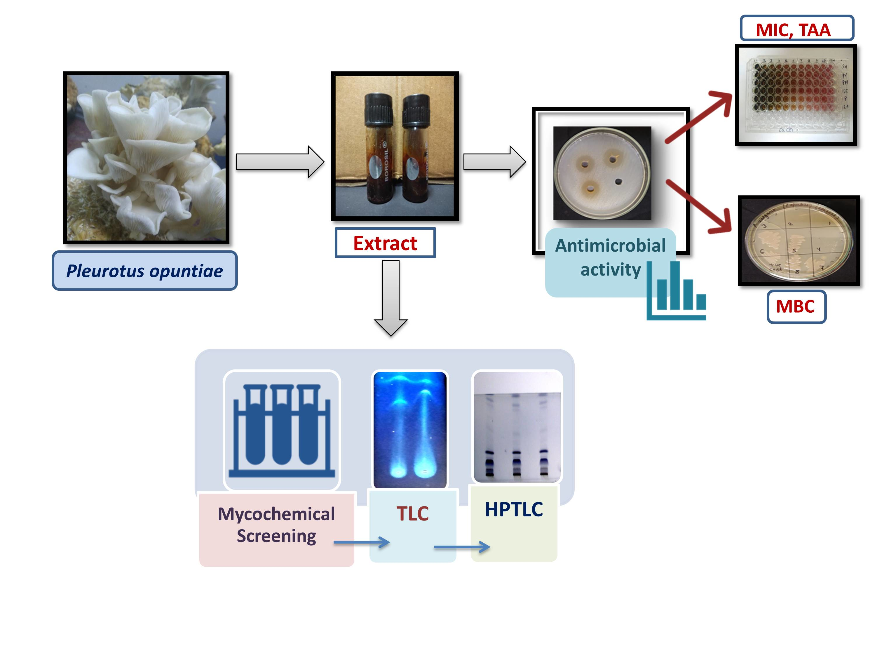
2. Materials and Methods

2.1. Collection of Mushroom

The culture of Pleurotus opuntiae (mushroom) was procured from Directorate of Mushroom Research (DMR), Solan (Himachal Pradesh, India), and was maintained on malt extract agar media at 25–28 °C and pH 6–6.5. The whole cultivation process was performed in-house at Centre of Biotechnology, University of Allahabad (Prayagraj, India). The fruiting bodies from the mushroom were washed thoroughly with distilled water. Following this, they were dried at room temperature, and ground to a fine powder and stored in airtight containers until further use.

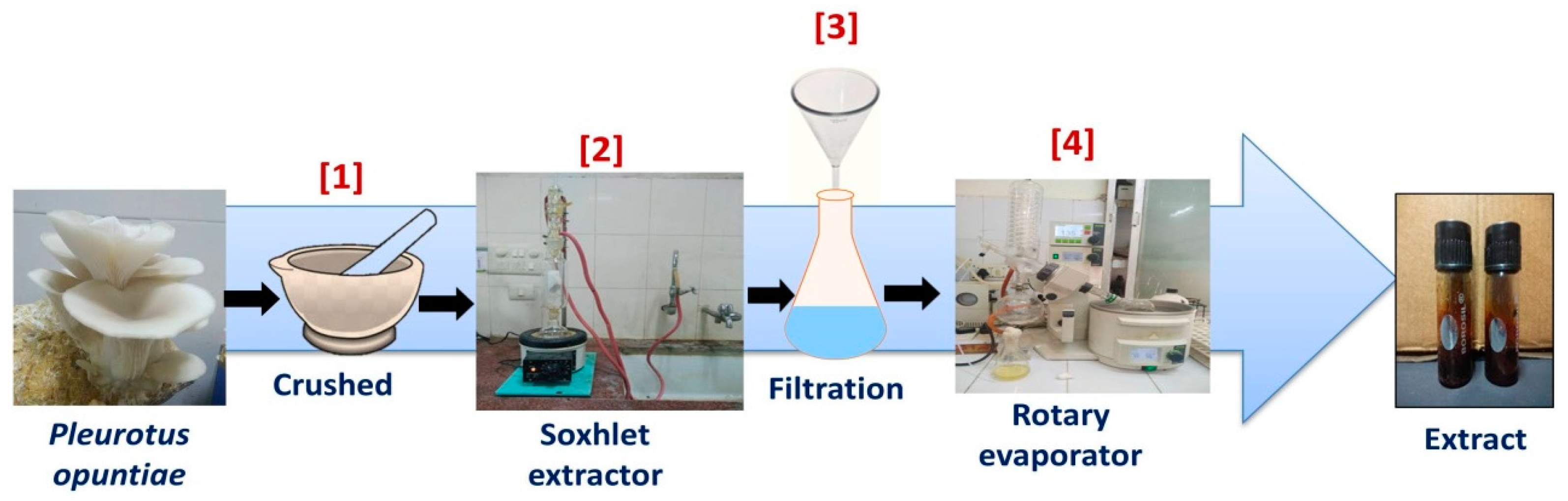
2.2. Preparation of Mushroom Extracts

The extraction methodology of mushroom was similar to the one used by Oyetayo et al. [16]. with minor modifications. A total of 10 g of powder was separately extracted with each 100 mL of ethanol and methanol by using a soxhlet extractor for 4 to 5 h at a temperature below the boiling point of the solvents. Using Whatman no. 1 filter paper, the extracts were filtered and the residual solvent was then removed by evaporation at 40 °C for 6–7 h using a rotatory evaporator. The residues obtained were stored in a sterile bottle under refrigeration (4 °C) prior to further analysis. The total percentage yield of extract was 40% w/w for methanol and 38.0% w/w for ethanol respectively [16]. The complete process for preparation of P. opuntiae extract is shown in Figure 1.

2.3. Bacterial Tested Organisms

A total of one Gram-positive (Staphylococcus aureus ATCC-25923) and six Gram-negative (Pseudomonas aeruginosa ATCC-27853, Proteus mirabilis NCIM-2300, Proteus vulgaris NCIM-5266, Serratia marcescens NCIM-2078, Shigella flexneri NCIM-5265, Moraxella sp. NCIM 2795) pathogenic bacterial strains were used in this study for testing the antimicrobial activity of the crude mushroom extracts. The microorganisms were procured from the Department of Microbiology, IMS BHU (Varanasi), and NCIM (Pune), India respectively.

2.4. Determination of Antibacterial Screening Test

Antimicrobial activity of methanol and ethanol extract of P. opuntiae was determined by the agar well diffusion method [16] with slight modification. Briefly, the methanol and ethanol extracts were dissolved in 3% dimethylsulfoxide (DMSO) at different concentrations (i.e., 100, 125 and 150 mg/mL). An overnight culture of each microbial isolate was inoculated in nutrient broth to turbidity which was equivalent to 0.5 McFarland (105 CFU/mL). To determine the antimicrobial activity of the extracts, an aliquot of test culture was evenly swabbed over the surface of the solidified Muller Hinton agar with the help of a sterile swab. Next, small wells (5 mm in diameter) were made in the agar plates by sterile cork borer. Quantities of 100 µL of both extracts were loaded into the different wells at different concentrations, respectively. Ciprofloxacin (5 μg/mL) for Gram-negative and penicillin (10 µg/mL) for Gram-positive dissolved in distilled water were used as the control for tested microorganisms. All the plates with respective extract and tested organisms were incubated at 37 °C, for 24 h. After 24 h, the zone of inhibition was measured in millimeters (mm) by the antibiotic zone scale. All the tests were carried out in triplicate and their means recorded.

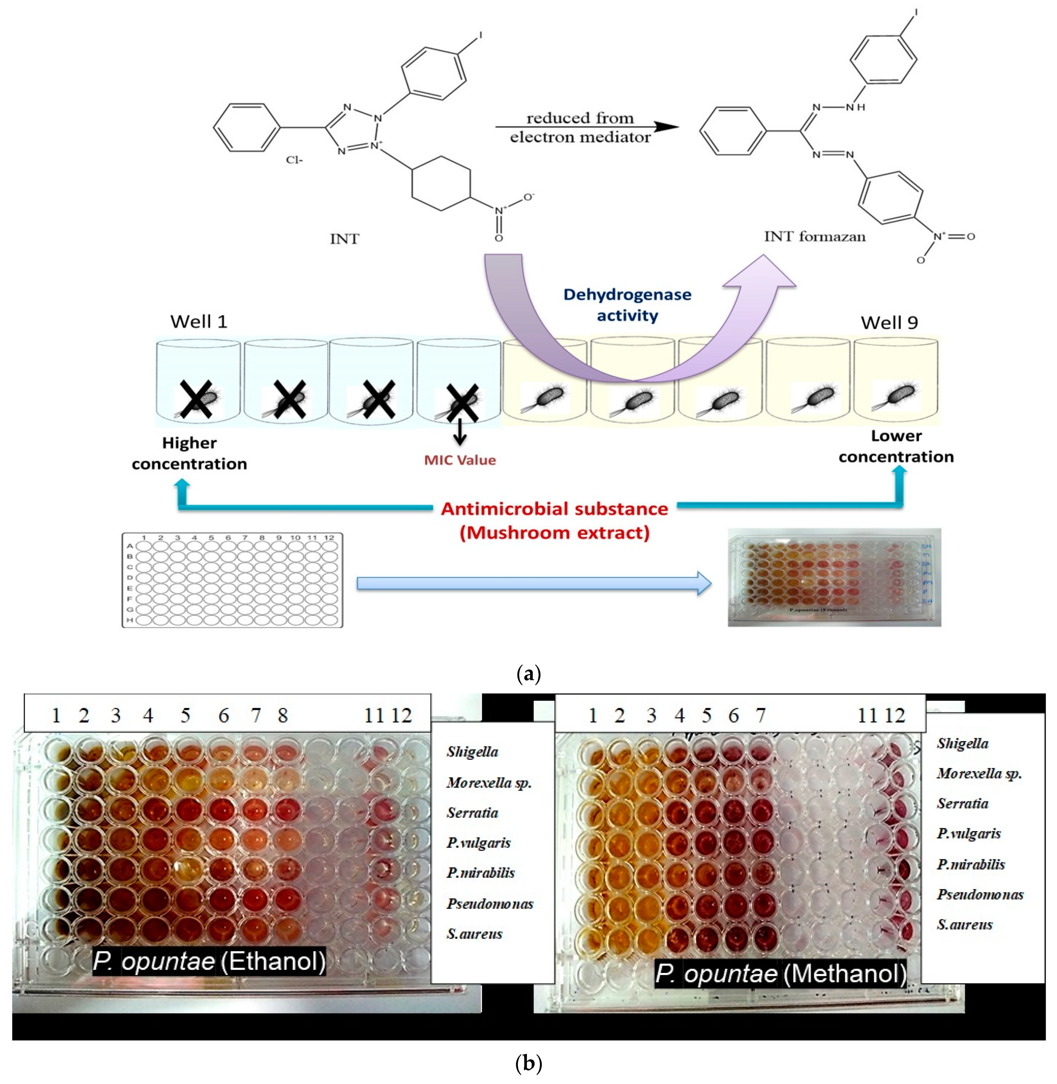
2.5. Determination of Minimum Inhibitory Concentration (MIC)

Minimum inhibitory concentrations of the extracts were determined by using the INT microdilution colorimetric assay method described by [6,17] with slight modifications. The stock solution of each extract was prepared by dissolving 250 mg/mL of extract in dimethyl sulfoxide (DMSO). Briefly, 96-well microtiter plates were prepared by dispensing 100 μL of nutrient broth into each well. A 100 μL from the stock solution (250 mg/mL) of tested extract was added into the first row of the plates. Then, twofold serial dilutions were performed by transferring 100 μL of solution from one row to another, using a pipette. The obtained concentration range was from 250 to 1.95 mg/mL for each extracts. Microbial inoculum size of 50 μL (1.0 × 10⁵ CFU/mL) was added to each well. Whereas in positive control 100 μL of nutrient broth and 50 μL of bacterial suspension were added and in the negative control, only the nutrient broth and extract was added. Then inoculated microtiter plates were incubated at 37 °C for 24 h. After 24 h the MIC was determined by adding 30 μL (2 mg/mL) of 0.02% p-Iodonitrotetrazolium chloride (INT) and incubated at 37 °C for 30 min. INT was used as an indicator for bacterial growth as bacteria metabolizes INT and changes it to pink color. Lack of color change after the addition of INT indicated no growth of microorganisms and were chosen as MIC values.

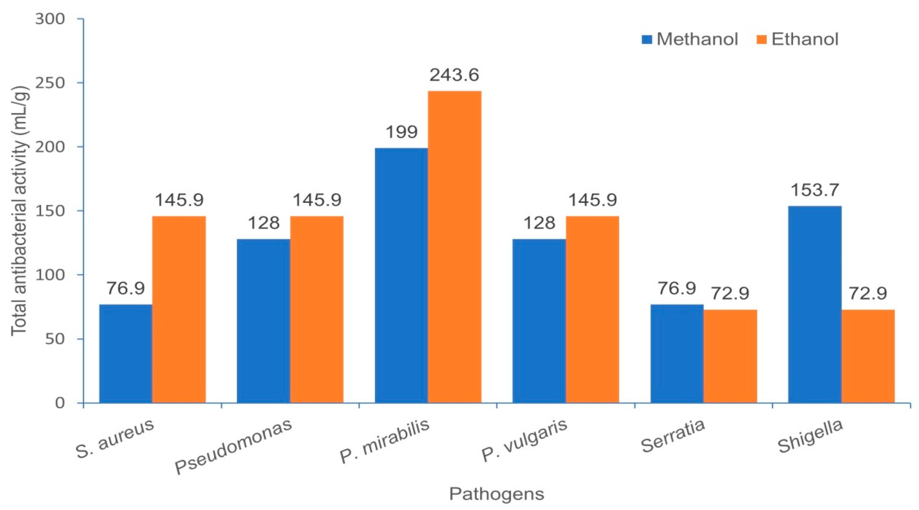
2.6. Total Antibacterial Activity (TAA)

The total antibacterial activity (TAA), is a function of the extraction yield in milligram per 1 g of P. opuntiae and the minimal inhibitory concentration (MIC), expressed in milliliter per gram (mL/g) [18]. TAA indicates the volume of water or solvent that is added to 1 g of the extract that will still inhibit the growth of the pathogen [19].

2.7. Determination of Minimum Bactericidal Concentration (MBC)

The minimum bactericidal concentration of Pleurotus opuntiae extracts was determined by modifying microbroth dilution method described by Gebreyohannes and team [6]. Micro-titer plates (96-wells) were each filled with 100 μL of sterile nutrient broth and serially diluted with extract concentrations within the range 250 to 1.95 mg/mL, respectively following the same procedure described above for MIC determination. An inoculum size of 50 μL (1.0 × 10⁵ CFU/mL) of test organisms were added to the appropriately labelled wells and incubated at 37 °C for 24 h. Post-incubation, a loopful of inoculum from each well of the microtiter plate with clear content (without any color change) was taken and streaked it on nutrient agar plates. Then plates were incubated for 24 h at 37 °C. Then, the MBCs were determined as the lowest concentration of the extract that permits no growth of bacteria. All tests were performed in triplicates to validate the results.

2.8. Mycochemical Screening

The methanol and ethanol extracts of P. opuntiae were screened to detect the presence of mycoconstituents such as alkaloids, carbohydrate, phenols, diterpenes, phytosterols (triterpenes), proteins, amino acids, tannins, anthraquinones, flavonoids, phlobatamins, terpenes, cardiac glycosides, saponins following standard protocols [20,21].

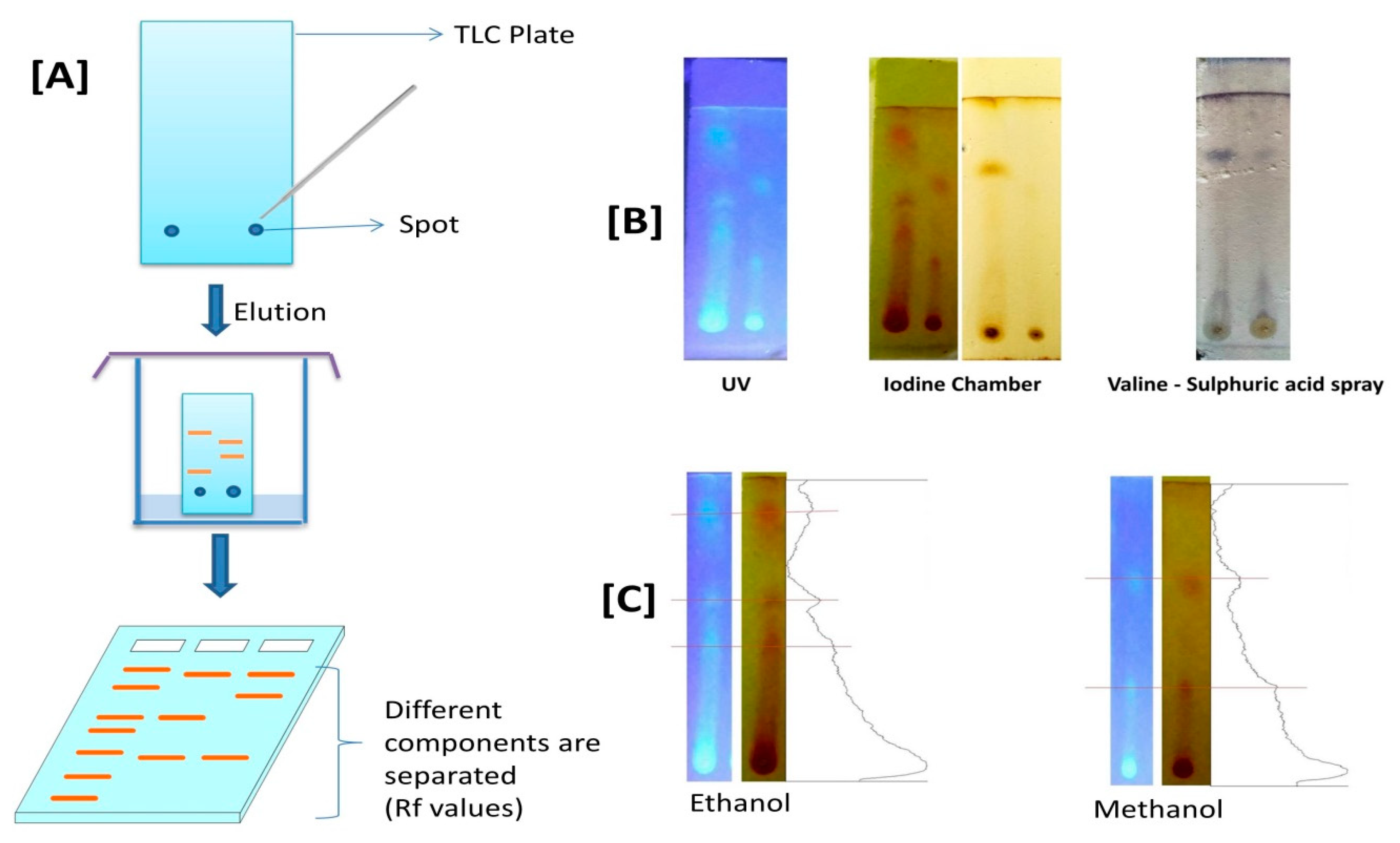
2.9. Thin-Layer Chromatography (TLC)

The methanol and ethanol extracts of P. opuntiae were investigated using TLC as described by [22]. Thin layer plates were prepared by spreading the mixture of silica gel (silica gel 60 GF254 (Merck) dissolved in distilled water (thick slurry) on clean glass slides. These plates were then dried and activated by heating in an oven for 30 min at 110 °C [23]. 100 mg of each extract was dissolved in 1 mL ethanol and 1 mL methanol, respectively, and applied as spots with the aid of capillary tubes on a silica gel coated plate about 1 cm from the base. The spotted plates were run in different solvent systems. Then developed plates were observed under the UV chamber and iodine chamber and were then sprayed with vanillin–sulfuric acid for the detection of the different spots after derivatization. Distances between the spots were measured and the Retention factor (Rf) values were calculated. The machine learning toolbox has recently helped decipher scientific data collected through a broad variety of medical information in the area of antimicrobial technology. In line with this, TLC chromatograms were analyzed using NIH ImageJ software [24].
Another method to develop prototype formulation was used with some modification known as bioautography method. In this method 100 mg of each extract was dissolved in 1 mL ethanol and applied as a coating layer (in the form of spot) on the silica gel coated plate at about 1 cm from the base. The spotted plates were run in developed solvent system (8 mL chloroform + 2 mL hexane). Then compounds in the extract got separated to form a thin coating layer (liquid coating) form in the TLC plate. The chromatogram (silica coated plate) was dried for up to 24 h at room temperature under a stream of air to remove the remaining solvent. The plates were then sprayed (spray coating) with bacterial suspension and incubated for 24 h. After 24 h it was further sprayed with a 2 mg/mL solution of p-Iodonitrotetrazolium violet (Sigma-Aldrich, St. Louis, MO, USA) (INT) and incubated for 4 to 6 h. Clear zone or white area of inhibition was observed against a purple background. White areas indicated reduction of INT to the coloured formazan did not take place due to the presence of compounds that inhibited the growth of tested pathogen [25].

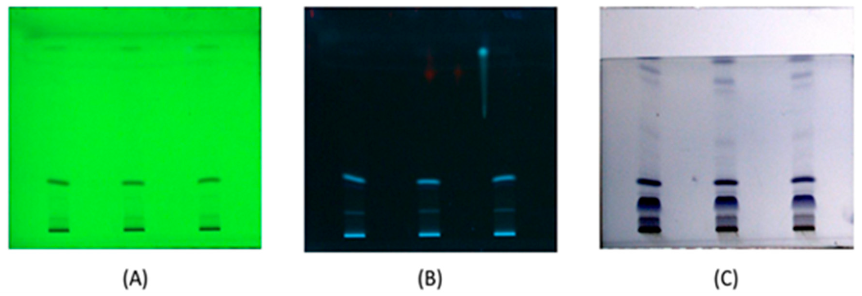
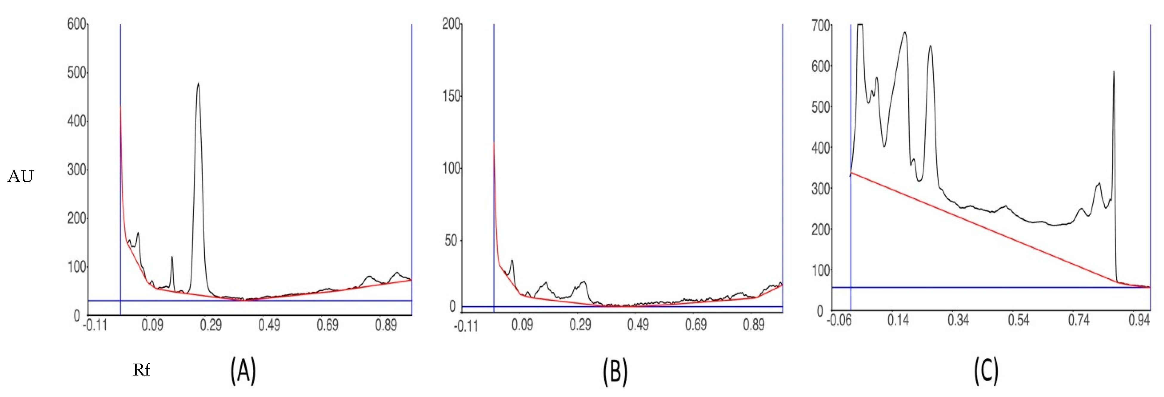
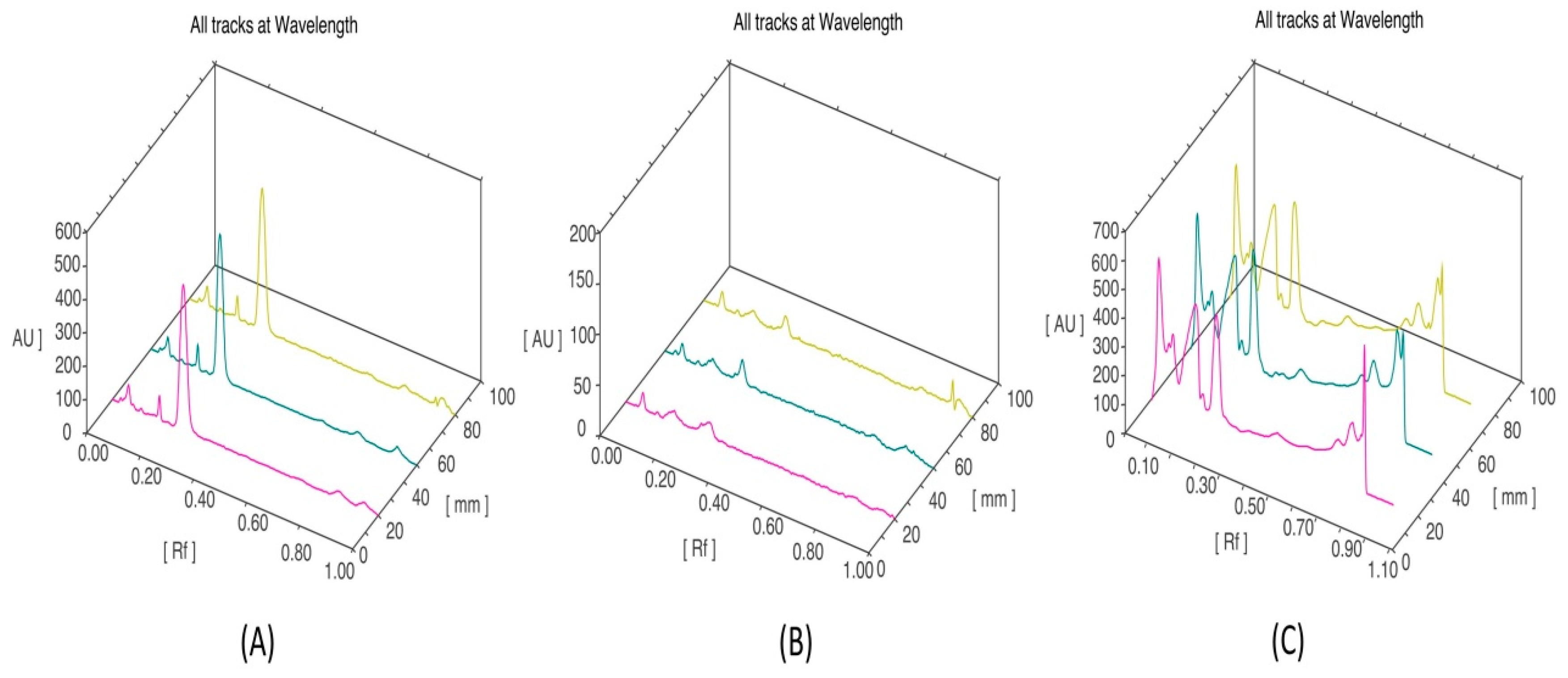
2.10. High-Performance Thin Layer Chromatography (HPTLC) Analysis

HPTLC analysis of ethanol extract of P. opuntiae was performed at CSIR-Central Institute of Medicinal and Aromatic Plants, CIMAP, Lucknow, India. The chromatographic analysis was performed using aluminum sheets precoated with silica gel 60 F254 as a stationary phase. Aliquots of 2 μL of extract (100 mg of extract dissolved in 1 mL ethanol) were taken for sample loading. The mobile phase used was chloroform: hexane (8:2, v/v) and further derivatized in vanillin–sulfuric acid reagent for detection of different spots present in the extract. Densitometric scanning was performed at wavelengths 254, 366 and 540 nm.

2.11. Statistical Analysis

Data obtained from the study were analyzed with Graph Pad Prism Version 5.0 for Windows (Graph Pad Software, Inc., San Diego, CA, USA statistical package program. Values are expressed as mean ± SEM (Standard Error Mean). Results obtained were statistically analyzed by using one-way ANOVA (Analysis of variance) followed by Bonferroni post hoc test, p < 0.05 was considered a significant value. The quantitative evaluation of different fractions of chromatograms (TLC) for both extracts was performed using the ImageJ software package.

3. Results

3.1. Antimicrobial Activity of P. opuntiae

In this work, antimicrobial activity of ethanol and methanol extract of P. opuntiae was tested against a prominent pathogenic Gram-positive bacteria (S. aureus ATCC 25923) and six Gram-negative bacteria (P. aeruginosa ATCC 27853, P. mirabilis NCIM 2300, P. vulgaris NCIM 5266, Serratia marcescens NCIM 2078, Shigella flexeneri NCIM 5265, Moraxella sp. NCIM 2795).
Results of antimicrobial activity of the extracts suggest that both extracts show a broad range of antimicrobial activity against different pathogenic strains. Mushroom used in this analysis indicates varying degree of antimicrobial activity against the tested microorganisms. Antimicrobial activity of P. oputiae extracts at varying concentrations is described in (Table 1 and Supplementary Material, Figure S1A,B. All the data obtained are statistically significant (p-value ≤ 0.05). It was observed that as the concentration of extract increases (100, 125, 150 mg/mL) the zone of inhibition also increases. Ethanol and methanol extracts of P. opuntiae were the effective in retarding microbial growth of all tested pathogenic bacteria by giving a clear zone of inhibition. The ethanol extract showed significantly higher antimicrobial activity against P. vulgaris (23.6 to 25.6 mm) and the least activity against P. aeruginosa and Shigella and Serratia. Methanol extract gave highest activity against P. aeruginosa (18.6 to 21.6 mm) and least activity against Serratia (10.0 to 15.3 mm) at all concentrations. The activity against S. aureus was negligible at concentration 100 mg/mL in methanol extract and gives the least activity at 150 mg/mL, whereas ethanol extract showed potent antimicrobial activity at all concentrations (100, 125, 150 mg/mL). Moraxella sp. shows no activity against both extracts of P. opuntiae at all concentrations. The standard antibiotic ciprofloxacin (5 µg) for Gram-negative bacteria and penicillin (10 µg) for Gram-positive bacteria was taken as control.

3.2. Minimum Inhibitory Concentration (MIC), Minimum Bactericidal Concentration (MBC), MBC/MIC Ratio and Total Antimicrobial Activity (TAA)

The MIC of methanol and ethanol extracts of P. opuntiae were employed by using INT colorimetric assay and MBC by microbroth dilution method to evaluate their bacteriostatic and bactericidal properties. The concentration effect of both the extracts were reported in Table 2 and illustrated in Figure 2. MIC values of ethanol extract were lower than those of methanol extract of P. opuntiae. Ethanol extract showed MIC between 15.6 and 52.08 mg/mL, whereas methanol extract show MIC between 20.81 and 52.08 mg/mL against different bacterial pathogens. Activity against S. aureus was noted at MIC 26.03 mg/mL for ethanol extract and 52.08 mg/mL for methanol respectively. P. mirabilis gave the lowest MIC value at 20.81 mg/mL for methanol and 15.6 mg/mL for ethanol extract. Similarly, ethanol extract showed the highest MIC value at 52.08 mg/mL against Serratia and Shigella.
MBC of the ethanol extract of P. opuntiae against all pathogens were found to fall between 26.03 and 62.5 mg/mL and for methanol from 52.08 to 125 mg/mL, respectively. The highest MBC value 125 mg/mL was by methanol extract against Serratia, whereas all other pathogens gave MBC value of 52.08 mg/mL. S. aureus showed 62.5 mg/mL MBC value for methanol and 52.08 mg/mL for ethanol extract, respectively. P. aeruginosa and P. mirabilis gave the lowest MBC value at 26.03 mg/mL for ethanol extract. Unlike MIC, ethanol extracts showed lower MBC values as compared to methanol extract. The obtained results are promising as both extracts were bactericidal as their MBC/MIC value of both extracts is ≤4. MBC/MIC ratio is shown in Table 2. The value of total antibacterial activity (TAA) of both extracts of P. opuntiae was recorded against all the pathogens which show their broad antimicrobial potential. The higher the TAA value, the more efficacious the extract. For S. aureus, Pseudomonas, P. mirabilis, and P. vulgaris ethanol extract show high TAA as compared to methanol extract, whereas methanol extract showed high TAA against Serratia and Shigella (Figure 3).

3.3. Mycochemical Screening from Pleurotus opuntiae

In addition to the in vitro antimicrobial activity, mycochemical analysis was also carried for ethanol and methanol extracts of P. opuntiae. Tests for alkaloids, carbohydrate, phenols, diterpenes, phytosterols (triterpenes), proteins, amino acids, tannins, anthraquinones, flavonoids, phlobatamins, terpenes, cardiac glycosides, saponins were carried out and their results are shown in Table 3.
It was found that alkaloids and diterpenes are present in ethanol extract in a greater amount as compared to methanol extract. Triterpenes are only present in methanol extract. Tannins, cardiac glycosides, and anthraquinones are absent in both the extract. The antibacterial activity of the P. opuntiae extract in this study can be attributed to the presence of various bioactive components. Presence of different classes of secondary metabolites: phenols, flavonoids, terpenoid, alkaloids, and saponins could be considered as the reason for the observed antimicrobial activities in this study.

3.4. Thin Layer Chromatography (TLC) Profile

Chemical constituents of both extracts were separated on TLC plates by standardizing in different solvent systems under saturated conditions, which are shown in Table 1, Supplementary Material. The Rf values of different eluted compounds were calculated. Amongst all the used solvent systems, the best result was given by Chloroform + Hexane (8:2) which eluted out 5 different compounds (spots) of P. opuntiae with different Rf values (Table 4). Apart from it, all the other solvent systems gave only 2 to 3 clear spots of separated compounds for both the extracts respectively. Although in 100% chloroform solvent system, 2 spots with the same Rf values (0.6, 0.7) in both extracts were observed which is also analyzed and confirmed by ImageJ software (Figures S2 and S3, Supplementary Material). In the solvent system, Chloroform + Hexane (8:2) ethanol extract gives 3 active bands (spots) with different Rf values 0.6, 0.3, 0.7, and methanol extract gives 2 active bands with Rf values 0.7 and 0.5 when visualized under UV and iodine chamber. In Vanillin-sulphuric acid, spray ethanol again gives 3 active bands with Rf values 0.7, 0.4, 0.2, and methanol gives 2 active bands with Rf values 0.7, 0.2. A total of five active bands were found in both the extract. Active bands of P. opuntiae in TLC result and their analysis and confirmation by ImageJ software is given in Table 4 and illustrated in Figure 4A–C). In the bioautography analysis of ethanol extract of P. opuntiae, only two pathogens Shigella flexneri and Proteus vulgaris gave positive antimicrobial activity results with Rf values 0.3 and 0.7, the supporting figure is given in Supplementary Material, Figure S4.

3.5. HPTLC Fingerprinting Analysis of Ethanol Extract of P. opuntiae

The HPTLC chromatogram showed that ethanol extract constituents were clearly separated without any tailing and or being diffuse. The chromatogram can be best observed under daylight, UV 254, 366 and 540 nm before and after derivatization (Figure 5). The HPTLC densitogram under UV–254, 366 and 540 nm of ethanol extract of P. opuntiae revealed several peaks which are presented in Figure 6 and Figure 7. The densitometry results of extract for 10 μL sample size, under UV–254 nm (high energy zone) revealed 7 spots, for 25 μL sample size, under UV–366 nm revealed 3 spots and for 25 μL sample size, under 540 nm revealed 12 spots with Rf max values (Table 5, Table 6 and Table 7). For 254 nm major spot with the Rf max 0.33 along with the percentage area of 76.75%, for 366 nm major spot with the Rf max 0.34 along with the percentage area of 49% and for 540 nm major spot with the Rf max 0.2 along with the percentage area of 21.77% were recorded respectively. These are found to be principal components or marker compounds of mushroom extract. Thus, the developed chromatogram will be specific with selected solvent system chloroform:hexane (8:2) and serves as the better tool for standardization of the compound from mushroom (P. opuntiae).

4. Discussion

Mycoconstituents display a wide range of biological activities against pathogenic microorganisms [26]. The findings obtained from present study affirm this further with a focus on antibacterial activity. Similar antimicrobial potentials have been observed in P. ostreatus [27] and many other species [21] but have never been reported in P. opuntiae despite having medicinal properties. Bacterial strains included in this study were chosen based on their significance and etiology in clinical microbiology and in different infectious processes [28]. Eloff; 1998 [29] studied the antimicrobial activity of different plant extracts on different test organisms by using colorimetric assays of various tetrazolium salts as a reagent. The result of his experiment indicated that the formation of formazan product was most stable, particularly in the case of INT formazan tetrazolium salt assay. Hence, in our study, we rationally used INT tetrazolium salt assay for the MIC determination of P. opuntiae. Both extracts showed potential bactericidal activity against the tested pathogenic bacteria. Antimicrobial activity is usually regarded as bactericidal if the MBC/MIC ratio is ≤4 and bacteriostatic if MBC/MIC > 4 [30]. The obtained results from both the extracts were bactericidal as their MBC/MIC value were ≤4.
Both extracts showed various degrees of antimicrobial activity against test pathogens. Variation in MIC of different extracts may arise from differences in their chemical constituents and volatile nature of their constituents [22]. Furthermore, the type of extraction solvent, the origin of the mushrooms, their age, species, as well as the type of bacterial strain, can be attributed to the differences in the antimicrobial activity [5]. The higher bioactive content in the mushroom extracts can also be one of the major factors correlating with different ranges of MIC values as some could exhibit synergistic effects as well. From the above result, it was concluded that ethanol extract gives the highest total antimicrobial activity with lower MIC and MBC values as compared to methanol extract of P. opuntiae. During mycochemical screening, the ethanol extract of P. opuntiae gave a large number of mycoconstituents compared to methanol extract. Such a difference between two extracts is generally correlated with the affinity of a particular bioactive compound towards a solvent which in turn depends on the polarity. By specific separation and purification strategies, the type of components specific to the solvents can be identified. The presence of a higher quantity of compounds in the ethanol extract of P. opuntiae is responsible for the antimicrobial activity at lower MIC values against test microorganisms [31,32]. The preliminary screening tests can be helpful in the identification of bioactive compounds in the mushroom and can help in the discovery and production of drugs [21]. Groups of mycochemical compounds are commonly associated with combating microbial resistance and having antimicrobial behavior in edible mushroom. Currently, many of the pathogenic bacteria exhibit multidrug-resistance to commonly used antibiotics causing different diseases [33]. Due to these contributory factors, the search for a new drug is important in current times, and P. opuntiae has been proven as a potential source of bioactive molecules. The isolation of these bioactive compounds needs different analytical methodologies which include thin layer chromatography as one of the procedures for preliminary screening [33].
Since silica gel retains more polar compounds, the nonpolar compounds are eluted first and moved further up the TLC plate. Hence, the more polar the compound, shows lower Rf (Retention factor) value, and the less polar the compound, show larger the Rf value. Thus, it can be concluded that the mushroom extract of P. opuntiae studied here has both polar and nonpolar compounds [22]. This could also justify the difference in the extraction efficiency and antimicrobial activity between the two solvent extracts. Though methanol and ethanol exhibit closer polarity index, there is however a significant difference between these two solvents. As a result, bioactive compounds with a very specific affinity towards ethanol are extracted with ethanol, unlike methanol. According to Bubueanu (2017 [34]) the Pleurotus ostreatus extract was characterized by two major spots, one was ferulic acid (Rf ~0.9 and ~0.55) and other was coumarins (Rf~0.1, 0.6 and 0.8). The presence of ergosterol (T3) with four major spots (Rf~0.3–0.9) was also found in different solvent systems. Thus, the present study on P. opuntiae elicits similar findings with major spots corresponding to the same Rf values as stated above. It was also found that in P. opuntiae extract, the highest percentage area (76.75% and 49.37%) covered by the Rf value 0.3, matched to the Rf value of ergosterol as mentioned above. This can further be justified by the higher extraction efficiency of ethanol, as ergosterol shows relatively lower polarity and hence soluble to a greater extent in ethanol compared to methanol. So, these findings suggest strong evidence-based similarities for presence of ergosterol, ferulic acid and coumarins along with other compounds respectively. Further analysis by digital chromatographic techniques like HPTLC posted strong scientific evidence validating the TLC results. The differentiation in HPTLC profile is an important and powerful procedure that has often been used for detection of compounds [35]. Huizhen Li [36] in his HPTLC study of Pleurotus ostreatus recorded Rf = 0.34 & Rf = 0.31 and found it to be corresponding with saponin which is also same with our HPTLC results as the maximum area at wavelength 366 nm is being Rf = 0.34, which is 49.37%, and the maximum area at wavelength 540 nm is being Rf = 0.34 which is 14.13%. This result is also being supported by the mycochemical screening test for the presence of saponins in the extract. As a result, the presence of saponin is confirmed in higher amounts in P. opuntiae extract [36].
As per the definition of antimicrobial coating from the literature, it should be some synthetic resin or epoxide or a nano-emulsion that may release certain chemicals to inhibit the growth of pathogens; hence, to prove this release of antimicrobial chemicals from the extract we used a TLC plate to show the separated antimicrobial compounds. We used mushroom extract for natural antimicrobial coating applications. The silica has been used as the antibacterial surface design with the incorporation or adhesion of mushroom extracts due to their properties of being non-toxic, eco-friendly, cheap, easily available and not containing heavy metals, presenting support to silica-based surfaces by natural adjuvants to promote its activity. Some papers had reported that the natural extracts need to be used in an optimized amount to have the best performance for the coating. The natural additives included biopolymers like chitosan, gluten, gelatin, etc. Mushroom extracts on the other hand presented itself as “self-sustained edible coatings”. Different biopolymers, such as proteins, lipids, polysaccharides or their combinations, have been used as carriers to produce edible coatings with antimicrobial properties. Since it is well reported that mushroom have large amount of polysaccharide (glucans, chitin, mannas, galactans, xylans, etc.) the purpose of edible coating is well fulfilled [37].
The main coating techniques in food packaging are spraying, dipping or spreading, which have all been carried out by us at different stages of experimentation. In this context, we propose that P. opuntiae, an edible mushroom, can act as an edible antimicrobial coating in future. Antimicrobial edible coatings are ready to provide an effective alternative in active packaging materials to improve the safety of processed food products for commercial purposes [38,39]. The mushroom antimicrobial coating can be used in two ways: one by immobilizing it on a packaging film or the other relatively easy method by spreading a layer of extract on the food material. The mushroom releases its active compounds by forming a matrix and releasing active mycochemicals presenting a layer or coat over the food. Our results have shown that mushroom extracts exhibit stronger antibacterial properties against several pathogens. They could be incorporated into an edible film and coating formulation as antimicrobial agents, which will lead to improved food safety [40].
It is well known from the literature that basidiomycete comprises numerous bioactive compounds that facilitate a range of functional properties, acting individually and/or synergistically. A. blazei includes several other compounds, such as ergosterol, other forms of fatty acids, polysaccharides and alkaline compounds, which can also play an important role in synergistic antimicrobial activity [41]. Sudirman et al. [42] purified the antimicrobial compound fraction of Rf value 0.73 from Lentinus cladopus LC4, which is also similar with the results of our TLC but the structure of that compound is still unknown [42]. The developed HPTLC method gave strong fingerprints for species authentication through direct band comparison and can confirm the existence of bioactive compounds. Thus, the present study provides sufficient information about the therapeutic efficacy of the medication and in the identification, standardization and quality assurance of the specific natural product. In conclusion, the findings of the qualitative assessment of the HPTLC fingerprint images should aid in the detection and quality assurance of the medication and ensure therapeutic efficacy [43].
Our evidence-based study provides facts about the use of mushroom as a functional food. It contains different components (bioactives) which might be acting “synergistically” in an in-vivo physiological system to produce different medicinal attributes which may not be “individually” present in desired compounds if they are isolated and tested “in-vitro”. Since mushrooms, and specifically Pleurotus opuntiae, are widely used as edible food supplement in the Indian sub-continent and the Asian diaspora worldwide, this study is a preliminary attempt to indicate the potential antimicrobial activity of the mushroom, identifying it as a super-food with tremendous medicinal properties [3].
This research is one of the few studies identifying the antimicrobial action of the fungal extracts derived from their fruiting body. Our findings also support the possible application of fungal metabolites in pharmaceutical products and the pharmaceutical industry [26]. With an increasing number of bacteria developing resistance to commercial antibiotics or MDR, extracts and derivatives from mushrooms hold a great promise for novel medicines in modern times [2,44,45]. In the light of the current global pandemic, more multidisciplinary methods, such as phyto-micro/nanotechnology-enabled strategies, may become front-runners in the battle against infectious diseases and global initiative for the prevention and treatment of these infections [3,45]. Lastly, we need to scrutinize the recently evolving diverse cellular machinery of microbial flora [46,47,48] and resistant viruses by extensive research into natural-based therapeutics to improve preventative foresight.

5. Conclusions

The results from our study present an insight on the prospective use of the mycopharmaceuticals in the management and treatment of infectious diseases. The data obtained focuses on the importance of primary screening strategies as a putative step by presenting new avenues for clinical as well as pharmaceutical research and development. The best total antibacterial activity (TAA) in the present study was shown by ethanolic extract of P. opuntiae against P. mirabilis. Our study findings support mushroom as a potential source of new chemical entities (NCEs) which in turn presents a possible new class of antibiotics; yet the conformational and structural prediction of the active compounds remain unexplored. With this, the conventional antibiotics can now be replaced with newer and effective natural antimicrobials developed from mushroom extracts to treat several deadly and multi-drug resistant infectious diseases. Additionally, this provides further scope in directly utilizing the extracts by exploiting the possible synergetic effects of the bioactives by eliminating the need to individual component synthesis and characterization. The mushroom used in our study was found to have a great degree of antimicrobial effect against the selected pathogens. However, more extensive analytical studies are needed in future to appropriately use these mushroom antimicrobials in medicine as an active pharmaceutical ingredient (API). Furthermore, in future these mushrooms which are edible could be used in the form of nano-encapsulating edible coatings for food and drug safety. Thus, this functional food extract could act tremendously in naturally curbing off infections and providing a holistic approach to therapeutics.

Supplementary Materials

The following are available online at https://www.mdpi.com/article/10.3390/coatings11040484/s1, Figure S1: (A) Shows antimicrobial activity of P. oputiae (Ethanol extract) against pathogens; (B) Shows antimicrobial activity of P. opuntiae (Methanol extract) against pathogens, Figure S2: Shows TLC profile of ethanol and methnol extracts of P. opuntiaein 100% Chloroform solvent system, Figure S3: TLC and ImageJ program chromatograms of (A) Ethanol extract fraction and (B) methanol fraction of P. opuntie in 100% chloroform which shows only 2 spots respectively (Total 4 spots), Figure S4: Shows the bioautography result of Proteus vulgaris (before spraying and after spraying INT) and Shigella flexneri (after spraying INT) in silca gel coated plate, Table S1: Standardization of different solvent systems for TLC to separate compounds in form of total no. of spots in ethanol and methanol extracts of P. opuntiae.

Author Contributions

Conceptualization, M.P.S. and A.T.P.; methodology, A.T.P. and I.P.; software, A.K.; validation, B.G.-C., S.B. and S.P.D.; formal analysis, A.V. and S.P.D.; investigation, M.P.S. and A.T.P.; resources, M.P.S. and I.P.; data curation, A.K. and A.T.P.; writing—original draft preparation, A.T.P. and I.P.; visualization, S.B. and A.T.P.; supervision, M.P.S.; funding acquisition, M.P.S. and S.B. All authors have read and agreed to the published version of the manuscript.

Funding

This research was funded by University of Allahabad for providing necessary funding in the form of UGC-CRET fellowship and institutional facilities. This work was partly supported by IRGC Cycle-05 Grant # IRGC-05-SI-18-360 from Hamad Medical Corporation, Qatar.

Institutional Review Board Statement

Not applicable.

Informed Consent Statement

Not applicable.

Data Availability Statement

All datasets generated for this study are included in the article/Supplementary Material.

Acknowledgments

Group Leader Mohan Prasad Singh gratefully acknowledge funding from Department of Biotechnology, Govt. of India under the RRFSP. The authors also acknowledge the support of Hamad Medical Corporation, Qatar.

Conflicts of Interest

The authors have no conflict of interest.

References

  1. Akova, M. Epidemiology of antimicrobial resistance in bloodstream infections. Virulence 2016, 7, 252–266. [Google Scholar] [CrossRef]
  2. Zaman, S.B.; Hussain, M.A.; Nye, R.; Mehta, V.; Mamun, K.T.; Hossain, N. A review on antibiotic resistance: Alarm bells are ringing. Cureus 2017, 9, e1403. [Google Scholar] [CrossRef]
  3. Pandey, A.T.; Pandey, I.; Hachenberger, Y.; Krause, B.C.; Haidar, R.; Laux, P.; Luch, A.; Singh, M.P.; Vikram Singh, V.A. Emerging paradigm against global antimicrobial resistance via bioprospecting of mushroom into novel nanotherapeutics development. Trends Food Sci. Technol. 2020, 106, 333–344. [Google Scholar] [CrossRef]
  4. Centers for Disease Control and Prevention (CDC). Recommendations of CDC and health care infection control practices advisory committee. Atlanta 2003, 52, 1–42. [Google Scholar]
  5. Alves, M.J.; Ferreira, I.C.F.R.; Martins, A.; Pintado, M. Antimicrobial activity of wild mushroom extracts against clinical isolates resistant to different antibiotics. J. Appl. Microbiol. 2012, 113, 466–475. [Google Scholar] [CrossRef]
  6. Gebreyohannes, G.; Nyerere, A.; Bii, C.; Berhe, S.D. Determination of antimicrobial activity of extracts of indigenous wild mushrooms against pathogenic organisms. Evid.-Based Complementary Altern. Med. 2019, 2019, 6212673. [Google Scholar] [CrossRef]
  7. Wolff, E.R.; Wisbeck, E.; Silveira, M.L.; Gern, R.M.; Pinho, M.S.; Furlan, S.A. Antimicrobial and antineoplasic activity of Pleurotus ostreatus. Appl. Biochem. Biotechnol. 2008, 151, 402–412. [Google Scholar] [CrossRef]
  8. Vamanu, E.; Pelinescu, D.; Avram, I. Antioxidative effects of phenolic compounds of mushroom mycelia in simulated regions of the human colon, in vitro study. Pol. J. Food Nutr. Sci. 2018, 68, 83–90. [Google Scholar] [CrossRef]
  9. Panti, M. The antibacterial activity of coriolus versicolor methanol extract and its effect on ultrastructural changes of staphylococcus aureus and salmonella enteritidis. Front. Microbiol. 2016, 7, 1226. [Google Scholar]
  10. Elisashvili, V. Submerged cultivation of medicinal mushrooms: Bioprocesses and products (review). Int. J. Med. Mushrooms 2012, 14, 211–239. [Google Scholar] [CrossRef]
  11. Ren, L.; Hemar, Y.; Perera, C.O.; Lewis, G.; Krissansen, G.W.; Buchanan, P.K. Antibacterial and antioxidant activities of aqueous extracts of eight edible mushrooms. Bioact. Carbohydr. Diet. Fibre. 2014, 3, 41–51. [Google Scholar] [CrossRef]
  12. Gashaw, G.; Fassil, A.; Redi, F. Evaluation of the antibacterial activity of Pleurotus spp. cultivated on different agricultural wastes in Chiro, Ethiopia. Int. J. Microbiol. 2020, 2020, 9312489. [Google Scholar] [CrossRef]
  13. Saritha, K.; Rajesh, A.; Manjulatha, K.; Setty, O.H.; Yenugu, S. Mechanism of antibacterial action of the alcoholic extracts of Hemidesmus indicus (L.) R. Br. ex Schult, Leucas aspera (Wild.), Plumbago zeylanica L., and Tridax procumbens (L.) R. Br. ex Schult. Front. Microbiol. 2015, 6, 577. [Google Scholar] [CrossRef]
  14. Camacho, M.; Guzmán, G.; Guzmán-Dávalos, L. Pleurotus opuntiae (Durieu et Lév.) Sacc. (higher Basidiomycetes) and other species related to Agave and Opuntia plants in Mexico-Taxonomy, distribution, and applications. Int. J. Med. Mushrooms 2012, 14, 65–78. [Google Scholar] [CrossRef]
  15. Zervakis, G.I.; Venturella, G.; Fryssouli, V.; Inglese, P.; Polemis, E.; Gargano, M.L. Pleurotus opuntiae revisited—An insight to the phylogeny of dimitic Pleurotus species with emphasis on the P. djamor complex. Fungal Biol. 2019, 123, 188–199. [Google Scholar] [CrossRef]
  16. Oyetayo, V.O.; Dong, C.H.; Yao, Y.J. Antioxidant and antimicrobial properties of aqueous extract from Dictyophora indusiata. Open Mycol. J. 2009, 3, 20–26. [Google Scholar] [CrossRef]
  17. Boakye, Y.D. Anti-infective properties and time-kill kinetics of phyllanthus muellerianus and its major constituent, Geraniin. Med. Chem. 2016, 6, 95–104. [Google Scholar] [CrossRef]
  18. Eloff, J.N. Quantification the bioactivity of plant extracts during screening and bioassay guided fractionation. Phytomedicine 2004, 11, 370–371. [Google Scholar] [CrossRef]
  19. Elisha, I.L.; Botha, F.S.; McGaw, L.J.; Eloff, J.N. The antibacterial activity of extracts of nine plant species with good activity against Escherichia coli against five other bacteria and cytotoxicity of extracts. BMC Complementary Altern. Med. 2017, 17, 133. [Google Scholar] [CrossRef]
  20. Prakash, K.D.; Sayali, G. Current quality control methods for standardization of herbal drugs. Int. J. Pharm. 2017, 5, 82–95. [Google Scholar]
  21. Bains, A.; Tripathi, A. Antibacterial activity and phytochemical screening of wild edible mushroom pleurotus ostreatus collected from himachal pradesh. Int. J. Curr. Adv. Res. 2016, 4, 2320–5407. [Google Scholar]
  22. Appiah, T.; Boakye, Y.D.; Agyare, C. Antimicrobial activities and time-kill kinetics of extracts of selected ghanaian mushrooms. Evid.-Based Complementary Altern. Med. 2017, 2017, 4534350. [Google Scholar] [CrossRef] [PubMed]
  23. Bele, A.A.; Khale, A.; Archana, M.; Bele, A. An overview on thin layer chromatography. Int. J. Pharm. Sci. Res. 2011, 2, 256–267. [Google Scholar]
  24. Singh, A.V.; Ansari, M.H.D.; Rosenkranz, D.; Maharjan, R.S.; Kriegel, F.L.; Gandhi, K.; Kanase, A.; Singh, R.; Laux, P.; Luch, A. Artificial intelligence and machine learning in computational nanotoxicology: Unlocking and empowering nanomedicine. Adv. Healthc. Mater. 2020, 9, 1901862. [Google Scholar] [CrossRef]
  25. Suleiman, M.M.; McGaw, L.J.; Naidoo, V.; Eloff, J.N. Detection of antimicrobial compounds by bioautography of different extracts of leaves of selected south african tree species. AJTCAM 2010, 1, 64–78. [Google Scholar] [CrossRef] [PubMed]
  26. Synytsya, A.; Monkai, J.; Bleha, R.; Macurkova, A.; Ruml, T.; Juhee, A.; Chukeatirote, E. Antimicrobial activity of crude extracts prepared from fungal mycelia. Asian Pac. J. Trop. Biomed. 2017, 7, 257–261. [Google Scholar] [CrossRef]
  27. Chowdhury, H.M.H.; Kubra, K.; Ahmed, R.R. Screening of antimicrobial, antioxidant properties and bioactive compounds of some edible mushrooms cultivated in Bangladesh. Ann. Clin. Microbiol. Antimicrob. 2015, 14, 8. [Google Scholar] [CrossRef]
  28. Pandey, T.A.; Pandey, I.; Zamboni, P.; Gemmati, D.; Kanase, A.; Singh, A.V.; Singh, M.P. Traditional herbal remedies with a multifunctional therapeutic approach as an implication in COVID-19. Coatings 2020, 10, 761. [Google Scholar] [CrossRef]
  29. Eloff, J.N. A sensitive and quick microplate method to determine the minimal inhibitory concentration of plant extracts for bacteria. Planta Med. 1998, 64, 711–713. [Google Scholar] [CrossRef]
  30. Keepers, T.R.; Gomez, M.; Celeri, C.; Nichols, W.W.; Krause, K.M. Bactericidal activity, absence of serum effect, and time-kill kinetics of ceftazidime-avibactam against β-lactamase-producing Enterobacteriaceae and Pseudomonas aeruginosa. Antimicrob. Agents Chemother. 2014, 58, 5297–5305. [Google Scholar] [CrossRef]
  31. Mostafa, A.A.; Al-Askar, A.A.; Almaary, K.S.; Dawoud, T.M.; Sholkamy, E.N.; Bakri, M.M. Antimicrobial activity of some plant extracts against bacterial strains causing food poisoning diseases. Saudi J. Biol. Sci. 2018, 25, 361–366. [Google Scholar] [CrossRef] [PubMed]
  32. González-Palma, I.; Escalona-Buendía, H.B.; Ponce-Alquicira, E.; Tellez, M.T.; Gupta, K.V.; Diaz-Godinez, G.; Soriano-Santos, J. Evaluation of the antioxidant activity of aqueous and methanol extracts of Pleurotus ostreatus in different growth stages. Front. Microbiol. 2016, 7, 1099. [Google Scholar] [CrossRef] [PubMed]
  33. Nagoor Meeran, M.F.; Javed, H.; Taee, H.A.; Azimullah, S.; Ojha, S.K. Pharmacological properties and molecular mechanisms of thymol: Prospects for its therapeutic potential and pharmaceutical development. Front. Pharmacol. 2017, 8, 380. [Google Scholar] [CrossRef]
  34. Bubueanu, C.; Grigore, A.; Serban, E.; Popa, G.; Cornea, P.C. HPTLC identification of bioactive compounds and antioxidant activity of Pleurotus ostreatus and Lentinus edodes extracts. Sci. Bull. Ser. F Biotechnol. 2017, XXI, 343–348. [Google Scholar]
  35. Gunalan, G.; Saraswathy, A.; Vijayalakshmi, K. HPTLC fingerprint profile of Bauhinia variegata Linn. leaves. Asian Pac. J. Trop. Dis. 2012, 2, S21–S25. [Google Scholar] [CrossRef]
  36. Li, H.; Zhao, Y.; Yang, W.; Zhang, Z. Characterization of astragaloside I-IV based on the separation of HPTLC from Pleurotus ostreatus cultivated with Astragalus. J. Food Sci. 2020, 85, 3183–3190. [Google Scholar] [CrossRef]
  37. Lima, C.U.J.O.; Gris, E.F.; Karnikowski, M.G.O. Antimicrobial properties of the mushroom Agaricus blazei—Integrative review. Braz. J. Pharmacogn. 2016, 26, 780–786. [Google Scholar] [CrossRef]
  38. Ong, G.; Kasi, R.; Subramaniam, R. A review on plant extracts as natural additives in coating applications. Prog. Org. Coat. 2021, 151, 106091. [Google Scholar] [CrossRef]
  39. Valdés, A.; Ramos, M.; Beltrán, A.; Jiménez, A.; Garrigós, M.C. State of the art of antimicrobial edible coatings for food packaging applications. Coatings 2017, 7, 56. [Google Scholar] [CrossRef]
  40. Ulbin-Figlewicz, N.; Zimoch, A.; Jarmoluk, A. Plant extracts as components of edible antimicrobial protective coatings. Czech J. Food Sci. 2013, 6, 596–600. [Google Scholar] [CrossRef]
  41. Irkin, R.; Esmer, O.K. Novel food packaging systems with natural antimicrobial agents. J. Food Sci. Technol. 2015, 10, 6095–6111. [Google Scholar] [CrossRef]
  42. Sudirman, I.L. Partial purification of antimicrobial compounds isolated from mycelia of tropical lentinus cladopus LC4. HAYATI 2010, 17, 63–67. [Google Scholar] [CrossRef]
  43. Murugesan, S.; Bhuvaneswari, S. HPTLC fingerprint profile of methanol extract of the marine red alga Portieria hornemannii (Lyngbye) (Silva). Int. J. Pharm. Anal. 2016, 3, 2320–4923. [Google Scholar]
  44. Dwivedi, C.; Pandey, I.; Pandey, H.; Patil, S.; Mishra, S.B.; Pandey, A.C.; Zamboni, P.; Ramteke, P.W.; Singh, A.V. In vivo diabetic wound healing with nanofibrous scaffolds modified with gentamicin and recombinant human epidermal growth factor. J. Biomed. Mater. Res. 2018, 106, 641–651. [Google Scholar] [CrossRef] [PubMed]
  45. Singh, A.V.; Ansari, M.H.D.; Laux, P.; Luch, A. Micro-nanorobots: Important considerations when developing novel drug delivery platforms. Expert Opin. Drug Del. 2019, 16, 1259–1275. [Google Scholar] [CrossRef] [PubMed]
  46. Singh, A.V.; Sitti, M. Targeted drug delivery and imaging using mobile milli/microrobots: A promising future towards theranostic pharmaceutical design. Curr. Pharm. Des. 2016, 22, 1418. [Google Scholar] [CrossRef]
  47. Singh, A.V.; Maharjan, R.; Kanase, A.; Siewert, K.; Rosenkranz, D.; Singh, R.; Laux, P.; Luch, A. Machine-learning-based approach to decode the influence of nanomaterial properties on their interaction with cells. ACS Appl. Mater. Interfaces 2021, 13, 1943−1955. [Google Scholar] [CrossRef]
  48. Singh, A.V.; Rosenkranz, D.; Hasan, M.; Ansari, D.; Singh, R.; Kanase, A.; Singh, S.P.; Johnston, B.; Tentschert, J.; Laux, P.; et al. Artificial intelligence and machine learning empower advanced biomedical material design to toxicity prediction. Adv. Intell. Syst. 2020, 2, 2000084. [Google Scholar] [CrossRef]
Figure 1. Schematic representation for the preparation of P. opuntiae extract.
Coatings 11 00484 g001
Figure 2. (a) Mechanism of formation of INT to INT formazan and determination of MIC value in INT colorimetric assay, (b) INT assay showing MIC values of methanol and ethanol extract of P. opuntiae against pathogens. 1—250 mg; 2—125 mg; 3—62.5 mg; 4—31.25 mg; 5—15. 6 mg; 6—7.8 mg; 7—3.9 mg; 8—1.95 mg/mL; 11—positive (+ve) control and 12—negative (–ve) control for ethanol; 11—negative (–ve) control and 12—positive (+ve) control for methanol.
Coatings 11 00484 g002
Figure 3. Total antibacterial activity (TAA) of ethanol and methanol extracts of P. opuntiae against the pathogens.
Coatings 11 00484 g003
Figure 4. (A) Schematic representation of Thin Layer Chromatography (TLC), (B) TLC profile of ethanol and methanol extracts of P. opuntiae in Chloroform + Hexane (8:2) solvent system, (C) TLC and ImageJ-based chromatograms of ethanol extract and methanol extract fraction of P. opuntie in Chloroform + Hexane (8:2) which shows 3 and 2 spots respectively.
Coatings 11 00484 g004
Figure 5. (A) HPTLC chromatogram image at 254 nm, (B) HPTLC chromatogram image at 366 nm and (C) HPTLC chromatogram image after derivatization.
Coatings 11 00484 g005
Figure 6. HPTLC densitogram of ethanol extracts of P. opuntiae at wavelength (A) 254 nm, (B) 366 nm and (C) 540 nm. y-axis shows AU- area under curve and x-axis shows Rf values of different compounds.
Coatings 11 00484 g006
Figure 7. 3D HPTLC fingerprint of ethanol extracts of P. opuntiae at wavelength (A) 254 nm, (B) 366 nm and (C) 540 nm. AU—Area under curve (measured the uniform areas under the curves in between wavelengths 254, 366 and 540 nm, Rf—Retention factor (shows Rf values of different compounds), mm-shows length of chromatogram run.
Coatings 11 00484 g007
Table 1. Antimicrobial Activity of P. opuntiae at different concentrations against pathogenic strains.
PathogensEthanolMethanolAntibiotics (Control)
100 mg/mL125 mg/mL150 mg/mL100 mg/mL125 mg/mL150 mg/mL-
Zone of Inhibition (mm)
S. aureus14.6 ± 0.317.3 ± 0.317.6 ± 0.310.0 ± 0.011.0 ± 0.011.6 ± 0.321.6 ± 0.3
P. aeruginosa11.3 ± 0.312.6 ± 0.314.3 ± 0.318.6 ± 0.319.6 ± 0.321.6 ± 0.320.6 ± 0.3
P. mirabilis14.6 ± 0.316.3 ± 0.319.3 ± 0.315.6 ± 0.317.6 ± 0.319.0 ± 0.024.3 ± 0.3
P. vulgaris23.6 ± 0.324.6 ± 0.325.6 ± 0.316.3 ± 0.318.6 ± 0.319.6 ± 0.333.6 ± 0.3
S. marcescens10.0 ± 0.014.0 ± 0.014.6 ± 0.310.0 ± 0.013.6 ± 0.315.3 ± 0.333.6 ± 0.3
S. flexneri11.6 ± 0.313.6 ± 0.316.3 ± 0.317.6 ± 0.319.6 ± 0.319.6 ± 0.325.6 ± 0.3
Morexella sp.0.0 ± 0.00.0 ± 0.00.0 ± 0.00.0 ± 0.00.0 ± 0.00.0 ± 0.033.6 ± 0.3
Results are shown as mean ± SEM (n = 3); Values are significant at p < 0.05; followed by one way ANOVA then Bonferroni post hoc. Comparison; compared to control. Control: ciprofloxcin 5 µg/mL (Gram-negative) and penicillin 10 µg/mL (Gram-positive).
Table 2. MIC, MBC and bactericidal potential (MBC/MIC) of methanol and ethanol extract of P. opuntiae against pathogens.
PathogensEthanolMethanol
MIC (mg/mL)MBC (mg/mL)* MBC/MICMIC (mg/mL)MBC (mg/mL)* MBC/MIC
S. aureus26.03 ± 7.352.08 ± 14.72.0052.08 ± 14.762.5 ± 0.01.20
P. aeruginosa26.03 ± 7.326.03 ± 7.31.0031.25 ± 0.052.08 ± 14.71.66
P. mirabilis15.60 ± 0.026.03 ± 7.31.6620.81 ± 7.352.08 ± 14.72.50
P. vulgaris26.03 ±7.362.5 ± 0.02.4031.25 ± 0.052.08 ± 14.71.66
S. marcescens52.08 ± 14.752.08 ± 14.71.0052.08 ± 14.7125.0 ± 0.02.40
S. flexneri52.08 ± 14.762.5 ± 0.01.2026.03 ± 7.352.08 ± 14.72.00
Moraxella sp. NANANANA
MIC: minimum inhibitory concentrations; MBC: minimum bactericidal concentrations; NA: No Activity; Results are shown as mean ± SD (n = 3); Values are significant at p < 0.05; followed by one way ANOVA then Bonferroni post hoc. multiple Comparison; compared to control. Control: ciprofloxcin 5 µg/mL (Gram-negative) and penicillin 10 µg/mL (Gram-positive), * MBC/MIC ratio is ≤4 bactericidal and bacteriostatic if >4.
Table 3. Mycochemical screening of ethanol and methanol extract of P. opuntiae.
MycoconstituentsMycochemical TestsPleurotus opuntiae (Mushroom)
EthanolMethanol
AlkaloidsMayer’s test+++
Wagner’s test+++
Dragendroff’s test+++
Carbohydrate (Reducing sugar)Benedict’s test++
Barfold’s test++
DiterpenesCopper acetate test+++
Phytosterol (triterpenes)Salkowski’s test+
PhenolsFerric chloride test++
FlavonoidsAlkaline reagent test
ProteinsXanthoproteic test++
Amino acidsNinhydrin test++
TanninsGelatin test
Braymer’s test
Cardiac glycosidesKeller-Kilian’s test
AnthraquinonesBorntrager’s test
Phlobatamins-++
TerpensVanillin-sulfuric acid spray++
SaponinsFroth test++
Foam test++
++: Present in high concentration, +: Present in low concentration, −: Absent.
Table 4. TLC Results of Ethanol and Methanol Extract of P. opuntiae.
Solvent SystemExtractsRf ValuesActive BandTotal Band
Visualised in Iodine Chamber
8 mL chloroform +
2 mL hexane
Ethanol0.6, 0.3, 0.735
Methanol0.7, 0.52
after Vanillin + Sulphuric Acid Spray
Ethanol0.7, 0.4, 0.235
Methanol0.7, 0.22
Table 5. HPTLC profile of ethanol extract of Pleurotus opuntiae at 254 nm.
PeakStart RfStart HeightMax RfMax HeightMax %End RfEnd HeightAreaArea %
10.020.50.0315.72.460.049.6105.10.83
20.0410.20.0665.010.180.090.2867.66.87
30.100.20.1110.51.640.120.177.30.61
40.143.60.1871.911.240.190.2628.24.98
50.230.30.27435.268.090.332.49688.676.75
60.810.30.8520.63.220.902.5671.95.32
70.910.50.9520.23.170.994.8585.64.64
Table 6. HPTLC profile of ethanol extract of Pleurotus opuntiae at 366 nm.
PeakStart RfStart HeightMax RfMax HeightMax %End RfEnd HeightAreaArea %
10.030.00.0616.136.840.090.0175.116.43
20.153.80.1812.027.460.222.1364.434.20
30.274.30.3115.635.710.342.0526.149.37
Table 7. HPTLC profile of ethanol extract of Pleurotus opuntiae at 540 nm.
PeakStart RfStart HeightMax RfMax HeightMax %End RfEnd HeightAreaArea %
10.005.20.03501.317.050.06187.912,608.313.74
20.06188.50.07221.77.540.08210.33122.33.40
30.08212.30.09259.38.820.1196.85401.75.89
40.1296.80.18398.013.540.2078.319,978.221.77
50.2080.30.2196.33.280.2347.91657.31.81
60.2348.10.27391.213.310.3431.012,969.814.13
70.3724.20.4139.91.360.4238.11402.51.53
80.4847.30.5275.02.550.5561.94264.34.65
90.6160.50.6574.12.520.6672.53313.73.61
100.6773.60.77144.64.920.79132.910,724.011.68
110.79133.10.83225.17.660.85170.09183.010.01
120.85170.10.88513.417.460.890.07153.87.79
Publisher’s Note: MDPI stays neutral with regard to jurisdictional claims in published maps and institutional affiliations.

