

Epidemiology of Staphylococcus aureus Non-Susceptible to Vancomycin in South Asia

 , ,
, ,
Abstract
1. Introduction
2. Antibiotic Resistance in S. aureus
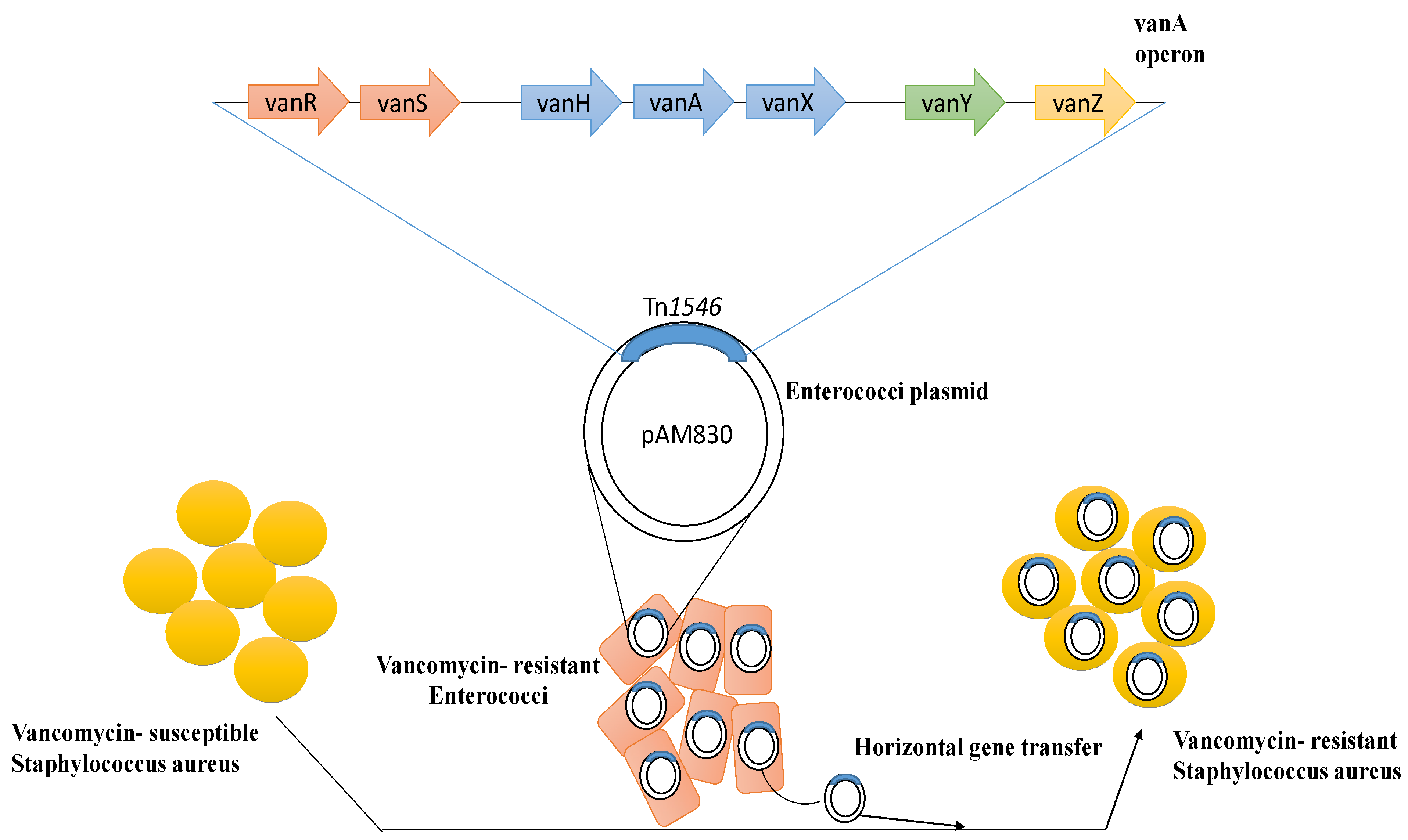
2.1. Vancomycin’s Mechanism of Action against S. aureus and the Emergence of Resistance
2.2. Interpretation Criteria for Vancomycin-Susceptible S. aureus
2.3. Selection Criteria
3. Vancomycin Non-Susceptible S. aureus in South Asia
3.1. Pakistan
3.2. India
3.3. Bangladesh
3.4. Sri Lanka
3.5. Nepal
3.6. Afghanistan

| Country | Study Period | Isolation Site | No. of Positive VRSA/Total | No. of Positive VISA/Total | No. of Positive hVISA/Total | Genetic Marker (vanA/vanB/icaA) | Molecular Typing | Reference |
|---|---|---|---|---|---|---|---|---|
| Pakistan | 2020 | Pus, skin wound, CSF | 113/200 | --- | --- | --- | --- | [69] |
| Pakistan | February 2017–March 2018 | Pus swabs from diabetic foot ulcers, wounds, breast abscesses | 565/6780 | 792/6780 | --- | --- | --- | [67] |
| Pakistan | January 2010–December 2010 | Pus, urine, blood, vaginal swab, and other secretions | 1/174 | 4/174 | --- | --- | --- | [66] |
| Pakistan | 2016 | Pus | 22/110 | 5/110 | --- | --- | --- | [70] |
| Pakistan | 2017–2018 | Blood, body fluid, pus, skin wound | 14/100 | --- | --- | vanA | --- | [101] |
| Pakistan | 2016 | Pus from ear, skin wound | 11/150 | --- | --- | vanA | --- | [64] |
| Pakistan | 2011 | Blood | 1/1 | --- | --- | vanA, icaA | --- | [65] |
| Pakistan | 2015 | Wound, pus swab | 5/51 | --- | --- | --- | --- | [102] |
| Nepal | Blood, urine, sputum, catheter swab, pus, and body fluids | 2/57 | 31/57 | --- | --- | --- | [103] | |
| Nepal | 2010 | Lacrimal swabs from chronic dacryocystitis | 22/27 | --- | --- | --- | --- | [92] |
| Nepal | November 2011–May 2012 | Urine, blood, and body fluids | --- | 4/45 | --- | --- | --- | [93] |
| India | January 1997–March 2000 | Clinical isolates | --- | --- | 5/80 | --- | --- | [76] |
| India | August 2002–July 2005 | Pus, urine, wound swabs, catheters, blood, sputum, and CSF | 2/783 | 6/783 | --- | 0 | --- | [104] |
| India | 2016 | Dental caries | 27/150 | --- | --- | 13 (vanA) 2(vanB) | --- | [105] |
| India | July 2010–September 2012 | Pus, urine, wound swabs, catheters, blood, and sputum | 7/115 | 53/115 | --- | --- | --- | [74] |
| India | March 2008–October 2008 | Blood, urine and throat swabs, wounds, and ear swabs | 7/358 | 16/358 | --- | 6 (vanA) | --- | [106] |
| India | May 2013–October 2013 | Clinical samples | 3/100 | 12/100 | 6/100 | --- | --- | [107] |
| India | July 2009–December 2012 | Surgical site infection | 0/267 | 3/267 | --- | --- | --- | [108] |
| India | January 2014–December 2016 | Clinical samples | --- | --- | 66/500 | --- | SCCmec III (8%), SCCmec IV (17.7%), SCCmec V (50%) | |
| India | July 2015–June 2016 | Pus, respiratory tract, urine, blood, body fluids, and catheter tips | --- | 18/266 | 15/266 | --- | NA | [75] |
| India | February 2019–March 2020 | Pus, tissue, blood, catheter | --- | --- | 14/220 | --- | --- | [80] |
| Sri Lanka | April 2002 | Surgical site infection | --- | 1/1 | --- | --- | --- | [87] |
| Bangladesh | August 2010–July 2011 | Clinical samples | 114/122 | --- | --- | --- | ||
| Bangladesh | January 2010–December 2011 | Clinical samples | 3/38 | --- | --- | --- | --- | [109] |
| Bangladesh | July 2011–June 2012 | Wound swabs | 2/15 | --- | --- | vanB | --- | [81] |
| Bangladesh | April 2012–January 2013 | Burn wounds | --- | 16/29 | --- | --- | --- | [19] |
4. Conclusions
Author Contributions
Funding
Institutional Review Board Statement
Informed Consent Statement
Data Availability Statement
Conflicts of Interest
References
- Hemmadi, V.; Biswas, M. An overview of moonlighting proteins in Staphylococcus aureus infection. Arch. Microbiol. 2020, 203, 481–498. [Google Scholar] [CrossRef]
- Lowy, F.D. Staphylococcus aureus infections. N. Engl. J. Med. 1998, 339, 2025–2027. [Google Scholar] [CrossRef]
- Abbasian, S.; Farahani, N.; Mir, Z.; Alinejad, F.; Haeili, M.; Dahmardehei, M.; Mirzaii, M.; Khoramrooz, S.; Nasiri, M.; Darban-Sarokhalil, D. Genotypic characterization of Staphylococcus aureus isolated from a burn centre by using agr, spa and SCCmec typing methods. New Microbes New Infect. 2018, 26, 15–19. [Google Scholar] [CrossRef]
- Mitchell, D.H.; Howden, B. Diagnosis and management of Staphylococcus aureus bacteraemia. Intern. Med. J. 2005, 35, S17–S24. [Google Scholar] [CrossRef]
- McGuinness, W.A.; Malachowa, N.; DeLeo, F.R. Focus: Infectious diseases: Vancomycin resistance in Staphylococcus aureus. Yale J. Biol. Med. 2017, 90, 269. [Google Scholar]
- Huttner, A.; Harbarth, S.; Carlet, J.; Cosgrove, S.; Goossens, H.; Holmes, A.; Jarlier, V.; Voss, A.; Pittet, D. Antimicrobial resistance: A global view from the 2013 World Healthcare-Associated Infections Forum. Antimicrob. Resist. Infect. Control 2013, 2, 31. [Google Scholar] [CrossRef]
- Bamigboye, B.T.; Olowe, O.A.; Taiwo, S.S. Phenotypic and molecular identification of vancomycin resistance in clinical Staphylococcus aureus isolates in Osogbo, Nigeria. Eur. J. Microbiol. Immunol. 2018, 8, 25–30. [Google Scholar] [CrossRef]
- Makgotlho, P.E.; Kock, M.; Hoosen, A.; Lekalakala, R.; Omar, S.V.; Dove, M.; Ehlers, M.M. Molecular identification and genotyping of MRSA isolates. FEMS Immunol. Med. Microbiol. 2009, 57, 104–115. [Google Scholar] [CrossRef]
- Chen, C.-J.; Huang, Y.-C. New epidemiology of Staphylococcus aureus infection in Asia. Clin. Microbiol. Infect. 2014, 20, 605–623. [Google Scholar] [CrossRef]
- Nelwan, E.J.; Andayani, D.; Clarissa, G.; Pramada, T. Vancomycin-resistant Staphylococcus aureus infection post-liposuction in South Korea. Cureus 2021, 13, e14357. [Google Scholar] [CrossRef]
- Hanaki, H.; Hososaka, Y.; Yanagisawa, C.; Nakae, T.; Sunakawa, K.; Otsuka, Y.; Nagasawa, Z. Occurrence of vancomycin-intermediate-resistant Staphylococcus aureus in Japan. J. Infect. Chemother. 2007, 13, 118–121. [Google Scholar] [CrossRef] [PubMed]
- Berglee, R. World Regional Geography: People, Places and Globalization; University of Minnesota Libraries Publishing: Minneapolis, MN, USA, 2012. [Google Scholar]
- Worldometer. Southern Asia Population. 4 April 2021. Available online: https://www.worldometers.info/world-population/southern-asia-population/ (accessed on 4 April 2022).
- Zaidi, S.; Saligram, P.; Ahmed, S.; Sonderp, E.; Sheikh, K. Expanding access to healthcare in South Asia. Br. Med. J. 2017, 357, j1645. [Google Scholar] [CrossRef] [PubMed]
- Cong, Y.; Yang, S.; Rao, X. Vancomycin resistant Staphylococcus aureus infections: A review of case updating and clinical features. J. Adv. Res. 2020, 21, 169–176. [Google Scholar] [CrossRef]
- Saha, B.; Singh, A.K.; Ghosh, A.; Bal, M. Identification and characterization of a vancomycin-resistant Staphylococcus aureus isolated from Kolkata (South Asia). J. Med. Microbiol. 2008, 57, 72–79. [Google Scholar] [CrossRef] [PubMed]
- Wu, Q.; Sabokroo, N.; Wang, Y.; Hashemian, M.; Karamollahi, S.; Kouhsari, E. Systematic review and meta-analysis of the epidemiology of vancomycin-resistance Staphylococcus aureus isolates. Antimicrob. Resist. Infect. Control 2021, 10, 101. [Google Scholar] [CrossRef] [PubMed]
- Riaz, S.; Hussain, A.; Sohail, M.; Rehman, S.U.; Javed, N.; Abbas, Z. Isolation and characterization of Vancomycin-Resistant Staphylococcus aureus (VRSA) from Intensive Care Units (ICU) of different hospitals in Lahore, Pakistan. Adv. Life Sci. 2021, 8, 339–344. [Google Scholar]
- Hasan, R.; Acharjee, M.; Noor, R. Prevalence of Vancomycin-Resistant Staphylococcus aureus (VRSA) in Methicillin-Resistant S. aureus (MRSA) strains isolated from burn wound infections. Tzu Chi Med. J. 2016, 28, 49–53. [Google Scholar] [CrossRef]
- Shariati, A.; Dadashi, M.; Moghadam, M.T.; van Belkum, A.; Yaslianifard, S.; Darban-Sarokhalil, D. Global prevalence and distribution of vancomycin resistant, vancomycin intermediate and heterogeneously vancomycin intermediate Staphylococcus aureus clinical isolates: A systematic review and meta-analysis. Sci. Rep. 2020, 10, 12698. [Google Scholar] [CrossRef]
- Stepanović, S.; Dakić, I.; Djukić, S.; Lozuk, B.; Svabić-Vlahović, M. Surgical wound infection associated with Staphylococcus sciuri. Scand. J. Infect. Dis. 2002, 34, 685–686. [Google Scholar] [CrossRef]
- Gill, S.R.; Fouts, D.E.; Archer, G.L.; Mongodin, E.F.; DeBoy, R.T.; Ravel, J.; Paulsen, I.T.; Kolonay, J.F.; Brinkac, L.; Beanan, M.; et al. Insights on evolution of virulence and resistance from the complete genome analysis of an early methicillin-resistant Staphylococcus aureus strain and a biofilm-producing methicillin-resistant Staphylococcus epidermidis strain. J. Bacteriol. 2005, 187, 2426–2438. [Google Scholar] [CrossRef]
- Chambers, H.F.; DeLeo, F.R. Waves of resistance: Staphylococcus aureus in the antibiotic era. Nat. Rev. Microbiol. 2009, 7, 629–641. [Google Scholar] [CrossRef] [PubMed]
- Rammelkamp, C.H.; Maxon, T. Resistance of Staphylococcus aureus to the action of penicillin. Proc. Soc. Exp. Biol. Med. 1942, 51, 386–389. [Google Scholar] [CrossRef]
- Olsen, J.E.; Christensen, H.; Aarestrup, F.M. Diversity and evolution of blaZ from Staphylococcus aureus and coagulase-negative staphylococci. J. Antimicrob. Chemother. 2006, 57, 450–460. [Google Scholar] [CrossRef] [PubMed]
- Jevons, M.P. “Celbenin”-resistant staphylococci. Br. Med. J. 1961, 1, 124. [Google Scholar] [CrossRef]
- Baede, V.O.; David, M.Z.; Andrasevic, A.T.; Blanc, D.S.; Borg, M.; Brennan, G.; Catry, B.; Chabaud, A.; Empel, J.; Enger, H.; et al. MRSA surveillance programmes worldwide: Moving towards a harmonised international approach. Int. J. Antimicrob. Agents 2022, 59, 106538. [Google Scholar] [CrossRef] [PubMed]
- Klein, E.; Smith, D.L.; Laxminarayan, R. Hospitalizations and deaths caused by methicillin-resistant Staphylococcus aureus, United States, 1999–2005. Emerg. Infect. Dis. 2007, 13, 1840. [Google Scholar] [CrossRef]
- Tang, J.; Hu, J.; Kang, L.; Deng, Z.; Wu, J.; Pan, J. The use of vancomycin in the treatment of adult patients with methicillin-resistant Staphylococcus aureus (MRSA) infection: A survey in a tertiary hospital in China. Int. J. Clin. Exp. Med. 2015, 8, 19436. [Google Scholar]
- Klevens, R.M.; Morrison, M.A.; Nadle, J.; Petit, S.; Gershman, K.; Ray, S.; Harrison, L.H.; Lynfield, R.; Dumyati, G.; Townes, J.M.; et al. Invasive methicillin-resistant Staphylococcus aureus infections in the United States. J. Am. Med. Assoc. 2007, 298, 1763–1771. [Google Scholar] [CrossRef] [PubMed]
- Tsuzuki, S.; Matsunaga, N.; Yahara, K.; Gu, Y.; Hayakawa, K.; Hirabayashi, A.; Kajihara, T.; Sugai, M.; Shibayama, K.; Ohmagari, N. National trend of blood-stream infection attributable deaths caused by Staphylococcus aureus and Escherichia coli in Japan. J. Infect. Chemother. 2020, 26, 367–371. [Google Scholar] [CrossRef]
- Ruhe, J.J.; Smith, N.; Bradsher, R.W.; Menon, A. Community-onset methicillin-resistant Staphylococcus aureus skin and soft-tissue infections: Impact of antimicrobial therapy on outcome. Clin. Infect. Dis. 2007, 44, 777–784. [Google Scholar] [CrossRef]
- Foster, T.J.; Geoghegan, J.A. Staphylococcus aureus. In Molecular Medical Microbiology, 4th ed.; Baron, S., Ed.; University of Texas Medical Branch: Galveston, TX, USA, 2015; pp. 655–674. [Google Scholar]
- Japoni, A.; Jamalidoust, M.; Farshad, S.; Ziyaeyan, M.; Alborzi, A.; Japoni, S.; Rafaatpour, N. Characterization of SCCmec types and antibacterial susceptibility patterns of methicillin-resistant Staphylococcus aureus in Southern Iran. Jpn. J. Infect. Dis. 2011, 64, 28–33. [Google Scholar] [CrossRef] [PubMed]
- Liu, J.; Chen, D.; Peters, B.M.; Li, L.; Li, B.; Xu, Z.; Shirliff, M.E. Staphylococcal chromosomal cassettes mec (SCCmec): A mobile genetic element in methicillin-resistant Staphylococcus aureus. Microb. Pathog. 2016, 101, 56–67. [Google Scholar] [CrossRef] [PubMed]
- Foster, T.J. Antibiotic resistance in Staphylococcus aureus. Current status and future prospects. FEMS Microbiol. Rev. 2017, 41, 430–449. [Google Scholar] [CrossRef]
- Chongtrakool, P.; Ito, T.; Ma, X.X.; Kondo, Y.; Trakulsomboon, S.; Tiensasitorn, C.; Hiramatsu, K. Staphylococcal cassette chromosome mec (SCCmec) typing of methicillin-resistant Staphylococcus aureus strains isolated in 11 Asian countries: A proposal for a new nomenclature for SCCmec elements. Antimicrob. Agents Chemother. 2006, 50, 1001–1012. [Google Scholar] [CrossRef]
- Peng, H.; Liu, D.; Ma, Y.; Gao, W. Comparison of community- and healthcare-associated methicillin-resistant Staphylococcus aureus isolates at a Chinese tertiary hospital, 2012–2017. Sci. Rep. 2018, 8, 17916. [Google Scholar] [CrossRef] [PubMed]
- Kateete, D.P.; Bwanga, F.; Seni, J.; Mayanja, R.; Kigozi, E.; Mujuni, B.; Ashaba, F.K.; Baluku, H.; Najjuka, C.F.; Källander, K.; et al. CA-MRSA and HA-MRSA coexist in community and hospital settings in Uganda. Antimicrob. Resist. Infect. Control 2019, 8, 94. [Google Scholar] [CrossRef] [PubMed]
- Hu, Q.; Cheng, H.; Yuan, W.; Zeng, F.; Shang, W.; Tang, D.; Xue, W.; Fu, J.; Zhou, R.; Zhu, J.; et al. Panton-Valentine Leukocidin (PVL)-positive health care-associated methicillin-resistant Staphylococcus aureus isolates are associated with skin and soft tissue infections and colonized mainly by infective PVL-encoding bacteriophages. J. Clin. Microbiol. 2015, 53, 67–72. [Google Scholar] [CrossRef]
- Rehm, S.J.; Tice, A. Staphylococcus aureus: Methicillin-susceptible S. aureus to methicillin-resistant S. aureus and vancomycin-resistant S. aureus. Clin. Infect. Dis. 2010, 51 (Suppl. S2), S176–S182. [Google Scholar]
- Hiramatsu, K.; Hanaki, H.; Ino, T.; Yabuta, K.; Oguri, T.; Tenover, F.C. Methicillin-resistant Staphylococcus aureus clinical strain with reduced vancomycin susceptibility. J. Antimicrob. Chemother. 1997, 40, 135–136. [Google Scholar] [CrossRef]
- Goldrick, B. First reported case of VRSA in the United States: An alarming development in microbial resistance. AJN Am. J. Nurs. 2002, 102, 17. [Google Scholar] [CrossRef]
- Centers for Disease Control and Prevention. Update: Staphylococcus aureus with reduced susceptibility to vancomycin—United States, 1997. MMWR. Morb. Mortal. Wkly. Rep. 1997, 46, 813–815.
- Hallin, M.; Friedrich, A.W.; Struelens, M.J. spa typing for epidemiological surveillance of Staphylococcus aureus. Mol. Epidemiol. Microorg. Methods Protocols 2009, 551, 189–202. [Google Scholar]
- Larsen, M.V.; Cosentino, S.; Rasmussen, S.; Friis, C.; Hasman, H.; Marvig, R.L.; Jelsbak, L.; Sicheritz-Pontéen, T.; Ussery, D.W.; Aarestrup, F.M.; et al. Multilocus sequence typing of total-genome-sequenced bacteria. J. Clin. Microbiol. 2012, 50, 1355–1361. [Google Scholar] [CrossRef]
- Shekarabi, M.; Hajikhani, B.; Chirani, A.S.; Fazeli, M.; Goudarzi, M. Molecular characterization of vancomycin-resistant Staphylococcus aureus strains isolated from clinical samples: A three-year study in Tehran, Iran. PLoS ONE 2017, 12, e0183607. [Google Scholar] [CrossRef]
- Monecke, S.; Coombs, G.; Shore, A.C.; Coleman, D.C.; Akpaka, P.; Borg, M.; Chow, H.; Ip, M.; Jatzwauk, L.; Jonas, D.; et al. A Field Guide to Pandemic, Epidemic and sporadic clones of methicillin-resistant Staphylococcus aureus. PLoS ONE 2011, 6, e17936. [Google Scholar]
- Bozdogan, B.; Ednie, L.; Credito, K.; Kosowska, K.; Appelbaum, P.C. Derivatives of a vancomycin-resistant Staphylococcus aureus strain isolated at Hershey Medical Center. Antimicrob. Agents Chemother. 2004, 48, 4762–4765. [Google Scholar] [CrossRef] [PubMed]
- Howden, B.P.; Seemann, T.; Harrison, P.F.; McEvoy, C.R.; Stanton JA, L.; Rand, C.J.; Stinear, T.P. Complete genome sequence of Staphylococcus aureus strain JKD6008, an ST239 clone of methicillin-resistant Staphylococcus aureus with intermediate-level vancomycin resistance. J. Bacteriol. 2010, 192, 5848–5849. [Google Scholar] [CrossRef] [PubMed]
- Howden, B.P.; Davies, J.K.; Johnson, P.D.R.; Stinear, T.P.; Grayson, M.L. Reduced vancomycin susceptibility in Staphylococcus aureus, including vancomycin-intermediate and heterogeneous vancomycin-intermediate strains: Resistance mechanisms, laboratory detection, and clinical implications. Clin. Microbiol. Rev. 2010, 23, 99–139. [Google Scholar] [CrossRef]
- Arthur, M.; Molinas, C.; Depardieu, F.; Courvalin, P. Characterization of Tn1546, a Tn3-related transposon conferring glycopeptide resistance by synthesis of depsipeptide peptidoglycan precursors in Enterococcus faecium BM4147. J. Bacteriol. 1993, 175, 117–127. [Google Scholar] [CrossRef] [PubMed]
- Périchon, B.; Courvalin, P. VanA-type vancomycin-resistant Staphylococcus aureus. Antimicrob. Agents Chemother. 2009, 53, 4580–4587. [Google Scholar] [CrossRef] [PubMed]
- Gilmore, M.S.; Clewell, D.B.; Ike, Y.; Shankar, N. Enterococci: From Commensals to Leading Causes of Drug Resistant Infection [Internet]; Massachusetts Eye and Ear Infirmary: Boston, MA, USA, 2014. [Google Scholar]
- Weinstein, R.A.; Fridkin, S.K. Vancomycin-intermediate and -resistant Staphylococcus aureus: What the infectious disease specialist needs to know. Clin. Infect. Dis. 2001, 32, 108–115. [Google Scholar] [CrossRef] [PubMed]
- Pillai, S.K.; Wennersten, C.; Venkataraman, L.; Eliopoulos, G.M.; Moellering, J.R.C.; Karchmer, A.W. Development of reduced vancomycin susceptibility in methicillin-susceptible Staphylococcus aureus. Clin. Infect. Dis. 2009, 49, 1169–1174. [Google Scholar] [CrossRef] [PubMed]
- Gardete, S.; Kim, C.; Hartmann, B.M.; Mwangi, M.; Roux, C.M.; Dunman, P.M.; Chambers, H.F.; Tomasz, A. Genetic pathway in acquisition and loss of vancomycin resistance in a Methicillin Resistant Staphylococcus aureus (MRSA) strain of clonal type USA300. PLoS Pathog. 2012, 8, e1002505. [Google Scholar]
- Wang, W.-Y.; Lee, S.-Y.; Chiueh, T.-S.; Lu, J.-J. Molecular and phenotypic characteristics of methicillin-resistant and vancomycin-intermediate Staphylococcus aureus isolates from patients with septic arthritis. J. Clin. Microbiol. 2009, 47, 3617–3623. [Google Scholar]
- Holmes, N.E.; Turnidge, J.D.; Munckhof, W.J.; Robinson, J.O.; Korman, T.; O’Sullivan, M.; Anderson, T.L.; Roberts, S.A.; Gao, W.; Christiansen, K.J.; et al. Antibiotic choice may not explain poorer outcomes in patients with Staphylococcus aureus bacteremia and high vancomycin minimum inhibitory concentrations. J. Infect. Dis. 2011, 204, 340–347. [Google Scholar] [CrossRef] [PubMed]
- Haripur District Demographics. 17 May 2022. Available online: https://kp.gov.pk/page/haripurdistrictdemographics (accessed on 17 May 2022).
- Wang, G.; Hindler, J.F.; Ward, K.W.; Bruckner, D.A. Increased vancomycin MICs for Staphylococcus aureus clinical isolates from a university hospital during a 5-Year Period. J. Clin. Microbiol. 2006, 44, 3883–3886. [Google Scholar] [CrossRef]
- Tenover, F.C.; Moellering, R.C., Jr. The rationale for revising the Clinical and Laboratory Standards Institute vancomycin minimal inhibitory concentration interpretive criteria for Staphylococcus aureus. Clin. Infect. Dis. 2007, 44, 1208–1215. [Google Scholar] [CrossRef] [PubMed]
- Edition, A.S.N. CLSI Document M07-A9; Clinical and Laboratory Standards Institute: Wayne, PA, USA, 2012. [Google Scholar]
- Azhar, A.; Rasool, S.; Haque, A.; Shan, S.; Saeed, M.; Ehsan, B.; Haque, A. Detection of high levels of resistance to linezolid and vancomycin in Staphylococcus aureus. J. Med. Microbiol. 2017, 66, 1328–1331. [Google Scholar] [CrossRef]
- Mirani, Z.A.; Jamil, N. Effect of sub-lethal doses of vancomycin and oxacillin on biofilm formation by vancomycin intermediate resistant Staphylococcus aureus. J. Basic Microbiol. 2011, 51, 191–195. [Google Scholar] [CrossRef]
- Taj, Y.; Abdullah, F.E.; Kazmi, S.U. Current pattern of antibiotic resistance in Staphylococcus aureus clinical isolates and the emergence of vancomycin resistance. J. Coll. Physicians Surg. Pak. 2010, 20, 728–732. [Google Scholar]
- Ahmad, S.; Ahmed, S.; Sabir, M.; Khan, H.; Rehman, M.; Niaz, Z. Frequency and comparison among antibiotic resistant Staphylococcus aureus strains in selected hospitals of Peshawar, Pakistan. J. Pak. Med. Assoc. 2020, 70, 1199–1202. [Google Scholar] [CrossRef]
- Hakim, S.T.; Arshed, S.; Iqbal, M.; Javaid, S.G. Vancomycin sensitivity of Staphylococcus aureus isolates from hospital patients in Karachi, Pakistan. Libyan J. Med. 2007, 2, 176–179. [Google Scholar] [CrossRef] [PubMed]
- Saeed, A.; Ahsan, F.; Nawaz, M.; Iqbal, K.; Rehman, K.U.; Ijaz, T. Incidence of vancomycin resistant phenotype of the Methicillin Resistant Staphylococcus aureus isolated from a tertiary care hospital in Lahore. Antibiotics 2019, 9, 3. [Google Scholar] [CrossRef] [PubMed]
- Ghias, W.; Sharif, M.; Yazdani, F.A.; Rabbani, M. Isolation and identification of methicillin and vancomycin resistance Staphylococcus aureus from pus samples of injured skin patients in Lahore, Pakistan. Biomed. Lett. 2016, 2, 103–112. [Google Scholar]
- Hanif, E.; Hassan, S.A. Evaluation of antibiotic resistance pattern in clinical isolates of Staphylococcus aureus. Pak. J. Pharm. Sci. 2019, 32, 1749–1753. [Google Scholar] [PubMed]
- Banerjee, T.; Anupurba, S. Colonization with vancomycin-intermediate Staphylococcus aureus strains containing the vanA resistance gene in a tertiary-care center in North India. J. Clin. Microbiol. 2012, 50, 1730–1732. [Google Scholar] [CrossRef] [PubMed]
- Goud, R.; Gupta, S.; Neogi, U.; Agarwal, D.; Naidu, K.; Chalannavar, R.; Subhaschandra, G. Community prevalence of methicillin and vancomycin resistant Staphylococcus aureus in and around Bangalore, southern India. Rev. da Soc. Bras. de Med. Trop. 2011, 44, 309–312. [Google Scholar] [CrossRef]
- Moses, V.K.; Kandi, V.; Rao, S.K.D. Minimum inhibitory concentrations of vancomycin and daptomycin against methicillin-resistant Staphylococcus aureus isolated from various clinical specimens: A study from South India. Cureus 2020, 12, e6749. [Google Scholar] [CrossRef]
- Mohanty, S.; Behera, B.; Sahu, S.; Praharaj, A.K. Recent pattern of antibiotic resistance in Staphylococcus aureus clinical isolates in Eastern India and the emergence of reduced susceptibility to vancomycin. J. Lab. Physicians 2019, 11, 340–345. [Google Scholar] [CrossRef] [PubMed]
- Song, J.-H.; Hiramatsu, K.; Suh, J.Y.; Ko, K.S.; Ito, T.; Kapi, M.; Kiem, S.; Kim, Y.-S.; Oh, W.S.; Peck, K.R.; et al. Emergence in Asian countries of Staphylococcus aureus with reduced susceptibility to vancomycin. Antimicrob. Agents Chemother. 2004, 48, 4926–4928. [Google Scholar] [CrossRef]
- Chung, D.R.; Lee, C.; Kang, Y.R.; Baek, J.Y.; Kim, S.H.; Ha, Y.E.; Kang, C.-I.; Peck, K.R.; Lee, N.Y.; Song, J.-H. Genotype-specific prevalence of heterogeneous vancomycin-intermediate Staphylococcus aureus in Asian countries. Int. J. Antimicrob. Agents 2015, 46, 338–341. [Google Scholar] [CrossRef]
- Sistla, S.; Amberpet, R.; Sugumar, M.; Nagasundaram, N.; Manoharan, M.; Parija, S.C. Detection of heterogeneous vancomycin-intermediate Staphylococcus aureus: A preliminary report from south India. Indian J. Med. Res. 2019, 150, 194–198. [Google Scholar] [CrossRef]
- Selvabai, A.P.; Sattar, S.B.A.; Jayaraman, P.; Shanmugam, P. Detection and characterisation of heteroresistant Vancomycin Intermediate Staphylococcus aureus (hVISA) using phenotypic and genotypic methods. J. Clin. Diagn. Res. 2019, 13, 1–5. [Google Scholar] [CrossRef]
- Sreejisha, M.; Mulki, S.S.; Shenoy, S.; Dhanashree, B.; Chakrapani, M.; Bhat, G. Heterogeneous vancomycin intermediate Staphylococcus aureus infections in diabetic and non-diabetic patients–A hospital-based comparative study. Infect. Drug Resist. 2023, 16, 9–17. [Google Scholar]
- Islam, T.A.B.; Shamsuzzaman, S. Prevalence and antimicrobial susceptibility pattern of methicillin-resistant, vancomycin-resistant, and Panton-Valentine Leukocidin positive Staphylococcus aureus in a tertiary care hospital Dhaka, Bangladesh. Tzu Chi Med. J. 2015, 27, 10–14. [Google Scholar] [CrossRef]
- Shahriar, M.; Shahid, S.; Katha, K.K.; Nasreen, W.; Bhuiyan, M.A. Vancomycin sensitivity of clinical isolates of Staphylococcus aureus from patients in Dhaka City, Bangladesh. Bangladesh Pharm. J. 2012, 15, 159–163. [Google Scholar] [CrossRef]
- Haque, N.; Aung, M.S.; Paul, S.K.; Bari, S.; Ahmed, S.; Sarkar, S.R.; Roy, S.; Nasreen, S.A.; Mahmud, M.C.; Hossain, M.A.; et al. Molecular epidemiological characterization of methicillin-susceptible and -resistant Staphylococcus aureus isolated from skin and soft tissue infections in Bangladesh. Microb. Drug Resist. 2019, 25, 241–250. [Google Scholar] [CrossRef]
- Taz, K.A.; Jobayer, M.; Shamsuzzaman, S.M. Nasal colonization of methicillin resistant Staphylococcus aureus among healthcare providers in a tertiary care hospital, Bangladesh. Mymensingh Med. J. MMJ 2019, 28, 627–633. [Google Scholar] [PubMed]
- Roy, S.; Barman, T.K.; Hossain, M.A.; Paul, S.K.; Haque, N.; Ahmed, S.; Nasreen, S.A.; Hossain, M.S.; Sarkar, S.R.; Kubayashi, N.; et al. Molecular-characterization of Methicillin-Resistance Staphylococcus aureus (MRSA) from different tertiary care hospitals in Bangladesh. Mymensingh Med. J. 2017, 26, 37–44. [Google Scholar]
- Parvez, M.A.K.; Ferdous, R.N.; Rahman, M.S.; Islam, S. Healthcare-associated (HA) and community-associated (CA) methicillin resistant Staphylococcus aureus (MRSA) in Bangladesh–Source, diagnosis and treatment. J. Genet. Eng. Biotechnol. 2018, 16, 473–478. [Google Scholar] [CrossRef] [PubMed]
- Song, J.-H.; Hsueh, P.-R.; Chung, D.R.; Ko, K.S.; Kang, C.-I.; Peck, K.R.; Yeom, J.-S.; Kim, S.-W.; Chang, H.-H.; Kim, Y.-S.; et al. Spread of methicillin-resistant Staphylococcus aureus between the community and the hospitals in Asian countries: An ANSORP study. J. Antimicrob. Chemother. 2011, 66, 1061–1069. [Google Scholar] [CrossRef] [PubMed]
- Jayaweera, J.; Karunarathne, M.; Kumbukgolla, W.W. The importance of timely introduction of vancomycin therapy against methicillin-resistant Staphylococcus aureus (MRSA) bacteremia and severity of MRSA bacteremia at teaching hospital, Anuradhapura, Sri Lanka. Int J. One Health 2017, 3, 7–11. [Google Scholar] [CrossRef]
- Samaranayake, W.; Karunanayake, L.; Patabendige, C. Characteristics of community acquired and hospital acquired methicillin resistant Staphylococcus aureus isolates in the National Hospital of Sri Lanka. Sri Lankan J. Infect. Dis. 2019, 9, 24. [Google Scholar] [CrossRef]
- McTavish, S.M.; Snow, S.J.; Cook, E.C.; Pichon, B.; Coleman, S.; Coombs, G.W.; Pang, S.; Arias, C.A.; Díaz, L.; Boldock, E.; et al. Genomic and epidemiological evidence of a dominant Panton-Valentine Leucocidin-positive methicillin resistant Staphylococcus aureus lineage in Sri Lanka and presence among isolates from the United Kingdom and Australia. Front. Cell. Infect. Microbiol. 2019, 9, 123. [Google Scholar] [CrossRef] [PubMed]
- Wijitha Weerakoon, S.D.A.; Gamage, D.S.; Wijeratne, M. Staphylococcus aureus with reduced susceptibility to vancomycin. Ceylon Med. J. 2003, 48, 58–59. [Google Scholar] [CrossRef]
- Chaudhary, M.; Bhattarai, A.; Adhikari, S.K.; Bhatta, D.R. Bacteriology and antimicrobial susceptibility of adult chronic dacryocystitis. Nepal. J. Ophthalmol. 2010, 2, 105–113. [Google Scholar] [CrossRef] [PubMed]
- Pahadi, P.C.; Shrestha, U.T.; Adhikari, N.; Shah, P.K.; Amatya, R. Growing resistance to vancomycin among methicillin resistant Staphylococcus aureus isolates from different clinical samples. J. Nepal Med. Assoc. 2014, 52, 977–981. [Google Scholar] [CrossRef]
- Raut, S.; Bajracharya, K.; Adhikari, J.; Pant, S.S.; Adhikari, B. Prevalence of methicillin resistant Staphylococcus aureus in Lumbini Medical College and Teaching Hospital, Palpa, Western Nepal. BMC Res. Notes 2017, 10, 187. [Google Scholar] [CrossRef]
- Shrestha, B.; Pokhrel, B.M.; Mohapatra, T.M. Phenotypic characterization of nosocomial isolates of Staphylococcus aureus with reference to MRSA. J. Infect. Dev. Ctries. 2009, 3, 554–560. [Google Scholar] [CrossRef]
- Kshetry, A.O.; Pant, N.D.; Bhandari, R.; Khatri, S.; Shrestha, K.L.; Upadhaya, S.K.; Poudel, A.; Lekhak, B.; Raghubanshi, B.R. Minimum inhibitory concentration of vancomycin to methicillin resistant Staphylococcus aureus isolated from different clinical samples at a tertiary care hospital in Nepal. Antimicrob. Resist. Infect. Control 2016, 5, 27. [Google Scholar] [CrossRef]
- Maharjan, M.; Sah, A.K.; Pyakurel, S.; Thapa, S.; Maharjan, S.; Adhikari, N.; Rijal, K.R.; Ghimire, P.; Shrestha, U.T. Molecular Confirmation of vancomycin-resistant Staphylococcus aureus with vanA gene from a hospital in Kathmandu. Int. J. Microbiol. 2021, 2021, 3847347. [Google Scholar] [CrossRef]
- Naimi, H.M.; Rasekh, H.; Noori, A.Z.; Bahaduri, M.A. Determination of antimicrobial susceptibility patterns in Staphylococcus aureus strains recovered from patients at two main health facilities in Kabul, Afghanistan. BMC Infect. Dis. 2017, 17, 737. [Google Scholar] [CrossRef]
- Naimi, H.M.; André, C.; Bes, M.; Tristan, A.; Gustave, C.-A.; Vandenesch, F.; Nazari, Q.A.; Laurent, F.; Dupieux, C. Antibiotic resistance profile and molecular characterization of Staphylococcus aureus strains isolated in hospitals in Kabul, Afghanistan. Eur. J. Clin. Microbiol. Infect. Dis. 2021, 40, 1029–1038. [Google Scholar] [CrossRef] [PubMed]
- Calhoun, J.H.; Murray, C.K.; Manring, M.M. Multidrug-resistant organisms in military wounds from Iraq and Afghanistan. Clin. Orthop. Relat. Res. 2008, 466, 1356–1362. [Google Scholar] [CrossRef] [PubMed]
- Liaqat, F.; Sheikh, A.A.; Nazir, J.; Hussain, T.; Rabbani, M.; Shaheen, A.Y.; Muhammad, J. Report-Isolation, identification and control of vancomycin resistant Staphylococcus aureus. Pak. J. Pharm. Sci. 2015, 28, 997–1004. [Google Scholar]
- Lama, U.; Shah, D.; Shrestha, U.T. Vancomycin resistant Staphylococcus aureus reported from tertiary care hospital in Nepal. Tribhuvan Univ. J. Microbiol. 2017, 4, 63–72. [Google Scholar] [CrossRef][Green Version]
- Tiwari, H.K.; Sen, M.R. Emergence of vancomycin resistant Staphylococcus aureus (VRSA) from a tertiary care hospital from northern part of India. BMC Infect. Dis. 2006, 6, 156. [Google Scholar] [CrossRef] [PubMed]
- Vellappally, S.; Divakar, D.D.; Al Kheraif, A.A.; Ramakrishnaiah, R.; Alqahtani, A.; Dalati, M.H.N.; Anil, S.; Khan, A.A.; Varma, P.R.H. Occurrence of vancomycin-resistant Staphylococcus aureus in the oral cavity of patients with dental caries. Acta Microbiol. Immunol. Hung. 2017, 64, 343–351. [Google Scholar] [CrossRef] [PubMed]
- Gaddad, S.M.; Thati, V.; Shivannavar, C.T. Vancomycin resistance among methicillin resistant Staphylococcus aureus isolates from intensive care units of tertiary care hospitals in Hyderabad. Indian J. Med. Res. 2011, 134, 704. [Google Scholar] [CrossRef]
- Bhattacharya, S.; Bir, R.; Majumdar, T. Evaluation of multidrug resistant Staphylococcus aureus and their association with biofilm production in a tertiary care hospital, Tripura, Northeast India. J. Clin. Diagn. Res. JCDR 2015, 9, DC01. [Google Scholar] [CrossRef]
- Bhattacharya, S.; Pal, K.; Jain, S.; Chatterjee, S.S.; Konar, J. Surgical site infection by methicillin resistant Staphylococcus aureus–On decline? J. Clin. Diagn. Res. JCDR 2016, 10, DC32. [Google Scholar] [CrossRef]
- Khanam, S.; Haq, J.A.; Shamsuzzaman, S.; Rahman, M.; Mamun, K.Z. Emergence of vancomycin resistant Staphylococcus aureus during hospital admission at a tertiary care hospital in Bangladesh. Bangladesh J. Infect. Dis. 2016, 3, 11–16. [Google Scholar] [CrossRef]
- Rehman, T.U.; Aslam, R.; Aqib, A.I.; Mohsin, M.; Manzoor, A.; Shoaib, M.; Naseer, M.A.; Hassan, A.; Sattar, H.; Kulyar, M.F.-E.; et al. Phylogeny of hospital acquired MRSA, and its comparative phenotypic clinico-epidemiology with vancomycin resistant S. aureus (VRSA). Microb. Pathog. 2020, 149, 104537. [Google Scholar] [CrossRef] [PubMed]

Disclaimer/Publisher’s Note: The statements, opinions and data contained in all publications are solely those of the individual author(s) and contributor(s) and not of MDPI and/or the editor(s). MDPI and/or the editor(s) disclaim responsibility for any injury to people or property resulting from any ideas, methods, instructions or products referred to in the content. |
© 2023 by the authors. Licensee MDPI, Basel, Switzerland. This article is an open access article distributed under the terms and conditions of the Creative Commons Attribution (CC BY) license (https://creativecommons.org/licenses/by/4.0/).
Share and Cite
Ejaz, M.; Syed, M.A.; Jackson, C.R.; Sharif, M.; Faryal, R. Epidemiology of Staphylococcus aureus Non-Susceptible to Vancomycin in South Asia. Antibiotics 2023, 12, 972. https://doi.org/10.3390/antibiotics12060972
Ejaz M, Syed MA, Jackson CR, Sharif M, Faryal R. Epidemiology of Staphylococcus aureus Non-Susceptible to Vancomycin in South Asia. Antibiotics. 2023; 12(6):972. https://doi.org/10.3390/antibiotics12060972
Chicago/Turabian StyleEjaz, Mohammad, Muhammad Ali Syed, Charlene R. Jackson, Mehmoona Sharif, and Rani Faryal. 2023. "Epidemiology of Staphylococcus aureus Non-Susceptible to Vancomycin in South Asia" Antibiotics 12, no. 6: 972. https://doi.org/10.3390/antibiotics12060972
APA StyleEjaz, M., Syed, M. A., Jackson, C. R., Sharif, M., & Faryal, R. (2023). Epidemiology of Staphylococcus aureus Non-Susceptible to Vancomycin in South Asia. Antibiotics, 12(6), 972. https://doi.org/10.3390/antibiotics12060972