
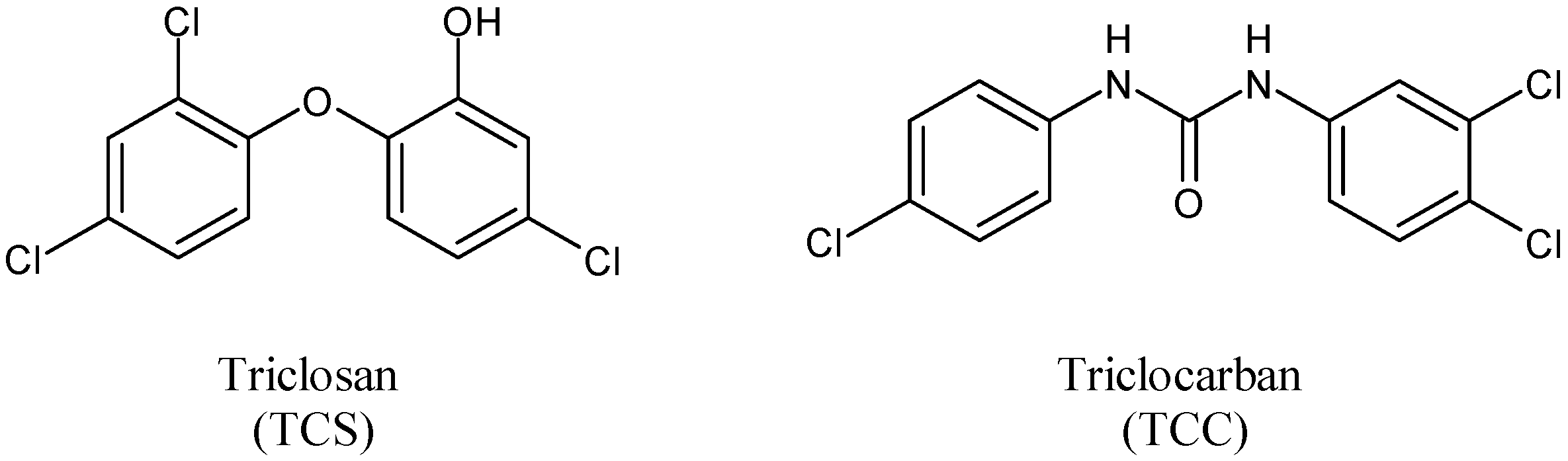
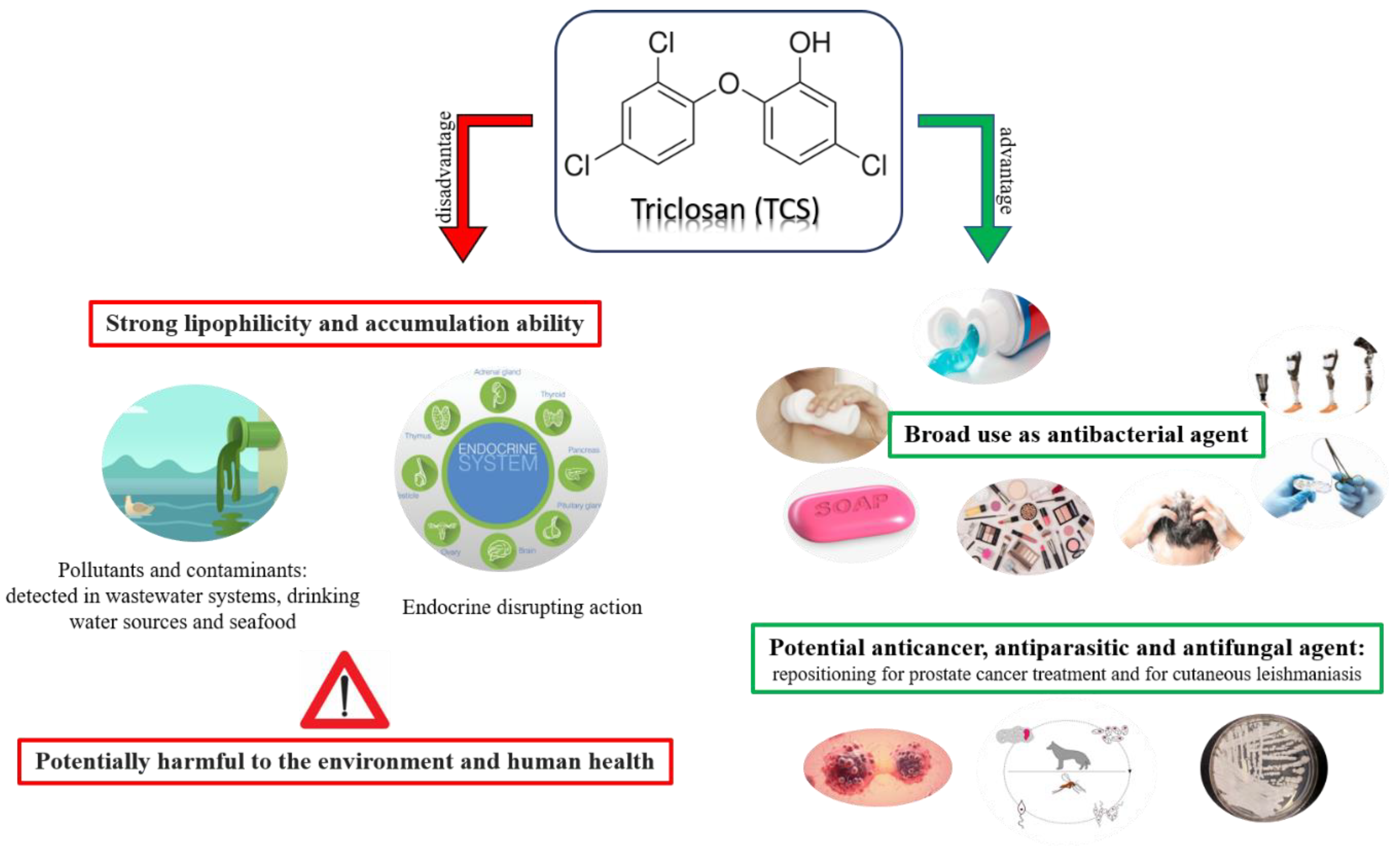
Triclosan: A Small Molecule with Controversial Roles

 ,
,  ,
,  ,
,  ,
, 
 ,
,  and
and 
Abstract
1. Introduction
2. Pharmacological Activities of TCS
2.1. Antimicrobial Activity
2.1.1. Antibacterial and Antifungal Activity
2.1.2. Antibiofilm Activity
2.1.3. Antiparasitic Activity
3. Studies on Toothpastes Containing TCS
4. Absorption, Distribution, Metabolism, and Excretion (ADME) Properties and Photodegradation
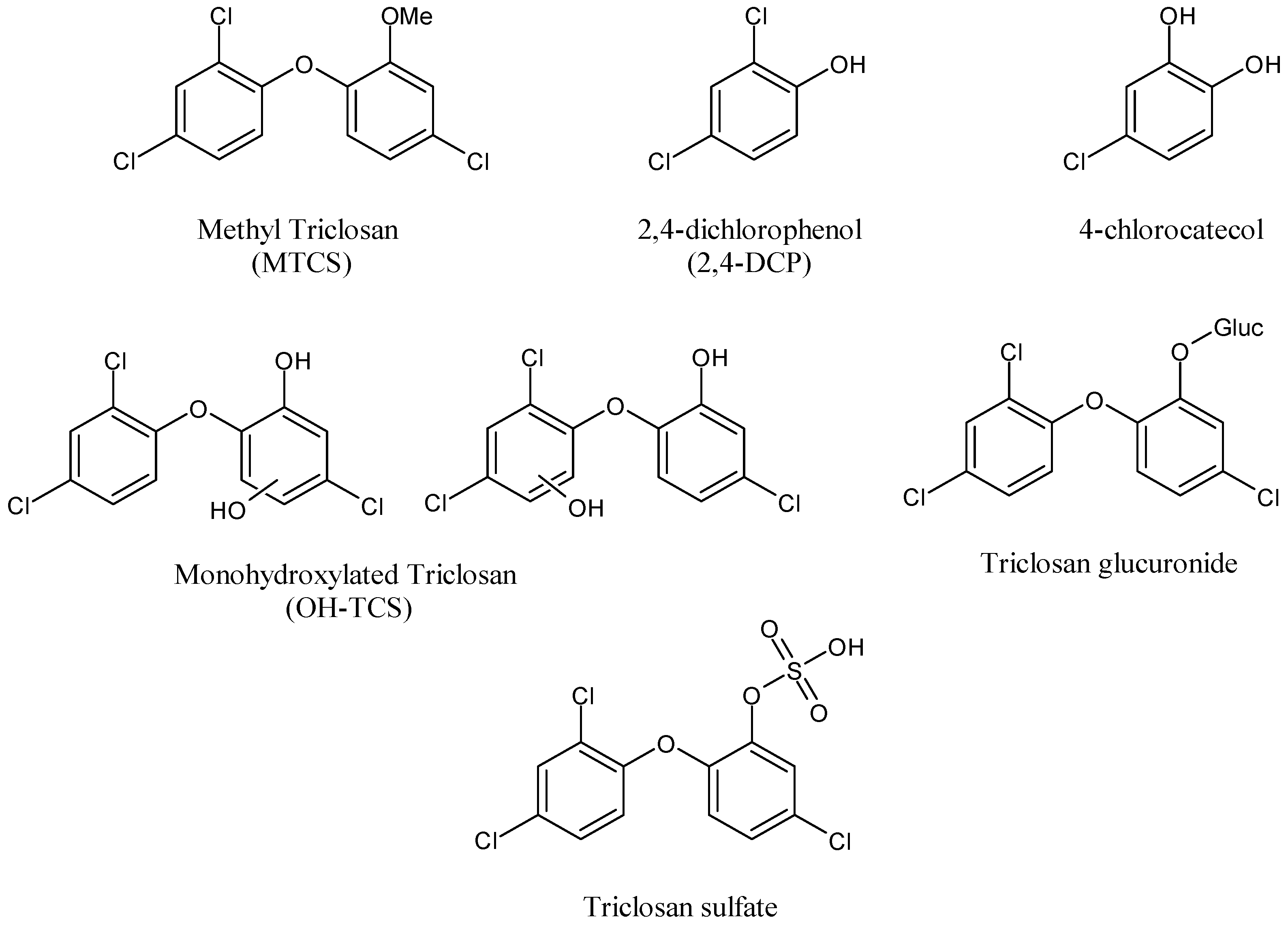
4.1. Metabolism and Transformation Products of TCS
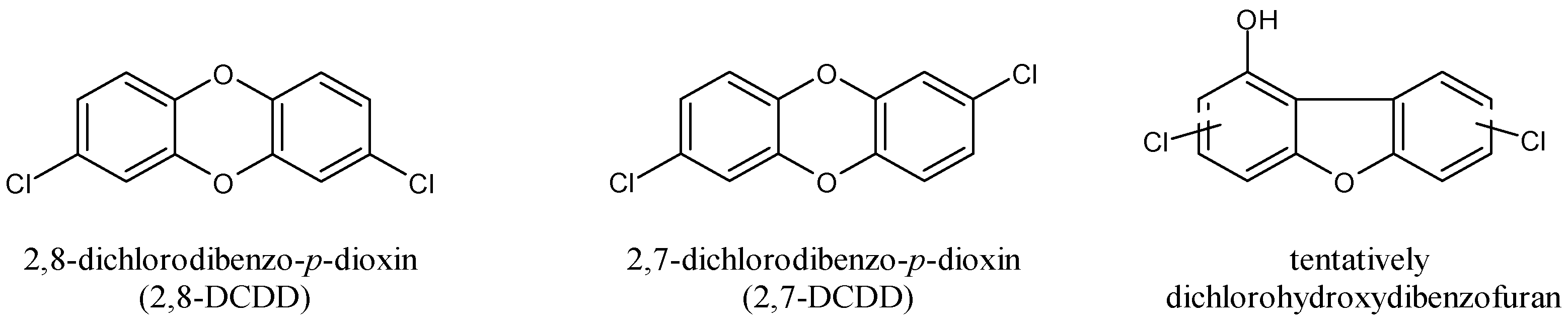
4.2. Photodegradation of TCS
5. Toxicity Studies on TCS
5.1. Effects on Hypothalamic-Pituitary-Thyroid Axis and Steroidogenesis
5.2. Effects on Semen
5.3. Studies on Prenatal Exposure to TCS
5.4. Studies on the Effects of TCS during Lactation
5.5. Neurotoxic and Hepatotoxic Effects of TCS
5.6. Metabolic Disorders, Nephrotoxicity, and Polycystic Ovary Syndrome (PCOS)
5.7. Immune Response, Asthma and Allergies
6. Gut Microbiota and Microbiome Involvement in TCS Exposure
7. Mechanism of Action in Pharmacological and Toxicity Studies
7.1. Toxicity Studies in Animal Models
7.2. Cytotoxicity Studies
8. Removal from Aquatic Environment/Degradation Techniques
9. Repositioning of TCS
10. Summary
Author Contributions
Funding
Institutional Review Board Statement
Informed Consent Statement
Data Availability Statement
Conflicts of Interest
Abbreviations
| AChE | Acetylcholinesterase |
| AhR | Aryl Hydrocarbon Receptor |
| AMH | Anti-Müllerian hormone |
| BALF | Bronchoalveolar Lavage Fluid |
| BMI | Body Mass Index |
| CEC | Contaminants of Emerging Concern |
| CNS | Central Nervous System |
| 2,8-DCDD | 2,8-dichlorodibenzo-p-dioxin |
| 2,4-DCP | 2,4-Dichlorophenol |
| EDCs | Endocrine Disrupting Chemicals |
| EDTA | Ethylenediaminetetraacetic Acid |
| ENR | Enoyl-acyl carrier Protein Reductase |
| EU | European Union |
| FASN | Fatty Acid Synthase Inhibitor |
| FDA | Food and Drug Administration |
| FSH | Follicle-stimulating hormone |
| FT3 | Triiodothyronine |
| FT4 | Thyroxine |
| GOT | Glutamic Oxaloacetic Transaminase |
| GPT | Glutamic Pyruvic Transaminase |
| HCC | Hepatocellular Carcinoma |
| HOME | Health Outcomes and Measures of the Environment |
| HUVEC | Human Umbilical Vein Endothelial Cell |
| IC50 | Half Minimal Inhibitory Concentration |
| IgE | Immunoglobulin E |
| IL-6 | Interleukin-6 |
| LU | Luteinizing hormone |
| LPS | Lipopolysaccharide |
| MIC | Minimum Inhibitory Concentration |
| MTCS | Methyltriclosan |
| NHANES | National Health and Nutrition Examination Surveys |
| OTC | Over-the-counter |
| PCOS | Polycystic Ovary Syndrome |
| PPCPs | Pharmaceutical and Personal Care Products |
| PVM/MA | Polyvinylmethyl Ether Maleic Acid |
| REACH | Registration, Evaluation, Authorisation and Restriction of Chemicals |
| ROS | Reactive Oxygen Species |
| SHIME | Simulator of the Human Intestinal Microbial Ecosystem |
| SSIs | Surgical Site Infections |
| TCC | Triclocarban |
| TCS | Triclosan |
| TNF-α | Tumor Necrosis Factor alpha |
| TP | Total Protein |
| TSH | Thyroid-stimulating hormone |
| UDP | Uridine diphosphate |
| UGTs | UDP-glucuronosyltransferases |
References
- Halden, R.U.; Lindeman, A.E.; Aiello, A.E.; Andrews, D.; Arnold, W.A.; Fair, P.; Fuoco, R.E.; Geer, L.A.; Johnson, P.I.; Lohmann, R.; et al. The Florence statement on triclosan and triclocarban. Environ. Health Perspect. 2017, 125, 064501. [Google Scholar] [CrossRef] [PubMed]
- Weatherly, L.M.; Gosse, J.A. Triclosan exposure, transformation, and human health effects. J. Toxicol. Environ. Health B Crit. Rev. 2017, 20, 447–469. [Google Scholar] [CrossRef] [PubMed]
- Gálvez-Ontiveros, Y.; Páez, S.; Monteagudo, C.; Rivas, A. Endocrine disruptors in food: Impact on gut microbiota and metabolic diseases. Nutrients 2020, 12, 1158. [Google Scholar] [CrossRef] [PubMed]
- Catalano, A.; Iacopetta, D.; Sinicropi, M.S.; Franchini, C. Diarylureas as antitumor agents. Appl. Sci. 2021, 11, 374. [Google Scholar] [CrossRef]
- Catalano, A. Diarylurea: A privileged scaffold in drug discovery and therapeutic development. Curr. Med. Chem. 2022, 29, 4301. [Google Scholar] [CrossRef]
- Iacopetta, D.; Catalano, A.; Ceramella, J.; Saturnino, C.; Salvagno, L.; Ielo, I.; Drommi, D.; Scali, E.; Plutino, M.R.; Rosace, G.; et al. The different facets of triclocarban: A review. Molecules 2021, 26, 2811. [Google Scholar] [CrossRef]
- Lee, J.D.; Lee, J.Y.; Kwack, S.J.; Shin, C.Y.; Jang, H.J.; Kim, H.Y.; Kim, M.K.; Seo, D.W.; Lee, B.M.; Kim, K.B. Risk assessment of triclosan, a cosmetic preservative. Toxicol. Res. 2019, 35, 137–154. [Google Scholar] [CrossRef]
- Wang, Y.; Liang, W. Occurrence, toxicity, and removal methods of triclosan: A timely review. Curr. Poll. Rep. 2021, 7, 31–39. [Google Scholar] [CrossRef]
- Gao, C.J.; Jia, L.L.; Guo, Y. Triclosan in over the counter medicines of South China. Environ. Monit. Assess. 2018, 190, 728. [Google Scholar] [CrossRef]
- Statista. Sales of the Leading Toothpaste Brands in the United States in 2019 (in Million U.S. Dollars). 2019. Available online: https://www.statista.com/statistics/195650/leading-us-toothpaste-brands-in-2007-and-2008-based-on-sales/ (accessed on 9 May 2022).
- Statista. Sales Growth of the Leading Toothpaste Brands in the United States in 2019 (Change to Prior Sales Year). 2019. Available online: https://www.statista.com/statistics/195651/us-sales-growth-of-toothpaste-brands-in-2007-and-2008/ (accessed on 9 May 2022).
- Al Habashneh, R.; Farasin, R.; Khader, Y. The effect of a triclosan/copolymer/fluoride toothpaste on plaque formation, gingivitis, and dentin hypersensitivity: A single-blinded randomized clinical study. Quintessence Int. 2017, 48, 123–130. [Google Scholar]
- Nachu, S.; Ravoori, S.; Pachava, S. Antiplaque efficacy of toothpaste–A systematic review and meta-analysis of randomized controlled trials. J. Ind. Associat. Public Health Dent. 2022, 20, 16. [Google Scholar] [CrossRef]
- Zhu, Q.; Wang, M.; Jia, J.; Hu, Y.; Wang, X.; Liao, C.; Jiang, G. Occurrence, distribution, and human exposure of several endocrine disrupting chemicals in indoor dust: A nationwide study. Environ. Sci. Technol. 2020, 54, 11333–11343. [Google Scholar] [CrossRef] [PubMed]
- Zhang, Y.; Li, T.-T.; Shiu, B.-C.; Sun, F.; Ren, H.-T.; Zhang, X.; Lou, C.-W.; Lin, J.-H. Eco-friendly versatile protective polyurethane/triclosan coated polylactic acid nonwovens for medical covers application. J. Clean. Prod. 2021, 282, 124455. [Google Scholar] [CrossRef]
- Ahmed, I.; Boulton, A.J.; Rizvi, S.; Carlos, W.; Dickenson, E.; Smith, N.-A.; Reed, M. The use of triclosan-coated sutures to prevent surgical site infections: A systematic review and meta-analysis of the literature. BMJ Open 2019, 9, e029727. [Google Scholar] [CrossRef] [PubMed]
- Daoud, F.C.; Coppry, M.; Moore, N.; Rogues, A.M. Do triclosan sutures modify the microbial diversity of surgical site infections? a systematic review and meta-analysis. Microorganisms 2022, 10, 927. [Google Scholar] [CrossRef]
- Alfhili, M.A.; Lee, M.H. Triclosan: An update on biochemical and molecular mechanisms. Oxid. Med. Cell. Longev. 2019, 2019, 1607304. [Google Scholar] [CrossRef]
- Iacopetta, D.; Ceramella, J.; Catalano, A.; Saturnino, C.; Pellegrino, M.; Mariconda, A.; Longo, P.; Sinicropi, M.S.; Aquaro, S. COVID-19 at a glance: An up-to-date overview on variants, drug design and therapies. Viruses 2022, 14, 573. [Google Scholar] [CrossRef]
- Chu, W.; Fang, C.; Deng, Y.; Xu, Z. Intensifed disinfection amid COVID-19 pandemic poses potential risks to water quality and safety. Environ. Sci. Technol. 2020, 55, 4084–4086. [Google Scholar] [CrossRef]
- Kumar, S.; Paul, T.; Shukla, S.P.; Kumar, K.; Karmakar, S.; Bera, K.K. Biomarkers-based assessment of triclosan toxicity in aquatic environment: A mechanistic review. Environ. Pollut. 2021, 286, 117569. [Google Scholar] [CrossRef]
- Tan, Q.; Chen, J.; Chu, Y.; Liu, W.; Yang, L.; Ma, L.; Zhang, Y.; Qui, D.; Wu, Z.; He, F. Triclosan weakens the nitrification process of activated sludge and increases the risk of the spread of antibiotic resistance genes. J. Hazard. Mater. 2021, 416, 126085. [Google Scholar] [CrossRef]
- Catalano, A.; Iacopetta, D.; Ceramella, J.; Scumaci, D.; Giuzio, F.; Saturnino, C.; Aquaro, S.; Rosano, C.; Sinicropi, M.S. Multidrug resistance (MDR): A widespread phenomenon in pharmacological therapies. Molecules 2022, 27, 616. [Google Scholar] [CrossRef] [PubMed]
- Usman, M.; Farooq, M.; Hanna, K. Environmental side effects of the injudicious use of antimicrobials in the era of COVID-19. Sci. Total Environ. 2020, 745, 141053. [Google Scholar] [CrossRef] [PubMed]
- Alvarez-Munoz, D.; Rodriguez-Mozaz, S.; Jacobs, S.; Serra-Compte, A.; Caceres, N.; Sioen, I.; Verbeke, W.; Barbosa, V.; Ferrari, F.; Fernandez-Tejedor, M.; et al. Pharmaceuticals and endocrine disruptors in raw and cooked seafood from european market: Concentrations and human exposure levels. Environ. Int. 2018, 119, 570–581. [Google Scholar] [CrossRef] [PubMed]
- Ates, G.; Goldberg, J.; Currais, A.; Maher, P. CMS121, a fatty acid synthase inhibitor, protects against excess lipid peroxidation and inflammation and alleviates cognitive loss in a transgenic mouse model of Alzheimer's disease. Redox Biol. 2020, 36, 101648. [Google Scholar] [CrossRef]
- European Commission, Scientific Committees on Consumer Safety (SCCS) Opinion on Triclosan, Antimicrobial Resistance. (SCCP/1251/09). Directorate-General for Health and Consumers, Opinion Approved 7th Plenary. 2010. Available online: http://ec.europa.eu/health/scientific_committees/consumer_safety/docs/sccs_o_023.pdf (accessed on 9 May 2022).
- Lubarsky, H.V.; Gerbersdorf, S.U.; Hubas, C.; Behrens, S.; Ricciardi, F.; Paterson, D.M. Impairment of the bacterial biofilm stability by triclosan. PLoS ONE 2012, 7, e31183. [Google Scholar] [CrossRef]
- Liu, X.; Tu, M.; Wang, S.; Wang, Y.; Wang, J.; Hou, Y.; Zheng, X.; Yan, Z. Research on freshwater water quality criteria, sediment quality criteria and ecological risk assessment of triclosan in China. Sci. Total Environ. 2021, 816, 151616. [Google Scholar] [CrossRef]
- Contardo-Jara, V.; Meinecke, S.; Feibicke, M.; Berghahn, R.; Schmidt, R.; Mohr, S. Fate, bioaccumulation and toxic effects of triclosan on a freshwater community–A mesocosm study. Environ. Adv. 2021, 5, 100100. [Google Scholar] [CrossRef]
- Mohan, S.; Balakrishnan, P. Triclosan in treated wastewater from a city wastewater treatment plant and its environmental risk assessment. Water Air Soil Pollut. 2019, 230, 69. [Google Scholar] [CrossRef]
- Dodson, R.E.; Boronow, K.E.; Susmann, H.; Udesky, J.O.; Rodgers, K.M.; Weller, D.; Woudneh, M.; Brody, J.G.; Rudel, R.A. Consumer behavior and exposure to parabens, bisphenols, triclosan, dichlorophenols, and benzophenone-3: Results from a crowdsourced biomonitoring study. Int. J. Hygiene Environ. Health 2020, 230, 113624. [Google Scholar] [CrossRef]
- Di Poi, C.; Costil, K.; Bouchart, V.; Halm-Lemeille, M.P. Toxicity assessment of five emerging pollutants, alone and in binary or ternary mixtures, towards three aquatic organisms. Environ. Sci. Pollut. Res. 2018, 25, 6122–6134. [Google Scholar] [CrossRef]
- Dar, O.I.; Aslam, R.; Pan, D.; Sharma, S.; Andotra, M.; Kaur, A.; Jia, A.Q.; Faggio, C. Source, bioaccumulation, degradability and toxicity of triclosan in aquatic environments: A review. Environ. Technol. Innov. 2022, 25, 102122. [Google Scholar] [CrossRef]
- Lu, S.; Yu, Y.; Ren, L.; Zhang, X.; Liu, G.; Yu, Y. Estimation of intake and uptake of bisphenols and triclosan from personal care products by dermal contact. Sci. Total Environ. 2018, 621, 1389–1396. [Google Scholar] [CrossRef] [PubMed]
- Skarha, J.; Mínguez-Alarcón, L.; Williams, P.L.; Korevaar, T.I.M.; de Poortere, R.A.; Broeren, M.A.C.; Ford, J.B.; Eliot, M.; Hauser, R.; Braun, J.M. Cross-sectional associations between urinary triclosan and serum thyroid function biomarker concentrations in women. Environ. Int. 2019, 122, 256–262. [Google Scholar] [CrossRef] [PubMed]
- Arbuckle, T.E.; Marro, L.; Davis, K.; Fisher, M.; Ayotte, P.; Belanger, P.; Dumas, P.; LeBlanc, A.; Berube, R.; Gaudreau, E.; et al. Exposure to free and conjugated forms of bisphenol A and triclosan among pregnant women in the MIREC cohort. Environ. Health Perspect. 2015, 123, 277–284. [Google Scholar] [CrossRef] [PubMed]
- Binder, A.M.; Corvalan, C.; Calafat, A.M.; Ye, X.; Mericq, V.; Pereira, A.; Michels, K.B. Childhood and adolescent phenol and phthalate exposure and the age of menarche in Latina girls. Environ. Health 2018, 17, 32. [Google Scholar] [CrossRef] [PubMed]
- Savage, J.H.; Matsui, E.C.; Wood, R.A.; Keet, C.A. Urinary levels of triclosan and parabens are associated with aeroallergen and food sensitization. J. Allergy Clin. Immunol. 2012, 130, 453–460.e7. [Google Scholar] [CrossRef] [PubMed]
- Alfhili, M.A.; Hussein, H.A.; Park, Y.; Lee, M.H.; Akula, S.M. Triclosan induces apoptosis in Burkitt lymphoma-derived BJAB cells through caspase and JNK/MAPK pathways. Apoptosis 2021, 26, 96–110. [Google Scholar] [CrossRef]
- Anderson, S.E.; Meade, B.J.; Long, C.M.; Lukomska, E.; Marshall, N.B. Investigations of immunotoxicity and allergic potential induced by topical application of triclosan in mice. J. Immunotoxicol. 2016, 13, 165–172. [Google Scholar] [CrossRef]
- Dinwiddie, M.T.; Terry, P.D.; Chen, J. Recent evidence regarding triclosan and cancer risk. Int. J. Environ. Res. Public Health 2014, 11, 2209–2217. [Google Scholar] [CrossRef]
- Singh, S.; Karthikeyan, C.; Moorthy, N.H.N. Recent advances in the development of fatty acid synthase inhibitors as anticancer agents. Mini Rev. Med. Chem. 2020, 20, 1820–1837. [Google Scholar] [CrossRef]
- Turanli, B.; Grøtli, M.; Boren, J.; Nielsen, J.; Uhlen, M.; Arga, K.Y.; Mardinoglu, A. Drug repositioning for effective prostate cancer treatment. Front. Physiol. 2018, 9, 500. [Google Scholar] [CrossRef] [PubMed]
- Montaseri, H.; Forbes, P.B.C. A review of monitoring methods for triclosan and its occurrence in aquatic environments. Trace Trend Anal. Chem. 2016, 85, 221–231. [Google Scholar] [CrossRef]
- Woodruff, T.J.; Zota, A.R.; Schwartz, J.M. Environmental chemicals in pregnant women in the United States: NHANES 2003–2004. Environ. Health Perspect. 2011, 119, 878–885. [Google Scholar] [CrossRef] [PubMed]
- Casas, L.; Fernandez, M.F.; Llop, S.; Guxens, M.; Ballester, F.; Olea, N.; Irurzun, M.B.; Rodriguez, L.S.; Riano, I.; Tardon, A.; et al. Urinary concentrations of phthalates and phenols in a population of Spanish pregnant women and children. Environ. Int. 2011, 37, 858–866. [Google Scholar] [CrossRef] [PubMed]
- FDA (U.S. Food and Drug Administration). 21 CFR Part 310 safety and effectiveness of consumer antiseptics. topical antimicrobial drug products for over-the-counter human use. Final rule. Fed. Reg. 2016, 81, 61106–61130.
- ECHA (European Chemicals Agency). Biocidal Products Committee (BPC): Opinion on the Application for Approval of the Active Substance: Triclosan Product-Type: 1. 2015. Available online: https://echa.europa.eu/documents/10162/efc985e4-8802-4ebb-8245-29708747a358 (accessed on 17 June 2016).
- EC (European Commission). Commission Implementing Decision (EU) 2016/110 of 27 January 2016 Not Approving Triclosan as an Existing Active Substance for Use in Biocidal Products for Product-Type 1. 2016. Available online: http://eur-lex.europa.eu/legal-content/EN/TXT/PDF/?uri=CELEX:32016D0110&from=EN (accessed on 23 January 2017).
- European Union. Official Journal L359—EUR-Lex. Available online: http://eur-lex.europa.eu/legal-content/EN/TXT/PDF/?uri=OJ:L:2014:107:FULL&from=EN (accessed on 9 May 2022).
- European Commission. Fitness Check on Endocrine Disruptors. Commission Staf Working Document. SWD (2020) 251 Fnal. Available online: https://ec.europa.eu/environment/pdf/chemicals/2020/10/SWD_on_Endocrines_disruptors.pdf (accessed on 10 January 2021).
- National Institute of Environmental Health Sciences (NIEHS). Endocrine Disruptors. 2020. Available online: https://www.niehs.nih.gov/health/topics/agents/endocrine/index.cfm (accessed on 5 January 2021).
- European Chemical Agency (ECHA). Substance Infocard, Triclosan. 2020. Available online: https://echa.europa.eu/substance-information/-/substanceinfo/100.020.167 (accessed on 10 January 2021).
- Ahmed, I.; Lin, H.; Zou, L.; Brody, A.L.; Li, Z.; Qazi, I.M.; Pavase, T.R.; Lv, L. A comprehensive review on the application of active packaging technologies to muscle foods. Food Control 2017, 82, 163–178. [Google Scholar] [CrossRef]
- Schumann, B.; Schmid, M. Packaging concepts for fresh and processed meat Recent progresses. Inn. Food Sci. Emerg. Technol. 2018, 47, 88–100. [Google Scholar] [CrossRef]
- Beiras, R.; Verdejo, E.; Campoy-López, P.; Vidal-Liñán, L. Aquatic toxicity of chemically defined microplastics can be explained by functional additives. J. Hazard. Mater. 2021, 406, 124338. [Google Scholar] [CrossRef]
- Petersen, R.C. Triclosan antimicrobial polymers. AIMS Mol. Sci. 2016, 31, 88–103. [Google Scholar] [CrossRef]
- Glaser, A. The ubiquitous triclosan. A common antibacterial agent exposed. Pest. You 2004, 24, 12–17. [Google Scholar]
- O'Neal, T.K. Identification and Characterization of Triclosan Resistant Bacteria. Ph.D. Dissertation, University of South Alabama, Mobile, AL, USA, 2019. [Google Scholar]
- European Commission. Commission Decision of 19 March 2010 Concerning the Non-Inclusion of 2,4,4’-trichloro-2’-hydroxydiphenyl ether in the Union List of Additives Which May Be Used in the Manufacture of Plastic Materials and Articles Intended to Come into Contact with Foodstuffs under Directive 2002/72/EC (Notified under Document C(2010) 1613) (Text with EEA Relevance) (2010/169/EU). Available online: https://www.legislation.gov.uk/eudn/2010/169/2010-03-19 (accessed on 9 May 2022).
- Marazuela, M.D.; Klaiber, M.; Moreno-Gordaliza, E.; Barata, A.; Gómez-Gómez, M.M. Safety assessment of commercial antimicrobial food packaging: Triclosan and microplastics, a closer look. Food Pack. Shelf Life 2022, 31, 100780. [Google Scholar] [CrossRef]
- Shrestha, P.; Zhang, Y.; Chen, W.J.; Wong, T.Y. Triclosan: Antimicrobial mechanisms, antibiotics interactions, clinical applications, and human health. J. Environ. Sci. Health Part. C 2020, 38, 245–268. [Google Scholar] [CrossRef] [PubMed]
- Adkins, J.M.; Ahmar, R.A.; Yu, H.D.; Musick, S.T.; Alberico, A.M. Comparison of antimicrobial activity between bacitracin-soaked sutures and triclosan coated suture. J. Surg. Res. 2022, 270, 203–207. [Google Scholar] [CrossRef] [PubMed]
- Orhan, M. Triclosan applications for biocidal functionalization of polyester and cotton surfaces. J. Eng. Fib. Fabr. 2020, 15, 1558925020940104. [Google Scholar] [CrossRef]
- Suarez, S.; Dodd, M.C.; Omil, F.; von Gunten, U. Kinetics of triclosan oxidation by aqueous ozone and consequent loss of antibacterial activity: Relevance to municipal wastewater ozonation. Water Res. 2007, 41, 2481–2490. [Google Scholar] [CrossRef]
- Querido, M.M.; Rosário, F.; Bessa, M.J.; Mendes, F.; Teixeira, J.C.; Teixeira, J.P.; Pereira, C.C. In vitro cyto- and genotoxicity assessment of antibacterial paints with triclosan and isoborneol. Toxics 2022, 10, 58. [Google Scholar] [CrossRef]
- Schweizer, H.P. Triclosan: A widely used biocide and its link to antibiotics. FEMS Microb. Lett. 2001, 202, 1–7. [Google Scholar] [CrossRef]
- Karnas, K.; Marotta, J.; Koseki, R.; Sherman, E.; Exton, L.P. Triclosan resistance derived across environmentally and clinically relevant Gram negative bacteria. J. Pennsylv. Acad. Sci. 2019, 93, 83–106. [Google Scholar] [CrossRef]
- Halden, R.U. On the need and speed of regulating triclosan and triclocarban in the United States. Environ. Sci. Technol. 2014, 48, 3603–3611. [Google Scholar] [CrossRef]
- Møretrø, T.; Høiby-Pettersen, G.S.; Habimana, O.; Heir, E.; Langsrud, S. Assessment of the antibacterial activity of a triclosan-containing cutting board. Int. J. Food. Microb. 2011, 146, 157–162. [Google Scholar] [CrossRef]
- Gowda, J.; Tavarageri, A.; Kulkarni, R.; Anegundi, R.T.; Janardhan, A.; Bhat, M.A. Comparative assessment of the antimicrobial efficacy of triclosan, amoxicillin and eugenol against Enterococcus faecalis. Int. J. Clin. Ped. Dent. 2021, 14, 59. [Google Scholar]
- Pozzi, C.; Ferrari, S.; Cortesi, D.; Luciani, R.; Stroud, R.M.; Catalano, A.; Costi, M.P.; Mangani, S. The structure of Enterococcus faecalis thymidylate synthase provides clues about folate bacterial metabolism. Acta Crystallogr. D Biol. Crystallogr. 2012, 68, 1232–1241. [Google Scholar] [CrossRef] [PubMed]
- Zeng, W.; Xu, W.; Xu, Y.; Liao, W.; Zhao, Y.; Zheng, X.; Xu, C.; Zhou, T.; Cao, J. The prevalence and mechanism of triclosan resistance in Escherichia coli isolated from urine samples in Wenzhou, China. Antimicrob. Resist. Infect. Control. 2020, 9, 161. [Google Scholar] [CrossRef] [PubMed]
- Rozman, U.; Pušnik, M.; Kmetec, S.; Duh, D.; Šostar Turk, S. Reduced Susceptibility and Increased Resistance of Bacteria against Disinfectants: A Systematic Review. Microorganisms 2021, 9, 2550. [Google Scholar] [CrossRef]
- Franklyne, J.S.; Ebenazer, A.; Mukherjee, A.; Chandrasekaran, N. Role of triclosan microemulsion against triclosan resistant clones of bacterial pathogens. J. Drug Deliv. Sci. Technol. 2021, 61, 102158. [Google Scholar] [CrossRef]
- Araujo, C.B.; Ribeiro, A.B.; Fortes, C.V.; Bueno, F.L.; De Wever, B.; Oliveira, V.C.; Macedo, A.P.; Paranhos, H.F.O.; Lovato da Silva, C.H. Effect of local hygiene protocols on denture-related stomatitis, biofilm, microbial load, and odor: A randomized controlled trial. J. Prosthet. Dent. 2021. [Google Scholar] [CrossRef]
- Jongsma, M.A.; Van Der Mei, H.C.; Atema-Smit, J.; Busscher, H.J.; Ren, Y. In vivo biofilm formation on stainless steel bonded retainers during different oral health-care regimens. Int. J. Oral Sci. 2015, 7, 42–48. [Google Scholar] [CrossRef][Green Version]
- Maiden, M.M.; Hunt, A.M.A.; Zachos, M.P.; Gibson, J.A.; Hurwitz, M.E.; Mulks, M.H.; Waters, C.M. Triclosan is an aminoglycoside adjuvant for eradication of Pseudomonas aeruginosa biofilms. Antimicrob. Agents Chemother. 2018, 62, e00146-18. [Google Scholar] [CrossRef]
- Maiden, M.M.; Waters, C.M. Triclosan depletes the membrane potential in Pseudomonas aeruginosa biofilms inhibiting aminoglycoside induced adaptive resistance. PLoS Pathog. 2020, 16, e1008529. [Google Scholar] [CrossRef]
- Ayyash, M.; Shehabi, A.A.; Mahmoud, N.N.; Al-Bakri, A.G. Antibiofilm properties of triclosan with EDTA or cranberry as Foley Catheter lock solutions. J. Appl. Microbiol. 2019, 127, 1876–1888. [Google Scholar] [CrossRef]
- Talaat, D.M.; Sharaf, A.A.E.A.; Ghoneim, M.A.E.M.; El-Shazly, S.A.; El Meligy, O.A.E.S. Efficacy of two mouth rinse sprays in inhibiting Streptococcus mutans growth on toothbrush bristles. Saudi Dent. J. 2018, 30, 365–372. [Google Scholar] [CrossRef] [PubMed]
- Yadav, S.; Mandal, H.; Saravanan, V.; Das, P.; Singh, S.K. In vitro and in silico analysis of L. donovani enoyl acyl carrier protein reductase-A possible drug target. J. Biomol. Srtuct. Dynam. 2021, 39, 6056–6069. [Google Scholar] [CrossRef] [PubMed]
- Vosatka, R.; Kratky, M.; Vinsova, J. Triclosan and its derivatives as antimycobacterial active agents. Eur. J. Pharm. Sci. 2018, 114, 318–331. [Google Scholar] [CrossRef] [PubMed]
- Bilsland, E.; van Vliet, L.; Williams, K.; Feltham, J.; Carrasco, M.P.; Fotoran, W.L.; Cubillos, E.F.G.; Wunderlich, G.; Grøtli, M.; Hollfelder, F.; et al. Plasmodium dihydrofolate reductase is a second enzyme target for the antimalarial action of triclosan. Sci. Rep. 2018, 8, 1038. [Google Scholar] [CrossRef]
- Chetty, S.; Armstrong, T.; Sharma Kharkwal, S.; Drewe, W.C.; De Matteis, C.I.; Evangelopoulos, D.; Bhakta, S.; Thomas, N.R. New InhA inhibitors based on expanded triclosan and di-triclosan analogues to develop a new treatment for tuberculosis. Pharmaceuticals 2021, 14, 361. [Google Scholar] [CrossRef]
- de Luco, J.F.; Recio-Balsells, A.I.; Ghiano, D.G.; Bortolotti, A.; Belardinelli, J.M.; Liu, N.; Hoffmann, P.; Lherbet, C.; Tonge, P.J.; Tekwani, W.; et al. Exploring the chemical space of 1,2,3-triazolyl triclosan analogs for discovery of new antileishmanial chemotherapeutic agents. RSC Med. Chem. 2021, 12, 120–128. [Google Scholar] [CrossRef]
- Walsh, L.J.; Healey, D.L. Prevention and caries risk management in teenage and orthodontic patients. Austr. Dent. J. 2019, 64, S37–S45. [Google Scholar] [CrossRef]
- Hall, P.J.; Green, A.K.; Horay, C.P.; de Brabander, S.; Beasley, T.J.; Cromwell, V.J.; Holt, J.S.; Savage, D.J. Plaque antibacterial levels following controlled food intake and use of a toothpaste containing 2% zinc citrate and 0.3% Triclosan. Int. Dent. J. 2003, 53 (Suppl. S1), 379–384. [Google Scholar] [CrossRef]
- Riley, P.; Lamont, T. Triclosan/copolymer containing toothpastes for oral health (Review). Cochrane Database Syst. Rev. 2013, 12, CD010514. [Google Scholar]
- Singh, S.; Chaknis, P.; DeVizio, W.; Petrone, M.; Panagakos, F.S.; Proskin, H.M. A Clinical investigation of the efficacy of three commercially available dentifrices for controlling established gingivitis and supragingival plaque. J. Clin. Dent. 2010, 21, 105–110. [Google Scholar]
- West, N.X.; He, T.; Hellin, N.; Claydon, N.; Seong, J.; Macdonald, E.; Farrell, S.; Eusebio, R.; Wilberg, A. Randomized in situ clinical trial evaluating erosion protection efficacy of a 0.454% stannous fluoride dentifrice. Int. J. Dent. Hyg. 2019, 17, 261–267. [Google Scholar] [CrossRef] [PubMed]
- Stewart, B.; Shibli, J.A.; Araujo, M.; Figueiredo, L.C.; Panagakos, F.; Matarazzo, F.; Mairink, R.; Onuma, T.; Faveri, M.; Retamal-Valdes, B.; et al. Effects of a toothpaste containing 0.3% triclosan on periodontal parameters of subjects enrolled in a regular maintenance program: A secondary analysis of a 2-year randomized clinical trial. J. Periodontol. 2020, 91, 596–605. [Google Scholar] [CrossRef] [PubMed]
- Fine, D.H.; Sreenivasan, P.K.; McKiernan, M.; Tischio-Bereski, D.; Furgang, D. Whole mouth antimicrobial effects after oral hygiene: Comparison of three dentifrice formulations. J. Clin. Periodont. 2012, 39, 1056–1064. [Google Scholar] [CrossRef] [PubMed]
- Panagakos, F.S.; Volpe, A.R.; Petrone, M.E.; DeVizio, W.; Davies, R.M.; Proskin, H.M. Advanced oral antibacterial/anti-inflammatory technology: A comprehensive review of the clinical benefits of a triclosan/copolymer/fluoride dentifrice. J. Clin. Dent. 2005, 16, S1–S19. [Google Scholar]
- Kerdvongbundit, V.; Wikesjö, U.M. Effect of triclosan on healing following non-surgical periodontal therapy in smokers. J. Clin. Periodontol. 2003, 30, 1024–1030. [Google Scholar] [CrossRef]
- Aminu, N.; Yam, M.F.; Chan, S.Y.; Bello, I.; Umar, N.M.; Nuhu, T.; Toh, S.M. The evaluation of healing effect of triclosan and flurbiprofen-loaded nanogels in experimental periodontitis in rats by morphometric analysis. Saudi Dent. J. 2021, 33, 554–559. [Google Scholar] [CrossRef]
- Shu, W.; Zhang, Y.; Zhang, C.; You, Q.; Zhou, H.; Wen, S. Triclosan inhibits the activation of human periodontal ligament fibroblasts induced by lipopolysaccharide from Porphyromonas gingivalis. J. Biomed. Res. 2021, 35, 206. [Google Scholar] [CrossRef]
- Ceramella, J.; Iacopetta, D.; Catalano, A.; Cirillo, F.; Lappano, R.; Sinicropi, M.S. A review on the antimicrobial activity of Schiff bases: Data collection and recent studies. Antibiotics 2022, 11, 191. [Google Scholar] [CrossRef]
- Pavez, L.; Tobar, N.; Chacon, C.; Arancibia, R.; Martínez, C.; Tapia, C.; Pastor, A.; Gonzàlez, M.; Martínez, J.; Smith, P.C. Chitosan-triclosan particles modulate inflammatory signaling in gingival fibroblasts. J. Periodont. Res. 2018, 53, 232–239. [Google Scholar] [CrossRef]
- Li, X.; Zhong, Y.; He, W.; Huang, S.; Li, Q.; Guo, C.; Ma, S.; Li, G.; Yu, Y. Co-exposure and health risks of parabens, bisphenols, triclosan, phthalate metabolites and hydroxyl polycyclic aromatic hydrocarbons based on simultaneous detection in urine samples from Guangzhou, South China. Environ. Pollut. 2021, 272, 115990. [Google Scholar] [CrossRef]
- Milanović, M.; Đurić, L.; Milošević, N.; Milić, N. Comprehensive insight into triclosan—from widespread occurrence to health outcomes. Environ. Sci. Pollut. Res. 2021, 1–22. [Google Scholar] [CrossRef] [PubMed]
- Allmyr, M.; Panagiotidis, G.; Sparve, E.; Diczfalusy, U.; Sandborgh-Englund, G. Human exposure to triclosan via toothpaste does not change CYP3A4 activity or plasma concentrations of thyroid hormones. Basic Clin. Pharmacol. Toxicol. 2009, 105, 339–344. [Google Scholar] [CrossRef] [PubMed]
- Sandborgh-Englund, G.; Adolfsson-Erici, M.; Odham, G.; Ekstrand, J. Pharmacokinetics of triclosan following oral ingestion in humans. J. Toxicol. Environ. Health A 2006, 69, 1861–1873. [Google Scholar] [CrossRef] [PubMed]
- Queckenberg, C.; Meins, J.; Wachall, B.; Doroshyenko, O.; Tomalik-Scharte, D.; Bastian, B.; Abdel-Tawab, M.; Fuhr, U. Absorption, pharmacokinetics, and safety of triclosan after dermal administration. Antimicrob. Agents Chemother. 2010, 54, 570–572. [Google Scholar] [CrossRef] [PubMed]
- Dhillon, G.S.; Kaur, S.; Pulicharla, R.; Brar, S.K.; Cledón, M.; Verma, M.; Surampalli, R.Y. TCS: Current status, occurrence, environmental risks and bioaccumulation potential. Int. J. Environ. Res. Publ. Health 2015, 12, 5657–5684. [Google Scholar] [CrossRef]
- Scientific Committee on Consumer Products (SCCP). Scientific Committee on Consumer Safety. Opinion on Triclosan Antimicrobial Resistance; SCCP/1251/09. Available online: https://ec.europa.eu/health/sites/health/files/scientific_committees/consumer_safety/docs/sccs_o_023.pdf (accessed on 9 May 2022).
- Bester, K. Fate of triclosan and triclosan-methyl in sewage treatment plants and surface waters. Arch. Environ. Contam. Toxicol. 2005, 49, 9–17. [Google Scholar] [CrossRef]
- Scientific Committee on Consumer Products (SCCP). Opinion on Triclosan (COLIPA No. P32). 2009. Available online: https://ec.europa.eu/health/ph_risk/committees/04_sccp/docs/sccp_o_166.pdf (accessed on 9 May 2022).
- Dix-Cooper, L.; Kosatsky, T. Use of antibacterial toothpaste is associated with higher urinary triclosan concentrations in Asian immigrant women living in Vancouver, Canada. Sci. Total Environ. 2019, 671, 897–904. [Google Scholar] [CrossRef]
- DeLorenzo, M.E.; Keller, J.M.; Arthur, C.D.; Finnegan, M.C.; Harper, H.E.; Winder, V.L.; Zdankiewicz, D.L. Toxicity of the antimicrobial compound triclosan and formation of the metabolite methyl-triclosan in estuarine systems. Environ. Toxicol. 2008, 23, 224–232. [Google Scholar] [CrossRef]
- Armstrong, D.L.; Lozano, N.; Rice, C.P.; Ramirez, M.; Torrents, A. Degradation of triclosan and triclocarban and formation of transformation products in activated sludge using benchtop bioreactors. Environ. Res. 2018, 161, 17–25. [Google Scholar] [CrossRef]
- Chen, X.; Casas, M.E.; Nielsen, J.L.; Wimmer, R.; Bester, K. Identification of Triclosan-O-Sulfate and other transformation products of Triclosan formed by activated sludge. Sci. Total Environ. 2015, 505, 39–46. [Google Scholar] [CrossRef]
- Lozano, N.; Rice, C.P.; Ramirez, M.; Torrents, A. Fate of Triclosan and Methyltriclosan in soil from biosolids application. Environ. Pollut. 2012, 160, 103–108. [Google Scholar] [CrossRef] [PubMed]
- Fu, J.; Tan, Y.X.R.; Gong, Z.; Bae, S. The toxic effect of triclosan and methyl-triclosan on biological pathways revealed by metabolomics and gene expression in zebrafish embryos. Ecotoxicol. Environ. Saf. 2020, 189, 110039. [Google Scholar] [CrossRef] [PubMed]
- Chen, H.C.; Chang, J.W.; Sun, Y.C.; Chang, W.T.; Huang, P.C. Determination of parabens, bisphenol a and its analogs, triclosan, and benzophenone-3 levels in human urine by isotope-dilution-UPLC-MS/MS method followed by supported liquid extraction. Toxics 2022, 10, 21. [Google Scholar] [CrossRef] [PubMed]
- Fang, J.L.; Stingley, R.L.; Beland, F.A.; Harrouk, W.; Lumpkins, D.L.; Howard, P. Occurrence, efficacy, metabolism, and toxicity of triclosan. J. Environ. Sci. Health Part. C 2010, 28, 147–171. [Google Scholar] [CrossRef] [PubMed]
- Zhang, H.; Sanidad, K.Z.; Zhu, L.; Parsonnet, J.; Haggerty, T.D.; Zhang, G.; Cai, Z. Frequent occurrence of triclosan hydroxylation in mammals: A combined theoretical and experimental investigation. J. Hazard. Mater. 2021, 407, 124803. [Google Scholar] [CrossRef]
- Latch, D.E.; Packer, J.L.; Arnold, W.A.; McNeill, K. Photochemical conversion of triclosan to 2, 8-dichlorodibenzo-p-dioxin in aqueous solution. J. Photochem. Photobiol. A Chem. 2003, 158, 63–66. [Google Scholar] [CrossRef]
- Ding, J.; Su, M.; Wu, C.; Lin, K. Transformation of triclosan to 2,8-dichlorodibenzo-p-dioxin by iron and manganese oxides under near dry conditions. Chemosphere 2015, 133, 41–46. [Google Scholar] [CrossRef]
- Buth, J.M.; Steen, P.O.; Sueper, C.; Blumentritt, D.; Vikesland, P.J.; Arnold, W.A.; McNeill, K. Dioxin photoproducts of triclosan and its chlorinated derivatives in sediment cores. Environ. Sci. Technol. 2010, 44, 4545–4551. [Google Scholar] [CrossRef]
- Rodrigues, F.; Lehmann, M.; doAmaral, V.S.; Reguly, M.L.; de Andrade, H.H.R. Genotoxicity of three mouthwash products, Cepacol®, Periogard®, and Plax®, in the Drosophila wing-spot test. Environ. Mol. Mutagen. 2007, 48, 644–649. [Google Scholar] [CrossRef]
- Carrisi, C.; Madeo, M.; Morciano, P.; Dolce, V.; Cenci, G.; Cappello, A.R.; Mazzeo, G.; Iacopetta, D.; Capobianco, L. Identification of the Drosophila melanogaster mitochondrial citrate carrier: Bacterial expression, reconstitution, functional characterization and developmental distribution. J. Biochem. 2008, 144, 389–392. [Google Scholar] [CrossRef]
- European Chemicals Agency (ECHA). Guidance on Information Requirements and Chemical Safety Assessment, Part C: PBT/vPvB assessment, Version 3.0. 2017. Available online: https://echa.europa.eu/information-on-chemicals/euclef (accessed on 9 May 2022).
- Li, L. Toxicity evaluation and by-products identifcation of triclosan ozonation and chlorination. Chemosphere 2021, 263, 128223. [Google Scholar] [CrossRef] [PubMed]
- Weiss, L.; Arbuckle, T.E.; Fisher, M.; Ramsay, T.; Mallick, R.; Hauser, R.; LeBlanc, A.; Walker, M.; Dumas, P.; Lang, C. Temporal variability and sources of triclosan exposure in pregnancy. Int. J. Hygiene Environ. Health 2015, 218, 507–513. [Google Scholar] [CrossRef] [PubMed]
- Johnson, P.I.; Koustas, E.; Vesterinen, H.M.; Sutton, P.; Atchley, D.S.; Kim, A.N.; Campbell, M.; Donald, J.M.; Sen, S.; Bero, L.; et al. Application of the Navigation Guide systematic review methodology to the evidence for developmental and reproductive toxicity of triclosan. Environ. Int. 2016, 92–93, 716–728. [Google Scholar] [CrossRef] [PubMed]
- Paul, K.B.; Thompson, J.T.; Simmons, S.O.; Vanden Heuvel, J.P.; Crofton, K.M. Evidence for triclosan-induced activation of human and rodent xenobiotic nuclear receptors. Toxicol. In Vitro 2013, 27, 2049–2060. [Google Scholar] [CrossRef]
- Koeppe, E.S.; Ferguson, K.K.; Colacino, J.A.; Meeker, J.D. Relationship between urinary triclosan and paraben concentrations and serum thyroid measures in NHANES 2007–2008. Sci. Total Environ. 2013, 445–446, 299–305. [Google Scholar] [CrossRef]
- Cullinan, M.P.; Palmer, J.E.; Carle, A.D.; West, M.J.; Seymour, G.J. Long term use of triclosan toothpaste and thyroid function. Sci. Total Environ. 2012, 416, 75–79. [Google Scholar] [CrossRef]
- Taha, M.; Marie, A.M.; Ahmed-Farid, O.A. Combined approaches for evaluation of xenoestrogen neural toxicity and thyroid dysfunction: Screening of oxido-nitrosative markers, DNA fragmentation, and biogenic amine degradation. J. Biochem. Mol. Toxicol. 2020, 34, e22521. [Google Scholar] [CrossRef]
- Zhang, P.; Yang, M.; Zeng, L.; Liu, C. P38/TRHr-dependent regulation of TPO in thyroid cells contributes to the hypothyroidism of triclosan-treated rats. Cell. Physiol. Biochem. 2018, 45, 1303–1315. [Google Scholar] [CrossRef]
- Cao, X.Y.; Hua, X.; Xiong, J.W.; Zhu, W.T.; Zhang, J.; Chen, L. Impact of triclosan on female reproduction through reducing thyroid hormones to suppress hypothalamic kisspeptin neurons in mice. Front. Mol. Neurosci. 2018, 11, 6. [Google Scholar] [CrossRef]
- Abd-Elhakim, Y.M.; Mohammed, A.T.; Ali, H.A. Impact of subchronic exposure to triclosan and/or fuoride on estrogenic activity in immature female rats: The expression pattern of calbindin-D9k and estrogen receptor α genes. J. Biochem. Mol. Toxicol. 2018, 32, e22027. [Google Scholar] [CrossRef]
- Jurewicz, J.; Wielgomas, B.; Radwan, M.; Karwacka, A.; Klimowska, A.; Dziewirska, E.; Korczak, K.; Zajdel, R.; Radwan, P.; Hanke, W. Triclosan exposure and ovarian reserve. Reprod. Toxicol. 2019, 89, 168–172. [Google Scholar] [CrossRef] [PubMed]
- Chen, W.; Yang, X.; Wang, B.; Wang, L.; Yu, X. The effects and possible mechanisms of triclosan on steroidogenesis in primary rat granulosa cells. Reprod. Toxicol. 2019, 83, 28–37. [Google Scholar] [CrossRef] [PubMed]
- Du, Y.; Wang, B.; Cai, Z.; Zhang, H.; Wang, B.; Liang, W.; Zhou, G.; Ouyang, F.; Wang, W. The triclosan-induced shift from aerobic to anaerobic metabolism link to increased steroidogenesis in human ovarian granulosa cells. Ecotoxicol. Environ. Safety 2021, 220, 112389. [Google Scholar] [CrossRef] [PubMed]
- Basini, G.; Bussolati, S.; Bertini, S.; Quintavalla, F.; Grasselli, F. Evaluation of triclosan effects on cultured swine luteal cells. Animals 2021, 11, 606. [Google Scholar] [CrossRef]
- Rehman, S.; Usman, Z.; Rehman, S.; Aldraihem, M.; Rehman, N.; Rehman, I.; Ahmad, G. Endocrine disrupting chemicals and impact on male reproductive health. Transl. Androl. Urol. 2018, 7, 490–503. [Google Scholar] [CrossRef]
- Gee, R.H.; Charles, A.; Taylor, N.; Darbre, P.D. Oestrogenic and androgenic activity of triclosan in breast cancer cells. J. Appl. Toxicol. 2008, 28, 78–91. [Google Scholar] [CrossRef]
- Yawer, A.; Sychrová, E.; Labohá, P.; Raška, J.; Jambor, T.; Babica, P.; Sovadinová, I. Endocrine-disrupting chemicals rapidly affect intercellular signaling in Leydig cells. Toxicol. Appl. Pharmacol. 2020, 404, 115177. [Google Scholar] [CrossRef]
- Kumar, V.; Chakraborty, A.; Kural, M.R.; Roy, P. Alteration of testicular steroidogenesis and histopathology of reproductive system in male rats treated with triclosan. Reprod. Toxicol. 2009, 27, 177–185. [Google Scholar] [CrossRef]
- Lan, Z.; Hyung Kim, T.; Shun Bi, K.; Hui Chen, X.; Sik Kim, H. Triclosan exhibits a tendency to accumulate in the epididymis and shows sperm toxicity in male sprague-dawley rats. Environ. Toxicol. 2015, 30, 83–91. [Google Scholar] [CrossRef]
- Priyanka, T.A.; Maske, P.; Mote, C.; Dighe, V. Gestational and lactational exposure to triclosan causes impaired fertility of F1 male offspring and developmental defects in F2 generation. Environ. Pollut. 2020, 257, 113617. [Google Scholar] [CrossRef]
- Ha, M.; Zhang, P.; Li, L.; Liu, C. Triclosan suppresses testicular steroidogenesis via the miR-6321/JNK/Nur77 cascade. Cell Physiol. Biochem. 2018, 50, 2029–2045. [Google Scholar] [CrossRef] [PubMed]
- Duan, P.; Huang, X.; Ha, M.; Li, L.; Liu, C. miR-142-5p/DAX1-dependent regulation of P450c17 contributes to triclosan-mediated testosterone suppression. Sci. Total Environ. 2020, 717, 137280. [Google Scholar] [CrossRef] [PubMed]
- Zhu, W.; Zhang, H.; Tong, C.; Xie, C.; Fan, G.; Zhao, S.; Yu, X.; Tian, Y.; Zhang, J. Environmental exposure to triclosan and semen quality. Int. J. Environ. Res. Public Health 2016, 13, 224. [Google Scholar] [CrossRef] [PubMed]
- Jurewicz, J.; Radwan, M.; Wielgomas, B.; Kałużny, P.; Klimowska, A.; Radwan, P.; Hanke, W. Environmental levels of triclosan and male fertility. Environ. Sci. Pollut. Res. Int. 2018, 25, 5484–5490. [Google Scholar] [CrossRef]
- Nassan, F.L.; Mínguez-Alarcón, L.; Williams, P.L.; Dadd, R.; Petrozza, J.C.; Ford, J.B.; Calafat, A.M.; Hauser, R. Urinary triclosan concentrations and semen quality among men from a fertility clinic. Environ. Res. 2019, 177, 108633. [Google Scholar] [CrossRef]
- Pernoncini, K.V.; Montagnini, B.G.; de Góes, M.L.M.; Garcia, P.C.; Gerardin, D.C.C. Evaluation of reproductive toxicity in rats treated with triclosan. Reprod. Toxicol. 2018, 75, 65–72. [Google Scholar] [CrossRef]
- Yan, J.; Joseph, M.A.; Reynolds, S.A.; Geer, L.A. Association between urinary triclosan and serum testosterone levels in U.S. adult males from NHANES, 2011–2012. Int. J. Environ. Res. Public Health 2020, 17, 7412. [Google Scholar] [CrossRef]
- Yuan, G.; Ma, Y.; Zeng, Y.; Pan, H.; Liu, P.; Liu, Y.; Liu, G.; Cheng, J.; Guo, Y. Associations between low-dose triclosan exposure and semen quality in a Chinese population. Environ. Pollut. 2022, 299, 118926. [Google Scholar] [CrossRef]
- Scinicariello, F.; Buser, M.C. Serum testosterone concentrations and urinary bisphenol A, benzophenon-3, triclosan, and paraben levels in male and female children and adolescents: NHANES 2011–2012. Environ. Health Perspect. 2016, 124, 1898–1904. [Google Scholar] [CrossRef]
- Gishti, O.; Jaddoe, V.W.V.; Duijts, L.; Steegers, E.; Reiss, I.; Hofman, A.; Wong, T.Y.; Ikram, M.K.; Gaillard, R. Impact of birth parameters and early life growth patterns on retinal microvascular structure in children: The Generation R Study. J. Hypertens. 2015, 33, 1429–1437. [Google Scholar] [CrossRef]
- Toemen, L.; de Jonge, L.L.; Gishti, O.; van Osch-Gevers, L.; Taal, H.R.; Steegers, E.A.P.; Hofman, A.; Helbing, W.A.; Jaddoe, V.W.V. Longitudinal growth during fetal life and infancy and cardiovascular outcomes at school-age. J. Hypertens. 2016, 34, 1396–1406. [Google Scholar] [CrossRef] [PubMed]
- Wang, X.; Chen, X.; Feng, X.; Chang, F.; Chen, M.; Xia, Y.; Chen, L. Triclosan causes spontaneous abortion accompanied by decline of estrogen sulfotransferase activity in humans and mice. Sci. Rep. 2015, 5, 18252. [Google Scholar] [CrossRef] [PubMed]
- Ouyang, F.; Tang, W.N.; Zhang, H.; Wang, X.; Zhao, S.; Wang, W.; Zhang, J.; Cheng, W. Maternal urinary triclosan level, gestational diabetes mellitus and birth weight in Chinese women. Sci. Total Environ. 2018, 626, 451–457. [Google Scholar] [CrossRef] [PubMed]
- Velez, M.P.; Arbuckle, T.E.; Fraser, W.D. Female exposure to phenols and phthalates and time to pregnancy: The Maternal-Infant Research on Environmental Chemicals (MIREC) Study. Fertil. Steril. 2015, 103, 1011–1020.e2. [Google Scholar] [CrossRef]
- Wang, C.; Chen, L.; Zhao, S.; Hua, Y.; Zhou, Y.; Gao, Y.; Wang, W.; Zhang, J.; Tian, Y. Impacts of prenatal triclosan exposure on fetal reproductive hormones and its potential mechanism. Environ. Int. 2018, 111, 279–286. [Google Scholar] [CrossRef]
- Zhong, Q.; Peng, M.; He, J.; Yang, W.; Huang, F. Association of prenatal exposure to phenols and parabens with birth size: A systematic review and meta-analysis. Sci. Total Environ. 2020, 703, 134720. [Google Scholar] [CrossRef]
- Khoshhali, M.; Amin, M.; Fatehizadeh, A.; Ebrahimi, A.; Taheri, E.; Kelishadi, R. Impact of prenatal triclosan exposure on gestational age and anthropometric measures at birth: A systematic review and meta-analysis. J. Res. Med. Sci. 2020, 2, 61. [Google Scholar]
- Patti, M.A.; Henderson, N.B.; Gajjar, P.; Eliot, M.; Jackson-Browne, M.; Braun, J.M. Gestational triclosan exposure and infant birth weight: A systematic review and meta-analysis. Environ. Intern. 2021, 157, 106854. [Google Scholar] [CrossRef]
- Lassen, T.H.; Frederiksen, H.; Kyhl, H.B.; Swan, S.H.; Main, K.M.; Andersson, A.M.; Lind, D.V.; Husby, S.; Wohlfahrt-Veje, C.; Skakkebæk, N.E.; et al. Prenatal triclosan exposure and anthropometric measures including anogenital distance in danish infants. Environ. Health Perspect. 2016, 124, 1261–1268. [Google Scholar] [CrossRef]
- Geer, L.A.; Pycke, B.F.G.; Waxenbaum, J.; Sherer, D.M.; Abulafia, O.; Halden, R.U. Association of birth outcomes with fetal exposure to parabens, triclosan and triclocarban in an immigrant population in Brooklyn, New York. J. Hazard. Mater. 2017, 323 Pt A, 177–183. [Google Scholar] [CrossRef]
- Huo, W.; Xia, W.; Wu, C.; Zhu, Y.; Zhang, B.; Wan, Y.; Zhou, A.; Qian, Z.; Chen, Z.; Jiang, Y.; et al. Urinary level of triclosan in a population of Chinese pregnant women and its association with birth outcomes. Environ. Pollut. 2018, 233, 872–879. [Google Scholar] [CrossRef] [PubMed]
- Lester, F.; Arbuckle, T.E.; Peng, Y.; McIsaac, M.A. Impact of exposure to phenols during early pregnancy on birth weight in two Canadian cohort studies subject to measurement errors. Environ. Int. 2018, 120, 231–237. [Google Scholar] [CrossRef] [PubMed]
- Tran, D.N.; Jung, E.M.; Yoo, Y.M.; Lee, J.H.; Jeung, E.B. Perinatal exposure to triclosan results in abnormal brain development and behavior in mice. Int. J. Mol. Sci. 2020, 21, 4009. [Google Scholar] [CrossRef] [PubMed]
- Chen, D.; Liu, J.; Yan, W.; Fang, K.; Xia, Y.; Lv, W.; & Shi, Z. Associations of prenatal exposure to triclosan and maternal thyroid hormone levels: A systematic review and meta-analysis. Front. Endocrinol. 2021, 11, 607055. [Google Scholar] [CrossRef] [PubMed]
- Aker, A.M.; Johns, L.; McElrath, T.F.; Cantonwine, D.E.; Mukherjee, B.; Meeker, J.D. Associations between maternal phenol and paraben urinary biomarkers and maternal hormones during pregnancy: A repeated measures study. Environ. Int. 2018, 113, 341–349. [Google Scholar] [CrossRef] [PubMed]
- Wang, X.; Ouyang, F.; Feng, L.; Wang, X.; Liu, Z.; Zhang, J. Maternal urinary triclosan concentration in relation to maternal and neonatal thyroid hormone levels: A prospective study. Environ. Health Perspect. 2017, 125, 067017. [Google Scholar] [CrossRef]
- Berger, K.; Gunier, R.B.; Chevrier, J.; Calafat, A.M.; Ye, X.; Eskenazi, B.; Harley, K.G. Associations of maternal exposure to triclosan, parabens, and other phenols with prenatal maternal and neonatal thyroid hormone levels. Environ. Res. 2018, 165, 379–386. [Google Scholar] [CrossRef]
- Braun, J.M.; Chen, A.; Hoofnagle, A.; Papandonatos, G.D.; Jackson-Browne, M.; Hauser, R.; Romano, M.E.; Karagas, M.R.; Yolton, K.; Zoeller, R.T.; et al. Associations of early life urinary triclosan concentrations with maternal, neonatal, and child thyroid hormone levels. Horm. Behav. 2018, 101, 77–84. [Google Scholar] [CrossRef]
- Ley, C.; Pischel, L.; Parsonnet, J. Triclosan and triclocarban exposure and thyroid function during pregnancy-A randomized intervention. Reprod. Toxicol. 2017, 74, 143–149. [Google Scholar] [CrossRef]
- Li, J.; Quan, X.; Zhang, Y.; Yu, T.; Lei, S.; Huang, Z.; Wang, Q.; Song, W.; Yang, X.; Xu, P. PPARγ regulates triclosan induced placental dysfunction. Cells 2022, 11, 86. [Google Scholar] [CrossRef]
- Derakhshan, A.; Shu, H.; Peeters, R.P.; Kortenkamp, A.; Lindh, C.H.; Demeneix, B.; Bornehag, C.G.; Korevaar, T.I. Association of urinary bisphenols and triclosan with thyroid function during early pregnancy. Environ. Int. 2019, 133, 105123. [Google Scholar] [CrossRef] [PubMed]
- Jackson-Browne, M.S.; Papandonatos, G.D.; Chen, A.; Calafat, A.M.; Yolton, K.; Lanphear, B.P.; Braun, J.M. Identifying vulnerable periods of neurotoxicity to triclosan exposure in children. Environ. Health Perspect. 2018, 126, 057001. [Google Scholar] [CrossRef] [PubMed]
- Jackson-Browne, M.S.; Papandonatos, G.D.; Chen, A.; Yolton, K.; Lanphear, B.P.; Braun, J.M. Early-life triclosan exposure and parent-reported behavior problems in 8-year-old children. Environ. Int. 2019, 128, 446–456. [Google Scholar] [CrossRef] [PubMed]
- Mandal, T.K.; Parvin, N.; Joo, S.W.; Roy, P. Risk assessment of cosmetics using triclosan on future generation’s germ cell maturation via lactating mother rats. Int. J. Environ. Res. Public Health 2020, 17, 1143. [Google Scholar] [CrossRef]
- Ruszkiewicz, J.A.; Li, S.; Rodriguez, M.B.; Aschner, M. Is triclosan a neurotoxic agent? J. Toxicol. Environ. Health B 2017, 20, 104–117. [Google Scholar] [CrossRef]
- Szychowski, K.A.; Wnuk, A.; Kajta, M.; Wójtowicz, A.K. Triclosan activates aryl hydrocarbon receptor (AhR)-dependent apoptosis and affects Cyp1a1 and Cyp1b1 expression in mouse neocortical neurons. Environ. Res. 2016, 151, 106–114. [Google Scholar] [CrossRef]
- Park, B.K.; Gonzales, E.L.; Yang, S.M.; Bang, M.; Choi, C.S.; Shin, C.Y. Effects of triclosan on neural stem cell viability and survival. Biomol. Ther. 2016, 24, 99–107. [Google Scholar] [CrossRef]
- Zhu, L.; Shao, Y.; Xiao, H.; Santiago-Schübel, B.; Meyer-Alert, H.; Schiwy, S.; Yin, D.; Hollert, H.; Küppers, S. Electrochemical simulation of triclosan metabolism and toxicological evaluation. Sci. Total Environ. 2018, 622, 1193–1201. [Google Scholar] [CrossRef]
- Falisse, E.; Voisin, A.S.; Silvestre, F. Impacts of triclosan exposure on zebrafish early-life stage: Toxicity and acclimation mechanisms. Aquat. Toxicol. 2017, 189, 97–107. [Google Scholar] [CrossRef]
- Ho, J.C.; Hsiao, C.D.; Kawakami, K.; William, K.F. Triclosan (TCS) exposure impairs lipid metabolism in zebrafish embryos. Aquat. Toxicol. 2016, 173, 29–35. [Google Scholar] [CrossRef]
- Kim, J.; Oh, H.; Ryu, B.; Kim, U.; Lee, J.M.; Jung, C.R.; Kim, C.; Park, J.H. Triclosan affects axon formation in the neural development stages of zebrafish embryos (Danio rerio). Environ. Pollut. 2018, 236, 304–312. [Google Scholar] [CrossRef] [PubMed]
- Pullaguri, N.; Nema, S.; Bhargava, Y.; Bhargava, A. Triclosan alters adult zebrafish behavior and targets acetylcholinesterase activity and expression. Environ. Toxicol. Pharmacol. 2020, 75, 103311. [Google Scholar] [CrossRef] [PubMed]
- Pullaguri, N.; Grover, P.; Abhishek, S.; Rajakumara, E.; Bhargava, Y.; Bhargava, A. Triclosan affects motor function in zebrafish larva by inhibiting ache and syn2a genes. Chemosphere 2021, 266, 128930. [Google Scholar] [CrossRef] [PubMed]
- Hao, Y.; Meng, L.; Zhang, Y.; Chen, A.; Zhao, Y.; Lian, K.; Guo, X.; Wang, X.; Du, Y.; Wang, X.; et al. Effects of chronic triclosan exposure on social behaviors in adult mice. J. Hazard. Mater. 2022, 424, 127562. [Google Scholar] [CrossRef] [PubMed]
- Haggard, D.E.; Noyes, P.D.; Waters, K.M.; Tanguay, R.L. Phenotypically anchored transcriptome profiling of developmental exposure to the antimicrobial agent, triclosan, reveals hepatotoxicity in embryonic zebrafish. Toxicol. Appl. Pharmacol. 2016, 308, 32–45. [Google Scholar] [CrossRef]
- Huang, W.; Cao, G.; Deng, C.; Chen, Y.; Wang, T.; Chen, D.; Cai, Z. Adverse effects of triclosan on kidney in mice: Implication of lipid metabolism disorders. J. Environ. Sci. 2023, 124, 481–490. [Google Scholar] [CrossRef]
- Kalloo, G.; Calafat, A.M.; Chen, A.; Yolton, K.; Lanphear, B.P.; Braun, J.M. Early life triclosan exposure and child adiposity at 8 years of age: A prospective cohort study. Environ. Health 2018, 17, 24. [Google Scholar] [CrossRef]
- Xie, X.; Lu, C.; Wu, M.; Liang, J.; Ying, Y.; Liu, K.; Huang, X.; Zheng, S.; Du, X.; Liu, D.; et al. Association between triclocarban and triclosan exposures and the risks of type 2 diabetes mellitus and impaired glucose tolerance in the National Health and Nutrition Examination Survey (NHANES 2013–2014). Environ. Int. 2020, 136, 105445. [Google Scholar] [CrossRef]
- Lankester, J.; Patel, C.; Cullen, M.R.; Ley, C.; Parsonnet, J. Urinary triclosan is associated with elevated body mass index in NHANES. PLoS ONE 2013, 8, e80057. [Google Scholar] [CrossRef]
- Cai, S.; Zhu, J.; Sun, L.; Fan, C.; Zhong, Y.; Shen, Q.; Li, Y. Association between urinary triclosan with bone mass density and osteoporosis in US adult women, 2005–2010. J. Clin. Endocrinol. Metab. 2019, 104, 4531–4538. [Google Scholar] [CrossRef]
- Han, M.; Wang, Y.; Tang, C.; Fang, H.; Yang, D.; Wu, J.; Wang, H.; Chen, Y.; Jiang, Q. Association of triclosan and triclocarban in urine with obesity risk in Chinese school children. Environ. Int. 2021, 157, 106846. [Google Scholar] [CrossRef] [PubMed]
- Nasab, H.; Mirzaee, M.; Ebrahimpour, K.; Hashemi, M. Association of urinary triclosan and methyl-triclosan levels with predictive indicators of cardiovascular disease and obesity in children and adolescents in 2020 (case study: Kerman, Iran). Environ. Health Eng. Manag. J. 2021, 8, 187–195. [Google Scholar] [CrossRef]
- Ye, J.; Zhu, W.; Liu, H.; Mao, Y.; Jin, F.; Zhang, J. Environmental exposure to triclosan and polycystic ovary syndrome: A cross-sectional study in China. BMJ Open 2018, 8, e019707. [Google Scholar] [CrossRef] [PubMed]
- Goodman, M.; Naiman, D.Q.; Lakind, J.S. Systematic review of the literature on triclosan and health outcomes in humans. Crit. Rev. Toxicol. 2018, 48, 1–51. [Google Scholar] [CrossRef]
- Bertelsen, R.J.; Longnecker, M.P.; Løvik, M.; Calafat, A.M.; Carlsen, K.H.; London, S.J.; Carlsen, K.C.L. Triclosan exposure and allergic sensitization in Norwegian children. Allergy 2013, 68, 84–91. [Google Scholar] [CrossRef]
- Baur, R.; Gandhi, J.; Marshall, N.B.; Lukomska, E.; Weatherly, L.M.; Shane, H.L.; Hu, G.; Anderson, S.E. Dermal exposure to the immunomodulatory antimicrobial chemical triclosan alters the skin barrier integrity and microbiome in mice. Toxicol. Sci. 2021, 184, 223–235. [Google Scholar] [CrossRef]
- Bera, K.K.; Kumar, S.; Paul, T.; Prasad, K.P.; Shukla, S.P.; Kumar, K. Triclosan induces immunosuppression and reduces survivability of striped catfish Pangasianodon hypophthalmus during the challenge to a fish pathogenic bacterium Edwardsiella tarda. Environ. Res. 2020, 186, 109575. [Google Scholar] [CrossRef]
- Tobar, S.; Tordesillas, L.; Berin, M.C. Triclosan promotes epicutaneous sensitization to peanut in mice. Clin. Transl. Allergy 2016, 6, 4–7. [Google Scholar] [CrossRef]
- Lin, M.H.; Chiu, S.Y.; Ho, W.C.; Chi, K.H.; Liu, T.Y.; Wang, I.J. Effect of triclosan on the pathogenesis of allergic diseases among children. J. Exp. Sci. Environ. Epidemiol. 2022, 32, 60–68. [Google Scholar] [CrossRef]
- Olaniyan, L.W.B.; Mkwetshana, N.; Okoh, A.I. Triclosan in water, implications for human and environmental health. SpringerPlus 2016, 5, 1639. [Google Scholar] [CrossRef]
- Di Maio, A.C.; Basile, G.; Iacopetta, D.; Catalano, A.; Ceramella, J.; Cafaro, D.; Saturnino, C.; Sinicropi, M.S. The significant role of nutraceutical compounds in ulcerative colitis treatment. Curr. Med. Chem. 2022, 29, 4216–4234. [Google Scholar]
- Shim, J.O. Recent advance in very early onset inflammatory bowel disease. Pediatr. Gastroenterol. Hepatol. Nutr. 2019, 22, 41–49. [Google Scholar] [CrossRef] [PubMed]
- Sanidad, K.Z.; Xiao, H.; Zhang, G. Triclosan, a common antimicrobial ingredient, on gut microbiota and gut health. Gut Microb. 2019, 10, 434–437. [Google Scholar] [CrossRef] [PubMed]
- Narrowe, A.B.; Albuthi-Lantz, M.; Smith, E.P.; Bower, K.J.; Roane, T.M.; Vajda, A.M.; Miller, C.S. Perturbation and restoration of the fathead minnow gut microbiome after low-level triclosan exposure. Microbiome 2015, 3, 6. [Google Scholar] [CrossRef]
- Gaulke, C.A.; Barton, C.L.; Proffitt, S.; Tanguay, R.L.; Sharpton, T.J. Triclosan exposure is associated with rapid restructuring of the microbiome in adult zebrafish. PLoS ONE 2016, 11, e0154632. [Google Scholar] [CrossRef] [PubMed]
- Hu, J.; Raikhel, V.; Gopalakrishnan, K.; Fernandez-Hernandez, H.; Lambertini, L.; Manservisi, F.; Falcioni, L.; Bua, L.; Belpoggi, F.; Teitelbaum, S.L.; et al. Effect of postnatal low-dose exposure to environmental chemicals on the gut microbiome in a rodent model. Microbiome 2016, 4, 26. [Google Scholar] [CrossRef]
- Gao, B.; Tu, P.; Bian, X.; Chi, L.; Ru, H.; Lu, K. Profound perturbation induced by triclosan exposure in mouse gut microbiome: A less resilient microbial community with elevated antibiotic and metal resistomes. BMC Pharmacol. Toxicol. 2017, 18, 46. [Google Scholar] [CrossRef]
- Ma, Y.; Guo, Y.; Ye, H.; Zhang, J.; Ke, Y. Perinatal triclosan exposure in the rat induces long-term disturbances in metabolism and gut microbiota in adulthood and old age. Environ. Res. 2020, 182, 109004. [Google Scholar] [CrossRef]
- Bever, C.S.; Rand, A.A.; Nording, M.; Taft, D.; Kalanetra, K.M.; Mills, D.A.; Breck, M.A.; Smilowitz, J.T.; German, J.B.; Hammock, B.D. Effects of triclosan in breast milk on the infant fecal microbiome. Chemosphere 2018, 203, 467–473. [Google Scholar] [CrossRef]
- Ribado, J.V.; Ley, C.; Haggerty, T.D.; Tkachenko, E.; Bhatt, A.S.; Parsonnet, J. Household triclosan and triclocarban effects on the infant and maternal microbiome. EMBO Mol. Med. 2017, 9, 1732–1741. [Google Scholar] [CrossRef]
- Mahalak, K.K.; Firrman, J.; Lee, J.J.; Bittinger, K.; Nuñez, A.; Mattei, L.M.; Zhang, H.; Fett, B.; Bobokalonov, J.; Arango-Argoty, G.; et al. Triclosan has a robust, yet reversible impact on human gut microbial composition in vitro. PLoS ONE 2020, 15, e0234046. [Google Scholar]
- Yang, H.; Wang, W.; Romano, K.A.; Gu, M.; Sanidad, K.Z.; Kim, D.; Yang, J.; Schmidt, B.; Panigrahy, D.; Pei, R.; et al. A common antimicrobial additive increases colonic inflammation and colitis-associated colon tumorigenesis in mice. Sci. Translat. Med. 2018, 10, eaan4116. [Google Scholar] [CrossRef] [PubMed]
- Zhang, P.; Zheng, L.; Duan, Y.; Gao, Y.; Gao, H.; Mao, D.; Luo, Y. Gut microbiota exaggerates triclosan-induced liver injury via gut-liver axis. J. Hazard. Mater. 2022, 421, 126707. [Google Scholar] [CrossRef]
- Zang, L.; Ma, Y.; Huang, W.; Ling, Y.; Sun, L.; Wang, X.; Zeng, A.; Dahlgren, R.A.; Wang, C.; Wang, H. Dietary Lactobacillus plantarum ST-III alleviates the toxic effects of triclosan on zebrafish (Danio rerio) via gut microbiota modulation. Fish. Shellfish Immunol. 2019, 84, 1157–1169. [Google Scholar] [CrossRef] [PubMed]
- Maksymowicz, M.; Machowiec, P.A.; Ręka, G.; Korzeniowska, A.; Leszczyk, P.; Piecewicz Szczęsna, H. Mechanism of action of triclosan as an endocrine-disrupting chemical with its impact on human health–literature review. J. Preclin. Clin. Res. 2021, 15, 169–175. [Google Scholar] [CrossRef]
- Yang, L.; Zhang, C.; Huang, F.; Liu, J.; Zhang, Y.; Yang, C.; Ren, C.; Chu, L.; Liu, B.; Liu, J. Triclosan-based supramolecular hydrogels as nanoantibiotics for enhanced antibacterial activity. J. Control Release 2020, 324, 354–365. [Google Scholar] [CrossRef]
- Rodricks, J.V.; Swenberg, J.A.; Borzelleca, J.F.; Maronpot, R.R.; Shipp, A.M. Triclosan: A critical review of the experimental data and development of margins of safety for consumer products. Crit Rev. Toxicol. 2010, 40, 422–484. [Google Scholar] [CrossRef]
- Bhargava, H.N.; Leonard, P.A. Triclosan: Applications and safety. Am. J. Infect. Control 1996, 24, 209–218. [Google Scholar] [CrossRef]
- Weatherly, L.M.; Shim, J.; Hashmi, H.N.; Kennedy, R.H.; Hess, S.T.; Gosse, J.A. Antimicrobial agent triclosan is a proton ionophore uncoupler of mitochondria in living rat and human mast cells and in primary human keratinocytes. J. Appl. Toxicol. 2016, 36, 777–789. [Google Scholar] [CrossRef]
- Weatherly, L.M.; Nelson, A.J.; Shim, J.; Riitano, A.M.; Gerson, E.D.; Hart, A.J.; de Juan-Sanz, J.; Ryan, T.A.; Sher, R.; Hess, S.T.; et al. Antimicrobial agent triclosan disrupts mitochondrial structure, revealed by super-resolution microscopy, and inhibits mast cell signaling via calcium modulation. Toxicol. Appl. Pharmacol. 2018, 349, 39–54. [Google Scholar] [CrossRef]
- Sangroula, S.; Vasquez, A.Y.B.; Raut, P.; Obeng, B.; Shim, J.K.; Bagley, G.D.; West, B.E.; Burnell, J.E.; Kinney, M.S.; Potts, C.M.; et al. Triclosan disrupts immune cell function by depressing Ca2+ influx following acidification of the cytoplasm. Toxicol. Appl. Pharmacol. 2020, 405, 115205. [Google Scholar] [CrossRef] [PubMed]
- González-Pleiter, M.; Rioboo, C.; Reguera, M.; Abreu, I.; Leganés, F.; Cid, Á.; Fernández-Piñas, F. Calcium mediates the cellular response of Chlamydomonas reinhardtii to the emerging aquatic pollutant triclosan. Aquat. Toxicol. 2017, 186, 50–66. [Google Scholar] [CrossRef] [PubMed]
- Pullaguri, N.; Kagoo, A.R.; Bhargava, A. New insights into inhibitory nature of triclosan on acetylcholinesterase activity. Toxicology 2022, 466, 153080. [Google Scholar] [CrossRef] [PubMed]
- Zhao, C.; Xie, R.; Qian, Q.; Yan, J.; Wang, H.; Wang, X. Triclosan induced zebrafish immunotoxicity by targeting miR-19a and its gene socs3b to activate IL-6/STAT3 signaling pathway. Sci. Total Environ. 2022, 815, 152916. [Google Scholar] [CrossRef]
- Kwon, J.T.; Yang, Y.S.; Kang, M.S.; Seo, G.B.; Lee, D.H.; Yang, M.J.; Shim, I.; Kim, H.M.; Kim, P.; Choi, K.; et al. Pulmonary toxicity screening of triclosan in rats after intratracheal instillation. J. Toxicol. Sci. 2013, 38, 471–475. [Google Scholar] [CrossRef]
- Yang, Y.S.; Kwon, J.T.; Shim, I.; Kim, H.M.; Kim, P.; Kim, J.C.; Lee, K. Evaluation of toxicity to triclosan in rats following 28 days of exposure to aerosol inhalation. Reg. Toxicol. Pharmacol. 2015, 71, 259–268. [Google Scholar] [CrossRef]
- Liang, Y.; Zhang, H.; Cai, Z. New insights into the cellular mechanism of triclosan-induced dermal toxicity from a combined metabolomic and lipidomic approach. Sci. Total Environ. 2021, 757, 143976. [Google Scholar] [CrossRef]
- Zhang, M.; Zhu, R.; Zhang, L. Triclosan stimulates human vascular endothelial cell injury via repression of the PI3K/Akt/mTOR axis. Chemosphere 2020, 241, 125077. [Google Scholar] [CrossRef]
- Yoon, D.S.; Choi, Y.; Cha, D.S.; Zhang, P.; Choi, S.M.; Alfhili, M.A.; Polli, J.R.; Pendergrass, D.; Taki, F.A.; Kapalavavi, B.; et al. Triclosan disrupts SKN-1/Nrf2-mediated oxidative stress response in C. elegans and human mesenchymal stem cells. Sci. Rep. 2017, 7, 12592. [Google Scholar] [CrossRef]
- Bao, S.; He, C.; Ku, P.; Xie, M.; Lin, J.; Lu, S.; Nie, X. Effects of triclosan on the RedoximiRs/Sirtuin/Nrf2/ARE signaling pathway in mosquitofish (Gambusia affinis). Aquat. Toxicol. 2021, 230, 105679. [Google Scholar] [CrossRef]
- Hemalatha, D.; Rangasamy, B.; Nataraj, B.; Ramesh, M. Assessment of triclosan impact on enzymatic biomarkers in an Indian major carp, Catla catla. J. Basic Appl. Zool. 2019, 80, 23. [Google Scholar] [CrossRef]
- Zhang, Q.; Hao, L.; Hong, Y. Exploring the multilevel effects of triclosan from development, reproduction to behavior using Drosophila melanogaster. Sci. Total Environ. 2021, 762, 144170. [Google Scholar] [CrossRef] [PubMed]
- Liu, M.; Ai, W.; Sun, L.; Fang, F.; Wang, X.; Chen, S.; Wang, H. Triclosan-induced liver injury in zebrafish (Danio rerio) via regulating MAPK/p53 signaling pathway. Comp. Biochem. Physiol. C Toxicol. Pharmacol. 2019, 222, 108–117. [Google Scholar] [CrossRef] [PubMed]
- Sharma, S.; Dar, O.I.; Singh, K.; Kaur, A.; Faggio, C. Triclosan elicited biochemical and transcriptomic alterations in Labeo rohita larvae. Environ. Toxicol. Pharmacol. 2021, 88, 103748. [Google Scholar] [CrossRef] [PubMed]
- Kim, J.Y.; Yi, B.R.; Go, R.E.; Hwang, K.A.; Nam, K.H.; Choi, K.C. Methoxychlor and triclosan stimulates ovarian cancer growth by regulating cell cycle- and apoptosis-related genes via an estrogen receptor-dependent pathway. Environ. Toxicol. Pharmacol. 2014, 37, 1264–1274. [Google Scholar] [CrossRef]
- Wang, Z.; Li, X.; Klaunig, J.E. Investigation of the mechanism of triclosan induced mouse liver tumors. Regul. Toxicol. Pharm. 2017, 86, 137–147. [Google Scholar] [CrossRef]
- Wu, M.; Zhao, G.; Zhuang, X.; Zhang, T.; Zhang, C.; Zhang, W.; Zhang, Z. Triclosan treatment decreased the antitumor effect of sorafenib on hepatocellular carcinoma cells. OncoTarg. Ther. 2018, 11, 2945–2954. [Google Scholar] [CrossRef]
- Kim, S.H.; Hwang, K.A.; Shim, S.M.; Choi, K.C. Growth and migration of LNCaP prostate cancer cells are promoted by triclosan and benzophenone-1 via an androgen receptor signaling pathway. Environ. Toxicol. Pharmacol. 2015, 39, 568–576. [Google Scholar] [CrossRef]
- Lee, H.-R.; Hwang, K.-A.; Nam, K.-H.; Kim, H.-C.; Choi, K.-C. Progression of breast cancer cells was enhanced by endocrine-disrupting chemicals, triclosan and octylphenol, via an estrogen receptor-dependent signaling pathway in cellular and mouse xenograft models. Chem. Res. Toxicol. 2014, 27, 834–842. [Google Scholar] [CrossRef]
- Kim, S.H.; Hwang, K.A.; Choi, K.C. Treatment with kaempferol suppresses breast cancer cell growth caused by estrogen and triclosan in cellular and xenograft breast cancer models. J. Nutr. Biochem. 2016, 28, 70–82. [Google Scholar] [CrossRef]
- Lee, G.A.; Choi, K.C.; Hwang, K.A. Treatment with phytoestrogens reversed triclosan and bisphenol A-induced anti-apoptosis in breast cancer cells. Biomol. Ther. 2018, 26, 503–511. [Google Scholar] [CrossRef] [PubMed]
- Farasani, A.; Darbre, P.D. Long-term exposure to triclosan increases migration and invasion of human breast epithelial cells in vitro. J. Appl. Toxicol. 2021, 41, 1115–1126. [Google Scholar] [CrossRef] [PubMed]
- Wu, A.H.; Franke, A.A.; Wilkens, L.R.; Tseng, C.; Conroy, S.M.; Li, Y.; Sangaramoorthy, M.; Polfus, L.M.; DeRouen, M.C.; Caberto, C.; et al. Risk of breast cancer and prediagnostic urinary excretion of bisphenol A, triclosan and parabens: The multiethnic cohort study. Int. J. Cancer 2021, 149, 1426–1434. [Google Scholar] [CrossRef] [PubMed]
- Szychowski, K.A.; Skóra, B.; Bar, M.; Piechowiak, T. Triclosan (TCS) affects the level of DNA methylation in the human oral squamous cell carcinoma (SCC-15) cell line in a nontoxic concentration. Biomed. Pharmacother. 2022, 149, 112815. [Google Scholar] [CrossRef] [PubMed]
- Bilal, M.; Barceló, D.; Iqbal, H.M. Persistence, ecological risks, and oxidoreductases-assisted biocatalytic removal of triclosan from the aquatic environment. Sci. Total Environ. 2020, 735, 139194. [Google Scholar] [CrossRef]
- Solá-Gutiérrez, C.; Schröder, S.; San-Román, M.F.; Ortiz, I. Critical review on the mechanistic photolytic and photocatalytic degradation of triclosan. J. Environ. Manag. 2020, 260, 110101. [Google Scholar] [CrossRef]
- Balakrishnan, P.; Mohan, S. Treatment of triclosan through enhanced microbial biodegradation. J. Hazard. Mater. 2021, 420, 126430. [Google Scholar] [CrossRef]
- Hu, B.; Hu, S.; Vymazal, J.; Chen, Z. Application of arbuscular mycorrhizal fungi for pharmaceuticals and personal care productions removal in constructed wetlands with different substrate. J. Clean. Product. 2022, 339, 130760. [Google Scholar] [CrossRef]
- Awad, A.M.; Shaikh, S.M.R.; Jalab, R.; Gulied, M.H.; Nasser, M.S.; Benamor, A.; Adham, S. Adsorption of organic pollutants by natural and modified clays: A comprehensive review. Sep. Purif. Technol. 2019, 228, 115719. [Google Scholar] [CrossRef]
- Vidovix, T.B.; Januário, E.F.D.; Araújo, M.F.; Bergamasco, R.; Vieira, A.M.S. Investigation of two new low-cost adsorbents functionalized with magnetic nanoparticles for the efficient removal of triclosan and a synthetic mixture. Environ. Sci. Pollut. Res. 2022, 1–17. [Google Scholar] [CrossRef]
- Hua, Y.; Dai, X.; Xu, Y.; Xing, G.; Liu, H.; Lu, T.; Chen, Y.; Zhang, Y. Drug repositioning: Progress and challenges in drug discovery for various diseases. Eur. J. Med. Chem. 2022, 234, 114239. [Google Scholar] [CrossRef] [PubMed]
- Catalano, A.; Iacopetta, D.; Pellegrino, M.; Aquaro, S.; Franchini, C.; Sinicropi, M.S. Diarylureas: Repositioning from antitumor to antimicrobials or multi-target agents against new pandemics. Antibiotics 2021, 10, 92. [Google Scholar] [CrossRef] [PubMed]
- Catalano, A.; Iacopetta, D.; Ceramella, J.; Mariconda, A.; Rosano, C.; Scumaci, D.; Saturnino, C.; Longo, P.; Sinicropi, M.S. The new challenges for the treatment of triple-negative breast cancer. Appl. Sci. 2022, submitted.
- Sadowski, M.C.; Pouwer, R.H.; Gunter, J.H.; Lubik, A.A.; Quinn, R.J.; Nelson, C.C. The fatty acid synthase inhibitor triclosan: Repurposing an anti-microbial agent for targeting prostate cancer. Oncotarget 2014, 5, 9362–9381. [Google Scholar] [CrossRef] [PubMed]
- Bahmad, H.F.; Demus, T.; Moubarak, M.M.; Daher, D.; Alvarez Moreno, J.C.; Polit, F.; Lopea, O.; Mehre, A.; Abou-Kheir, W.; Nieder, A.M.; et al. Overcoming drug resistance in advanced prostate cancer by drug repurposing. Med. Sci. 2022, 10, 15. [Google Scholar] [CrossRef] [PubMed]
- Mesquita, J.T.; Romanelli, M.M.; de Melo Trinconi Trinconi, C.M.; Guerra, J.M.; Taniwaki, N.N.; Uliana, S.R.B.; Reimaão, J.Q.; Tempone, A.G. Repurposing topical triclosan for cutaneous leishmaniasis: Preclinical efficacy in a murine Leishmania (L.) amazonensis model. Drug Develop. Res. 2022, 83, 285–295. [Google Scholar] [CrossRef] [PubMed]
- Otero, E.; García, E.; Palacios, G.; Yepes, L.M.; Carda, M.; Agut, R.; Vélez, I.D.; Cardona, W.I.; Robledo, S.M. Triclosan-caffeic acid hybrids: Synthesis, leishmanicidal, trypanocidal and cytotoxic activities. Eur. J. Med. Chem. 2017, 141, 73–83. [Google Scholar] [CrossRef]
- Duarte, S.S.; de Moura, R.O.; da Silva, P.M. Effect of antiprotozoal molecules on hypnospores of Perkinsus spp. parasite. Expe. Parasitol. 2018, 192, 25–35. [Google Scholar] [CrossRef]
- Vergara, S.; Carda, M.; Agut, R.; Yepes, L.M.; Velez, I.D.; Robledo, S.M.; Galeano, W.C. Synthesis, antiprotozoal activity and cytotoxicityinU-937macrophages of triclosan–hydrazone hybrids. Med. Chem. Res. 2017, 26, 3262–3273. [Google Scholar] [CrossRef]


Publisher’s Note: MDPI stays neutral with regard to jurisdictional claims in published maps and institutional affiliations. |
© 2022 by the authors. Licensee MDPI, Basel, Switzerland. This article is an open access article distributed under the terms and conditions of the Creative Commons Attribution (CC BY) license (https://creativecommons.org/licenses/by/4.0/).
Share and Cite
Sinicropi, M.S.; Iacopetta, D.; Ceramella, J.; Catalano, A.; Mariconda, A.; Pellegrino, M.; Saturnino, C.; Longo, P.; Aquaro, S. Triclosan: A Small Molecule with Controversial Roles. Antibiotics 2022, 11, 735. https://doi.org/10.3390/antibiotics11060735
Sinicropi MS, Iacopetta D, Ceramella J, Catalano A, Mariconda A, Pellegrino M, Saturnino C, Longo P, Aquaro S. Triclosan: A Small Molecule with Controversial Roles. Antibiotics. 2022; 11(6):735. https://doi.org/10.3390/antibiotics11060735
Chicago/Turabian StyleSinicropi, Maria Stefania, Domenico Iacopetta, Jessica Ceramella, Alessia Catalano, Annaluisa Mariconda, Michele Pellegrino, Carmela Saturnino, Pasquale Longo, and Stefano Aquaro. 2022. "Triclosan: A Small Molecule with Controversial Roles" Antibiotics 11, no. 6: 735. https://doi.org/10.3390/antibiotics11060735
APA StyleSinicropi, M. S., Iacopetta, D., Ceramella, J., Catalano, A., Mariconda, A., Pellegrino, M., Saturnino, C., Longo, P., & Aquaro, S. (2022). Triclosan: A Small Molecule with Controversial Roles. Antibiotics, 11(6), 735. https://doi.org/10.3390/antibiotics11060735