Article

Anti-Salmonella and Antibiofilm Potency of Salvia officinalis L. Essential Oil against Antibiotic-Resistant Salmonella enterica

1
Department of Clinical Laboratory Sciences, College of Applied Medical Sciences, Jouf University, Sakaka 72388, Saudi Arabia
2
Department of Medical Microbiology and Parasitology, Faculty of Medicine, King Abdulaziz University, Jeddah 21589, Saudi Arabia
3
Department of Clinical Pharmacy, College of Pharmacy, Najran University, Najran 55461, Saudi Arabia
4
Hematology/Pediatric Oncology, Yousef Abdulatif Jameel Scientific Chair of Prophetic Medicine Application, Faculty of Medicine, King Abdulaziz University, Jeddah 21589, Saudi Arabia
5
Department of Medical Microbiology, Faculty of Medicine, University of Tabuk, Tabuk 71491, Saudi Arabia
6
Department of Clinical Laboratory Sciences, College of Applied Medical Sciences at Al-Quriat, Jouf University, Al-Quriat 77454, Saudi Arabia
7
Department of Biology, College of Science and Arts at Khulis, University of Jeddah, Jeddah 21959, Saudi Arabia
8
Botany and Microbiology Department, Faculty of Science, Suez Canal University, Ismailia 41522, Egypt
*
Authors to whom correspondence should be addressed.
Antibiotics 2022, 11(4), 489; https://doi.org/10.3390/antibiotics11040489
Submission received: 23 February 2022 / Revised: 2 April 2022 / Accepted: 4 April 2022 / Published: 6 April 2022
(This article belongs to the Special Issue Antimicrobial Activities of Plant Extracts)

Abstract

:
Raw milk is a significant vehicle for the transmission of different infections. In the present study, we focused on Salmonella enterica from raw milk and its resistance to various antibacterial drugs. Furthermore, we have investigated the antimicrobial and antibiofilm effects of essential oil (EO) obtained from Salvia officinalis L. leaves that were collected from the Aljouf region, Saudi Arabia, against S. enterica. One-dozen strains of S. enterica were found in a batch of a hundred milk samples, and those S. enterica strains were shown to be resistant to several antibiotics, particularly the β-lactam group of antimicrobial drugs. Against multidrug-resistant S. enterica, the inhibitory zones for EO from S. officinalis leaves were found to be 21 mm in diameter. S. officinalis EO at 5% concentration showed a remarkable in vitro inhibitory activity toward the biofilm growth of different S. enterica isolates. Analysis of EO by GC–MS identified 21 distinct components, accounting for 89.94% of the total oil component. The most prominent compounds were 1,8-cineole (39.18%), β-caryophyllene (12.8%), and α--terpineol (10.3%). Taken together, our results unequivocally confirm that the S. officinalis EOs exert numerous bioactivities. Thus, the well-deserved attention on S. officinalis EO usage as a food preservative and adjunctive remedy for bacterial food-borne diseases is justified.

1. Introduction

Several bacteria live and thrive in milk, making it a potentially dangerous food source [1]. Toxic bacteria in milk can be transmitted by direct contact with contaminated sources on dairy farms and discharge from diseased animals’ udders [2]. The present controversy over the production of dairy products from unpasteurized milk directs current research to once again highlight the cost of consumption of such products on human health and to look for additional ways to make dairy products safer to use [2]. It is no secret that dairy farms are a major source of food-borne diseases [1]. Because most food-borne infections live in the ruminant digestive system, milking cows are an obvious source of Salmonella, Campylobacter, and E. coli that produce the Shiga toxin. Various studies have shown that the frequency of Salmonella species in bulk tank milk can vary from one percent to over 9%, and Salmonella species have been related to sickness in both animals and people, with the human food-borne disease as the common complaint [3,4,5,6].
Over 91,000 cases of Salmonella enterica-related salmonellosis have been reported in the EU so far this year, making it the second most common bacterial food-borne disease in Europe [7]. There are approximately 2600 different serotypes of S. enterica, a Gram-negative bacteria, and these may be divided into two major categories based on their pathogenicity: typhoidal and nontyphoidal [8,9]. Typhoidal Salmonella is the most common cause of human mortality; on the other hand, nontyphoidal Salmonella infections are self-limiting and do not require antibiotic treatment [10,11]. An essential part of treating a serious disease involves the utilization of safe antimicrobial medicines, such as fluoroquinolones and third-generation cephalosporin antibiotics that may be prescribed for both adults and children [12]. Human health may be harmed more by infection with multidrug-resistant Salmonella than by less-resistant bacteria. In the 21st century, antibiotic resistance is a serious public health concern, according to the World Health Organization (WHO). Antibiotic-resistant microorganisms are responsible for the deaths of more than 70,000 individuals worldwide each year, and according to predictions, this issue will soon impact millions of individuals [13]. As bacterial illnesses become more common, antibiotic resistance becomes a significant factor. Therefore, public health organizations need to practice design, implementation, and assessment to understand the degree of antibiotic resistance in different bacterial species [13]. Animals can transmit antibiotic resistance to humans either directly (through direct contact with blood or saliva), or indirectly (by consumption of infected commodities such as eggs, meat, or dairy products) [14]. Many factors contribute to the presence of antibiotic-resistant bacteria in the food supply chain, including the overuse of antibiotics in aquaculture, animal husbandry, and crop production, in addition to the transmission of antibiotic-resistant bacteria through the environment and other food sources [14,15]. According to research [16,17], antibiotic-resistant strains of Salmonella in humans have been linked to animals that generate food such as fish, poultry, eggs, meat, and dairy products that can all contain different Salmonella serovars.
Antimicrobial therapies are not recommended for self-limiting simple Salmonella infections in individuals without underlying comorbidities or in patients who are not immunocompromised, but they are required for invasive Salmonella infections [10]. Enteric fever has a 30% mortality rate if left untreated; however, it can be reduced to as low as 0.5% with adequate antibiotic therapy [9,10]. Due to the widespread emergence of strains resistant to traditional first-line antibiotics such as ampicillin, chloramphenicol, and trimethoprim, current recommendations suggest treating invasive and severe Salmonella infections with fluoroquinolones (e.g., ciprofloxacin) or extended-spectrum cephalosporins (e.g., ceftriaxone). The widespread use of fluoroquinolones, on the other hand, has been linked to a reduction in sensitivity and resistance to these medications. As a result, new antimicrobial agents were required to treat multiresistant Salmonella strains.
Sage, a member of the Lamiaceae family (Salvia officinalis L.), also known as common sage, is extensively used as a medicinal and culinary plant. Sage may be found in its natural condition as well as in extracts and EOs [18,19]. In addition to its traditional uses in food and medicine, sage includes many compounds with a wide range of intriguing characteristics. Its biological characteristics, including anti-inflammatory, antibiotic, fungistatic, virostatic, astringent, eupeptic, and antihydrotic, have an impact on human health [20,21,22,23]. Their hypoglycemic and antimutagenic properties as well as their use in treating Alzheimer’s disease, may make them useful in the pharmaceutical and medical industries [24,25]. It is popular in the pharmaceutical and cosmetics industries to employ EOs which are also used in food preparation and aromatherapy [26,27,28]. Sage EOs are combinations of a number of different active components. Sage EO chemical composition has been researched in several areas across the world, including Tunisia, Greece, and Algeria as well as the Balkans, Iran, the Middle East (Kashmir), Turkey, Croatia (Croatia), and Saudi Arabia (Saudi Arabia) [29,30,31,32]. Since these aspects are so important, researchers have investigated sage EO’s chemical composition and biological activity [28,33,34,35]. Although Saudi Arabia is a desirable location for sage cultivation because of its large botanical variety, all available information on the herb comes from sources outside Saudi Arabia [36]. Therefore, the objective of the current investigation was to detect S. enterica in the raw milk of cows and to study the biological activities of the EO of the leaves of the common sage, S. officinalis L. obtained from the Aljouf region in Saudi Arabia on the S. enterica strains that demonstrated resistance to several antibiotics.

2. Materials and Methods

2.1. Chemicals

All solvents (DMSO and methanol) and drugs (ampicillin, chloramphenicol, cefotaxime, ciprofloxacin, gentamicin, nalidixic acid, penicillin, and oxytetracycline) were obtained from Sigma Aldrich Co. Ltd. (St. Louis, MO, USA).

2.2. Samples Collection and Bacterial Isolation

One hundred samples of raw cow’s milk were gathered for analysis. As soon as the microbial isolation procedure began, all samples were stored at 4 °C until needed. In total, 0.1 mL was injected as a selective pre-enrichment broth into 10 mL of Rappaport-Vasiliadis and selenite cystine broth. Before enrichment, 1 mL of the broth was subcultured into the medium of the xylose lysine deoxycholate (XLD) and the MacConkey agar, respectively. Antisera specific to somatic, (BD Difco™ Salmonella O Antisera, USA) and flagella (BD Difco Salmonella H Antisera, Becton, NJ, USA) antigens were used to identify S. enterica strains according to the manufacturer’s instructions.

2.3. Testing for Microorganism Sensitivity

The original stock cultures of S. enterica were used in all tests to avoid the loss of antibiotic resistance that might occur when frequently subculturing. From the pure and fresh S. enterica growth, 0.5 McFarland suspensions were prepared using sterile normal saline [31]. Then, the suspension was inoculated on Müeller–Hinton agar for modified Kirby–Bauer disk-diffusion susceptibility analysis. Following the National Committee for Clinical Laboratory standards (NCCLS), S. enterica was tested using the disk diffusion technique. Discs of antibiotics (ampicillin (10 µg), chloramphenicol (10 µg), cefotaxime (5 µg), ciprofloxacin (10 µg), gentamicin (10 µg), nalidixic acid (10 µg), penicillin (10U), and oxytetracycline (30 µg)) were placed on the agar surface after the inoculum had dried. After 24 h of incubation at 37 °C, the diameter in mm of clear zones around the antibiotic discs suggesting bacterial growth inhibition was evaluated.

2.4. Collection and Identification of Plants

S. officinalis L. leaves (Figure 1) were collected from the Sakaka (29°58′11″ N 40°12′00″ E), Aljouf Botanical Gardens, maintained by the Ministry of Agriculture, Aljouf province, Kingdom of Saudi Arabia. The institutional botanist identified the plants and confirmed them by Dr. Ahmed Elbanhawy as Salvia officinalis L. At least five representative samples have been vouched and deposited at SCUI herbarium under the accession numbers Am_18012021/5. As a result, no permits were needed to gather the plant because it grew wild in public gardens and the desert and was not listed as endangered or protected. For 7 days, the samples were air-dried in the shade at room temperature.

2.5. Extraction of Salvia Essential Oil (SEO)

Hydrodistillation is used to extract essential oils from S. officinalis L. leaves using a Clevenger-type apparatus. A glass flask (2 L) is filled with 100 g of leaves material and 1500 mL of water, then shaken. After the first drop of distillate is seen at the condenser’s exit, the extraction time is recorded and the distillation completed. Calcium chloride is used to dry the oil and keep it cool (4 °C) in a firmly sealed, opaque glass flask [31]. The following equation pronounces the essential oil yield.
E s s e n t i a l   o i l   y i e l d   ( % ) = W e i g h t   o f   e x t r a c t e d   o i l × 100 W e i g h t   o f   p l a n t   m a t e r i a l s

2.6. GC and GC-MS Analysis Conditions of EO

GC analysis was performed on a Hewlett Packard 5890 II gas chromatograph equipped with an FID and HP-5 MS capillary column (bonded and cross-linked 5% phenyl-methylpolysiloxane 30 m × 0.25 mm, (film thickness 0.25 mL). Injector and detector temperatures were set at 220 and 290 °C, respectively. The oven temperature was held at 50 °C for 3 min, then programmed to 240 °C at a rate of 3 °C/min. Helium was the carrier gas at a flow rate of 1 mL/min. Diluted samples (1/100 in acetone, v/v) of 1.0 lL were injected manually and in the splitless mode. Quantitative data were obtained electronically from FID area percent data. GC-MS analysis of the EO was performed under the same conditions with GC (column, oven temperature, flow rate of the carrier gas) using a Hewlett Packard 5890 II gas chromatograph equipped with a Hewlett Packard 5972 mass selective detector in the electron impact mode (70 eV). Injector and MS transfer line temperatures were set at 220 and 290 °C, respectively. The components were identified based on the comparison of their relative retention time and mass spectra with those of standards, NBS75K library data of the GC-MS system, and literature data [37,38]. Alkanes were used as standards in calculating relative retention indices (RRI).

2.7. Anti-Salmonella Activity and Minimum Inhibitory Concentration (MIC) Assay

A well diffusion approach was used to assess the anti-Salmonella activity of EO. Spreading 100 μL of the 108 CFU mL–1 test culture with a sterile glass spreader inoculates the agar plate surface. The culture was allowed to dry for 3–5 min. A sterile cork borer was used to make 5 mm-diameter holes on the agar’s surface. The plates were incubated at 28 °C for 48 h with 50 μL of 30 mg mL–1 of each sage oil solution in each well. The inhibitory zone’s diameter was measured in millimeters after incubation. It was necessary to do the test five times. There was a positive control, which was ampicillin, and sterile water as a negative control. The MIC was determined by preparing a range of EO strengths (0 to 5%) by dissolving in DMSO and then injecting into Müeller–Hinton agar wells (8 mm in diameter). There was 2 h of cooling in the fridge, then 24 h of incubation at 37 °C. For recording the inhibition zones to actual concentrations of EO, Vernier calipers were used.

2.8. In Vitro Time-Kill Curve of EO on the Growth of Salmonella

The Salmonella strain was inoculated in XLD broth with 5% v/v of EO and incubated at 37 °C [21]. The bacterial burden was assessed regularly until the growth curve of the control samples reached the stationary phase. As part of the analysis, a series of decimal dilutions of peptone water were prepared, and then 100 µL of each was pour-plated onto XLD (Oxiod, UK). Each plate was kept at 37 °C for 24 h. Each test was carried out in three separate sessions and valued as CFU/mL.

2.9. Antibiofilm Tests

The impact of EO on Salmonella biofilm development was qualitatively assessed as described Xiao et al. [39]. In 96-well cell culture plates, 40 µL of exponentially developing cells was dispersed. The wells were incubated for 24 h at 37 °C with the addition of 5% v/v EO. Unattached cells were removed from the solution and rinsed with Phosphate Buffer Saline after incubation (PBS). Once it had cured, the plate was dyed with 0.1% (v/w) crystal violet (Sigma-Aldrich, Darmstadt, Germany). Each test was performed in three separated replica.

2.10. Statistical Analysis

In order to perform all assessments, we used SPSS version 20.0 to import the antibacterial effect statistics from the study on sage.

3. Results

3.1. Drug Resistance of S. enterica Isolates toward Antimicrobial Drugs

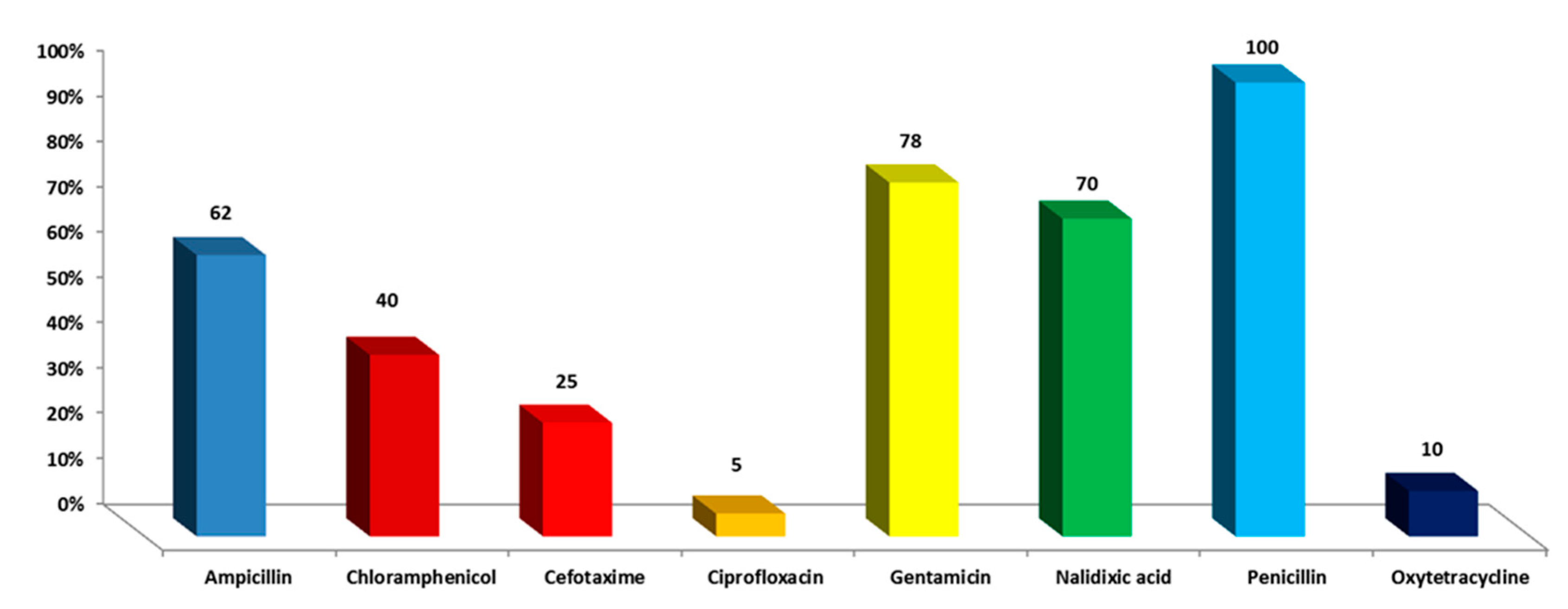
Out of 100 raw milk samples tested, 12 strains of S. enterica were found, representing a 12.5% prevalence. Resistance to penicillin was 100%, and gentamicin resistance was 78% in the isolates that were recovered. As shown in Figure 2, resistance to nalidixic acid was 70%, ampicillin 62%, and chloramphenicol 40%.

3.2. Antibacterial Activity of S. officinalis L. EO on S. enterica Isolates

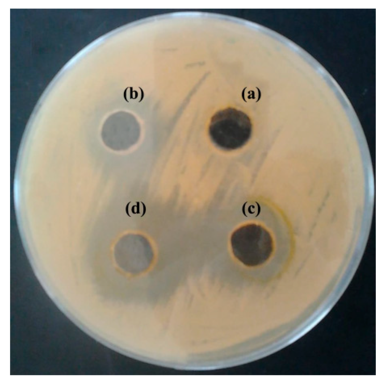
The MIC of the EO were engaged using the well diffusion technique to assess their bacteriostatic and bactericidal capabilities. The levels of the effective EO were reported in Table 1 and depicted in Figure 3. The inhibitory action of EO of S. officinalis began at 1% v/v with inhibition zones of 9 mm against S. enterica whereas inhibited bacterial growth of these strains at concentration of 5% v/v with inhibition zones of 21 mm.

3.3. Effect of S. officinalis L. EO on Viability of S. enterica

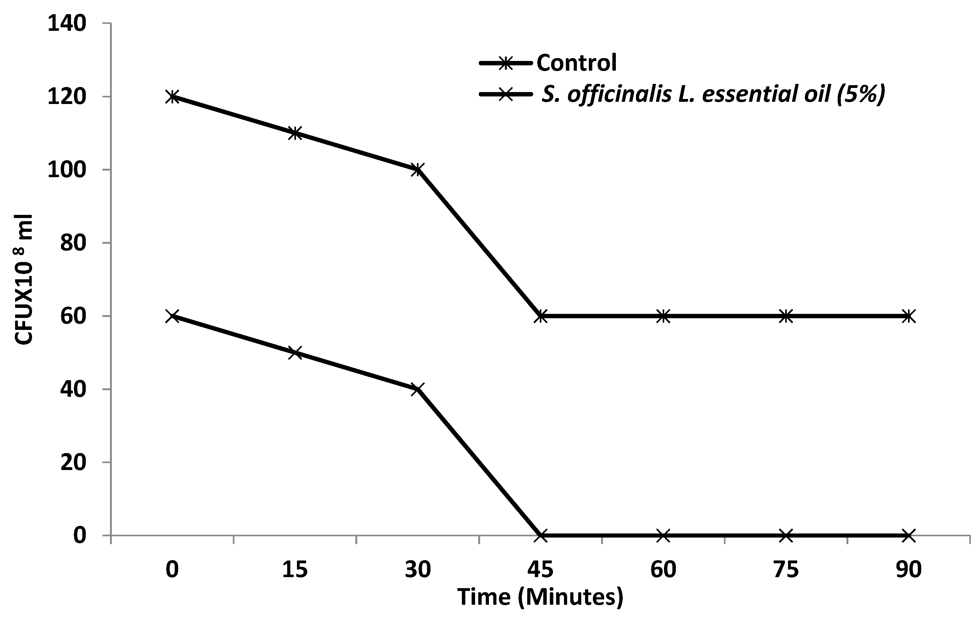
Based on these results, we have focused our efforts to investigate the inhibitory potency of the EO on viability and biofilm formation of S. enterica. Toward this, S. officinalis L. EO at 5% concentration has a possible antibacterial impact on the viability of S. enterica cells, as evidenced by the influence on cell viability. The cell viability of S. enterica was completely inhibited by S. officinalis L. EO exposure durations of 5% at 45 min (Figure 4).

3.4. Effect of S. officinalis L. EO on Biofilm Growth of S. enterica Isolates

Biofilms formed by twelve S. enterica isolates were removed after one hour of contact to 5% EO from S. officinalis L. (Figure 5).

3.5. Chemical Characterization of S. officinalis L. EO by GC

The EO obtained from a sage was analyzed for the presence, in percentages, of their constituents. The experimental findings demonstrate that a particle size of 2 mm, a condensation flow rate of 1.4 mL/min, and an extraction period of 180 min resulted in a high yield of 0.8%. The identified compounds, qualitative and quantitative analytical results by GC and GC/MS are shown in Table 2, according to their elution order on a HP-5 MS capillary column. The GC–MS analysis of the EO led to the identification of 20 different components, representing 89.94% of total oil constituents (Table 2). A total of 21 constituents representing 89.94% of the oil were identified: 1,8-cineole (39.18%), β-caryophyllene (12.8%), α-terpineol (10.32%), α-humulene (7.37%), and carvone (4.25%) were the main components of the oil. A portion (0.23%) of the total composition was not identified.

4. Discussion

One of the most common food-borne illnesses is caused by the bacterium S. enterica [40]. Studies have shown that antimicrobial resistance to β-lactams, aminoglycosides, and tetracyclines is higher, while resistance to ciprofloxacin is lower [41]. Another study found a similar result for β-lactam and macrolide antibiotics (52.9–100%), while resistance to chloramphenicol was lower (3.13%), and higher resistance to ciprofloxacin and β-lactams (68.75%), tetracycline (65.62%), and colistin sulphate (46.87) was reported [36]. Recent research suggests that increased resistance to β-lactams, tetracyclines, and fluoroquinolones may be due in part to the widespread use of antibiotics without proper supervision.
Detailed data on the chemical analysis of sage EO are available from a variety of different sources [42]. The chemotypic elements of the S. officinalis EO were discovered to include thujone (α -thujone), 1,8-cineole, and camphor [42]. EO yields and components may be affected by a variety of factors, including seasonal fluctuations, geographic location variances in oil quality and yields, EO extraction processes, and EOs acquired via various drying methods [43,44,45]. According to the aforementioned factors, significant variability in sage EO components have been observed. When dried in an oven at 45 or 65 °C, as well as in the microwave oven (500 W), the sage EO’s chemotypic products, 1,8-cineole and camphor, considerably differed from batch to batch [46]. EO batches derived from plants harvested at various times of the year and from different geographic regions included varying amounts of α -thujone and camphor [47].
It has been speculated that the EOs of S. officinalis might contain beneficial substances, and as previously stated, the sage EO has antimicrobial properties, supported by this study [48]. Therefore, the antibacterial properties of sage EO can be attributed to certain oil components, including camphor, 1,8-cineole, and α-pinene, all of which are found in high concentrations [49]. These substances are well known for their antibacterial action against many microbes [50]. According to previous research, phenolic compounds have also been linked to antibacterial activity [51]. The results show that this natural substance may be used in various sectors, including food preservation and medical treatment [52]. Its antimicrobial activity is affected by the variability of the chemical composition of S. officinalis EOs. This oil depends on several factors (such as genetic background, region, environmental conditions, season, plant parts used for extraction, and the extraction method) [53,54]. To fully understand Saudi S. officinalis L. and its active constituents in antimicrobial pharmaceuticals both locally and systemically, further research is required.

5. Conclusions

Providing a safe and nutrient-dense food supply is challenging due to several variables involved from farm to fork. There is a clear connection between food-borne pathogens such as S. enterica and dairy food safety. In the current study, we have investigated the inhibitory activity of S. officinalis. L. extract and EO on S. enterica. A selective inhibition against potentially pathogenic Salmonella and a significant inhibition of the biofilm formation were shown by the S. officinalis L. EO. To our knowledge, this is the first report on the characterization of S. officinalis EO for its antibiofilm properties against Salmonella. According to our findings, S. officinalis L. EO gathered in the Aljouf region, Saudi Arabia, may be used in the future for the development of antibacterial drugs against multidrug-resistant S. enterica.

Author Contributions

Conceptualization, S.S., M.S.A., H.A.,S.K.A.J. and M.W.; methodology, S.S., M.S.A., S.K.A.J. and M.W.; software, S.S., H.A., N.H. and F.M.S.; validation, S.S., M.S.A., H.A. and N.H.; formal analysis, M.S.A., H.A., N.H., F.M.S. and M.W.; investigation, S.S., M.S.A., H.A., S.K.A.J. and M.W.; resources, S.S., and M.W.; data curation, S.S., M.S.A., F.M.S., S.K.A.J. and M.W.; writing—original draft preparation, S.S., M.S.A., N.H., S.K.A.J. and M.W.; writing—review and editing, S.S., M.S.A., H.A., N.H., F.M.S. and M.W.; visualization, S.S., H.A., N.H. and M.W.; supervision, S.S., M.S.A. and M.W.; project administration, S.S., M.S.A. and M.W.; funding acquisition, S.S. and M.W. All authors have read and agreed to the published version of the manuscript.

Funding

This work was funded by the Deanship of Scientific Research at Jouf University under grant No (DSR-2021-01-0343).

Data Availability Statement

Data is contained within the article.

Acknowledgments

This work was funded by the Deanship of Scientific Research at Jouf University under grant No (DSR-2021-01-0343). We want to extend our thanks and appreciation to Ahmed Elbanhawy (Botany Department, Faculty of Sciences, Suez Canal University, Egypt) for helping in the identification and confirmation of the plant material used in the manuscript.

Conflicts of Interest

The authors declare no conflict of interest.

References

  1. Dhanashekar, R.; Akkinepalli, S.; Nellutla, A. Milk-Borne Infections. An Analysis of Their Potential Effect on the Milk Industry. Germs 2012, 2, 101–109. [Google Scholar] [CrossRef]
  2. Oliver, S.P.; Jayarao, B.M.; Almeida, R.A. Foodborne Pathogens in Milk and the Dairy Farm Environment: Food Safety and Public Health Implications. Foodborne Pathog. Dis. 2005, 2, 115–129. [Google Scholar] [CrossRef] [Green Version]
  3. Todd, E.C.D. Foodborne Disease and Food Control in the Gulf States. Food Control. 2017, 73, 341–366. [Google Scholar] [CrossRef]
  4. Rotimi, V.O.; Jamal, W.; Pal, T.; Sonnevend, A.; Dimitrov, T.S.; Albert, M.J. Emergence of Multidrug-Resistant Salmonella spp. and Isolates with Reduced Susceptibility to Ciprofloxacin in Kuwait and the United Arab Emirates. Diagn. Microbiol. Infect. Dis. 2008, 60, 71–77. [Google Scholar] [CrossRef]
  5. World Health Organization. Brucellosis in Humans and Animals; Weltgesundheitsorganisation, FAO, OIE—World Organisation for Animal Health, Eds.; World Health Organization: Geneva, Switzerland, 2006; ISBN 978-92-4-154713-0. [Google Scholar]
  6. Bin-Jumah, M.; Abdel-Fattah, A.-F.M.; Saied, E.M.; El-Seedi, H.R.; Abdel-Daim, M.M. Acrylamide-Induced Peripheral Neuropathy: Manifestations, Mechanisms, and Potential Treatment Modalities. Environ. Sci Pollut Res. 2021, 28, 13031–13046. [Google Scholar] [CrossRef]
  7. Ezzati, M.; Lopez, A.D.; Rodgers, A.; Vander Hoorn, S.; Murray, C.J. Selected Major Risk Factors and Global and Regional Burden of Disease. Lancet 2002, 360, 1347–1360. [Google Scholar] [CrossRef]
  8. Gal-Mor, O.; Boyle, E.C.; Grassl, G.A. Same Species, Different Diseases: How and Why Typhoidal and Non-Typhoidal Salmonella Enterica Serovars Differ. Front. Microbiol. 2014, 5, 391. [Google Scholar] [CrossRef] [Green Version]
  9. Eng, S.-K.; Pusparajah, P.; Ab Mutalib, N.-S.; Ser, H.-L.; Chan, K.-G.; Lee, L.-H. Salmonella: A Review on Pathogenesis, Epidemiology and Antibiotic Resistance. Front. Life Sci. 2015, 8, 284–293. [Google Scholar] [CrossRef] [Green Version]
  10. Chen, H.-M.; Wang, Y.; Su, L.-H.; Chiu, C.-H. Nontyphoid Salmonella Infection: Microbiology, Clinical Features, and Antimicrobial Therapy. Pediatr. Neonatol. 2013, 54, 147–152. [Google Scholar] [CrossRef] [Green Version]
  11. Black, P.H.; Kunz, L.J.; Swartz, M.N. Salmonellosis—A Review of Some Unusual Aspects. N. Engl. J. Med. 1960, 262, 811–817. [Google Scholar] [CrossRef]
  12. Klein, N.C.; Cunha, B.A. Third-Generation Cephalosporins. Med. Clin. N. Am. 1995, 79, 705–719. [Google Scholar] [CrossRef]
  13. Ventola, C.L. The Antibiotic Resistance Crisis: Part 1: Causes and Threats. Pharm. Ther. 2015, 40, 277–283. [Google Scholar]
  14. Founou, L.L.; Founou, R.C.; Essack, S.Y. Antibiotic Resistance in the Food Chain: A Developing Country-Perspective. Front. Microbiol. 2016, 7, 1881. [Google Scholar] [CrossRef] [PubMed]
  15. Collenburg, L.; Beyersdorf, N.; Wiese, T.; Arenz, C.; Saied, E.M.; Becker-Flegler, K.A.; Schneider-Schaulies, S.; Avota, E. The Activity of the Neutral Sphingomyelinase Is Important in T Cell Recruitment and Directional Migration. Front. Immunol. 2017, 8. [Google Scholar] [CrossRef] [PubMed] [Green Version]
  16. Marshall, B.M.; Levy, S.B. Food Animals and Antimicrobials: Impacts on Human Health. Clin. Microbiol. Rev. 2011, 24, 718–733. [Google Scholar] [CrossRef] [Green Version]
  17. Ruvalcaba-Gómez, J.M.; Villagrán, Z.; Valdez-Alarcón, J.J.; Martínez-Núñez, M.; Gomez-Godínez, L.J.; Ruesga-Gutiérrez, E.; Anaya-Esparza, L.M.; Arteaga-Garibay, R.I.; Villarruel-López, A. Non-Antibiotics Strategies to Control Salmonella Infection in Poultry. Animals 2022, 12, 102. [Google Scholar] [CrossRef]
  18. Wall, B.A. Dynamics and Epidemiology of Antimicrobial Resistance in Animal Production; Food and Agriculture Organization of the United Nations Drivers (FAO): Rome, Italy, 2016. [Google Scholar]
  19. Altindal, D.; Altindal, N. Chapter 81—Sage (Salvia officinalis) Oils. In Essential Oils in Food Preservation, Flavor and Safety; Preedy, V.R., Ed.; Academic Press: San Diego, CA, USA, 2016; pp. 715–721. ISBN 978-0-12-416641-7. [Google Scholar]
  20. Ghorbani, A.; Esmaeilizadeh, M. Pharmacological Properties of Salvia Officinalis and Its Components. J. Tradit. Complement. Med. 2017, 7, 433–440. [Google Scholar] [CrossRef]
  21. Jery, A.E.; Hasan, M.; Rashid, M.M.; Mesfer, M.K.A.; Danish, M.; Rebah, F.B. Phytochemical Characterization, and Antioxidant and Antimicrobial Activities of Essential Oil from Leaves of the Common Sage Salvia officinalis L. from Abha, Saudi Arabia. Asian Biomed. 2020, 14, 261–270. [Google Scholar] [CrossRef]
  22. Baricevic, D.; Sosa, S.; Della Loggia, R.; Tubaro, A.; Simonovska, B.; Krasna, A.; Zupancic, A. Topical Anti-Inflammatory Activity of Salvia officinalis L. Leaves: The Relevance of Ursolic Acid. J. Ethnopharmacol. 2001, 75, 125–132. [Google Scholar] [CrossRef]
  23. Saied, E.; Eid, A.M.; Hassan, S.E.-D.; Salem, S.S.; Radwan, A.A.; Halawa, M.; Saleh, F.M.; Saad, H.A.; Saied, E.M.; Fouda, A. The Catalytic Activity of Biosynthesized Magnesium Oxide Nanoparticles (MgO-NPs) for Inhibiting the Growth of Pathogenic Microbes, Tanning Effluent Treatment, and Chromium Ion Removal. Catalysts 2021, 11, 821. [Google Scholar] [CrossRef]
  24. Ovais, M.; Zia, N.; Ahmad, I.; Khalil, A.T.; Raza, A.; Ayaz, M.; Sadiq, A.; Ullah, F.; Shinwari, Z.K. Phyto-Therapeutic and Nanomedicinal Approaches to Cure Alzheimer’s Disease: Present Status and Future Opportunities. Front. Aging Neurosci. 2018, 10, 284. [Google Scholar] [CrossRef] [PubMed] [Green Version]
  25. Alagiakrishnan, K.; Sankaralingam, S.; Ghosh, M.; Mereu, L.; Senior, P. Antidiabetic Drugs and Their Potential Role in Treating Mild Cognitive Impairment and Alzheimer’s Disease. Discov. Med. 2013, 16, 277–286. [Google Scholar] [PubMed]
  26. Wells, R.; Truong, F.; Adal, A.M.; Sarker, L.S.; Mahmoud, S.S. Lavandula Essential Oils: A Current Review of Applications in Medicinal, Food, and Cosmetic Industries of Lavender. Nat. Prod. Commun. 2018, 13, 1403–1417. [Google Scholar] [CrossRef] [Green Version]
  27. Ali, B.; Al-Wabel, N.A.; Shams, S.; Ahamad, A.; Khan, S.A.; Anwar, F. Essential Oils Used in Aromatherapy: A Systemic Review. Asian Pac. J. Trop. Biomed. 2015, 5, 601–611. [Google Scholar] [CrossRef] [Green Version]
  28. Khalifa, S.A.M.; Shedid, E.S.; Saied, E.M.; Jassbi, A.R.; Jamebozorgi, F.H.; Rateb, M.E.; Du, M.; Abdel-Daim, M.M.; Kai, G.-Y.; Al-Hammady, M.A.M.; et al. Cyanobacteria—From the Oceans to the Potential Biotechnological and Biomedical Applications. Mar. Drugs 2021, 19, 241. [Google Scholar] [CrossRef]
  29. Charles, D.J. Antioxidant Properties of Spices, Herbs and Other Sources; Springer: New York, NY, USA, 2013; ISBN 978-1-4614-4309-4. [Google Scholar]
  30. Candela, R.G.; Rosselli, S.; Bruno, M.; Fontana, G. A Review of the Phytochemistry, Traditional Uses and Biological Activities of the Essential Oils of Genus Teucrium. Planta Med. 2021, 87, 432–479. [Google Scholar] [CrossRef]
  31. Zaouali, Y.; Messaoud, C.; Salah, A.B.; Boussaïd, M. Oil Composition Variability among Populations in Relationship with Their Ecological Areas in Tunisian Rosmarinus officinalis L. Flavour Fragr. J. 2005, 20, 512–520. [Google Scholar] [CrossRef]
  32. National Committee for Clinical Laboratory Standards. Performance Standards for Antimicrobial Disk Susceptibility Tests. Approved Standard M2-A6; National Committee for Clinical Laboratory Standards: Wayne, PA, USA, 1998. [Google Scholar]
  33. Khedher, M.R.B.; Khedher, S.B.; Chaieb, I.; Tounsi, S.; Hammami, M. Chemical Composition and Biological Activities of Salvia Officinalis Essential Oil from Tunisia. EXCLI J. 2017, 16, 160–173. [Google Scholar] [CrossRef]
  34. Saied, E.M.; El-Maradny, Y.A.; Osman, A.A.; Darwish, A.M.G.; Abo Nahas, H.H.; Niedbała, G.; Piekutowska, M.; Abdel-Rahman, M.A.; Balbool, B.A.; Abdel-Azeem, A.M. A Comprehensive Review about the Molecular Structure of Severe Acute Respiratory Syndrome Coronavirus 2 (SARS-CoV-2): Insights into Natural Products against COVID-19. Pharmaceutics 2021, 13, 1759. [Google Scholar] [CrossRef]
  35. Gaber, A.; Alsanie, W.F.; Kumar, D.N.; Refat, M.S.; Saied, E.M. Novel Papaverine Metal Complexes with Potential Anticancer Activities. Molecules 2020, 25, 5447. [Google Scholar] [CrossRef]
  36. Ansari, M.J.; Al-Ghamdi, A.; Khan, K.A.; Adgaba, N.; El-Ahmady, S.H.; Gad, H.A.; Roshan, A.; Meo, S.A.; Kolyali, S. Validation of Botanical Origins and Geographical Sources of Some Saudi Honeys Using Ultraviolet Spectroscopy and Chemometric Analysis. Saudi J. Biol. Sci. 2018, 25, 377–382. [Google Scholar] [CrossRef] [PubMed]
  37. Jemia, M.B.; Formisano, C.; Bancheva, S.; Bruno, M.; Senatore, F. Chemical Composition of the Essential Oils of Centaurea formanekii and C. orphanidea ssp. thessala, Growing Wild in Greece. Nat. Prod. Commun. 2012, 7, 1083–1086. [Google Scholar] [CrossRef] [PubMed] [Green Version]
  38. Khan, M.; Mousa, A.A.; Syamasundar, K.V.; Alkhathlan, H.Z. Determination of Chemical Constituents of Leaf and Stem Essential Oils of Artemisia monosperma from Central Saudi Arabia. Nat. Prod. Commun. 2012, 7, 1079–1082. [Google Scholar] [CrossRef] [PubMed] [Green Version]
  39. Xiao, J.; Zuo, Y.; Liu, Y.; Li, J.; Hao, Y.; Zhou, X. Effects of Nidus Vespae Extract and Chemical Fractions on Glucosyltransferases, Adherence and Biofilm Formation of Streptococcus Mutans. Arch. Oral Biol. 2007, 52, 869–875. [Google Scholar] [CrossRef]
  40. Giuffrida, A.; Giarratana, F.; Valenti, D.; Muscolino, D.; Parisi, R.; Parco, A.; Marotta, S.; Ziino, G.; Panebianco, A. A New Approach to Predict the Fish Fillet Shelf-Life in Presence of Natural Preservative Agents. Ital. J. Food Saf. 2017, 6, 6768. [Google Scholar] [CrossRef] [Green Version]
  41. Bhuvaneswa, M.; Shanmughap, S.; Natarajase, K. Prevalence of Multidrug-Resistant (MDR) Salmonella Enteritidis in Poultry and Backyard Chicken from Tiruchirappalli, India. Microbiol. J. 2015, 5, 28–35. [Google Scholar] [CrossRef] [Green Version]
  42. Khan, N.A.; Nazar, R.; Iqbal, N.; Anjum, N.A. Phytohormones and Abiotic Stress Tolerance in Plants; Springer: Berlin/Heidelberg, Germany, 2012; ISBN 978-3-642-25828-2. [Google Scholar]
  43. Craft, J.D.; Satyal, P.; Setzer, W.N. The Chemotaxonomy of Common Sage (Salvia officinalis) Based on the Volatile Constituents. Medicines 2017, 4, 47. [Google Scholar] [CrossRef] [Green Version]
  44. Perry, N.B.; Anderson, R.E.; Brennan, N.J.; Douglas, M.H.; Heaney, A.J.; McGimpsey, J.A.; Smallfield, B.M. Essential Oils from Dalmatian Sage (Salvia officinalis L.): Variations among Individuals, Plant Parts, Seasons, and Sites. J. Agric. Food Chem. 1999, 47, 2048–2054. [Google Scholar] [CrossRef]
  45. Reverchon, E.; Taddeo, R.; Porta, G. Extraction of Sage Oil by Supercritical CO2: Influence of Some Process Parameters. J. Supercrit. Fluids 1995, 8, 302–309. [Google Scholar] [CrossRef]
  46. Glisic, S.; Ivanovic, J.; Ristic, M.; Skala, D. Extraction of Sage (Salvia officinalis L.) by Supercritical CO2: Kinetic Data, Chemical Composition and Selectivity of Diterpenes. J. Supercrit. Fluids 2010, 52, 62–70. [Google Scholar] [CrossRef]
  47. Sellami, I.H.; Rebey, I.B.; Sriti, J.; Rahali, F.Z.; Limam, F.; Marzouk, B. Drying Sage (Salvia officinalis L.) Plants and Its Effects on Content, Chemical Composition, and Radical Scavenging Activity of the Essential Oil. Food Bioprocess Technol. 2012, 5, 2978–2989. [Google Scholar] [CrossRef]
  48. Ismail, M.A.; Amin, M.A.; Eid, A.M.; Hassan, S.E.-D.; Mahgoub, H.A.; Lashin, I.; Abdelwahab, A.T.; Azab, E.; Gobouri, A.A.; Elkelish, A. Comparative Study between Exogenously Applied Plant Growth Hormones versus Metabolites of Microbial Endophytes as Plant Growth-Promoting for Phaseolus Vulgaris L. Cells 2021, 10, 1059. [Google Scholar] [CrossRef] [PubMed]
  49. Longaray Delamare, A.P.; Moschen-Pistorello, I.T.; Artico, L.; Atti-Serafini, L.; Echeverrigaray, S. Antibacterial Activity of the Essential Oils of Salvia officinalis L. and Salvia triloba L. Cultivated in South Brazil. Food Chem. 2007, 100, 603–608. [Google Scholar] [CrossRef]
  50. El Euch, S.K.; Hassine, D.B.; Cazaux, S.; Bouzouita, N.; Bouajila, J. Salvia officinalis Essential Oil: Chemical Analysis and Evaluation of Anti-Enzymatic and Antioxidant Bioactivities. S. Afr. J. Bot. 2019, 120, 253–260. [Google Scholar] [CrossRef]
  51. Garcia-Gutierrez, E.; Mayer, M.J.; Cotter, P.D.; Narbad, A. Gut Microbiota as a Source of Novel Antimicrobials. Gut Microbes 2018, 10, 1–21. [Google Scholar] [CrossRef] [Green Version]
  52. Bouarab-Chibane, L.; Forquet, V.; Lantéri, P.; Clément, Y.; Léonard-Akkari, L.; Oulahal, N.; Degraeve, P.; Bordes, C. Antibacterial Properties of Polyphenols: Characterization and QSAR (Quantitative Structure–Activity Relationship) Models. Front. Microbiol. 2019, 10, 829. [Google Scholar] [CrossRef]
  53. Amit, S.K.; Uddin, M.d.M.; Rahman, R.; Islam, S.M.R.; Khan, M.S. A Review on Mechanisms and Commercial Aspects of Food Preservation and Processing. Agric. Food Secur. 2017, 6, 51. [Google Scholar] [CrossRef]
  54. Ilie, C.; Golet, I.; Craciunescu, M.; Hogea, E.; Popescu, R.; Horhat, F.G. Chemical Composition and Antimicrobial Activity of Essential Oil of Western Romanian Salvia officinalis. Rev. Chim. 2016, 67, 131–133. [Google Scholar]
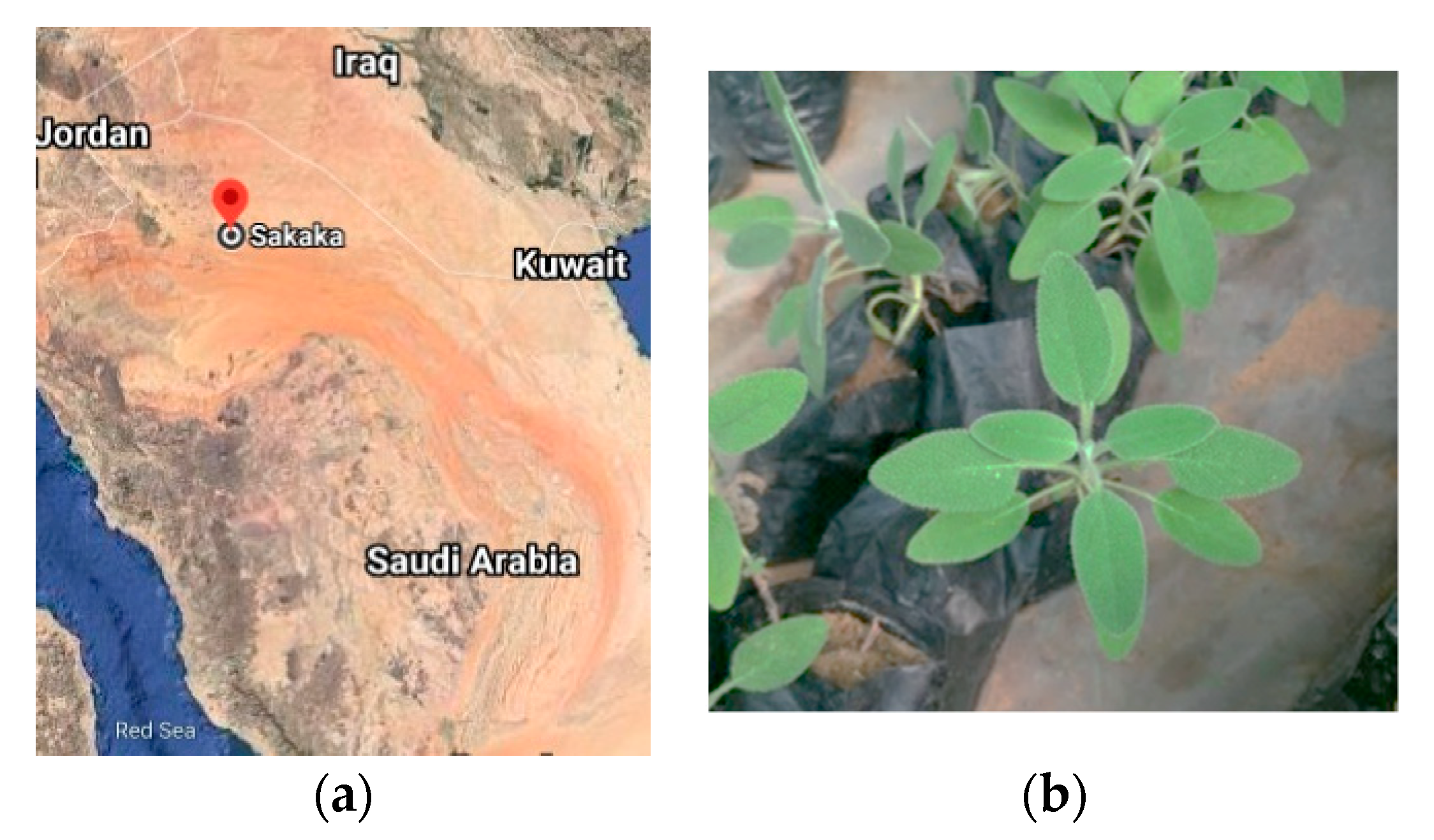
Figure 1. (a) Map of Kingdom of Saudi Arabia showing sampling site of the Sakaka, Aljouf region area and (b) S. officinalis L. leaves.
Antibiotics 11 00489 g001
Figure 2. Comparison of drug resistance patterns of S. enterica isolates against studied antimicrobial agents.
Antibiotics 11 00489 g002
Figure 3. The inhibition zone of essential oil of S. officinalis L. leaves against S. enterica, at a concentration of (a) 0% represents the negative control, (b) 1% v/v (c) 2% v/v, and (d) 5% v/v in dimethylsulfoxide (DMSO).
Antibiotics 11 00489 g003
Figure 4. Effect of EO of S. officinalis at 5% v/v concentration on the viability of S. enterica. Values are the average of three individual replicates (means ± S.D.).
Antibiotics 11 00489 g004
Figure 5. Qualitative biofilm assessment of S. enterica by 96-well microtiter plate evaluating (A) the point of wells with biofilm establishment and (B) specifies wells with no biofilm formation in the treatment by S. officinalis L. EO (5% v/v).
Antibiotics 11 00489 g005
Table 1. Inhibition zone diameter and MIC of S. officinalis L. EO against S. enterica.
ItemSalmonella enterica
Inhibition Zone Diameter (mm)MIC (µg/mL)
MICMIC50MIC90
Essential Oil a21 ± 1.09125
DMSO (Negative control)----
a Concentration 5% v/v EO in DMSO. Sensitive (Inhibition Zone Diameter ≥11 mm: Bauer et al. [31]). Results are Mean ± S.D. of triplicate experiments.
Table 2. Chemical composition of S. officinalis L. EO.
Peak NoCompound aRI bKI cComposition (%)
1α-Pinene9389400.74
2Camphene9549540.86
3Myrcene9949923.36
4α-Terpinene101810190.18
5p-Cymene102610250.72
6Limonene103010300.42
71,8-Cineol1035103339.18
8β-Ocimene104010410.14
9γ-Terpinene106210630.12
10α-Terpinolene109010890.09
11Linalool109810980.99
12Borneol106811700.36
13α-Terpineol1188118910.32
14Myrtenol119311943.13
15Carvone124612484.25
16Bornyl acetate128512860.15
17β-Caryophyllene1419141912.8
18α-humulene145514567.37
19Germacrene-B153515360.38
20Viridiflorol159115914.15
21UD e169516950.23
Identified components (%)89.94
Monoterpene hydrocarbons6.28
Oxygenated monoterpenes55.73
Sesquiterpene hydrocarbons20.55
Oxygenated sesquiterpenes4.15
Oil yield (%) (v/w)
0.45
a Components were identified through KI and GC–MS (gas chromatograph coupled with mass spectrometry) and listed according to their elution on HP-5 MS capillary column (30 m). b RI: Retention Index. c KI: Kovats indexes on HP-Innowax capillary column in reference to C9-C28 n-alkanes [37,40]. d UD: not identified compound.
Publisher’s Note: MDPI stays neutral with regard to jurisdictional claims in published maps and institutional affiliations.