Photoactivatable Aptamer-Based Biosensors for Point-of-Care Testing: Advances and Applications


Abstract
1. Introduction
2. The Mechanism of Action of Photoactivatable Aptamers
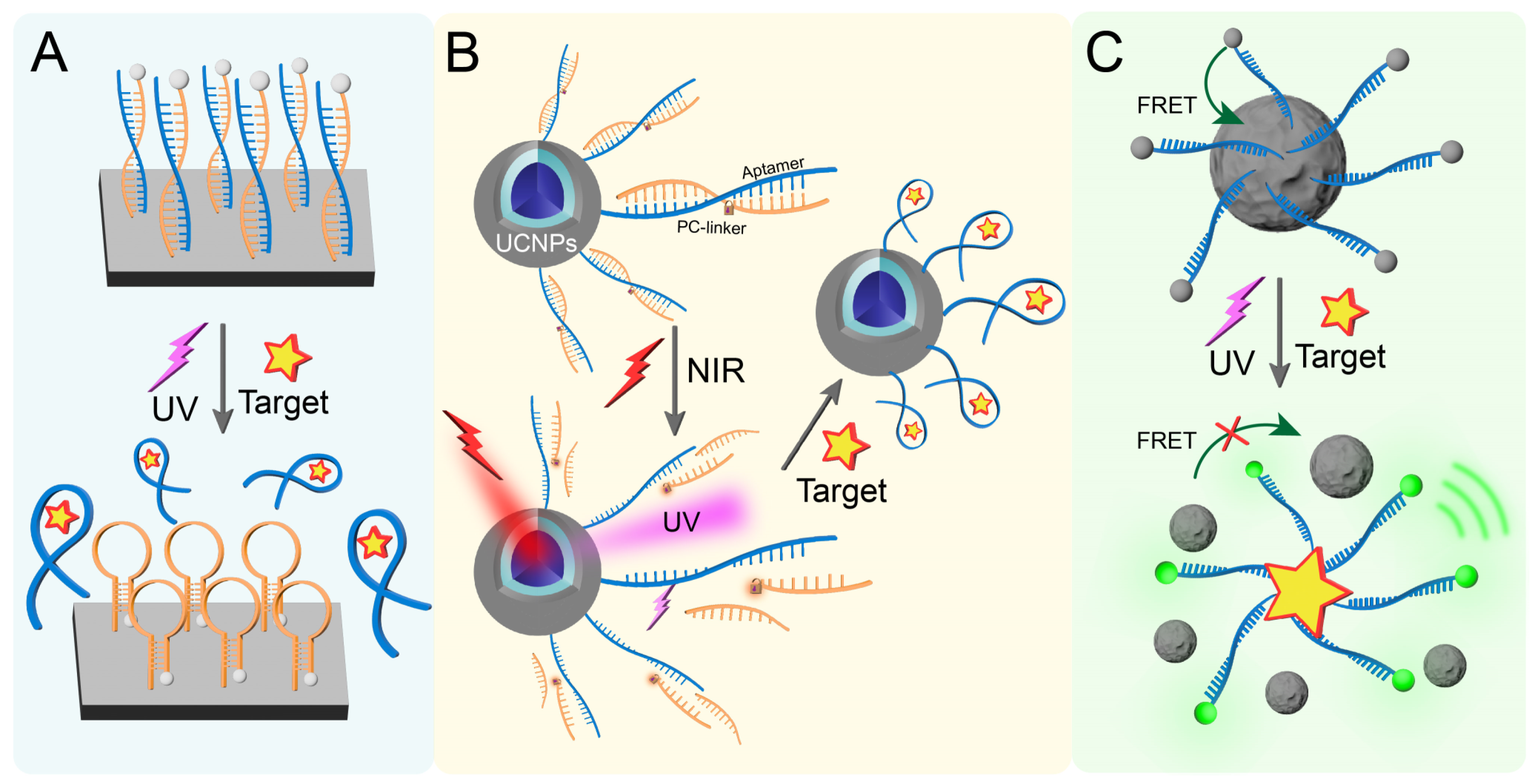
2.1. Optical Control of Aptamer
2.2. Optical Control of Materials
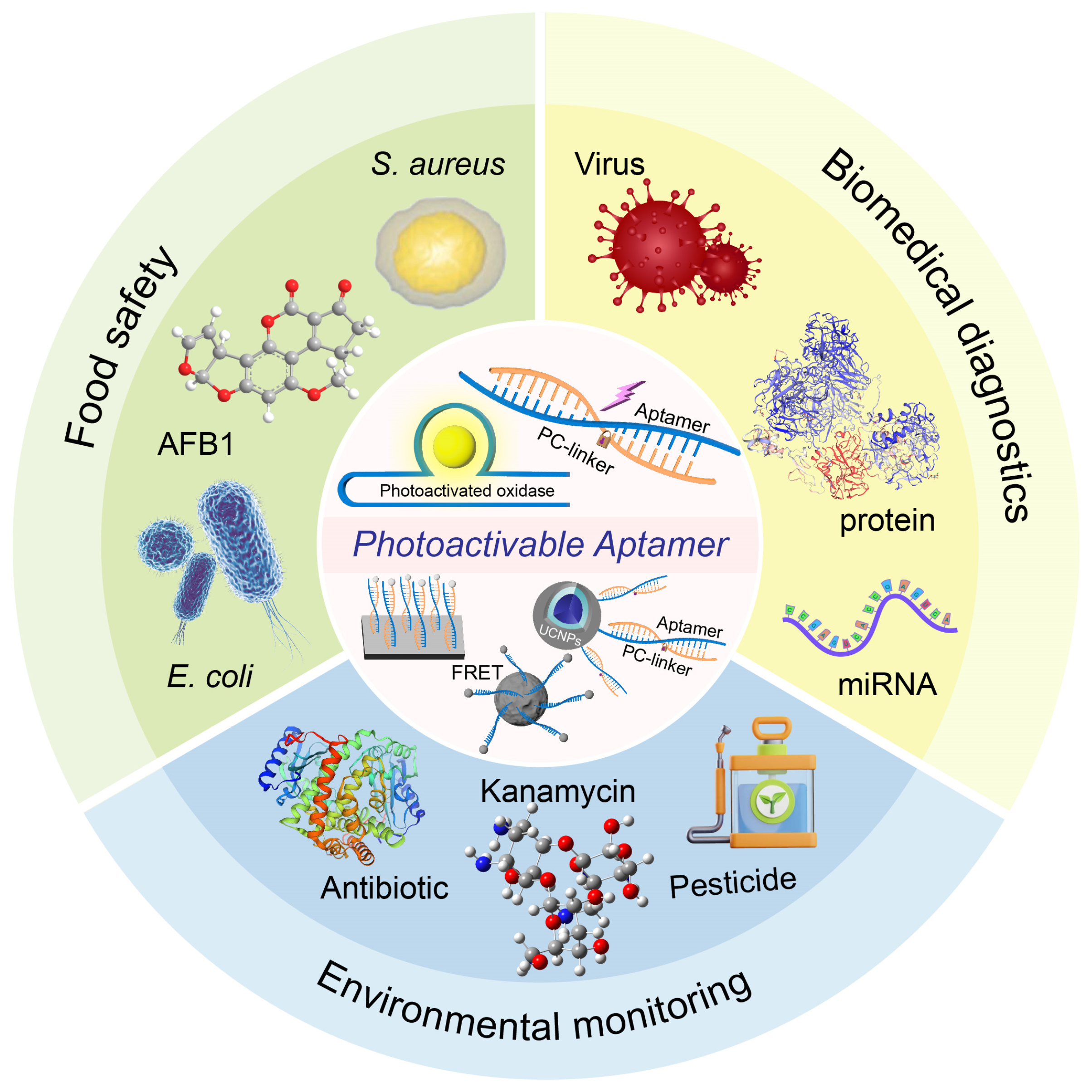
3. The Application of POCT Based on Photoactivatable Aptamers in Different Fields
3.1. Food Safety
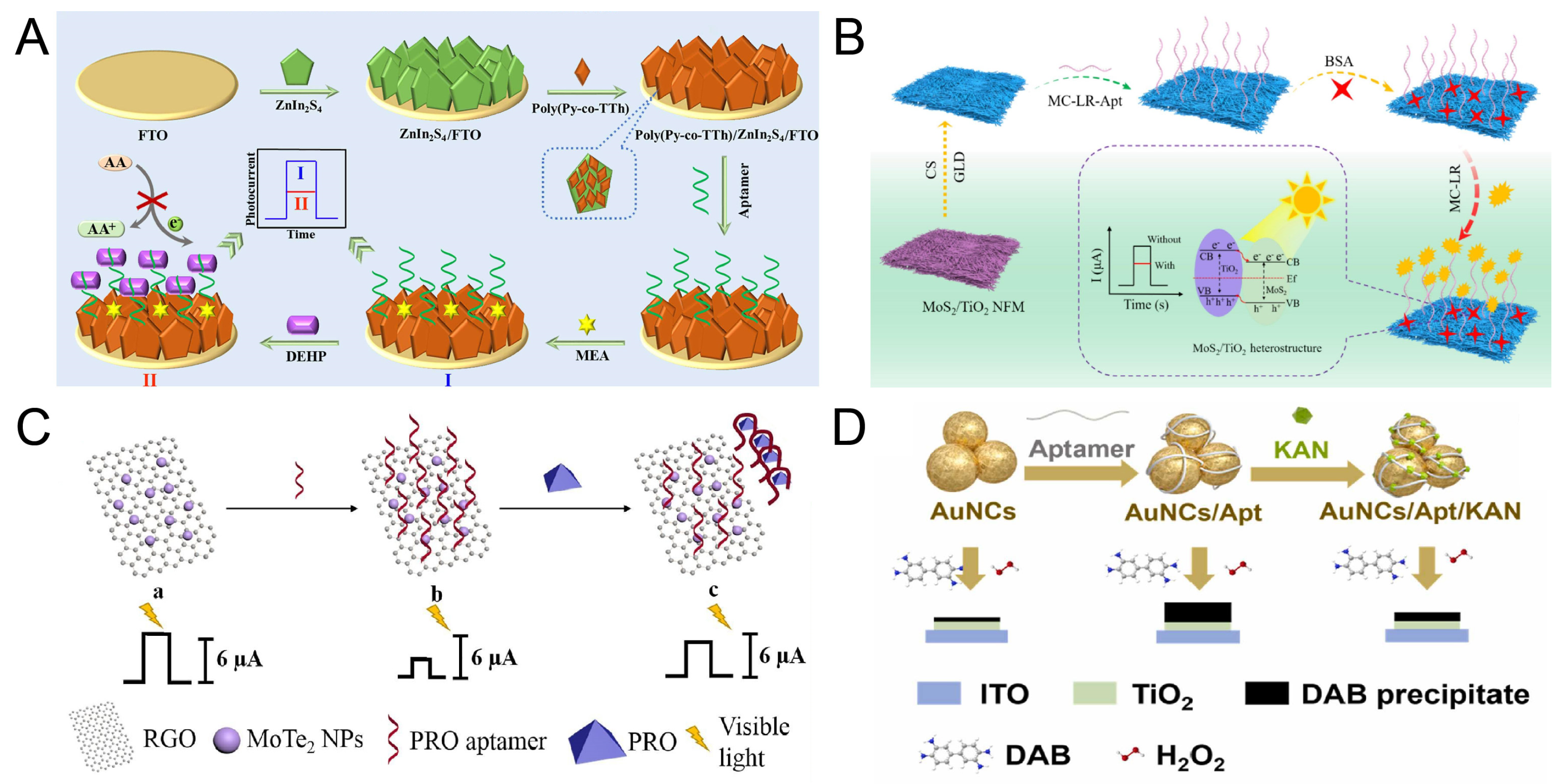
3.2. Environmental Monitoring
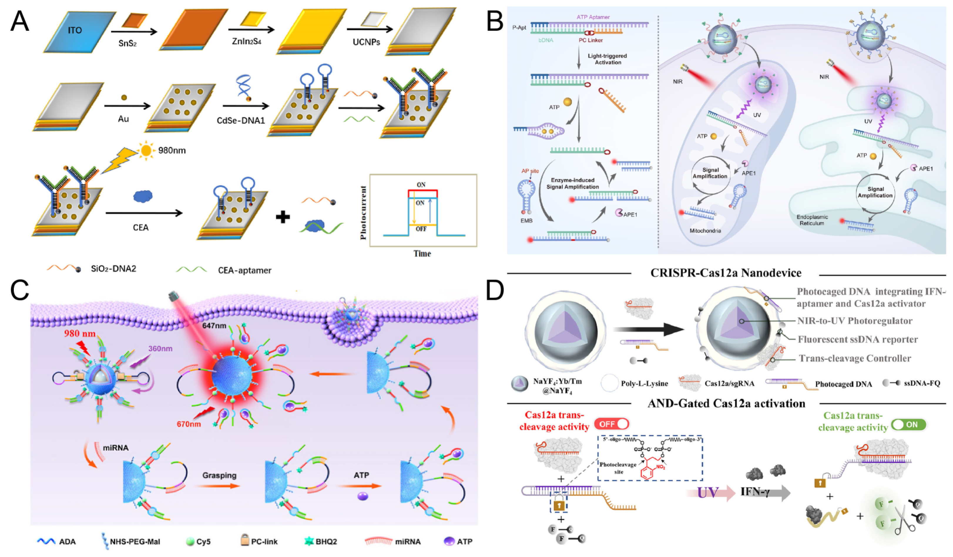
3.3. Biomedical Diagnostics
4. Summary and Outlook
Author Contributions
Funding
Conflicts of Interest
References
- Mu, Y.; Chen, Z.; Zhan, J.; Zhang, J. Recent Advances in Aptamer-Based sensors for in vitro detection of small molecules. Anal. Sens. 2024, 4, e202400027. [Google Scholar] [CrossRef]
- Liu, R.; Liu, Z.; Chen, M.; Xing, H.; Zhang, P.; Zhang, J. Cooperatively designed aptamer-PROTACs for spatioselective degradation of nucleocytoplasmic shuttling protein for enhanced combinational therapy. Chem. Sci. 2024, 15, 134–145. [Google Scholar] [CrossRef] [PubMed]
- Zhang, J.; Lan, T.; Lu, Y. Overcoming major barriers to developing successful sensors for practical applications using functional nucleic acids. Annu. Rev. Anal. Chem. 2022, 15, 151–171. [Google Scholar] [CrossRef]
- Ellington, A.D.; Szostak, J.W. In vitro selection of RNA molecules that bind specific ligands. Nature 1990, 346, 818–822. [Google Scholar] [CrossRef]
- Tuerk, C.; Gold, L. Systematic evolution of ligands by exponential enrichment: RNA ligands to bacteriophage T4 DNA polymerase. Science 1990, 249, 505–510. [Google Scholar] [CrossRef]
- Shi, Y.; Yun, Y.; Wang, R.; Liu, Z.; Wu, Z.; Xiang, Y.; Zhang, J. Engineering covalent aptamer chimeras for enhanced autophagic degradation of membrane proteins. Angew. Chem. Int. Ed. 2025, 64, e202425123. [Google Scholar] [CrossRef]
- Wong, K.-Y.; Liu, Y.; Wong, M.-S.; Liu, J. Cornea-SELEX for aptamers targeting the surface of eyes and liposomal drug delivery. Exploration 2024, 4, 20230008. [Google Scholar] [CrossRef]
- Wang, J.; Chen, D.; Huang, W.; Yang, N.; Yuan, Q.; Yang, Y. Aptamer-functionalized field-effect transistor biosensors for disease diagnosis and environmental monitoring. Exploration 2023, 3, 20210027. [Google Scholar] [CrossRef]
- Song, Y.; Zhu, Z.; An, Y.; Zhang, W.; Zhang, H.; Liu, D.; Yu, C.; Duan, W.; Yang, C.J. Selection of DNA aptamers against epithelial cell adhesion molecule for cancer cell imaging and circulating tumor cell capture. Anal. Chem. 2013, 85, 4141–4149. [Google Scholar] [CrossRef]
- Zheng, L.; Ye, Q.; Wang, M.; Sun, F.; Chen, Q.; Yu, X.; Wang, Y.; Liang, P. Research progress in small-molecule detection using aptamer-based SERS techniques. Biosensors 2025, 15, 29. [Google Scholar] [CrossRef]
- Liu, Y.; Wang, J.; Cui, G.; Wang, X.; Xiang, S.; Huang, W.; Liu, C. RNA aptamer-based CRISPR-Cas12a system for enhanced small molecule detection and point-of-care testing. Int. J. Biol. Macromol. 2025, 303, 140675. [Google Scholar] [CrossRef] [PubMed]
- Tan, J.H.; Fraser, A.G. Quantifying metabolites using structure-switching aptamers coupled to DNA sequencing. Nat. Biotechnol. 2025, 1–4. [Google Scholar] [CrossRef] [PubMed]
- Liu, H.; Peng, J.-M.; Zha, C.-J.; Su, M.; Ying, Z.-M. Split T7 switch-based orthogonal logic operation of fluorogenic RNA aptamer for small molecules detection. Spectrochim. Acta Part A 2025, 336, 126044. [Google Scholar] [CrossRef]
- Hayat, M.; Bukhari, S.A.R.; Raza, M.; Aslam, A.; Liu, Z. Nanostructured aptasensors for ricin detection and tumor therapy: Exploring aptamer-protein interactions and conformational stability in biological complexities. Int. J. Biol. Macromol. 2025, 310, 143282. [Google Scholar] [CrossRef]
- Filius, M.; Fasching, L.; van Wee, R.; Rwei, A.Y.; Joo, C. Decoding aptamer-protein binding kinetics for continuous biosensing using single-molecule techniques. Sci. Adv. 2025, 11, eads9687. [Google Scholar] [CrossRef]
- Cui, S.; Wang, F.; Yang, W. Highly sensitive electrochemical PDGF-BB sensor based on protein-templated split aptamer ligation reaction. Sens. Actuators B 2025, 425, 136996. [Google Scholar] [CrossRef]
- Li, D.; Chen, Q.; Li, Y.; Yuan, R.; Xiang, Y. Programming catalytic nucleic acid amplification cascade for highly sensitive electrochemical aptamer-based angiopoietin like protein 4 biosensor. Sens. Actuators B 2025, 427, 137207. [Google Scholar] [CrossRef]
- Feng, Z.Y.; Liu, R.; Li, X.; Zhang, J. Harnessing the CRISPR-Cas13d system for protein detection by dual-aptamer-based transcription amplification. Chem.—Eur. J. 2023, 29, e202202693. [Google Scholar] [CrossRef]
- Kalsoom, I.; Shehzadi, K.; Irfan, M.; Qiu, L.; Wang, Y.; Xu, Z.; Meng, Z. Structure switching aptamer enhance sensitivity and specificity of photonic crystal-based sensors for RSV-G protein detection. Biosens. Bioelectron. 2025, 273, 117091. [Google Scholar] [CrossRef]
- Wang, J.; Zhao, L.; Li, X.; Gao, Y.; Yong, W.; Jin, Y.; Dong, Y. Development of aptamer-based lateral flow devices for rapid detection of SARS-CoV-2 S protein and uncertainty assessment. Talanta 2025, 281, 126825. [Google Scholar] [CrossRef]
- Goto, K.; Amano, R.; Ichinose, A.; Michishita, A.; Hamada, M.; Nakamura, Y.; Takahashi, M. Generation of RNA aptamers against chikungunya virus E2 envelope protein. J. Virol. 2025, 29, e02095-24. [Google Scholar] [CrossRef] [PubMed]
- Nourry, J.; Chevalier, P.; Laurenceau, E.; Cattoen, X.; Bertrand, X.; Peres, B.; Oukacine, F.; Peyrin, E.; Choisnard, L. Whole-cell aptamer-based techniques for rapid bacterial detection: Alternatives to traditional methods. J. Pharm. Biomed. Anal. 2025, 255, 116661. [Google Scholar] [CrossRef] [PubMed]
- Fatah, S.A.; Omer, K.M. Aptamer-modified MOFs (Aptamer@ MOF) for efficient detection of bacterial pathogens: A Review. ACS Appl. Mater. Interfaces 2025, 17, 11578–11594. [Google Scholar] [CrossRef]
- Liu, R.; Yun, Y.; Feng, Z.-Y.; Chen, M.; Zhang, J. Rational design of trident aptamer scaffold for rapid and accurate monitoring of 25-Hydroxyvitamin D3 metabolism in living cells. Anal. Chem. 2023, 95, 10322–10329. [Google Scholar] [CrossRef]
- Tang, Y.; Li, Y.; Chen, P.; Zhong, S.; Yang, Y. Nucleic acid aptamer-based sensors for bacteria detection: A Review. BioEssays 2025, 47, e202400111. [Google Scholar] [CrossRef]
- Li, L.; Wan, J.; Wen, X.; Guo, Q.; Jiang, H.; Wang, J.; Ren, Y.; Wang, K. Identification of a new DNA aptamer by tissue-SELEX for cancer recognition and imaging. Anal. Chem. 2021, 93, 7369–7377. [Google Scholar] [CrossRef]
- Hsu, Y.-W.; Ma, L.; Tang, Y.; Li, M.; Zhou, C.; Geng, Y.; Zhang, C.; Wang, T.; Guo, W.; Li, M. The application of aptamers in the repair of bone, nerve, and vascular tissues. J. Mater. Chem. B 2025, 13, 1872–1889. [Google Scholar] [CrossRef]
- Chen, Y.; Li, F.; Zhang, S.; Liu, F.; Mao, C.; Li, M.; Jiang, J.; Zhang, Y.; Fan, C.; Zuo, X. DNA Framework-ensembled aptamers enhance fluid stability in circulating tumor cells capture for tumor treatment evaluation. Angew. Chem. Int. Ed. 2025, e202425252. [Google Scholar] [CrossRef]
- Qin, X.; Li, D.; Qin, X.; Chen, F.; Guo, H.; Gui, Y.; Zhao, J.; Jiang, L.; Luo, D. Electrochemical detection of the cardiac biomarker cardiac troponin I. View 2024, 5, 20240025. [Google Scholar] [CrossRef]
- Xiao, Q.; Zhang, D.; Yang, M.; Liu, S.; Fang, Y.; Huang, S. A Highly sensitive electrochemical aptasensor for kanamycin: Leveraging recJf exonuclease-assisted target recycling and hybridization chain reaction signal amplification. J. Anal. Test. 2025, 9, 96–108. [Google Scholar] [CrossRef]
- Adachi, T.; Nakamura, Y. Aptamers: A review of their chemical properties and modifications for therapeutic application. Molecules 2019, 24, 4229. [Google Scholar] [CrossRef] [PubMed]
- Ni, S.; Zhuo, Z.; Pan, Y.; Yu, Y.; Li, F.; Liu, J.; Wang, L.; Wu, X.; Li, D.; Wan, Y. Recent progress in aptamer discoveries and modifications for therapeutic applications. ACS Appl. Mater. Interfaces 2020, 13, 9500–9519. [Google Scholar] [CrossRef] [PubMed]
- Bhardwaj, T.; Sharma, T.K. Aptasensors for full body health checkup. Biosens. Bioelectron. 2022, 11, 100199. [Google Scholar] [CrossRef]
- Farid, S.; Ghosh, S.; Dutta, M.; Stroscio, M.A. Aptamer-based optical and electrochemical sensors: A review. Chemosensors 2023, 11, 569. [Google Scholar] [CrossRef]
- Wang, W.; He, Y.; He, S.; Deng, L.; Wang, H.; Cao, Z.; Feng, Z.; Xiong, B.; Yin, Y. A brief review of aptamer-based biosensors in recent years. Biosensors 2025, 15, 120. [Google Scholar] [CrossRef]
- Chen, Z.; Chen, M.; Liu, R.; Fan, H.; Zhang, J. A cocktail therapeutic strategy based on clofarabine-containing aptamer-PROTAC for enhanced cancer therapy. Chem. Comm. 2023, 59, 11560–11563. [Google Scholar] [CrossRef]
- Yu, H.; Alkhamis, O.; Canoura, J.; Liu, Y.; Xiao, Y. Advances and challenges in small-molecule DNA aptamer isolation, characterization, and sensor development. Angew. Chem. Int. Ed. 2021, 60, 16800–16823. [Google Scholar] [CrossRef]
- Ali, G.K.; Omer, K.M. Molecular imprinted polymer combined with aptamer (MIP-aptamer) as a hybrid dual recognition element for bio (chemical) sensing applications. Review. Talanta 2022, 236, 122878. [Google Scholar] [CrossRef]
- Wang, K.; Wang, M.; Ma, T.; Li, W.; Zhang, H. Review on the selection of aptamers and application in paper-based sensors. Biosensors 2022, 13, 39. [Google Scholar] [CrossRef]
- Chung, Y.-D.; Tsai, Y.-C.; Wang, C.-H.; Lee, G.-B. Aptamer selection via versatile microfluidic platforms and their diverse applications. Lab Chip 2025, 25, 1047–1080. [Google Scholar] [CrossRef]
- Yuan, M.; Li, C.; Wang, M.; Cao, H.; Ye, T.; Hao, L.; Wu, X.; Yin, F.; Yu, J.; Xu, F. Low-cost, portable, on-site fluorescent detection of As (III) by a paper-based microfluidic device based on aptamer and smartphone imaging. Microchim. Acta 2023, 190, 109. [Google Scholar] [CrossRef] [PubMed]
- Inês, A.; Cosme, F. Biosensors for Detecting Food Contaminants—An Overview. Processes 2025, 13, 380. [Google Scholar] [CrossRef]
- Shaban, S.M.; Kim, D.-H. Recent advances in aptamer sensors. Sensors 2021, 21, 979. [Google Scholar] [CrossRef]
- Chen, Y.; Morihiro, K.; Nemoto, Y.; Ichimura, A.; Ueki, R.; Sando, S.; Okamoto, A. Selective inhibition of cancer cell migration using a pH-responsive nucleobase-modified DNA aptamer. Chem. Sci. 2024, 15, 17097–17102. [Google Scholar] [CrossRef]
- Liang, Q.; Luo, Y.; Zeng, J.; Han, S.; Wang, Y.; Su, X.; Li, X.; Liang, T.; Liu, J.; Qu, P. Temporal responsive vesicle-INFγ aptamer-PEI/HA system targeted activation of B-CD4T-Tfh-CD8T cells cascade inhibits TNBC progression. Nano Today 2025, 62, 102708. [Google Scholar] [CrossRef]
- Wang, C.; O’Hagan, M.P.; Li, Z.; Zhang, J.; Ma, X.; Tian, H.; Willner, I. Photoresponsive DNA materials and their applications. Chem. Soc. Rev. 2022, 51, 720–760. [Google Scholar] [CrossRef]
- Aqib, R.M.; Umer, A.; Li, J.; Liu, J.; Ding, B. Light responsive DNA nanomaterials and their biomedical applications. Chem.-Asian J. 2024, 19, e202400226. [Google Scholar] [CrossRef]
- Zhang, X.; Shi, C.; Liu, Q.; Zhong, Y.; Zhu, L.; Zhao, Y. Combination of adenosine blockade and ferroptosis for photo-immunotherapy of triple negative breast cancer with aptamer-modified copper sulfide. J. Mater. Chem. B 2025, 13, 2504–2519. [Google Scholar] [CrossRef]
- Cesarini, V.; Appleton, S.L.; de Franciscis, V.; Catalucci, D. The recent blooming of therapeutic aptamers. Mol. Asp. Med. 2025, 102, 101350. [Google Scholar] [CrossRef]
- Meng, L.; Zhao, T.; Wang, S.; Wang, W. A biomimetic 3D DNA nanoplatform for enhanced capture and high-purity isolation of stem cell exosomes. Anal. Methods 2025, 17, 388–394. [Google Scholar] [CrossRef]
- Chen, S.; Li, J.; Liang, H.; Lin, X.H.; Li, J.; Yang, H.H. Light-induced activation of c-Met signalling by photocontrolled DNA assembly. Chem.-Eur. J. 2018, 24, 15988–15992. [Google Scholar] [CrossRef] [PubMed]
- Hong, S.; Zhang, X.; Lake, R.J.; Pawel, G.T.; Guo, Z.; Pei, R.; Lu, Y. A photo-regulated aptamer sensor for spatiotemporally controlled monitoring of ATP in the mitochondria of living cells. Chem. Sci. 2020, 11, 713–720. [Google Scholar] [CrossRef] [PubMed]
- Wu, Y.; Yang, Z.; Lu, Y. Photocaged functional nucleic acids for spatiotemporal imaging in biology. Curr. Opin. Chem. Biol. 2020, 57, 95–104. [Google Scholar] [CrossRef]
- Zhao, D.; Yang, G.; Liu, Q.; Liu, W.; Weng, Y.; Zhao, Y.; Qu, F.; Li, L.; Huang, Y. A photo-triggerable aptamer nanoswitch for spatiotemporal controllable siRNA delivery. Nanoscale 2020, 12, 10939–10943. [Google Scholar] [CrossRef]
- Liu, R.; Jiang, D.; Yun, Y.; Feng, Z.; Zheng, F.; Xiang, Y.; Fan, H.; Zhang, J. Photoactivatable engineering of CRISPR/Cas9-inducible DNAzyme probe for in situ imaging of nuclear zinc ions. Angew. Chem. Int. Ed. 2024, 63, e202315536. [Google Scholar] [CrossRef]
- Wang, S.; Wang, J.; Li, B.; Zhang, J. Photoactivable CRISPR for biosensing and cancer therapy. ChemBioChem 2024, 25, e202400685. [Google Scholar] [CrossRef]
- Xiao, H.; Chen, Y.; Yuan, E.; Li, W.; Jiang, Z.; Wei, L.; Su, H.; Zeng, W.; Gan, Y.; Wang, Z. Obtaining more accurate signals: Spatiotemporal imaging of cancer sites enabled by a photoactivatable aptamer-based strategy. ACS Appl. Mater. Interfaces 2016, 8, 23542–23548. [Google Scholar] [CrossRef]
- Ranzani, A.T.; Buchholz, K.; Blackholm, M.; Kopkin, H.; Möglich, A. Induction of bacterial expression at the mRNA level by light. Nucleic Acids Res. 2024, 52, 10017–10028. [Google Scholar] [CrossRef]
- Motohashi, Y.; Nishihara, T.; Tanabe, K. Preparation of a multifunctional photoactivated prodrug on a streptavidin scaffold bearing a DNA aptamer. Bioorg. Med. Chem. Lett. 2022, 71, 128819. [Google Scholar] [CrossRef]
- Ye, M.; Hu, S.; Zhou, L.; Tang, X.; Zhao, S.; Zhao, J. Fluidic membrane accelerating the kinetics of photoactivatable hybridization chain reaction for accurate imaging of tumor-derived exosomes. Anal. Chem. 2022, 94, 17645–17652. [Google Scholar] [CrossRef]
- Zhan, J.; Wang, S.; Li, X.; Zhang, J. Molecular engineering of functional DNA molecules toward point-of-care diagnostic devices. Chem. Commun. 2025, 61, 4316–4338. [Google Scholar] [CrossRef] [PubMed]
- Dong, T.; Yu, C.; Han, F.; Mao, Q.; Jing, W.; Yang, Z.; Pires, N.M.M.; Correia, J.H.; Lin, Q.; Hu, F. Cutting-edge RNA quantitative detection methods for rapid and on-site analysis. TrAC Trends Anal. Chem. 2025, 190, 118274. [Google Scholar] [CrossRef]
- Khan, A.R.; Hussain, W.L.; Shum, H.C.; Hassan, S.U. Point-of-care testing: A critical analysis of the market and future trends. Front. Lab Chip Technol. 2024, 3, 1394752. [Google Scholar] [CrossRef]
- Han, G.-R.; Goncharov, A.; Eryilmaz, M.; Ye, S.; Palanisamy, B.; Ghosh, R.; Lisi, F.; Rogers, E.; Guzman, D.; Yigci, D. Machine learning in point-of-care testing: Innovations, challenges, and opportunities. Nat. Commun. 2025, 16, 3165. [Google Scholar] [CrossRef]
- Liu, Q.; Lu, C.; Lv, Q.; Lei, L. Emerging point-of-care testing technology for the detection of animal pathogenic microorganisms. Chem. Eng. J. 2025, 512, 162548. [Google Scholar] [CrossRef]
- Raja, M.; Musi, R.; Fattorini, M.; Piva, E.; Putoto, G. Point of care testing and transfusion safety in resource limited settings: A review. J. Blood Disord. Transfus. 2015, 6, 269. [Google Scholar]
- Fonseca, W.T.; Parra Vello, T.; Lelis, G.C.; Ferreira Deleigo, A.V.r.; Takahira, R.K.; Martinez, D.S.T.; de Oliveira, R.F. Chemical sensors and biosensors for point-of-care testing of pets: Opportunities for individualized diagnostics of companion animals. ACS Sens. 2025, 10, 3222–3238. [Google Scholar] [CrossRef]
- Futane, A.; Narayanamurthy, V.; Jadhav, P.; Srinivasan, A. Aptamer-based rapid diagnosis for point-of-care application. Microfluid. Nanofluid. 2023, 27, 15. [Google Scholar] [CrossRef]
- Zhao, L.; Li, L.; Zhao, Y.; Zhu, C.; Yang, R.; Fang, M.; Luan, Y. Aptamer-based point-of-care-testing for small molecule targets: From aptamers to aptasensors, devices and applications. TrAC Trends Anal. Chem. 2023, 169, 117408. [Google Scholar] [CrossRef]
- Lan, Y.; He, B.; Tan, C.S.; Ming, D. Applications of smartphone-based aptasensor for diverse targets detection. Biosensors 2022, 12, 477. [Google Scholar] [CrossRef]
- Yin, B.; Wan, X.; Sohan, A.M.F.; Lin, X. Microfluidics-based POCT for SARS-CoV-2 diagnostics. Micromachines 2022, 13, 1238. [Google Scholar] [CrossRef] [PubMed]
- Du, Z.; Chen, L.; Yang, S. Advancements in the research of finger-actuated POCT chips. Microchim. Acta 2024, 191, 65. [Google Scholar] [CrossRef] [PubMed]
- Fan, Y.; Li, J.; Amin, K.; Yu, H.; Yang, H.; Guo, Z.; Liu, J. Advances in aptamers, and application of mycotoxins detection: A review. Food Res. Int. 2023, 170, 113022. [Google Scholar] [CrossRef]
- Liu, G.; Wang, X.; Su, X.; Ji, S.; Ma, Z.; Gao, Y.; Song, X. The development potential of AuNPs-based lateral flow technology combined with other advanced technologies in POCT. Appl. Biochem. Biotechnol. 2025, 197, 2867–2886. [Google Scholar] [CrossRef]
- Song, W.; Hu, J.-J.; Song, S.-J.; Xu, Y.; Yang, H.; Yang, F.; Zhou, Y.; Yu, T.; Qiu, W.-X. Aptamer-gold nanocage composite for photoactivated immunotherapy. ACS Appl. Mater. Interfaces 2022, 14, 42931–42939. [Google Scholar] [CrossRef]
- Huang, X.; Zhu, J.; Dong, C.; Li, Y.; Yu, Q.; Wang, X.; Chen, Z.; Li, J.; Yang, Y.; Wang, H. Polyvalent aptamer-functionalized NIR-II quantum dots for targeted theranostics in high PD-L1-expressing tumors. ACS Appl. Mater. Interfaces 2024, 16, 21571–21581. [Google Scholar] [CrossRef]
- Prante, M.; Segal, E.; Scheper, T.; Bahnemann, J.; Walter, J. Aptasensors for point-of-care detection of small molecules. Biosensors 2020, 10, 108. [Google Scholar] [CrossRef]
- Liu, S.; Xu, Y.; Jiang, X.; Tan, H.; Ying, B. Translation of aptamers toward clinical diagnosis and commercialization. Biosens. Bioelectron. 2022, 208, 114168. [Google Scholar] [CrossRef]
- Zhang, J.; Lan, T.; Lu, Y. Translating in vitro diagnostics from centralized laboratories to point-of-care locations using commercially-available handheld meters. TrAC Trends Anal. Chem. 2020, 124, 115782. [Google Scholar] [CrossRef]
- Xiong, Y.; Zhang, J.; Yang, Z.; Mou, Q.; Ma, Y.; Xiong, Y.; Lu, Y. Functional DNA regulated CRISPR-Cas12a sensors for point-of-care diagnostics of non-nucleic-acid targets. J. Am. Chem. Soc. 2019, 142, 207–213. [Google Scholar] [CrossRef]
- Duan, Z.; Tan, L.; Duan, R.; Chen, M.; Xia, F.; Huang, F. Photoactivated biosensing process for dictated ATP detection in single living cells. Anal. Chem. 2021, 93, 11547–11556. [Google Scholar] [CrossRef] [PubMed]
- Huang, F.; Lin, M.; Duan, R.; Lou, X.; Xia, F.; Willner, I. Photoactivated specific mRNA detection in single living cells by coupling “signal-on” fluorescence and “signal-off” electrochemical signals. Nano Lett. 2018, 18, 5116–5123. [Google Scholar] [CrossRef] [PubMed]
- Jia, Y.; Han, B.; Zhou, G.; Li, Z.; Xue, M.; Liang, B.; Liu, P.; Cheng, Y. Light-activated aptasensor for plug-and-play detection of Aflatoxin B1 in food samples. Anal. Chim. Acta 2025, 1346, 343786. [Google Scholar] [CrossRef]
- Quillin, A.L.; Karloff, D.B.; Ayele, T.M.; Flores, T.F.; Chen, G.; McEachin, Z.T.; Valdez-Sinon, A.N.; Heemstra, J.M. Imaging and tracking RNA in live mammalian cells via fluorogenic photoaffinity labeling. ACS Chem. Biol. 2025, 20, 707–720. [Google Scholar] [CrossRef]
- Wakano, M.; Tsunoda, M.; Murayama, K.; Morimoto, J.; Ueki, R.; Aoyama-Ishiwatari, S.; Hirabayashi, Y.; Asanuma, H.; Sando, S. Reversible optical control of receptor tyrosine kinase activity and ERK dynamics using azobenzene-carrying DNA aptamer agonist. J. Am. Chem. Soc. 2025, 147, 11477–11484. [Google Scholar] [CrossRef]
- Guo, Z.; Yang, B.; Zhu, J.; Lou, S.; Hao, H.; Lu, W. Light-activated, dual-mode fluorescence and colorimetric detection of estradiol with high fidelity based on aptamer’s special recognition. Food Chem. 2024, 436, 137702. [Google Scholar] [CrossRef]
- Lu, W.; Lou, S.; Yang, B.; Guo, Z.; Tian, Z. Light-activated oxidative capacity of isoquinoline alkaloids for universal, homogeneous, reliable, colorimetric assays with DNA aptamers. Talanta 2024, 279, 126667. [Google Scholar] [CrossRef]
- Zhu, A.; Jiao, T.; Ali, S.; Xu, Y.; Ouyang, Q.; Chen, Q. SERS sensors based on aptamer-gated mesoporous silica nanoparticles for quantitative detection of staphylococcus aureus with signal molecular release. Anal. Chem. 2021, 93, 9788–9796. [Google Scholar] [CrossRef]
- Pourhajibagher, M.; Etemad-Moghadam, S.; Alaeddini, M.; Miri Mousavi, R.s.; Bahador, A. DNA-aptamer-nanographene oxide as a targeted bio-theragnostic system in antimicrobial photodynamic therapy against Porphyromonas gingivalis. Sci. Rep. 2022, 12, 12161. [Google Scholar] [CrossRef]
- Wu, X.; Zhu, J.; Wen, R.; Tian, J.; Lu, J. A photoelectrochemical aptasensor for omethoate determination based on a photocatalysis of CeO2@ MnO2 heterojunction for glucose oxidation. Anal. Chim. Acta 2024, 1293, 342284. [Google Scholar] [CrossRef]
- Zhao, J.; Tu, K.; Liu, Y.; Qin, Y.; Wang, X.; Qi, L.; Shi, D. Photo-controlled aptamers delivery by dual surface gold-magnetic nanoparticles for targeted cancer therapy. Mater. Sci. Eng. 2017, 80, 88–92. [Google Scholar] [CrossRef] [PubMed]
- Wang, Y.; Wu, J.; Chen, M.; Zhang, J.; Sun, X.; Zhou, H.; Gao, Z. Application of near-infrared-activated and ATP-responsive trifunctional upconversion nano-jelly for in vivo tumor imaging and synergistic therapy. Biosens. Bioelectron. 2024, 250, 116094. [Google Scholar] [CrossRef] [PubMed]
- Zhao, J.; Gao, J.; Xue, W.; Di, Z.; Xing, H.; Lu, Y.; Li, L. Upconversion luminescence-activated DNA nanodevice for ATP sensing in living cells. J. Am. Chem. Soc. 2018, 140, 578–581. [Google Scholar] [CrossRef]
- Zhang, J.; Zhao, P.; Li, W.; Ye, L.; Li, L.; Li, Z.; Li, M. Near-infrared light-activatable spherical nucleic acids for conditional control of protein activity. Angew. Chem. Int. Ed. 2022, 61, e202117562. [Google Scholar] [CrossRef]
- Shen, Y.; Wu, T.; Zhang, Y.; Ling, N.; Zheng, L.; Zhang, S.-L.; Sun, Y.; Wang, X.; Ye, Y. Engineering of a dual-recognition ratiometric fluorescent nanosensor with a remarkably large stokes shift for accurate tracking of pathogenic bacteria at the single-cell level. Anal. Chem. 2020, 92, 13396–13404. [Google Scholar] [CrossRef]
- Ye, Y.; Zheng, L.; Wu, T.; Ding, X.; Chen, F.; Yuan, Y.; Fan, G.-C.; Shen, Y. Size-dependent modulation of polydopamine nanospheres on smart nanoprobes for detection of pathogenic bacteria at single-cell level and imaging-guided photothermal bactericidal activity. ACS Appl. Mater. Interfaces 2020, 12, 35626–35637. [Google Scholar] [CrossRef]
- Wu, Q.; Jiang, S.; Huang, Y.; Zhang, L.; Li, Z.; Hou, Y.; Zhang, J.; Wang, Y.; Zhu, C.; Zhou, D. A one-pot method based on rolling circle amplification and light-activated CRISPR/Cas12a reaction for simple and highly sensitive detection of Staphylococcus aureus. Chem. Eng. J. 2023, 477, 146814. [Google Scholar] [CrossRef]
- Zhang, J.J.; Nie, C.; Fu, W.L.; Cheng, F.L.; Chen, P.; Gao, Z.F.; Wu, Y.; Shen, Y. Photoresponsive DNA-modified magnetic bead-assisted rolling circle amplification-driven visual photothermal sensing of Escherichia coli. Anal. Chem. 2022, 94, 16796–16802. [Google Scholar] [CrossRef]
- Ding, N.; Zhang, B.; Hamed, E.M.; Qin, M.; Ji, L.; Qi, S.; Li, S.F.Y.; Wang, Z. Aptamer-driven multifunctional nanoplatform for near-infrared fluorescence imaging and rapid in situ inactivation of salmonella typhimurium. Anal. Chem. 2025, 97, 1889–1899. [Google Scholar] [CrossRef]
- Zhang, H.; Li, S.; Zong, F.; Li, J.; Xu, Y.; Zhang, Z. Visible light-activated signal amplification PEC aptasensor for ultrasensitive zearalenone detection based on double Z-type BiOI/g-C3N4@ Au/Bi2S3 heterojunctions. Microchem. J. 2024, 207, 112159. [Google Scholar] [CrossRef]
- He, Z.; Zhang, J.; Liu, M.; Meng, Y. Polyvalent aptamer scaffold coordinating light-responsive oxidase-like nanozyme for sensitive detection of zearalenone. Food Chem. 2024, 431, 136908. [Google Scholar] [CrossRef] [PubMed]
- Zhao, Z.; Wu, Z.; Lin, X.; Han, F.; Liang, Z.; Huang, L.; Dai, M.; Han, D.; Han, L.; Niu, L. A label-free PEC aptasensor platform based on g-C3N4/BiVO4 heterojunction for tetracycline detection in food analysis. Food Chem. 2023, 402, 134258. [Google Scholar] [CrossRef] [PubMed]
- Ji, X.; Du, X.; Sun, J.; Zhang, B.; Lian, Y.; Zheng, M.; Zhu, R.; Geng, L. Persulfate mediated single-channel bimodal self-checking light-assisted self-powered aptasensor for highly accurate assay. Sens. Actuators B 2023, 397, 134647. [Google Scholar] [CrossRef]
- Yang, H.; Tu, C.; Zhuang, Y.; Li, Y.; Hao, Y.; Yang, J.; Zhang, L.; Zhang, Y.; Yu, J. Near-infrared light-driven photoelectrochemical aptasensor based on direct Z-scheme poly (pyrrole-co-thiophene)/ZnIn2S4 heterostructure for sensitive detection of di (2-ethylhexyl) phthalate. Sens. Actuators B 2025, 427, 137183. [Google Scholar] [CrossRef]
- Ding, L.; Wang, Y.; Pu, L.; Wang, T.; Liu, Y.; Zhou, X.; Wang, K. Visible light-responsive enrofloxacin PEC aptasensor based on CN QDs sensitized Bi4O5Br2 nanosheets. Anal. Chim. Acta 2025, 1337, 343545. [Google Scholar] [CrossRef]
- Ding, L.; Wei, J.; Qiu, Y.; Wang, Y.; Wen, Z.; Qian, J.; Hao, N.; Ding, C.; Li, Y.; Wang, K. One-step hydrothermal synthesis of telluride molybdenum/reduced graphene oxide with Schottky barrier for fabricating label-free photoelectrochemical profenofos aptasensor. Chem. Eng. J. 2021, 407, 127213. [Google Scholar] [CrossRef]
- Ye, Z.; Qin, H.; Wei, X.; Tao, T.; Li, Q.; Mao, S. Antibiotic residue detection by novel photoelectrochemical extended-gate field-effect transistor sensor. J. Hazard. Mater. 2025, 485, 136897. [Google Scholar] [CrossRef]
- Liao, X.; Liu, S.; Li, L.; Li, S.; Zhang, Y.; Zhao, F.; Tang, Y.; Liu, R. Visible light-activated photoelectrochemical aptasensor for microcystin-LR detection based on DFT-proved Type-Ⅱ heterojunction MoS2/TiO2 NFM. Microchem. J. 2025, 212, 113497. [Google Scholar] [CrossRef]
- Zhou, H.; Jiang, Y.; Zhao, W.; Zhang, S. Light-activated nanodevice for on-demand imaging of miRNA in living cells via logic assembly. ACS Appl. Mater. Interfaces 2022, 14, 13070–13078. [Google Scholar] [CrossRef]
- Ren, L.; Jiang, C.; Zhang, Y.; Li, M.; Zhang, Y.; Shi, X.; Wang, Q.; Zhang, S.; Song, X. Construction of a near-infrared photoswitched nanomachine powered by an endogenous trigger for activatable imaging of intracellular microRNA and amplified photodynamic therapy for cancer cells. ACS Appl. Mater. Interfaces 2023, 15, 56881–56894. [Google Scholar] [CrossRef]
- Zheng, Y.; Li, L.; Chen, Y.; Lin, Z.; Ruan, X.; Lin, Q.; Xing, C.; Lu, C. Construction of an exogenously and endogenously Co-activated DNA logic amplifier for highly reliable intracellular MicroRNA imaging. Biosens. Bioelectron. 2024, 259, 116409. [Google Scholar] [CrossRef] [PubMed]
- Cho, E.; Yun, D.; Jung, C. One-pot RPA/CRISPR-Cas12a assay with photomodulated aptamer-based inhibitors. Sens. Actuators B 2024, 412, 135790. [Google Scholar] [CrossRef]
- Zhang, X.; Song, C.; Yang, K.; Hong, W.; Lu, Y.; Yu, P.; Mao, L. Photoinduced regeneration of an aptamer-based electrochemical sensor for sensitively detecting adenosine triphosphate. Anal. Chem. 2018, 90, 4968–4971. [Google Scholar] [CrossRef]
- Feng, X.; Yi, D.; Li, L.; Li, M. Exogenously and endogenously sequential regulation of DNA nanodevices enables organelle-specific signal amplification in subcellular ATP profiling. Angew. Chem. Int. Ed. 2025, 64, e202422651. [Google Scholar] [CrossRef]
- Lin, Y.; Chen, X.; Li, Y.; Ye, Y.; Yang, L.; Liao, L.; Deng, Y.; Liu, J.; Zhang, J.; Zheng, C. Scandium-mediated photosensitization oxidation: A new strategy for fast and neutral pH colorimetric detection of cocaine by coupling split aptamer. Sens. Actuators B 2023, 380, 133349. [Google Scholar] [CrossRef]
- Liu, Z.; Duan, X.; Yun, Y.; Li, S.; Feng, Z.; Zhan, J.; Liu, R.; Li, Y.; Zhang, J. Photoactivatable aptamer-CRISPR nanodevice enables precise profiling of interferon-gamma release in humanized mice. ACS Nano 2024, 18, 3826–3838. [Google Scholar] [CrossRef]
- Liu, B.; Ge, Y.; Lu, Y.; Huang, Y.; Zhang, X.; Yuan, X. An NIR light-responsive “on-off-on” photoelectrochemical aptasensor for carcinoembryonic antigen assay based on Y-shaped DNA. Biosens. Bioelectron. 2023, 229, 115241. [Google Scholar] [CrossRef]
- Zhang, J.; Smaga, L.P.; Satyavolu, N.S.R.; Chan, J.; Lu, Y. DNA aptamer-based activatable probes for photoacoustic imaging in living mice. J. Am. Chem. Soc. 2017, 139, 17225–17228. [Google Scholar] [CrossRef]
- Kang, Q.; Xing, X.-y.; Zhang, S.-q.; He, L.; Li, J.-z.; Jiao, J.-b.; Du, X.-j.; Wang, S. A novel aptamer-induced CHA amplification strategy for ultrasensitive detection of Staphylococcus aureus and NIR-triggered photothermal bactericidal activity based on aptamer-modified magnetic Fe3O4@ AuNRs. Sens. Actuators B 2023, 382, 133554. [Google Scholar] [CrossRef]
- Yu, T.; Xu, H.; Zhao, Y.; Han, Y.; Zhang, Y.; Zhang, J.; Xu, C.; Wang, W.; Guo, Q.; Ge, J. Aptamer based high throughput colorimetric biosensor for detection of staphylococcus aureus. Sci. Rep. 2020, 10, 9190. [Google Scholar] [CrossRef]
- Taguchi, Y.; Toma, K.; Iitani, K.; Arakawa, T.; Iwasaki, Y.; Mitsubayashi, K. In vitro performance of a long-range surface plasmon hydrogel aptasensor for continuous and real-time vancomycin measurement in human serum. ACS Appl. Mater. Interfaces 2024, 16, 28162–28171. [Google Scholar] [CrossRef] [PubMed]
- Fan, R.; Luo, S.; He, Y.; Xiao, Y.; Liang, Y.; Zhang, L.; Li, W.; Zhang, Y.; Li, L. Simple and sensitive SERS platform for Staphylococcus aureus one-pot determination by photoactivated CRISPR/Cas12a cascade system and core–shell DNA tetrahedron@ AuNP@ Fe3O4 reporter. Microchim. Acta 2025, 192, 240. [Google Scholar] [CrossRef] [PubMed]
- Dou, X.; Wu, G.; Ding, Z.; Xie, J. Construction of a nanoscale metal-organic framework aptasensor for fluorescence ratiometric sensing of AFB1 in real samples. Food Chem. 2023, 416, 135805. [Google Scholar] [CrossRef]
- Zhou, Y.-J.; Zhang, J.; Cao, D.-X.; Tang, A.-N.; Kong, D.-M. Telomerase-activated Au@ DNA nanomachine for targeted chemo-photodynamic synergistic therapy. RSC Med. Chem. 2023, 14, 2268–2276. [Google Scholar] [CrossRef]
- Zhang, J.; Li, W.; Qi, Y.; Wang, G.; Li, L.; Jin, Z.; Tian, J.; Du, Y. PD-L1 aptamer-functionalized metal-organic framework nanoparticles for robust photo-immunotherapy against cancer with enhanced safety. Angew. Chem. Int. Ed. 2023, 62, e202214750. [Google Scholar] [CrossRef]
- Wang, D.; Liu, J.; Duan, J.; Ma, Y.; Gao, H.; Zhang, Z.; Liu, J.; Shi, J.; Zhang, K. Photocontrolled spatiotemporal delivery of DNA immunomodulators for enhancing membrane-targeted tumor photodynamic immunotherapy. ACS Appl. Mater. Interfaces 2022, 14, 44183–44198. [Google Scholar] [CrossRef]
- Zhu, J.; Peng, L.; Jehan, S.; Wang, H.; Chen, X.; Zhao, S.; Zhou, W. Activable photodynamic DNA probe with an “AND” logic gate for precision skin cancer therapy. Research 2024, 7, 0295. [Google Scholar] [CrossRef]
- Li, Z.; Yang, M.; Shu, Y.; Wang, J. DNA hairpin self-assembly on cell membrane triggered by Dual-aptamer logic circuit for cancer cell recognition and photodynamic therapy. Sens. Actuators B 2023, 391, 134063. [Google Scholar] [CrossRef]
- Zheng, L.-e.; Huang, M.; Liu, Y.; Bao, Q.; Huang, Y.; Ye, Y.; Liu, M.; Sun, P. Colorimetric aptasensor based on temporally controllable light-stimulated oxidase-mimicking fluorescein for the sensitive detection of exosomes in mild conditions. Anal. Methods 2024, 16, 3577–3586. [Google Scholar] [CrossRef]
- Liu, B.; Ma, R.; Zhao, J.; Zhao, Y.; Li, L. A smart DNA nanodevice for ATP-activatable bioimaging and photodynamic therapy. Sci. China Chem. 2020, 63, 1490–1497. [Google Scholar] [CrossRef]
- Yu, S.-Y.; Ruan, Y.-F.; Liu, Y.-L.; Han, D.-M.; Zhou, H.; Zhao, W.-W.; Jiang, D.; Xu, J.-J.; Chen, H.-Y. Photocontrolled nanopipette biosensor for ATP gradient electroanalysis of single living cells. ACS Sens. 2021, 6, 1529–1535. [Google Scholar] [CrossRef] [PubMed]
- Li, P.; Xie, G.; Liu, P.; Kong, X.-Y.; Song, Y.; Wen, L.; Jiang, L. Light-driven ATP transmembrane transport controlled by DNA nanomachines. J. Am. Chem. Soc. 2018, 140, 16048–16052. [Google Scholar] [CrossRef] [PubMed]
- Liu, J.-X.; Sun, X.-M.; Xin, M.-K.; Chen, Y.-L.; He, J.-W.; Liu, D.; Li, C.-Y. Light-gated cascade amplification DNA circuit under the encapsulation of a natural protein-derived facilitate nanocarrier for spatiotemporal sensing in living cells. Sens. Actuators B 2022, 373, 132715. [Google Scholar] [CrossRef]
- Xu, X.; Lu, Y.; Liu, D.; Zhang, L.; Zheng, L.; Nie, G. Highly efficient photoelectrochemical aptasensor based on CdS/CdTe QDs co-sensitized TiO2 nanoparticles designed for thrombin detection. Microchim. Acta 2024, 191, 216. [Google Scholar] [CrossRef]
- Duan, Z.; Ouyang, Y.; Fu, Y.; Huang, F.; Xia, F.; Willner, I. Optical and electrochemical probes for monitoring cytochrome c in subcellular compartments during apoptosis. Angew. Chem. Int. Ed. 2023, 62, e202301476. [Google Scholar] [CrossRef]
- Newman, S.S.; Wilson, B.; Zheng, L.; Eisenstein, M.; Soh, T. Multiplexed assay for small-molecule quantification via photo-cross-linking of structure switching aptamers. ACS Omega 2024, 9, 43785–43792. [Google Scholar] [CrossRef]
- Chen, G.; Qiu, H.; Prasad, P.N.; Chen, X. Upconversion nanoparticles: Design, nanochemistry, and applications in theranostics. Chem. Rev. 2014, 114, 5161–5214. [Google Scholar] [CrossRef]
- Li, C.; Zuo, J.; Zhang, L.; Chang, Y.; Zhang, Y.; Tu, L.; Liu, X.; Xue, B.; Li, Q.; Zhao, H. Accurate quantitative sensing of intracellular pH based on self-ratiometric upconversion luminescent nanoprobe. Sci. Rep. 2016, 6, 38617. [Google Scholar] [CrossRef]
- de Gruijl, F.R.; van Kranen, H.J.; Mullenders, L.H. UV-induced DNA damage, repair, mutations and oncogenic pathways in skin cancer. J. Photochem. Photobiol. B 2001, 63, 19–27. [Google Scholar] [CrossRef]
- MacKenzie, L.E.; Harvey, A.R. Oximetry using multispectral imaging: Theory and application. J. Opt. 2018, 20, 063501. [Google Scholar] [CrossRef]
- Mironova, K.; Khochenkov, D.; Generalova, A.; Rocheva, V.; Sholina, N.; Nechaev, A.; Semchishen, V.; Deyev, S.; Zvyagin, A.; Khaydukov, E. Ultraviolet phototoxicity of upconversion nanoparticles illuminated with near-infrared light. Nanoscale 2017, 9, 14921–14928. [Google Scholar] [CrossRef] [PubMed]
- Bianchi, L.; Mooney, R.; Cornejo, Y.R.; Schena, E.; Berlin, J.M.; Aboody, K.S.; Saccomandi, P. Thermal analysis of laser irradiation-gold nanorod combinations at 808 nm, 940 nm, 975 nm and 1064 nm wavelengths in breast cancer model. Int. J. Hyperth. 2021, 38, 1099–1110. [Google Scholar] [CrossRef] [PubMed]
- Li, X.; Kolemen, S.; Yoon, J.; Akkaya, E.U. Activatable photosensitizers: Agents for selective photodynamic therapy. Adv. Funct. Mater. 2017, 27, 1604053. [Google Scholar] [CrossRef]
- Allison, R.R. Photodynamic therapy: Oncologic horizons. Future Oncol. 2014, 10, 123–124. [Google Scholar] [CrossRef]
- Hu, R.; Zhang, X.; Zhao, Z.; Zhu, G.; Chen, T.; Fu, T.; Tan, W. DNA nanoflowers for multiplexed cellular imaging and traceable targeted drug delivery. Angew. Chem. Int. Ed. 2014, 126, 5931–5936. [Google Scholar] [CrossRef]
- Feng, Q.; Wang, J.; Zhao, K. A finger actuated POCT microfluidic chip for on-site multiple detection. Microchem. J. 2025, 212, 113390. [Google Scholar] [CrossRef]
- de Plaza, M.A.P.; Lambrakis, K.; Marmolejo-Ramos, F.; Beleigoli, A.; Archibald, M.; Yadav, L.; McMillan, P.; Clark, R.; Lawless, M.; Morton, E. Human-centred AI for emergency cardiac care: Evaluating RAPIDx AI with PROLIFERATE_AI. Int. J. Med. Inf. 2025, 196, 105810. [Google Scholar] [CrossRef]
- Rajput, Y.; Tarif, T.; Wolfe, A.; Dawson, E.; Fox, K. AI in Point-of-Care—A sustainable healthcare revolution at the edge. Pac. Symp. Biocomput. 2025, 30, 734–747. [Google Scholar]


| Application | Target | Target Category | Detection Method | Optically Controlled Strategy | LOD | Ref. |
|---|---|---|---|---|---|---|
| Food safety | S. aureus | Bacteria | FRET, Photothermal | Polydopamine nanospheres | 1.0 CFU/mL | [96] |
| S. aureus | Bacteria | Fluorescence | PC-linker | 2 CFU/mL | [97] | |
| S. aureus | Bacteria | FRET, Thermal | Electronic carbon nanoparticles | 1.0 CFU/mL | [95] | |
| E. coli | Bacteria | Photothermal | CuxS nanoparticles + PC-linker | 1.8 CFU/mL | [98] | |
| S. typhimurium | Bacteria | Fluorescence | Nanomaterials + Photosensitizer | 2 CFU/mL | [99] | |
| Zearalenone ZEN | Small molecule | Photoelectrochemical | Semiconductor materials | 0.087 fg/mL | [100] | |
| Zearalenone ZEN | Small molecule | Colorimetric | TiO2 NPs + PC-linker | 0.0087 ng/mL | [101] | |
| Aflatoxin | Small molecule | Fluorescence | PC-linker | 0.074 ng/mL | [83] | |
| Tetracycline | Small molecule | Photoelectrochemical | Semiconductor materials | 1.6 nM | [102] | |
| Environmental monitoring | Chloramphenicol | Small molecule | Electrochemical | Semiconductor materials | 0.17 pM | [103] |
| Di (2-ethylhexyl) phthalate | Small molecule | Photoelectrochemical | Semiconductor materials | 0.45 pM | [104] | |
| Enrofloxacin | Small molecule | Photoelectrochemical | CN QDs | 0.033 ng/mL | [105] | |
| Profenofos | Small molecule | Photoelectrochemical | Semiconductor materials | 0.33 pg·mL | [106] | |
| Omethoate | Small molecule | Photoelectrochemical | Photoactive nanomaterial | 0.0027 nM | [90] | |
| Kanamycin | Small molecule | Photoelectrochemical | Photoactive nanomaterial | 1.13 nM | [107] | |
| Microcystin toxin-LR | Small molecule | Photoelectrochemical | Semiconductor materials | 0.34 pM | [108] | |
| Biomedical diagnostics | let-7a | microRNA | Fluorescence | PC-linker | 1.0 pM | [109] |
| miRNA-21 | microRNA | Fluorescence | UCNPs + PC-linker | 18 pM | [110] | |
| miRNA-21 | microRNA | Fluorescence | PC-linker | 0.025 nM | [111] | |
| ASFV p72 gene | Virus | Fluorescence | PC-linker | 2.5 copies/µL | [112] | |
| ATP | Small molecule | Electrochemical | Electrode material | 0.5 nM | [113] | |
| ATP | Small molecule | Fluorescence | PC-linker | 3.7 μM | [52] | |
| ATP | Small molecule | Fluorescence | UCNPs + PC-linker | 0.44 μM | [114] | |
| Cocaine | Small molecule | Colorimetric | Photoactivated oxidases | 0.38 μM | [115] | |
| IFN-γ | Cytokine | Fluorescence | UCNPs + PC-linker | 0.45 pg/mg | [116] | |
| Estradiol | Small molecule | Colorimetric | Photoactivated oxidases | 326 nM | [87] | |
| Carcinoembryonic antigen | Protein | Photoelectrochemical | Semiconductor materials | 0.3 pg/L | [117] | |
| Thrombin | Protein | Fluorescence | FRET | 112 nM | [118] |
Disclaimer/Publisher’s Note: The statements, opinions and data contained in all publications are solely those of the individual author(s) and contributor(s) and not of MDPI and/or the editor(s). MDPI and/or the editor(s) disclaim responsibility for any injury to people or property resulting from any ideas, methods, instructions or products referred to in the content. |
© 2025 by the authors. Licensee MDPI, Basel, Switzerland. This article is an open access article distributed under the terms and conditions of the Creative Commons Attribution (CC BY) license (https://creativecommons.org/licenses/by/4.0/).
Share and Cite
Wang, S.; Cui, X.; Zhong, Z.; Zhang, J. Photoactivatable Aptamer-Based Biosensors for Point-of-Care Testing: Advances and Applications. Biosensors 2025, 15, 336. https://doi.org/10.3390/bios15060336
Wang S, Cui X, Zhong Z, Zhang J. Photoactivatable Aptamer-Based Biosensors for Point-of-Care Testing: Advances and Applications. Biosensors. 2025; 15(6):336. https://doi.org/10.3390/bios15060336
Chicago/Turabian StyleWang, Siyuan, Xinyun Cui, Zixuan Zhong, and Jingjing Zhang. 2025. "Photoactivatable Aptamer-Based Biosensors for Point-of-Care Testing: Advances and Applications" Biosensors 15, no. 6: 336. https://doi.org/10.3390/bios15060336
APA StyleWang, S., Cui, X., Zhong, Z., & Zhang, J. (2025). Photoactivatable Aptamer-Based Biosensors for Point-of-Care Testing: Advances and Applications. Biosensors, 15(6), 336. https://doi.org/10.3390/bios15060336