Review

Recent Progress on Rare-Earth-Doped Upconversion Nanomaterials for Bioassay Applications

State Key Laboratory on Integrated Optoelectronics, College of Electronic Science and Engineering, Jilin University, Changchun 130012, China
*
Authors to whom correspondence should be addressed.
These authors contributed equally to this work.
Biosensors 2025, 15(6), 335; https://doi.org/10.3390/bios15060335
Submission received: 2 April 2025 / Revised: 13 May 2025 / Accepted: 19 May 2025 / Published: 23 May 2025

Abstract

:
Rare-earth-doped upconversion nanoparticles (UCNPs) have been widely used in biological detection due to their unique anti-Stokes shift, stable chemical properties, tunable emission wavelengths, and low biotoxicity. However, their low fluorescence quantum yield remains a challenge. Constructing a high-performance detection platform based on UCNPs is therefore a critical consideration. Focusing on the biological detection applications of UCNPs, this paper introduces the fundamental principles of upconversion and the design of upconversion fluorescence probes. It then summarizes common strategies for enhancing upconversion luminescence and three biosensing platform formats: solution-based, strip-based, and plate-based. Finally, future directions for UCNPs in biological detection are discussed.

1. Introduction

Upconversion luminescence is an anti-Stokes process where sequential absorption of two or more low-energy photons leads to the emission of one high-energy photon. Bloembergen first observed this phenomenon in 1959 by exciting ZnS with 960 nm infrared light to produce green fluorescence [1]. Unlike quantum dots and organic fluorescent dyes, upconversion typically involves near-infrared excitation and visible light emission [2]. This unique optical property minimizes interference from biological autofluorescence, enabling high signal-to-noise ratios and deep tissue penetration. Due to these advantages, upconversion nanoparticles (UCNPs) have found broad applications in biological detection [3,4,5,6], bioimaging [7,8,9], and therapeutic applications [10,11,12,13].
Rare-earth-doped upconversion materials represent a major category of luminescent materials. Rare earth elements comprise lanthanides, scandium, and yttrium. Lanthanide ions possess unfilled 4f energy levels. When doped into crystal matrices, the absence of a centrosymmetric crystal field causes 4f configurations to mix with opposite-parity wave functions, enabling 4f–4f intrashell transitions that result in light emission [14], as shown in Figure 1. These transitions endow Ln3+ ions with abundant energy levels. The shielding effect of the outer 5s5p electron shells makes lanthanide ions chemically stable and minimally affected by external environments [15]. The upconversion materials primarily consist of three components: host matrix, activator, and sensitizer. The matrix serves as a doping host for lanthanide ions without participating in light emission. An optimal matrix must meet three key requirements: (1) low lattice phonon energy to minimize non-radiative transitions, (2) appropriate lattice spacing matching dopant ions, and (3) excellent chemical stability [16,17]. Fluoride matrices, with their characteristically low phonon energy (~350 cm⁻1) and compatible ionic radii (Na+, Ca2+, Y3+), are particularly suitable for lanthanide doping [18]. Among various fluorides, NaYF4 has demonstrated superior performance as a host matrix for blue and green upconversion emissions [19]. The activator, serving as the luminescence center, undergoes energy level transitions to emit fluorescence. Different lanthanide ions exhibit distinct 4f energy level distributions, enabling multicolor emissions. For instance, Tm3+ and Er3+ are commonly used activators for blue and green light emission, respectively. The upconversion quantum yield initially improves with increasing activator concentration. However, excessive doping reduces interionic distances in the lattice, enhancing non-radiative transitions through the concentration quenching effect. Optimal doping concentrations are typically below 0.5% for Tm3+ and 3% for Er3+ [20]. Due to their weak near-infrared absorption, activators often require sensitizers. Sensitizers possess large absorption cross-sections for efficient energy transfer. Yb3+ (2F7/22F5/2 transition) absorbs 980 nm light, while Nd3+ (4I9/22H9/2 transition) preferentially absorbs 808 nm laser, thereby mitigating the water-heating effects associated with 980 nm excitation. Unlike activators, sensitizers require higher doping concentrations, typically around 20 mol% [21]. Rare-earth-doped upconversion materials exhibit several advantages, including excellent chemical stability, tunable emission wavelengths, narrow emission peaks, and low biotoxicity. However, their practical applications are constrained by relatively low fluorescence quantum yields, primarily due to the forbidden nature of 4f–4f transitions, inefficient energy transfer processes, and various non-radiative decay pathways [22]. Consequently, enhancing the luminescence intensity remains a key research focus for rare-earth-doped upconversion materials.
While recent reviews have comprehensively examined the luminescence mechanisms, synthesis approaches, and surface modifications of rare-earth-doped upconversion materials, some focusing on biosensing applications have been categorized by detection targets [24,25]. However, a systematic review addressing practical material applications remains lacking. This review examines recent advances and key publications regarding rare-earth-doped upconversion materials for bio-detection applications. We introduce the luminescence principle and material composition of upconversion, discuss the design principle and fluorescence enhancement strategy of fluorescence probes, and then trace the development of materials from the solution phase to the solid phase. Our analysis may provide valuable guidance for the design and application of upconversion fluorescent probes.

2. Principles of Fluorescence Probe Construction

The construction of fluorescent probes requires the effective conjugation of upconversion nanoparticles (UCNPs) to target molecules. As most bioassays are conducted in aqueous environments, the initial challenge involves modifying the hydrophobic surface of oleic acid-coated UCNPs to improve water solubility. Several surface modification strategies can be employed: (1) ligand exchange with amphiphilic molecules (e.g., cPEG, PAA, or PEI); (2) oxidation of oleic acid’s C=C bonds to carboxyl groups; or (3) encapsulation within an amino-functionalized SiO₂ shell. These modifications enhance water solubility, tune the surface charge, and introduce specific functional groups.
UCNP-targeted conjugation can be achieved through either electrostatic adsorption or covalent bonding. For small-molecule detection in vitro, electrostatic adsorption offers a straightforward approach. By tailoring the surface charge (e.g., amino groups for a positive charge or carboxyl groups for a negative charge), UCNPs with opposite polarity to the target can be selected for probe formation. However, this method lacks specificity and is susceptible to interference from environmental ions and pH variations, limiting its application to rapid in vitro assays. For biological macromolecules like proteins and nucleic acids, covalent coupling provides superior stability and specificity. Proteins contain accessible amino and carboxyl groups that can form amide bonds with complementary functional groups on UCNPs. A classical preparation method involves activating carboxyl-modified UCNPs with EDC/NHS reagents before mixing with target proteins in a buffer solution. Alternatively, amino-modified UCNPs can be crosslinked to proteins via glutaraldehyde. Similarly, nucleic acid conjugation requires terminal amino or carboxyl group modification for covalent attachment. These covalent approaches maintain binding efficiency under mild reaction conditions while ensuring probe specificity, particularly when combined with antigen–antibody recognition systems.
Although upconversion nanoparticles (UCNPs) can be conjugated to biomolecules in close proximity, their optical properties minimize interference with biomolecular absorption. Specifically, UCNP luminescence primarily occurs in the visible region, whereas most biomolecules absorb in the UV range. Consequently, UCNPs typically cannot directly modulate their fluorescence signals. Establishing a quantitative relationship between analyte concentration and system fluorescence intensity is therefore crucial. As shown in Figure 2, fluorescence probe design principles can be categorized into four types based on their detection mechanisms and signal generation approaches.

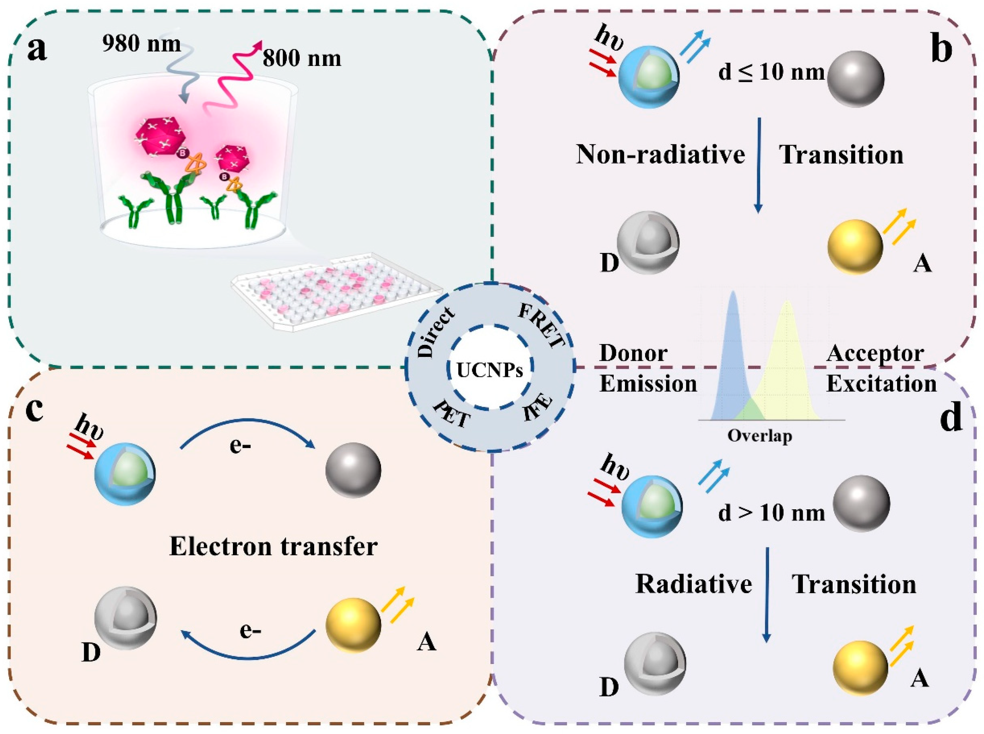
2.1. Direct Detection Method

Direct detection utilizes the luminescent properties of upconversion materials by conjugating fluorescent labels with target analytes, followed by fluorescence measurement after separation and enrichment for target localization or quantification. In contrast, this method has limited utility in complete solution-phase assays, primarily employed in imaging applications requiring fixed substrates. Al-Salihi et al. [26] synthesized NaYbF4@NaYF4:Er,Tm@NaYF4 NPs, and successfully realized fluorescent lifetime imaging of mouse cerebral vasculature using a triple-gate data acquisition method. Gorris’ team [27] used UCNP fluorescent probes to bind PSA antigens fixed on the pore plate by the sandwich method, and developed a new mode of simulating digital readings under fluorescence microscopy, raising the detection limit to sub-fM level. In addition to the integrated reading of combined detection, magnetic separation enhances sensitivity by combining target capture with enrichment. For instance, Song [28] employed Fe3O4 magnetic nanoparticles to bind clenbuterol hydrochloride, followed by aptamer-conjugated UCNPs to form MNP-CLB-UCNP complexes. Magnetic separation enabled detection at 0.304 ng mL−1.

2.2. Förster Resonance Energy Transfer (FRET)

Förster resonance energy transfer (FRET) is the most widely used fluorescence detection method. For effective FRET, three conditions must be satisfied: (1) the donor must be a fluorophore with a sufficiently long lifetime; (2) the donor’s emission spectrum must overlap with the acceptor’s absorption spectrum; (3) the donor–acceptor distance must be less than 10 nm. FRET detection primarily involves two approaches: modulating the donor–acceptor distance or selecting different acceptor types. The sub-10 nm dimensions of typical biomolecules (antigens, antibodies, and nucleic acids) make FRET particularly suitable for biological immunoassays. Signal generation occurs through two mechanisms: Aptamer-mediated acceptor binding that quenches upconversion fluorescence; and competitive target binding that restores donor fluorescence. In essence, the target concentration correlates with the number of donor fluorophores undergoing binding or dissociation events.
A representative example is the work by Lao et al. [29], who developed NaGdF4:Yb/Tm@NaYF4:Yb/Er nanoparticles with Tm3+/Er3+ co-doping for SARS-CoV-2 RNA detection. In this system, gold nanoparticles (Au NPs) served as acceptors to quench the Er3+ shell luminescence via FRET. Notably, the Tm3+-induced localized surface plasmon resonance (LSPR) of Au NPs was found to enhance the overall FRET efficiency. Although colloidal gold remains the most prevalent FRET acceptor, owing to its excellent chemical stability and broad absorption spectrum, its limited quenching efficiency has prompted the exploration of alternative acceptors. Recent studies have investigated various nanomaterials with larger surface areas or additional functionalities, including gold nanoflowers [30], MoS₂ nanosheets [31], MnO2 [32], and organic dyes [33]. These emerging materials represent promising directions for FRET-based detection systems. Therefore, optimal FRET system design requires careful consideration of both spectral overlap characteristics and specific signal requirements when selecting the appropriate acceptors. The choice of acceptor material should be guided by the intended application and desired detection performance.

2.3. Inner Filter Effect (IFE)

Unlike FRET, the inner filter effect (IFE) describes the reduction in fluorescence intensity when another substance in the system absorbs the donor’s emitted light. This phenomenon does not require close proximity between the donor and acceptor—spectral overlap alone is sufficient to induce IFE, significantly enhancing the system’s design flexibility and simplicity. IFE-based detection typically employs ratiometric fluorescence measurements, considering two aspects: On the one hand, whether there is a compound before and after that combines with the object to be measured can produce or eliminate absorption peaks that coincide with the emitted light. NaYF4:Yb,Er have become common UCNPs due to their emissions at 540 nm and 655 nm, which Zhang used to detect serum bilirubin [34], the original Fe3+ and sulfosalicylic acid (SSA) in the system. The combination of the two can quench green light at 540 nm, and the bilirubin and SSA compete to bind Fe3+ to restore green light. The IG/IR readout value is used to quantify the green light. Similarly, mexiletine in serum can restore green light by electrostatic association with the dye Bengal Rose (RB) [35]. On the other hand, it can be considered whether the chemical reaction of the object to be tested can produce an absorbent. For example, glucose oxidase can degrade glucose to produce H2O2, thus oxidizing the gallic acid-Fe2+ complex (GA-Fe (Ⅱ)) to GA-Fe (Ⅲ), thereby quenching fluorescence [36]. More generally, the target analyte can also be detected indirectly by immobilizing it with other IFE-reactive components. For example, Wu et al. [37] developed an assay using aldehyde-functionalized magnetic nanoparticles to capture sulfadimethoxine (SDM), followed by binding with biotin-labeled aptamers and HRP. The SDM concentration was then determined through oxidized TMB quantification. Beyond using intrinsic upconversion emission ratios, ratiometric fluorescence can also be constructed by incorporating carbon dots as complementary emitters [38]. While IFE significantly broadens detection capabilities, its specificity remains inferior to immunoassay methods.

2.4. Photo-Induced Electron Transfer (PET)

Photo-induced electron transfer (PET) probes consist of a fluorophore and a quenching group. Upon light excitation, electron transfer occurs from the donor’s highest occupied molecular orbital (HOMO) to the acceptor’s lowest unoccupied molecular orbital (LUMO), resulting in fluorescence quenching [39]. This quenching mechanism demonstrates higher efficiency compared to FRET and IFE. Li et al. [40] identified o-quinone as an effective small-molecule quencher for upconversion luminescence (UCL) via PET, enabling alkaline phosphatase (ALP) detection. The system utilizes ALP-mediated hydrolysis of O-phospho-L-tyrosine to L-tyrosine, followed by enzymatic oxidation to generate the quencher L-dopa-o-quinone. Ye [41] developed crown ether-functionalized anthracene derivatives as quenchers for triplet–triplet annihilation upconversion systems. These probes achieved Mg2+ detection with a 2.52 μM limit through PET inhibition via Mg2+-crown ether complexation.

3. Luminescence Enhancement Strategy

Despite their versatile applications, UCNPs face an inherent challenge of low luminescence efficiency. The main reasons include a low transition probability of Ln3+ 4f electrons, poor light absorption and energy transfer efficiency, and a significant non-radiative transition. Current strategies for enhancing upconversion luminescence can be divided into two categories: internal regulation and external regulation. The internal control is based on the composition of the material to change the lattice symmetry of the matrix, the concentration of doped ions, the structure of the material, etc. External regulation is to connect or modify new substances on the surface of the material to play the role of light field regulation. These approaches address the fundamental limitations while providing pathways for performance improvement.

3.1. Structural Composition Regulation

As previously discussed, the mixing of wave functions with opposite parity enables 4f–4f transitions in Ln3+ ions, while enhanced crystal field asymmetry significantly increases electric dipole transition probabilities. Doping with foreign ions to reduce local symmetry around lanthanides thus represents an effective approach for luminescence enhancement [42]. For oxide and fluoride substrates, Mn2+, Fe3+, Cu2+, Ni2+, Mg2+, Cr3+, and other transition-metal ions have been reported to be doped successfully to improve the luminescence intensity [43,44,45,46]. Figure 3a [47] shows a NIR-Ⅱ-responsive red single-band material by co-doping NaLuF4 with Er/Mn. The experiment found that when the content of Mn2+ increased from 0 to 40%, the upconversion intensity increased by 10 times, and the IR/IG increased significantly from 20 to about 300. The same phenomenon was also found in doping Mn2+ in Ba2Sc0.67Yb0.3Er0.03AlO5 phosphor [48], which proved that the inclusion of Mn2+ can enhance the luminescence of Er3+ through the energy transfer between Mn2+ and Er3+ and the energy transfer between Er3+ and the defect band.
The luminescence efficiency of upconversion materials initially improves with increasing lanthanide ion concentration (activators and sensitizers). However, excessive doping reduces interionic distances, inducing cross-relaxation that ultimately decreases the material’s brightness [49]. In response to this concentration quenching phenomenon, it was found that increasing the laser power [50,51] and constructing the inert shell could have a strong brightness at a relatively high activator concentration. However, high-power laser often fails to meet the needs of practical applications (such as biological monitoring). Zhang Hong’s team found that at a low temperature (40 K), the upconversion emission intensity of NaErF4@NaYF4 increased sharply by 170 times, compared with NaYF4:20%Yb,2%Er@NaYF4. Low temperatures limit the cross-relaxation between Er3+, and high doping can be applied in low-temperature imaging and temperature sensing [52]. In addition, the Zhou Bo group [53] designed the alloying NaEr0.01Yb0.99F4@NaYF4, which effectively reduces the distance between Er3+ and Yb3+ and promotes energy transfer, as shown in Figure 3b. Compared with the traditional doping content, under the laser power of 0.26 W/cm2, the emissions of green light and red light are increased by 2786 times and 89 times, respectively. This provides a new idea for high blending.
UCNPs exhibit significant surface quenching effects due to their high surface-to-volume ratio, where excitation energy from internal ions migrates to surface defects or is quenched by ligands and solvents. The external coating of the shell is a common surface passivation method. The most common shell types are homogeneous inert rare-earth shells [54,55] and SiO2 [56,57], among which SiO2 has good water dispersion and can also play the role of surface modification. The choice of shell material and structure, and the distribution of doped ions in the core–shell structure, will affect the brightness of the material. As shown in Figure 3c, Chen’s team constructed a sandwich nanocrystalline structure, β-NaYF4@NaYF4:Yb/Tm@NaYF4 [58]. Using the effective confined isolation protection of the inert material in the core and outer shell, they demonstrated the size-dependent effect of the quantum efficiency of the material’s upconversion by precisely regulating the thickness of the intermediate layer (1.2–13 nm). The epitaxial growth of ScF3 matched with the NaYF4 (111) crystal face on the α-NaYF4:Yb/Ln surface will give the material positive/negative thermal expansion properties, and unlike the traditional thermal quenching conversion, UCL will be enhanced with the increase of temperature [59].
Figure 3. Structural control for upconversion enhancement: (a) Scheme of Er3+/Mn2+ co-doping to prepare UCNPs with NIR-Ⅱ-responsive single-band red UCL emission. Reproduced from Ref. [47], with permission from John Wiley and Sons; copyright 2022. (b) Schematic of energy transfer upconversion in the erbium–ytterbium alloyed core–shell nanoparticles under 980 nm excitation. Reproduced from Ref. [53], with permission from the American Chemical Society; copyright 2024. (c) Schematic illustration of simplified lanthanide energy levels as well as the involved upconversion processes in the core–shell–shell nanostructure. Reproduced from Ref. [58], with permission from Springer Nature; copyright 2024.
Biosensors 15 00335 g003

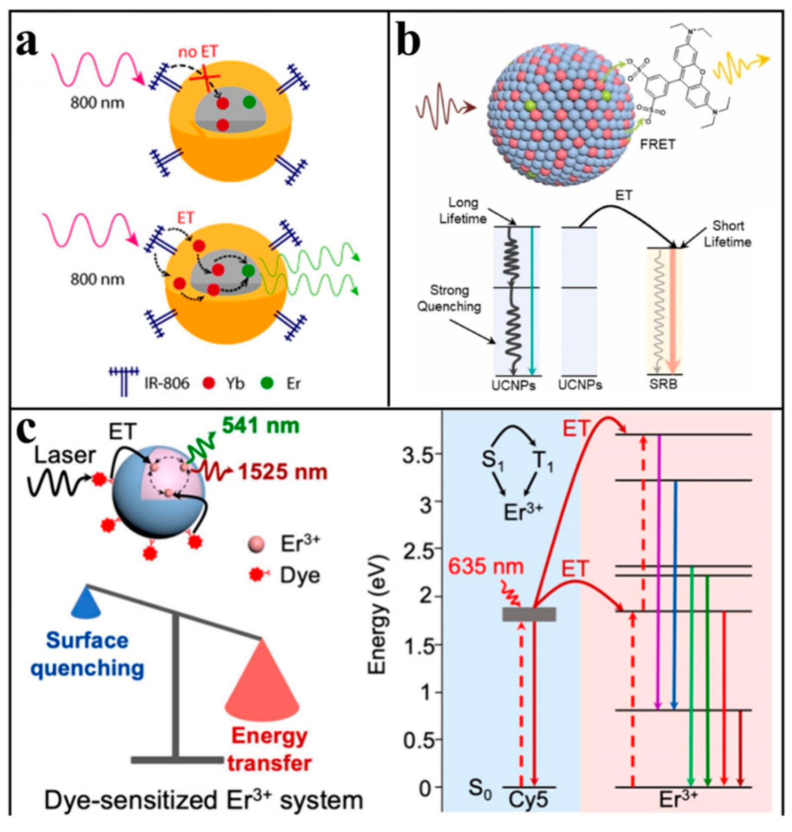
3.2. Dye Sensitization

Rare-earth-doped upconversion materials exhibit limited light absorption cross-sections. In comparison, organic dyes demonstrate superior light-harvesting capabilities, with absorption cross-sections 103–104 times greater than lanthanide ions [60,61]. To address this limitation, researchers developed dye-sensitized upconversion systems by conjugating organic dyes to UCNP surfaces as light-harvesting antennas.
As shown in Figure 4a, the usual pathway for dye delivery is first to the sensitizer and then to the activator. The effect of dye sensitization depends on the degree of spectral overlap between the dye and Ln3+ and the distance between the dye and the receptor, so there are limitations in dye selection. These parameters impose strict selection criteria for optimal dye performance.
Meng et al. [62] modified NaYF4:20%Yb,1%Ho@NaLuF4:10%Yb,40%Nd nanoparticles with excellent luminescence properties under 808 nm excitation by ICG. ICG transferred the energy of the 808 nm light source to Nd3+, and Nd3+ further transferred the energy to Yb3+ and finally to the luminescence center Ho3+. The experiment found that the sensitization of ICG enhanced the luminescence of the material at 540 nm by about 80 times. At the same time, dyes can also be used as receptors to enhance the upconversion efficiency. For example, Rhodamine sulfonyl B (SRB) is connected to the surface of NaGdF4@NaYF4: Gd3+, Yb3+,Er3+, and SRB will absorb the emitted light at 540 nm of the material into its own light at 645 nm. Researchers found that dyes acting directly as FRET receptors can reduce the non-radiative relaxation of the system, as seen in Figure 4b. The surface modification of SRB results in an energy transfer efficiency of up to 98.8%, and the luminescence intensity of SRB as a receptor is 4.6 times that of the UCNPs at a 541 nm emission reduction [63]. Liu’s team [64] proposed a new strategy for dye direct sensitization of the Er3+ emission. Different from traditional dye sensitization receptors Yb3+ and Nd3+, Er3+ can absorb from ultraviolet to near-infrared. The researchers used a series of cyanine dyes with absorption bands coincident with Er3+ as antenna molecules and found that they all play an enhanced role. Cy5, with the largest spectral overlap, enhanced UCL by a factor of 1942, which provided the idea for polychromatic excited dye sensitization; the corresponding process is reflected in Figure 4c.
Figure 4. Dye sensitization for upconversion enhancement: (a) Schematic diagram of the dye-enhanced upconversion luminescence principle. Reproduced from Ref. [61], with permission from the American Chemical Society; copyright 2016. (b) Schematic representation of organic dyes as receptors for sensitized upconversion emission. Reproduced from Ref. [63], with permission from Elsevier; copyright 2025. (c) Schematic illustration of energy transfer for direct sensitization of Er3+ by Cy5. Reproduced from Ref. [64], with permission from the American Chemical Society; copyright 2024.
Biosensors 15 00335 g004

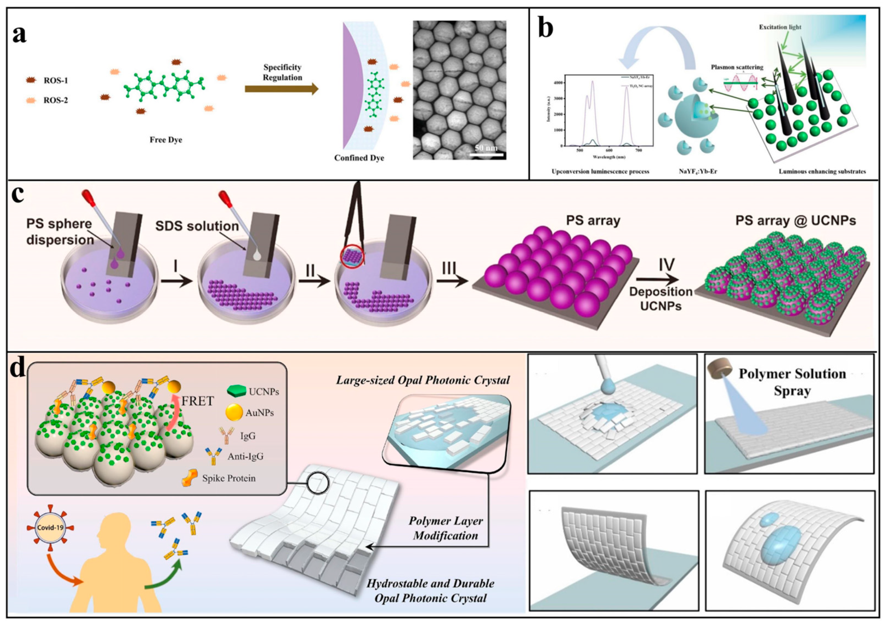
3.3. Local Field Control

To enhance upconversion luminescence (UCL), two primary external modulation approaches exist alongside internal rare-earth material modifications: surface plasmon resonance (SPR) and photonic crystal modulation. These local field modulation strategies offer broader applicability and operational convenience compared to component-specific modifications. By increasing the photonic state density at the material surface, they effectively boost UCL efficiency.
Localized surface plasmon resonance (LSPR) in noble metals involves collective electron oscillations at the metal surface under light excitation, generating propagating surface plasmons that create enhanced electromagnetic fields. The Purcell effect predicts optimal luminescence enhancement when the emitter is positioned within an optical cavity and the cavity resonance matches the emitter’s emission frequency [65]. Many works have been conducted to enhance UCL with various forms of precious metals or cavities with different structures; e.g., Figure 5a shows an optical cavity with silver nanocubes arranged on a gold film and which fills the nanoparticle gaps with UCNPs designed by Liu Xiaogang’s team [66], obtaining a 166-fold enhancement factor of luminescence at a low excitation power. In order to realize the resonant enhancement of the excitation and emission light at the same time, Xu et al. [67] designed a UCNPs/MgF2/Ag optical structure and controlled the coupling of light by adjusting the two-dimensional grating period, which confirms that the UCL can be maximally enhanced when the grating is coupled with the excitation and emission wavelengths of UCNPs at the same time.
Photonic crystals are periodic dielectric structures with alternating refractive indices that exhibit photonic bandgap properties. When incident light wavelengths match the bandgap, total reflection occurs, producing characteristic structural colors. The bandgap position depends primarily on the structural unit size (typically microspheres), enabling tunability through material dimension control. The main measure for photonic crystal enhancement of fluorescent materials is to make the bandgap position coincide with the excitation wavelength or emission wavelength. The former allows the excitation light to be repeatedly reflected and scattered between the gaps of the photonic crystal, which enhances the energy density of the excitation light; the latter plays an enhancement effect by modulating the direction of emission of the emitted light. Ju [68] designed an opal with a bandgap of 450 nm for the poly(methyl methacrylate) (PMMA) deposited photonic crystals to enhance the blue UCL of Tm3+ for glucose detection. As depicted in Figure 5b, our group explored the enhancement of the luminescence of UCNPs by double-stopband-bilayer opal photonic crystals and found that the modulation of the excitation field of the photonic crystal plays a dominant role, and that the combined action of the excitation and emission fields can have a superimposed enhancement effect, and that double-forbidden-band photonic crystals enhanced luminescence at 540 nm by a factor of 156 for NaYF4: Yb,Er [69]. Similarly, double-forbidden-band anti-opal photonic crystals have been applied in anti-counterfeit markers with bright fluorescence [70].
Figure 5. Local field control for upconversion enhancement: (a) Structural design of plasma cavity-coupled upconversion nanoparticles (UCNPs) and schematic SEM and photoluminescence spectra. Reproduced from Ref. [66], with permission from Springer Nature; copyright 2019. (b) Schematic structure of a double-stopband photonic crystal and the enhancement of a photonic crystal film with the excitation field emitting field in the opposite position. Reproduced from Ref. [69], with permission from Elsevier; copyright 2021.
Biosensors 15 00335 g005

4. Platform for UCNPs in Bioassays

4.1. Solution-Phased Detection

Solution-phase detection represents the most prevalent fluorescence assay format, offering homogeneous reaction conditions and stable detection sensitivity. However, this approach faces challenges, including operational complexity and a lack of portability.
The development direction of the upconversion solution form of detection is most notably to enhance the luminescence intensity of UCNPs, such as the structural modulation of the material itself and plasma coupling, mentioned above, which will not be reintroduced in this section. It is also possible to consider increasing the binding sites of the reaction and the probability of the reaction occurring, etc. The following highlights several innovative approaches in this field.
Hou et al. [71] first introduced the atom transfer radical polymerization (ATRP) reaction, which refers to the phenomenon of reversible transfer of an initiator between active and dormant species in the presence of a transition-metal complex. The modification of antibodies and UCNPs with initiator and reactive monomers, respectively, could make UCNPs accumulate on the surface of magnetic beads in large quantities by ATPR, which could enhance the fluorescence intensity. The system achieved a 38.7 fg/mL detection sensitivity for CYFRA21-1. Two-dimensional materials with a large specific surface area, such as MXene, are favorable for increasing the reaction sites. For example, Hao immobilized red- and blue-emitting UCNPs on Nb2CTx MXene [72], and the double-helix structure formed in the presence of the target was repelled away by the negatively charged MXene, and the FRET was invalidated so as to restore the fluorescence, which realized the simultaneous detection of the two substances and enhanced the accuracy of the detection. Their team also used Au-Au dimer as a novel FRET bursting agent [73]; a dimer form that generates a stronger electromagnetic field, improves the bursting efficiency, and extends the working distance. The accurate detection of long-chain oligonucleotides was realized, and the detection limit was even lower than the aM level, which provided a new idea for conventional FRET detection.

4.2. Lateral Flow Chromatography

Test strips represent a cost-effective biosensing platform with multiple advantages, including operational simplicity, rapid detection, user-friendliness, and field applicability, leading to their commercialization in immunoassays [74]. A standard test strip consists of a sample pad, conjugate pad, nitrocellulose membrane (with test [T] and control [C] lines), absorption pad, and backing card. In immunoassays, the T-line is coated with target-specific antibodies, while the C-line contains universal antibodies. After adding the sample, the target analytes will bind with detection antibodies in the conjugate pad, and the complexes migrate to the T-line via lateral flow. Unbound antibodies reach the C-line for quality control. Colloidal gold is the most common indicator for test strips and can be read directly with the naked eye. Still, this indication method has the problem of low sensitivity [75] and is typically applicable only for qualitative binary (positive/negative) analysis. Compared with conventional colorimetric methods, fluorescence reporter molecules can greatly improve detection sensitivity up to 100–10,000 times and better meet the quantitative requirements [76]. Upconversion nanoparticles (UCNPs) offer distinct advantages for bioassays, primarily due to their near-infrared (NIR) excitation that avoids the autofluorescence background associated with conventional UV excitation. Furthermore, multicolor UCNPs enable the simultaneous detection of multiple analytes. For high-precision applications, UCNP-based lateral flow assays present an optimal solution. While the inability to read UCNP signals with the naked eye increases operational complexity, this limitation can be overcome by employing a simple laser flashlight coupled with smartphone-based grayscale analysis, maintaining portability while digitizing the measurement results for point-of-care testing (POCT). Notably, several biotechnology companies, including Hotgen and Germany-based Miltenyi Biotec, have developed handheld UCNP immunoassay analyzers to facilitate the large-scale implementation of this technology. Recent advances demonstrate the successful integration of upconversion nanoparticles with test strips, achieving enhanced performance for biological detection.
The detection effect of the test strip is mainly affected by four aspects: target acquisition, signal conduction, signal separation, and signal analysis [77]. Jin [78] comprehensively optimized the signal reading device, the release efficiency of the UCNPs probe, the change in fluorescence intensity caused by the size of UCNPs, and the reference value of line C to reduce the deviation. The superimposed effect of multiple links made the detection limit of H. pylori nucleic acid reach 25 fM, a 105-fold increase. In view of the signal separation process, as shown in Figure 6a, Ji [79] innovatively took the emission light of NaYF4:Yb3+,Tm3+@NaYF4 at 800 nm as the signal, so that the excitation wavelength (980 nm) and emission wavelength of the material are both in the first biological window, avoiding the interference of biological self-fluorescence and colored plasma scattering. The detection limit of procalcitonin in serum reached 0.03 ng/mL. For signal analysis, a portable in situ detection biosensor combined with a smartphone has been invented [80], which overcomes the traditional lengthy pre-processing and fuzzy qualitative results, in which the computer vision algorithm improves the sensitivity and robustness of quantitative results, and the accuracy of quantitative detection results is increased by 17.4% compared with the standard quantitative detection process. The detection limit of methamphetamine can reach 0.1 ng/mL within 20 s.
The strip combines the advantages of solid and liquid sensors. On the one hand, it is easy to carry and easy to operate. On the other hand, due to the supporting role of the three-dimensional fiber structure of the cellulose nitrate film, the strip can provide a contact environment close to solution, which solves the problem of an insufficient reaction area and limited contact in-plane detection. Chen et al. [81] took NaYF4:Yb3+ and Er3+ UCNPs as fluorescence reporter molecules, and pre-coated them on the T-line of the nitrate cellulose film by drops. Using the FRET mode, Au NPs were replaced by Au-DTNB@Ag with Raman signals. With an increase in target material, the number of Au@Ag particles accumulated on the T-line increased, the fluorescence signal weakened, and the Raman signal strengthened (Figure 6b). This method was used to detect microRNA-21—a marker of periodontitis in saliva—and reached the dynamic range of 2 nM–1 fM, which greatly improved the detection sensitivity in this field. This method provides a good strategy for the simple and sensitive application of test strips.
The capillary action of the strip allows the liquid to flow completely from one side to the opposite side, which means that it can be spatially partitioned, allowing different targets to be combined in different locations and supplemented by upconversion reporter molecules of different colors, enabling simultaneous detection of multiple substances. For example, Sahar et al. [82] first explored the relationship between the content and ratio of CD44 and FKBPL proteins in serum and preeclampsia. The authors used the same system to construct their own test strip for these two proteins. Although there was no cross-reactivity and the sensitivity reached the pg/mL level, the operation was relatively complicated. Jin et al. [83] synthesized red, green, and blue upconversion fluorescent probes, designed a structure of test paper containing three T-lines and one C-line, as illustrated in Figure 6c, and realized the simultaneous detection of three different types of analytes, Hg2+, ochratoxin A, and Salmonella, in water samples. However, the number of lines was limited by the limited diffusion distance of the samples on the strip; thus, Miikka [84] chose to place both the Er3+-doped green light upconversion probe and Tm3+-doped blue light upconversion probe on the T-line to simultaneously test CA125 and CA15-3 proteins. Guo et al. [85] designed and synthesized NaErF4:Yb3+,Tm3+@NaYF4:Yb3+@NaNdF4:Yb3+ UCNPs with orthogonal emission properties, which produced red fluorescence at a 980 nm excitation and green light at an 808 nm excitation, achieving orthogonal emission. The intensity ratio (IG/IR) served as an internal reference, eliminating the need for a control line (C-line) and thereby simplifying the strip design.
Figure 6. Biological detection on test strips: (a) Schematic illustration of NIR to NIR lateral flow detection based on upconverted nanoparticles. Reproduced from Ref. [79], with permission from the American Chemical Society; copyright 2020. (b) Schematic diagram of the fluorescence and Raman dual-mode sensor of the FRET principle on the test strip and SEM image of the sandwich structure formed on the test strip. Reproduced from Ref. [81], with permission from Elsevier; copyright 2024. (c) Schematic illustration of LFAA for simultaneous multiple target detection. Reproduced from Ref. [83], with permission from Elsevier; copyright 2018.
Biosensors 15 00335 g006
Given the versatility of test strips as platforms for diverse sensing modalities, we conducted a comprehensive comparison between UCNP-based detection systems and conventional reporter molecules for multiple bioanalytes. The comparative performance metrics are systematically summarized in Table 1. Upconversion nanoparticles (UCNPs) demonstrate broad applicability for detecting diverse biological targets. Compared with the colorimetric and electrochemical methods, UCNP-based detection offers superior sensitivity while not matching the ultra-high sensitivity of Raman spectroscopy; rather, it provides a significantly wider dynamic range and better adaptability.

4.3. Plate

Plate-based biosensors confine the sensing process to a planar surface, offering the portability advantages of solid-state devices while typically exhibiting lower detection sensitivity. Research efforts, therefore, focus on two key challenges: (1) enhancing localized fluorescence intensity and (2) developing more sensitive detection modalities within these constrained areas.
Beatriz et al. [94] immobilized cysteine- and antibody-modified nanoparticles (UCNPs@Cys-Ab) on quartz substrates via layer-by-layer self-assembly using sodium dodecyl sulfate. Optimal signal intensity was achieved with six assembly layers, enabling Escherichia coli detection across a dynamic range of 2 × 10³–106 CFU/mL, with a detection limit of 34 CFU/mL. Chen Guanying’s team [95] developed ROS-responsive IR820 dye-sensitized UCNPs (Figure 7a), where steric hindrance restricted IR820 absorption exclusively to ·OH. Under 808 nm irradiation, ·OH quenched the upconversion monolayer fluorescence while the 980 nm emission remained unaffected, allowing ratiometric detection of endogenous H₂O₂ with a 0.1 nM detection limit. In addition to self-assembly, UCNPs can also be deposited on other polymer substrates. PDMS is a common solid substrate. Ouyang et al., for example, anchored UCNPs on amino-functionalized PDMS substrate [96] to achieve a sensitive detection of malachite green of 0.029 ng/mL. Similarly, Rong [97] encapsulated UCNPs in PDMS to create a solid-state sensor suitable for testing actual samples of acrylamide produced during frying, with a detection limit of 1 nM. In addition, Gorris has performed a number of assays using a 96-well plate as a substrate using the upper conversion link immunosorbent assay (ULISA). Similar to ELISA, ULISA uses UCNPs instead of traditional enzymes and fluorescence signals instead of colorimetric signals that are generated by enzymatic reactions, achieving better sensitivity. Compared with the analog ULISA, which is based solely on luminous intensity, the digital ULISA, which counts light spots under a fluorescence microscope, can further improve the sensitivity by 10 times [98].
Figure 7. Biological detection on different types of planes: (a) Schematic illustration of self-assembled monolayer UCNPs plates for specific selective detection of ROS. Reproduced from Ref. [95], with permission from John Wiley and Sons; copyright 2023. (b) Schematic diagram of Ti3O5 nanocone array with structural constraint effect and LSPR-enhanced upconversion luminescence. Reproduced from Ref. [99], with permission from the American Chemical Society; copyright 2024. (c) Structural diagram of the UCNP plate deposited on the PS microsphere array. Reproduced from Ref. [100], with permission from Elsevier; copyright 2024. (d) Schematic diagram of the detection of COVID-19 by highly hydrostable opal photonic crystal film deposited with UCNPs. Reproduced from Ref. [101], with permission from Elsevier; copyright 2023.
Biosensors 15 00335 g007
For the above example of UCNP itself or other polymers as solid substrates, the substrate itself does not have the enhancement effect; the following describes some signal enhancement substrates, mainly including the plasma effect and photonic crystal modulation. In view of the weak plasma effect of defective semiconductors, Xu et al. [99] designed the Ti3O5 nanopyramid array substrate (Figure 7b). By using the sharp structure of the nanopyramid, the excited light can be reflected several times inside the array and coupled with the surface plasma of the material itself, thus increasing the green and red luminescence of the upper UCNPs by 32 times and 40 times. It shows superior semiconductor enhancement performance and confirms that the special structure of the substrate can modulate the light field. Similarly, Wu [100] prepared the PS microsphere array @UCNPs’ flexible film by the self-assembly method (Figure 7c). Compared with the plane, the UCL strength of the array was increased by 10 times, and similar results were obtained by FDTD theoretical simulation, which was used to detect acetic acid gas, reaching a detection limit of 5.2 ppm. This enhancement was interpreted as Mie scattering on the surface of the PS microsphere. The photonic crystal is also an ordered arrangement of a microsphere array, which is different from a single layer. Furthermore, it behaves as a microsphere array in three-dimensional space. Intuitively, there is a photonic band gap in the transmission spectrum so that the enhancement factor can be further increased to tens of times. Our group successfully prepared a flexible film of photonic crystals by spraying polymers. As shown in Figure 7d, this method uses polymers to fill the fragmentary gaps in photonic crystals, acting as a bond, which solves the problem that photonic crystals are easily broken and do not have water stability on a macro level; thus, it can be used to detect biological samples. The detection sensitivity of 0.1 ng ·mL−1 for COVID-19 was achieved [101]. Photonic crystal enhancement is also used to detect glucose [68]. The author cleverly utilized the characteristic that H2O2, the oxidation product of glucose, can react with the quenching agent MnO2 to generate Mn2+, and constructed a fluorescence colorimetric dual-mode detection platform. The combination of photonic crystals and the metal plasma effect is also a common idea. Our research group has long explored the enhancement effect of gold bars deposited on opal photonic crystals [102], and found that when the SPR matches the excitation light spectrum, the plasma enhancement is the most significant, which can increase UCL by 3 orders of magnitude. Similarly, Chu [103] developed a flexible upconversion optical transducer using a dendritic gold inverse opal framework. This design demonstrated 48-fold luminescence enhancement at ultralow laser power, confirming synergistic interactions between photonic bandgap effects and localized surface plasmon resonance (LSPR). This device shows promising potential for implantable optogenetic applications.

4.4. Linkage with Other Technologies

To enhance the sensitivity and accuracy of bio-detection, in addition to enhancing the luminescence performance of UCNPs themselves, we can consider increasing the target analyte quantities and constructing multimodal detection modes. Some functional materials, such as chalcogenide quantum dots, nano-enzymes, and magnetic materials, have been developed into UCNP-based nanocomposites. In addition, nucleic acid amplification techniques such as catalytic hairpin self-assembly are also commonly used as a means of design. Table 2 summarizes the representative applications of these approaches.

5. Summary and Prospect

This paper introduces the composition of rare-earth-dope upconversion nanoparticles (UCNPs) based on upconversion luminescence principles. Focusing on bio-detection applications, we summarized four fluorescence probe design strategies as follows: direct detection, Förster resonance energy transfer (FRET), inner filter effect (IFE), and photo-induced electron transfer (PET). To overcome UCNPs’ low luminescence efficiency, three luminescence enhancement means, namely, structure composition modulation, dye sensitization, and local field modulation, are summarized. The structural composition modulation includes the doping of other metal ions, the proportion modulation between different compositions, and the construction of the core–shell structure; the local field modulation can be subdivided into the plasma resonance effect of the noble metals or semiconductors and the modulation effect of the photonic crystals on the excitation or emission light field. We also reviewed the bioassay work from recent years in the form of practical applications of UCNP detection, which is divided into three categories: solution environment, test strips, and plates. This review provides new perspectives for UCNP biosensing applications, highlighting both fundamental principles and practical implementations.
Despite their advantage of low biofluorescence background, UCNPs face several challenges, such as the photothermal effect of 980 nm excitation may cause tissue damage, while limited binding sites restrict detection capabilities. Recent research reveals three key development trends as follows: (1) The preparation of small and bright UCNPs: UCNPs with small and homogeneous particle sizes can provide more binding sites and easier access to biological tissues, but small particles increase the percentage of bursts and are often insufficiently bright; the modification of ligands and the excitation light source is a feasible strategy in this regard. (2) Construction of a multimodal detection system: The association of UCNPs with other materials, such as chalcogenides and nano-enzymes, can make the system’s characteristics more diverse. Relative to fluorescence unimodal detection, the introduction of detection modes, such as electrochemistry and Raman, can make the detection results more accurate, expanding the scope of application. (3) Development of corresponding detection equipment: How to transfer from laboratory research to a practical application is an important development direction for UCNPs in the future, and the development of a small portable device integrating a laser and a reading device is necessary.

Author Contributions

Writing—original draft, J.X. and H.C.; Writing—review & editing, L.S. and B.D.; Validation, C.W. and T.W.; Supervision, L.S. and B.D. All authors have read and agreed to the published version of the manuscript.

Funding

This work was supported by the Jilin Provincial Department of Science and Technology (grant numbers 20210204095YY).

Institutional Review Board Statement

Not applicable.

Informed Consent Statement

Not applicable.

Data Availability Statement

Not applicable.

Conflicts of Interest

The authors declare no conflicts of interest.

References

  1. Bloembergen, N. Solid State Infrared Quantum Counters. Phys. Rev. Lett. 1959, 2, 84–85. [Google Scholar] [CrossRef]
  2. Peltomaa, R.; Benito-Pena, E.; Gorris, H.H.; Moreno-Bondi, M.C. Biosensing based on upconversion nanoparticles for food quality and safety applications. Analyst 2021, 146, 13–32. [Google Scholar] [CrossRef]
  3. Gong, Y.; Yuan, W.; Song, F.; Zhang, Q.; Zhang, P.; Ding, C. A NIR light gated targeting nanoprobe based on DNA-modified upconversion nanoparticles with antifouling properties for ratiometric detection and imaging of microRNA-21. Anal. Chim. Acta 2022, 1235, 340554. [Google Scholar] [CrossRef]
  4. Han, Q.; Zhao, X.; Zhang, X.; Na, N.; Ouyang, J. A rationally designed triple-qualitative and double-quantitative high precision multi-signal readout sensing platform. Sens. Actuators B-Chem. 2022, 360, 131663. [Google Scholar] [CrossRef]
  5. Jin, B.; Du, Z.; Ji, J.; Bai, Y.; Tang, D.; Qiao, L.; Lou, J.; Hu, J.; Li, Z. Regulation of probe density on upconversion nanoparticles enabling high-performance lateral flow assays. Talanta 2023, 256, 124327. [Google Scholar] [CrossRef]
  6. Wang, Y.; Pan, Y.; Ma, Y.; Sun, X.; Zhang, Y.; Li, H. Simultaneous evolutions in composition, structure, morphology, and upconversion luminescence of BiOxFy:Yb/Er microcrystals and their application for ratiometric temperature sensing. J. Alloys Compd. 2024, 992, 174596. [Google Scholar] [CrossRef]
  7. Li, X.; Li, L.; Liang, R.; Li, P.; Wang, J.; Wang, F. A bright Ce-based downshifting luminescence nanoprobe for NIR-IIb vessel imaging. Chem. Eng. J. 2024, 496, 153917. [Google Scholar] [CrossRef]
  8. Yuan, H.; Sun, K.; Su, X.; Hu, D.; Luo, Y.; Sun, Y.; Liu, Q.; Chen, L.; Qiao, J.; Xu, M.; et al. A dark-state-dominated photochemical upconversion afterglow via triplet energy transfer relay. Sci. Adv 2025, 11, eadt1225. [Google Scholar] [CrossRef]
  9. Ye, M.; Zhang, J.; Jiang, D.; Tan, Q.; Li, J.; Yao, C.; Zhu, C.; Zhou, Y. A Hemicyanine-Assembled Upconversion Nanosystem for NIR-Excited Visualization of Carbon Monoxide Bio-Signaling In Vivo. Small 2022, 18, 2202263. [Google Scholar] [CrossRef]
  10. Li, Y.; Jin, X.; Sun, Y.; Guan, S.; Chang, B.; Li, Y. Dual-mode nanoprobes for reactive oxygen species scavenging and treatment of acute lung injury. Appl. Surf. Sci. 2023, 610, 155458. [Google Scholar] [CrossRef]
  11. Jiang, Y.; Hong, Y.; Liu, Y.-Y.; Guan, Y.; Zhou, J.; Wang, H.; Sun, L. Switching between upconversion luminescence imaging and therapy in vitro enabled by NIR excitation modulation of nanocomposites. J. Mater. Chem. C 2024, 12, 11938–11947. [Google Scholar] [CrossRef]
  12. Lv, W.; Wu, Y.; Chen, H. Orthogonal upconversion nanocarriers for combined photodynamic therapy and precisely triggered gene silencing in combating keloids. J. Control. Release 2025, 379, 1–13. [Google Scholar] [CrossRef]
  13. Molkenova, A.; Choi, H.E.; Lee, G.; Baek, H.; Kwon, M.; Lee, S.B.; Park, J.-M.; Kim, J.-H.; Han, D.-W.; Park, J.; et al. Cold-Responsive Hyaluronated Upconversion Nanoplatform for Transdermal Cryo-Photodynamic Cancer Therapy. Adv. Sci. 2024, 11, 202306684. [Google Scholar] [CrossRef]
  14. Dong, H.; Sun, L.-D.; Yan, C.-H. Local Structure Engineering in Lanthanide-Doped Nanocrystals for Tunable Upconversion Emissions. J. Am. Chem. Soc. 2021, 143, 20546–20561. [Google Scholar] [CrossRef]
  15. Chan, E.M.; Levy, E.S.; Cohen, B.E. Rationally Designed Energy Transfer in Upconverting Nanoparticles. Adv. Mater. 2015, 27, 5753–5761. [Google Scholar] [CrossRef]
  16. Wang, F.; Banerjee, D.; Liu, Y.; Chen, X.; Liu, X. Upconversion nanoparticles in biological labeling, imaging, and therapy. Analyst 2010, 135, 1839–1854. [Google Scholar] [CrossRef]
  17. Chen, J.; Zhao, J.X. Upconversion Nanomaterials: Synthesis, Mechanism, and Applications in Sensing. Sensors 2012, 12, 2414–2435. [Google Scholar] [CrossRef]
  18. Wang, G.; Peng, Q.; Li, Y. Upconversion Luminescence of Monodisperse CaF2:Yb3+/Er3+ Nanocrystals. J. Am. Chem. Soc. 2009, 131, 14200–14201. [Google Scholar] [CrossRef]
  19. Krämer, K.W.; Biner, D.; Frei, G.; Güdel, H.U.; Hehlen, M.P.; Lüthi, S.R. Hexagonal sodium yttrium fluoride based green and blue emitting upconversion phosphors. Chem. Mater. 2004, 16, 1244–1251. [Google Scholar] [CrossRef]
  20. Su, Q.; Feng, W.; Yang, D.; Li, F. Resonance Energy Transfer in Upconversion Nanoplatforms for Selective Biodetection. Acc. Chem. Res. 2017, 50, 32–40. [Google Scholar] [CrossRef]
  21. Wang, F.; Liu, X. Upconversion multicolor fine-tuning: Visible to near-infrared emission from lanthanide-doped NaYF4 nanoparticles. J. Am. Chem. Soc. 2008, 130, 5642–5643. [Google Scholar] [CrossRef]
  22. Wang, J.K.; Zhu, Y.L.; Grimes, C.A.; Nie, Z.; Cai, Q.Y. Eu,Sm,Mn-Doped CaS Nanoparticles with 59.3% Upconversion-Luminescence Quantum Yield: Enabling Ultrasensitive and Facile Smartphone-Based Sulfite Detection. Anal. Chem. 2018, 90, 8658–8664. [Google Scholar] [CrossRef]
  23. Pan, Q.; Cai, Z.; Yang, Y.; Yang, D.; Kang, S.; Chen, Z.; Qiu, J.; Zhan, Q.; Dong, G. Engineering Tunable Broadband Near-Infrared Emission in Transparent Rare-Earth Doped Nanocrystals-in-Glass Composites via a Bottom-Up Strategy. Adv. Opt. Mater 2019, 7, 1801482. [Google Scholar] [CrossRef]
  24. Gao, C.; Zheng, P.; Liu, Q.; Han, S.; Li, D.; Luo, S.; Temple, H.; Xing, C.; Wang, J.; Wei, Y.; et al. Recent Advances of Upconversion Nanomaterials in the Biological Field. Nanomaterials 2021, 11, 2474. [Google Scholar] [CrossRef]
  25. Ansari, A.A.; Thakur, V.K.; Chen, G. Functionalized upconversion nanoparticles: New strategy towards FRET-based luminescence bio-sensing. Coord. Chem. Rev. 2021, 436, 213821. [Google Scholar] [CrossRef]
  26. Al-Salihi, M.; Ghellab, S.E.; Li, Y.; Luo, C.; Kalsoom, U.-e.; Liu, L. Effective Rapid Fluorescence Lifetime Imaging of the Brain: A Novel Approach Using Upconversion Photoluminescence Lifetime Based on Gate-Width Acquisition. Nano Lett. 2024, 24, 14973–14982. [Google Scholar] [CrossRef]
  27. Mickert, M.J.; Farka, Z.; Kostiv, U.; Hlavacek, A.; Horak, D.; Skladal, P.; Gorris, H.H. Measurement of Sub-femtomolar Concentrations of Prostate-Specific Antigen through Single-Molecule Counting with an Upconversion-Linked Immunosorbent Assay. Anal. Chem. 2019, 91, 9435–9441. [Google Scholar] [CrossRef]
  28. Song, X.-J.; Ye, F.; Zhang, Y.; Sun, J.; Shentu, X.; Yu, X.; Li, W.; Wu, Y.-F. A clenbuterol detection method based on magnetic separation upconversion fluorescent probe. Food Chem.-X 2024, 24, 101911. [Google Scholar] [CrossRef] [PubMed]
  29. Lao, X.; Liu, Y.; Li, L.; Song, M.; Ma, Y.; Yang, M.; Chen, G.; Hao, J. Plasmon-enhanced FRET biosensor based on Tm3+/Er3+ co-doped core-shell upconversion nanoparticles for ultrasensitive virus detection. Aggregate 2024, 5, e448. [Google Scholar] [CrossRef]
  30. Chen, H.; Tian, P.; Guo, J.; Sun, M.; Zhu, W.; Li, Z.; Liu, Z. Synergistic synthesis of gold nanoflowers as upconversion near-infrared nanoprobe energy acceptor and recognition unit for improved hydrogen sulfide sensing. Talanta 2024, 273, 125908. [Google Scholar] [CrossRef]
  31. Che, D.; Cao, X.; Chen, C.; Yan, H. A point-of-care aptasensor based on the upconversion nanoparticles/MoS2 FRET system for the detection of Pseudomonas aeruginosa infection. Microchim. Acta 2024, 191, 61. [Google Scholar] [CrossRef] [PubMed]
  32. Wang, P.; Li, J.; Zhang, Y.; Xiao, D.; Zhou, C. Blue emission-dominated NaYbF4@NaYF4:2%Ho@NaYF4 upconversion nanoparticles for detecting ascorbic acid. Nanoscale 2024, 16, 18910–18917. [Google Scholar] [CrossRef]
  33. Wu, M.; Yi, J.; Yin, C.; Sun, Q.; Gao, L.; Niu, N.; Chen, L. An upconversion nanosensor with phenolic-like functionality for accurate identification of chlorpyrifos in grapes. Food Chem. 2023, 416, 135859. [Google Scholar] [CrossRef]
  34. Zhang, L.; Kang, X.; Yang, F.; Jia, W.; Yang, L.; Jiang, C. Zinc Doping-Induced Lattice Growth Regulation for Enhanced Upconversion Emission in Serum Bilirubin Detection. Anal. Chem. 2025, 97, 3515–3524. [Google Scholar] [CrossRef]
  35. Kang, X.; Zhang, L.; Yang, L.; Jiang, C. Ratiometric upconversion luminescence sensing platform for visual monitoring of antiarrhythmic drug level in serum. Chem. Eng. J. 2023, 476, 146923. [Google Scholar] [CrossRef]
  36. Shao, X.; Cao, L.; Lu, L. Ultrasensitive detection of glucose oxidase and alkaline phosphatase in milk based on valence regulated upconversion nanoprobes. Food Chem. 2024, 432, 137212. [Google Scholar] [CrossRef]
  37. Wu, W.; Ahmad, W.; Hassan, M.M.; Wu, J.; Ouyang, Q.; Chen, Q. An upconversion biosensor based on inner filter effect for dual-role recognition of sulfadimethoxine in aquatic samples. Food Chem. 2024, 437, 137832. [Google Scholar] [CrossRef]
  38. Yang, Y.; Wei, S.; Wang, J.; Li, J.; Tang, J.; Aaron, A.A.; Cai, Q.; Wang, N.; Li, Z. Highly sensitive and ratiometric detection of nitrite in food based on upconversion-carbon dots nanosensor. Anal. Chim. Acta 2023, 1263, 341245. [Google Scholar] [CrossRef]
  39. David, M.; Gutkin, S.; Nithun, R.V.; Jbara, M.; Shabat, D. Unprecedented Photoinduced-Electron-Transfer Probe with a Turn-ON Chemiluminescence Mode-of-Action. Angew. Chem.-Int. Ed. 2025, 64, 202417924. [Google Scholar] [CrossRef]
  40. Li, Y.; Jia, D.; Ren, W.; Shi, F.; Liu, C. A Versatile Photoinduced Electron Transfer-Based Upconversion Fluorescent Biosensing Platform for the Detection of Disease Biomarkers and Nerve Agent. Adv. Funct. Mater. 2019, 29, 1903191. [Google Scholar] [CrossRef]
  41. Li, L.; Ding, Y.; Zhang, C.; Xian, H.; Chen, S.; Dai, G.; Wang, X.; Ye, C. Ratiometric Fluorescence Detection of Mg2+ Based on Regulating Crown-Ether Modified Annihilators for Triplet-Triplet Annihilation Upconversion. J. Phys. Chem. B 2022, 126, 3276–3282. [Google Scholar] [CrossRef] [PubMed]
  42. Li, D.; Wen, S.; Kong, M.; Liu, Y.; Hu, W.; Shi, B.; Shi, X.; Jin, D. Highly Doped Upconversion Nanoparticles for In Vivo Applications Under Mild Excitation Power. Anal. Chem. 2020, 92, 10913–10919. [Google Scholar] [CrossRef]
  43. Hu, Y.; Liang, X.; Wang, Y.; Liu, E.; Hu, X.; Fan, J. Enhancement of the red upconversion luminescence in NaYF4:Yb3+, Er3+nanoparticles by the transition metal ions doping. Ceram. Int. 2015, 41, 14545–14553. [Google Scholar] [CrossRef]
  44. Du, K.; Xu, X.; Yao, S.; Lei, P.; Dong, L.; Zhang, M.; Feng, J.; Zhang, H. Enhanced upconversion luminescence and controllable phase/shape of NaYF4:Yb/Er crystals through Cu2+ ion doping. Crystengcomm 2018, 20, 1945–1953. [Google Scholar] [CrossRef]
  45. Yi, M.; Liu, Y.; Gao, H.; Huang, Z.; Liang, J.; Mao, Y. Upconversion effective enhancement of NaYF4:Yb3+/Er3+ nanoparticles by Ni2+ doping. J. Mater. Sci. 2018, 53, 1395–1403. [Google Scholar] [CrossRef]
  46. Tang, H.; Xu, Y.; Cheng, X. Growth and enhanced upconversion luminescence intensity of Mg2+ and Cr3+ co-doped β-NaYF4:Yb3+/Er3+ microcrystals. J. Solid State Chem. 2020, 285, 121229. [Google Scholar] [CrossRef]
  47. Bi, S.; Deng, Z.; Huang, J.; Wen, X.; Zeng, S. NIR-II Responsive Upconversion Nanoprobe with Simultaneously Enhanced Single-Band Red Luminescence and Phase/Size Control for Bioimaging and Photodynamic Therapy. Adv. Mater. 2023, 35, 2207038. [Google Scholar] [CrossRef]
  48. Liu, Y.; Duan, B.; Zhou, L.; Wu, Y.; Wang, F.; Ding, C.; Hu, J. Large enhancement of red upconversion luminescence in beta Ba2Sc0.67Yb0.3Er0.03AlO5 phosphor via Mn2+ ions doping for thermometry. Sci. Rep. 2024, 14, 8893. [Google Scholar] [CrossRef]
  49. Chen, B.; Wang, F. Combating Concentration Quenching in Upconversion Nanoparticles. Acc. Chem. Res. 2020, 53, 358–367. [Google Scholar] [CrossRef]
  50. Drees, C.; Raj, A.N.; Kurre, R.; Busch, K.B.; Haase, M.; Piehler, J. Engineered Upconversion Nanoparticles for Resolving Protein Interactions inside Living Cells. Angew. Chem.-Int. Ed. 2016, 55, 11668–11672. [Google Scholar] [CrossRef]
  51. Misiak, M.; Prorok, K.; Cichy, B.; Bednarkiewicz, A.; Strek, W. Thulium concentration quenching in the up-converting α-Tm3+/Yb3+ NaYF4 colloidal nanocrystals. Opt. Mater. 2013, 35, 1124–1128. [Google Scholar] [CrossRef]
  52. Tu, L.; Wu, K.; Luo, Y.; Wang, E.; Yuan, J.; Zuo, J.; Zhou, D.; Li, B.; Zhou, J.; Jin, D.; et al. Significant Enhancement of the Upconversion Emission in Highly Er3+-Doped Nanoparticles at Cryogenic Temperatures. Angew. Chem.-Int. Ed. 2023, 62, 202217100. [Google Scholar] [CrossRef] [PubMed]
  53. Yan, L.; Tao, L.; Zhang, Q.; Huang, H.; Zhang, Q.; Zhou, B. Amplifying Photon Upconversion in Alloyed Nanoparticles for a Near-Infrared Photodetector. Nano Lett. 2024, 24, 4580–4587. [Google Scholar] [CrossRef]
  54. Hu, Y.; Yu, S.; Deng, X.; Zhou, J.; Zhang, R.; Shao, Q. Opposite luminescence thermal behavior of upconversion core/shell nanocrystals for anticounterfeiting. Nanoscale 2023, 15, 15552–15557. [Google Scholar] [CrossRef]
  55. Grauel, B.; Wuerth, C.; Homann, C.; Krukewitt, L.; Andresen, E.; Roik, J.; Recknagel, S.; Haase, M.; Resch-Genger, U. Volume and surface effects on two-photonic and three-photonic processes in dry co-doped upconversion nanocrystals. Nano Res. 2022, 15, 2362–2373. [Google Scholar] [CrossRef]
  56. Li, Y.; Li, Y.; Bai, Y.; Wang, R. Activating ultralow upconversion nanothermometry in neodymium sublattice for heart tissue imaging rapid-responsive. Talanta 2023, 264, 124764. [Google Scholar] [CrossRef]
  57. Alzahrani, Y.A.; Alessa, A.M.; Almosaind, M.K.; Alarifi, R.S.; Alromaeh, A.; Alkahtani, M. 17Preparation and Characterization of Uniform and Controlled Silica Encapsulating on Lithium Yttrium Fluoride-Based Upconversion Nanoparticles. Nanomaterials 2024, 14, 685. [Google Scholar] [CrossRef]
  58. Li, F.; Tu, L.; Zhang, Y.; Huang, D.; Liu, X.; Zhang, X.; Du, J.; Fan, R.; Yang, C.; Kraemer, K.W.; et al. Size-dependent lanthanide energy transfer amplifies upconversion luminescence quantum yields. Nat. Photonics 2024, 18, 440–449. [Google Scholar] [CrossRef]
  59. Wang, Y.; Rui, J.; Song, H.; Yuan, Z.; Huang, X.; Liu, J.; Zhou, J.; Li, C.; Wang, H.; Wu, S.; et al. Antithermal Quenching Upconversion Luminescence via Suppressed Multiphonon Relaxation in Positive/Negative Thermal Expansion Core/Shell NaYF4:Yb/Ho@ScF3 Nanoparticles. J. Am. Chem. Soc. 2024, 146, 6530–6535. [Google Scholar] [CrossRef]
  60. Garfield, D.J.; Borys, N.J.; Hamed, S.M.; Torquato, N.A.; Tajon, C.A.; Tian, B.; Shevitski, B.; Barnard, E.S.; Suh, Y.D.; Aloni, S.; et al. Enrichment of molecular antenna triplets amplifies upconverting nanoparticle emission. Nat. Photonics 2018, 12, 402–407. [Google Scholar] [CrossRef]
  61. Wu, X.; Zhang, Y.; Takle, K.; Bilsel, O.; Li, Z.; Lee, H.; Zhang, Z.; Li, D.; Fan, W.; Duan, C.; et al. Dye-Sensitized Core/Active Shell Upconversion Nanoparticles for Optogenetics and Bioimaging Applications. ACS Nano 2016, 10, 1060–1066. [Google Scholar] [CrossRef] [PubMed]
  62. Meng, X.; Shen, T.; Zhang, W.; Luo, R.; Zhou, J.; Liao, R.; Zhao, R.; Cao, C. Energy Aggregation for Illuminating Upconversion Multicolor Emission Based on Ho3+ Ions. Acs Appl. Mater. Interfaces 2025, 17, 8191–8197. [Google Scholar] [CrossRef] [PubMed]
  63. Luo, Y.; Luo, Y.; Liu, Y.; Huang, Y.; Yu, P.; Ma, H.; Li, X.; Zhang, Z.; Zhang, C.; Chen, C.; et al. A hybrid strategy to enhance small-sized upconversion nanocrystals. Biosens. Bioelectron. 2025, 271, 117003. [Google Scholar] [CrossRef]
  64. Zhao, F.; Ling, H.; Zhang, W.; Zhang, Y.; Liu, Q. Dye-to-Er3+ Direct Energy Transfer for Enhancing Up- and Down-conversion Luminescence in Sub-10 nm NaErF4. Nano Lett. 2024, 24, 14838–14846. [Google Scholar] [CrossRef]
  65. Chikkaraddy, R.; de Nijs, B.; Benz, F.; Barrow, S.J.; Scherman, O.A.; Rosta, E.; Demetriadou, A.; Fox, P.; Hess, O.; Baumberg, J.J. Single-molecule strong coupling at room temperature in plasmonic nanocavities. Nature 2016, 535, 127–130. [Google Scholar] [CrossRef]
  66. Wu, Y.; Xu, J.; Poh, E.T.; Liang, L.; Liu, H.; Yang, J.K.W.; Qiu, C.-W.; Vallee, R.A.L.; Liu, X. Upconversion superburst with sub-2 μs lifetime. Nat. Nanotechnol. 2019, 14, 1110–1115. [Google Scholar] [CrossRef]
  67. Xu, F.; Li, Z.; Gu, Y.; Luo, W.; Sun, Z. 26Dual-resonance enhancement and polarization control of upconversion luminescence of NaYF4:Yb,Er nanoparticles on 2D metal gratings. J. Alloys Compd. 2025, 1010, 177919. [Google Scholar] [CrossRef]
  68. Ju, Z.; Wang, M.; Chen, Y.; Wang, Z.; Yang, M.; Meng, F.; Lv, R. An Optoelectronic Sensing Real-Time Glucose Detection Film Using Photonic Crystal Enhanced Rare Earth Fluorescence and Additive Manufacturing. Small 2025, 21, e2409725. [Google Scholar] [CrossRef]
  69. Hu, S.; Xu, H.; Zhou, B.; Xu, S.; Shen, B.; Dong, B.; Yin, Z.; Xu, S.; Sun, L.; Lv, J.; et al. Double Stopband Bilayer Photonic Crystal Based Upconversion Fluorescence PSA Sensor. Sens. Actuators B-Chem. 2021, 326, 128816. [Google Scholar] [CrossRef]
  70. Meng, Z.; Wu, Y.; Ren, J.; Li, X.; Zhang, S.; Wu, S. Upconversion Nanoparticle-Integrated Bilayer Inverse Opal Photonic Crystal Film for the Triple Anticounterfeiting. Acs Appl. Mater. Interfaces 2022, 14, 12562–12570. [Google Scholar] [CrossRef]
  71. Hou, M.; Ma, L.; Yang, H.; Si, F.; Liu, Y. Background-free and signal-amplified upconversion fluorescent biosensing platform for sensitive detection of CYFRA21-1. Talanta 2023, 262, 124659. [Google Scholar] [CrossRef] [PubMed]
  72. Song, M.; Ma, Y.; Li, L.; Wong, M.-C.; Wang, P.; Chen, J.; Chen, H.; Wang, F.; Hao, J. Multiplexed detection of SARS-CoV-2 based on upconversion luminescence nanoprobe/MXene biosensing platform for COVID-19 point-of-care diagnostics. Mater. Des. 2022, 223, 111249. [Google Scholar] [CrossRef] [PubMed]
  73. Ma, Y.; Song, M.; Li, L.; Lao, X.; Liu, Y.; Wong, M.-c.; Yang, M.; Chen, H.; Hao, J. Attomolar-level detection of respiratory virus long-chain oligonucleotides based on FRET biosensor with upconversion nanoparticles and Au-Au dimer. Biosens. Bioelectron. 2024, 243, 115778. [Google Scholar] [CrossRef]
  74. Huang, X.; Aguilar, Z.P.; Xu, H.; Lai, W.; Xiong, Y. Membrane-based lateral flow immunochromatographic strip with nanoparticles as reporters for detection: A review. Biosens. Bioelectron. 2016, 75, 166–180. [Google Scholar] [CrossRef]
  75. Lee, L.G.; Nordman, E.S.; Johnson, M.D.; Oldham, M.F. A low-cost, high-performance system for fluorescence lateral flow assays. Biosensors 2013, 3, 360–373. [Google Scholar] [CrossRef]
  76. Gong, X.; Cai, J.; Zhang, B.; Zhao, Q.; Piao, J.; Peng, W.; Gao, W.; Zhou, D.; Zhao, M.; Chang, J. A review of fluorescent signal-based lateral flow immunochromatographic strips. J. Mater. Chem. B 2017, 5, 5079–5091. [Google Scholar] [CrossRef]
  77. Jin, B.; Zhang, C.; Ma, C.; Yin, H.; Li, S.; Du, Z.; Zhao, G.; Huang, H.; Li, Z. Innovative strategies and approaches for enhancing performance in optical probe-based biosensors for point-of-care testing. Trac-Trends Anal. Chem. 2024, 176, 117775. [Google Scholar] [CrossRef]
  78. Jin, B.; Li, S.; Zhang, C.; Ma, C.; Hu, J.; Wang, J.; Li, Z. Systematic optimization of UCNPs-LFA for Helicobacter pylori nucleic acid detection at point-of-care. Microchim. Acta 2024, 191, 650. [Google Scholar] [CrossRef]
  79. Ji, T.; Xu, X.; Wang, X.; Cao, N.; Han, X.; Wang, M.; Chen, B.; Lin, Z.; Jia, H.; Deng, M.; et al. Background-Free Chromatographic Detection of Sepsis Biomarker in Clinical Human Serum through Near-Infrared to Near-Infrared Upconversion Immunolabeling. ACS Nano 2020, 14, 16864–16874. [Google Scholar] [CrossRef]
  80. Wang, W.; Ye, Z.; Ma, X.; Guo, J. Smartphone enabled upconversion nanoparticle-based lateral flow strip for ultra-low concentration of methamphetamine detection. Sens. Actuators B-Chem. 2022, 370, 132421. [Google Scholar] [CrossRef]
  81. Chen, C.; Hu, S.; Tian, L.; Qi, M.; Chang, Z.; Li, L.; Wang, L.; Dong, B. A versatile upconversion-based multimode lateral flow platform for rapid and ultrasensitive detection of microRNA towards health monitoring. Biosens. Bioelectron. 2024, 252, 116135. [Google Scholar] [CrossRef] [PubMed]
  82. Masoumeh Ghorbanpour, S.; Wen, S.; Kaitu’u-Lino, T.u.J.; Hannan, N.J.; Jin, D.; McClements, L. 9 Quantitative Point of Care Tests for Timely Diagnosis of Early-Onset Preeclampsia with High Sensitivity and Specificity. Angew. Chem.-Int. Ed. 2023, 62, 202301193. [Google Scholar] [CrossRef]
  83. Jin, B.; Yang, Y.; He, R.; Park, Y.I.; Lee, A.; Bai, D.; Li, F.; Lu, T.J.; Xu, F.; Lin, M. Lateral flow aptamer assay integrated smartphone-based portable device for simultaneous detection of multiple targets using upconversion nanoparticles. Sens. Actuators B-Chem. 2018, 276, 48–56. [Google Scholar] [CrossRef]
  84. Ekman, M.; Salminen, T.; Raiko, K.; Soukka, T.; Gidwani, K.; Martiskainen, I. Spectrally separated dual-label upconversion luminescence lateral flow assay for cancer-specific STn-glycosylation in CA125 and CA15-3. Anal. Bioanal. Chem. 2024, 416, 3251–3260. [Google Scholar] [CrossRef]
  85. Guo, X.; Yuan, Y.; Liu, J.; Fu, S.; Zhang, J.; Mei, Q.; Zhang, Y. Single-Line Flow Assay Platform Based on Orthogonal Emissive Upconversion Nanoparticles. Anal. Chem. 2021, 93, 3010–3017. [Google Scholar] [CrossRef]
  86. Ai, Z.; Cai, H.; Liu, C.; Zhao, Y.; Fu, Q.; Fan, N.; Li, Y.; Li, S.; Zhou, S.; Li, C.; et al. Ultrasensitive Bi-Mode Lateral-Flow Assay via UCNPs-Based Host-Guest Assembly of Fluorescent-Colorimetric Nanoparticles. Small 2025, 21, 2410947. [Google Scholar] [CrossRef]
  87. Mao, M.; Chen, X.; Cai, Y.; Yang, H.; Zhang, C.; Zhang, Y.; Wang, Z.; Peng, C. Accelerated and signal amplified nanozyme-based lateral flow assay of acetamiprid based on bivalent triple helix aptamer. Sens. Actuators B-Chem. 2023, 378, 133148. [Google Scholar] [CrossRef]
  88. Song, Z.; Guo, H.; Suo, Y.; Zhang, Y.; Zhang, S.; Qiu, P.; Liu, L.; Chen, B.; Cheng, Z. Enhanced NIR-II Fluorescent Lateral Flow Biosensing Platform Based on Supramolecular Host-Guest Self-Assembly for Point-of-Care Testing of Tumor Biomarkers. ACS Appl. Mater. Interfaces 2023, 15, 52038–52050. [Google Scholar] [CrossRef]
  89. Peng, S.; Fan, M.; Xiao, C.; Chen, Y.; You, R.; Xu, Y.; Chen, Y.; Liu, Y.; Xiao, X.; Feng, S.; et al. Portable SERS-based lateral flow immunoassay strips with self-calibration for detection of a prostate cancer biomarker. Sens. Actuators B-Chem. 2024, 401, 135012. [Google Scholar] [CrossRef]
  90. Sun, Y.; Mao, M.; Tai, S.; Chao, M.; Xu, H.; Cai, Y.; Peng, C.; Ma, W.; Wang, Z. Lateral flow assay with automatic signal amplification based on delayed substrate release. Talanta 2025, 286, 127557. [Google Scholar] [CrossRef]
  91. Nandhakumar, P.; Martin, C.M.S.; Arevalo, B.; Ding, S.; Lunker, M.; Vargas, E.; Djassemi, O.; Campuzano, S.; Wang, J. Redox Cycling Amplified Electrochemical Lateral-Flow Immunoassay: Toward Decentralized Sensitive Insulin Detection. ACS Sens. 2023, 8, 3892–3901. [Google Scholar] [CrossRef] [PubMed]
  92. Han, J.; Kim, S.; Kang, D.; Lee, S.-H.; Cho, A.Y.; Lee, H.; Kwon, J.-H.; Shin, Y.; Kim, Y.-P.; Lee, J. Near-Infrared Long-Lived Luminescent Nanoparticle-Based Time-Gated Imaging for Background-Free Detection of Avian Influenza Virus. ACS Sens. 2025, 10, 1312–1320. [Google Scholar] [CrossRef]
  93. Tu, Z.; Yang, X.; Dong, H.; Yu, Q.; Zheng, S.; Cheng, X.; Wang, C.; Rong, Z.; Wang, S. Ultrasensitive Fluorescence Lateral Flow Assay for Simultaneous Detection of Pseudomonas aeruginosa and Salmonella typhimurium via Wheat Germ Agglutinin-Functionalized Magnetic Quantum Dot Nanoprobe. Biosensors 2022, 12, 942. [Google Scholar] [CrossRef]
  94. Ramin, B.B.S.; Santos, W.G.; Messaddeq, Y.; Deffune, E.; Moraes, M.L.; Ribeiro, S.J.L. Layer-by-layer assembly of NaYF4:Yb3+/Er3+UCNPs@Cystein and UCNPs@Cys-Ab as hybrid bio-optical sensors for E. coli bacteria. J. Lumin. 2024, 271, 120590. [Google Scholar] [CrossRef]
  95. Wang, X.; Fu, J.; Jiang, C.; Liao, X.; Chen, Y.; Jia, T.; Chen, G.; Feng, X. Specific and Long-Term Luminescent Monitoring of Hydrogen Peroxide in Tumor Metastasis. Adv. Mater. 2023, 35, 2210948. [Google Scholar] [CrossRef]
  96. Ouyang, Q.; Liu, S.; Sun, R.; Zhang, M.; Riaz, T.; Chen, Q. Au@Ag NPs-based probes with enhanced quenching effect immobilized on polymer platforms as solid-phase fluorescent sensors for malachite green detection. Sens. Actuators B-Chem. 2024, 405, 135351. [Google Scholar] [CrossRef]
  97. Rong, Y.; Hassan, M.M.; Wu, J.; Chen, S.; Yang, W.; Li, Y.; Zhu, J.; Huang, J.; Chen, Q. Enhanced detection of acrylamide using a versatile solid-state upconversion sensor through spectral and visual analysis. J. Hazard. Mater. 2024, 466, 133369. [Google Scholar] [CrossRef]
  98. Brandmeier, J.C.; Jurga, N.; Grzyb, T.; Hlavácek, A.; Oborilová, R.; Skládal, P.; Farka, Z.; Gorris, H.H. Digital and Analog Detection of SARS-CoV-2 Nucleocapsid Protein via an Upconversion-Linked Immunosorbent Assay. Anal. Chem. 2023, 95, 4753–4759. [Google Scholar] [CrossRef]
  99. Xu, J.; Xu, Y.; Yan, J.; Wu, Y.; Zhang, Y.; Yang, Y.; Zhou, D.; Long, Z.; Wang, Q.; Qiu, J. Enhancement of Upconversion Luminescence through Three-Dimensional Design of Plasma Ti3O5-Coupled Structural Domain-Limiting Effects. ACS Appl. Mater. Interfaces 2024, 16, 24879–24888. [Google Scholar] [CrossRef]
  100. Wu, X.; Cheng, S.; Huang, G.; Zhan, S.; Nie, G.; Su, X.; Cheng, D.; Liu, Y. Flexible composite film with enhanced upconversion emission for ultrasensitive gas detection. Ceram. Int. 2024, 50, 3843–3851. [Google Scholar] [CrossRef]
  101. Hu, S.; Li, Y.; Dong, B.; Tang, Z.; Zhou, B.; Wang, Y.; Sun, L.; Xu, L.; Wang, L.; Zhang, X.; et al. Highly hydrostable and flexible opal photonic crystal film for enhanced up-conversion fluorescence sensor of COVID-19 antibody. Biosens. Bioelectron. 2023, 237, 115484. [Google Scholar] [CrossRef] [PubMed]
  102. Yin, Z.; Li, H.; Xu, W.; Cui, S.; Zhou, D.; Chen, X.; Zhu, Y.; Qin, G.; Song, H. Local Field Modulation Induced Three-Order Upconversion Enhancement: Combining Surface Plasmon Effect and Photonic Crystal Effect. Adv. Mater. 2016, 28, 2518–2525. [Google Scholar] [CrossRef] [PubMed]
  103. Chu, C.-Y.; Wu, P.-W.; Chen, J.-C.; Tsou, N.-T.; Lin, Y.-Y.; Lo, Y.-C.; Li, S.-J.; Chang, C.-W.; Chen, B.-W.; Tsai, C.-L.; et al. Flexible Optogenetic Transducer Device for Remote Neuron Modulation Using Highly Upconversion-Efficient Dendrite-Like Gold Inverse Opaline Structure. Adv. Healthc. Mater. 2022, 11, 2101310. [Google Scholar] [CrossRef]
  104. Wang, G.; Li, L.; Zheng, H.; Li, Q.; Huang, J.; Zhang, L.; Yang, H.; Cui, K.; Yu, J. Bifunctional Strategy toward Constructing Perovskite/Upconversion Lab-on-Paper Photoelectrochemical Device for Sensitive Detection of Malathion. Acs Nano 2023, 17, 13418–13429. [Google Scholar] [CrossRef]
  105. He, Y.; Rao, H.; Wang, J.; Wu, Y.; Han, C.; Yan, C.; Temple, H.; Zhang, L.; Chen, W.; Liu, Y. Perovskite quantum dots modulating upconversion nanomaterials for cancer early detections. Cancer Nanotechnol. 2023, 14, 52. [Google Scholar] [CrossRef]
  106. Yan, J.; Yin, B.; Zhang, Q.; Li, C.; Chen, J.; Huang, Y.; Hao, J.; Yi, C.; Zhang, Y.; Wong, S.H.D.; et al. A CRISPR-Cas12a-mediated dual-mode luminescence and colorimetric nucleic acid biosensing platform based on upconversion nanozyme. Biosens. Bioelectron. 2025, 270, 116963. [Google Scholar] [CrossRef]
  107. Cao, Y.; Song, Y.; Fan, X.; Ma, L.; Feng, T.; Zeng, J.; Xue, C.; Xu, J. A smartphone-assisted portable sensing hydrogel modules based on UCNPs and Co3O4 NPs for fluorescence quantitation of hypoxanthine in aquatic products. Talanta 2024, 276, 12625. [Google Scholar] [CrossRef]
  108. Ding, X.; Ahmad, W.; Rong, Y.; Wu, J.; Ouyang, Q.; Chen, Q. A dual-mode fluorescence and colorimetric sensing platform for efficient detection of ofloxacin in aquatic products using iron alkoxide nanozyme. Food Chem. 2024, 442, 13841. [Google Scholar] [CrossRef]
  109. Xu, Z.; Yu, S.; Xie, X.; Li, Q.; Ding, L.; Chen, M.; Tu, J.; Xing, K.; Cheng, Y. Target-triggered Fe3O4@NPC-UCNPs assembly for photoactivatable biosensing of Aflatoxin B1. Chem. Eng. J. 2023, 470, 144028. [Google Scholar] [CrossRef]
  110. Chen, J.; Ho, W.K.H.; Yin, B.; Zhang, Q.; Li, C.; Yan, J.; Huang, Y.; Hao, J.; Yi, C.; Zhang, Y.; et al. Magnetic-responsive upconversion luminescence resonance energy transfer (LRET) biosensor for ultrasensitive detection of SARS-CoV-2 spike protein. Biosens. Bioelectron. 2024, 248, 115969. [Google Scholar] [CrossRef]
  111. Liao, H.Z.; Ye, S.; Zhang, H.R.; Huang, R.H.; Zheng, T.N.; Wang, D.P. Core-satellite upconversion nanocrystals@metal organic frameworks composite for the detection of hydrogen peroxide. J. Alloys Compd. 2025, 1014, 17870. [Google Scholar] [CrossRef]
  112. Sharma, P.; Duhan, U.; Dubey, R.; Kumar, S.; Goswami, T. Upconverting Luminescent MOF for Highly Sensitive Dual-Mode Recognition of Synthetic Dyes. Inorg. Chem. 2024, 63, 23651–23661. [Google Scholar] [CrossRef]
  113. Lu, Y.; Zhao, X.; Yan, D.; Mi, Y.; Sun, P.; Yan, X.; Liu, X.; Lu, G. Upconversion-based chiral nanoprobe for highly selective dual-mode sensing and bioimaging of hydrogen sulfide in vitro and in vivo. Light-Sci. Appl. 2024, 13, 180. [Google Scholar] [CrossRef]
  114. Wang, Y.; Peng, Y.; Li, S.; Han, D.; Ren, S.; Qin, K.; Zhou, H.; Han, T.; Gao, Z. The development of a fluorescence/colorimetric biosensor based on the cleavage activity of CRISPR-Cas12a for the detection of non-nucleic acid targets. J. Hazard. Mater. 2023, 449, 131044. [Google Scholar] [CrossRef]
  115. Zhang, Y.; Luo, D.; Zhang, Y.; Zhang, Q.-H.; Ji, Q.-Y.; Zhou, S.-K.; Huang, S.; Li, L.-l.; Lu, F.; Yao, W.-F.; et al. DNAzymes-conjugated upconversion nanoamplicon for in-situ ultrasensitive detection and imaging of microRNA in vivo. Chem. Eng. J. 2023, 454, 140489. [Google Scholar] [CrossRef]
Figure 1. Abundant energy level transitions of positive Ln3+. Reproduced from Ref. [23], with permission from John Wiley and Sons; copyright 2019.
Biosensors 15 00335 g001
Figure 2. Construction of fluorescent probes: (a) direct detection; (b) Förster resonance energy transfer; (c) internal filter effect; (d) photo-induced electron transfer.
Biosensors 15 00335 g002
Table 1. Performance comparison of UCNPs with other biosensors (paper-based).
Biomass CategorySensing MethodLODLinear RangeRef
Organic small moleculesMethamphetamineUCNP-based47.25 pg/mL5.012 × 10–5.012 × 10⁵ pg/mL[86]
Acetamipridnanoenzymatic colorimetry25 cells/mL102–104 cells/mL[87]
AntigenPSAUCNP-based0.65 ng/mL1.5–80 ng/mL[88]
PSASERS0.08 ng/mL0–400 ng/mL[89]
Nucleic acidmiRNA-21UCNP-based1 fM1 fM–2 nM[81]
H1N1nanoenzymatic colorimetry0.02 nM0.02–50 nM[90]
ProteinFKBPLUCNP-based10 pg/mL0.025–100 ng/mL[82]
Insulinelectrochemical690 pM0–5 nM[91]
MicroorganismAvian influenza virusUCNP-based103.5 EID50/mL103.5–105.5 EID50/mL[92]
Salmonella typhimuriumQDs25 cells/mL102–104 cells/mL[93]
Table 2. Application of biosensors based on upconversion composite materials.
TypeMaterialsTargetLODMechanismRef.
PerovskiteDonor: CsPbBr2I@UCNP
Acceptor: NiMn-LDH/CdS
Malathion4.8 fg/LFRET
PET
[104]
Donor: UCNP@SiO2
Acceptor: CsPbX3
miRNA-15573.5 pMFRET[105]
Nano-enzymeDonor: UCNP@SiO2/CeO2
Acceptor: MGO
S-cDNAUCL:
320 fM
Colorimetric:
28.4 pM
FRET[106]
Donor: UCNPs
Acceptor: reaction product
Hypoxanthine0.69 mg/LIFE[107]
Donor: UCNPs
Acceptor: oxTMB
OfloxacinUCL:
0.048 μg/kg
Colorimetric:
0.165 μg/kg
IFE[108]
Donor: UCNPs
Acceptor: Fe3O4@NPC
Aflatoxin B10.56 pg/mLFRET[109]
Magnetic materialDonor: UCNPs
Acceptor: Fe3O4@Au
SARS-CoV-22.1 pg/mLFRET[110]
MOFDonor: UCNP@UiO-66-NH2
Acceptor: NiSx
H2O20.15 μMIFE[111]
Donor: 30%Yb3+-EuMOF
Acceptor: MG
Malachite green36.33 nMFRET[112]
Chiral materialDonor: UCNPs
Acceptor: CuxOS
H2SUCL:
43 μM
CD:
22 μM
FRET[113]
CHAUCNPs-ssDNA-BHQ1PSA0.648 pg/mLFRET[114]
UCNPs@DNAzyme-BHQ1miRNA-2131 fMFRET[115]
Disclaimer/Publisher’s Note: The statements, opinions and data contained in all publications are solely those of the individual author(s) and contributor(s) and not of MDPI and/or the editor(s). MDPI and/or the editor(s) disclaim responsibility for any injury to people or property resulting from any ideas, methods, instructions or products referred to in the content.

