In Situ Copolymerized Polyacrylamide Cellulose Supported Fe3O4 Magnetic Nanocomposites for Adsorptive Removal of Pb(II): Artificial Neural Network Modeling and Experimental Studies


Abstract
1. Introduction
2. Experimental Section
2.1. Chemicals
Amalgamation of Cell@Fe3O4 Nanoparticles
Fabrication of Poly(acrylamide)-g-Cell@Fe3O4 (PAC@Fe3O4) Nanocomposite
2.2. Instrumentation
2.3. Optimization Experiments
2.4. Isotherm Studies
2.5. Models for Kinetic Studies
2.6. Thermodynamic Aspect of Adsorption
2.7. Non-Direct Chi-Square (χ2) Test
2.8. Desorption Experiments
2.9. Definition of ANN
3. Results and Discussion
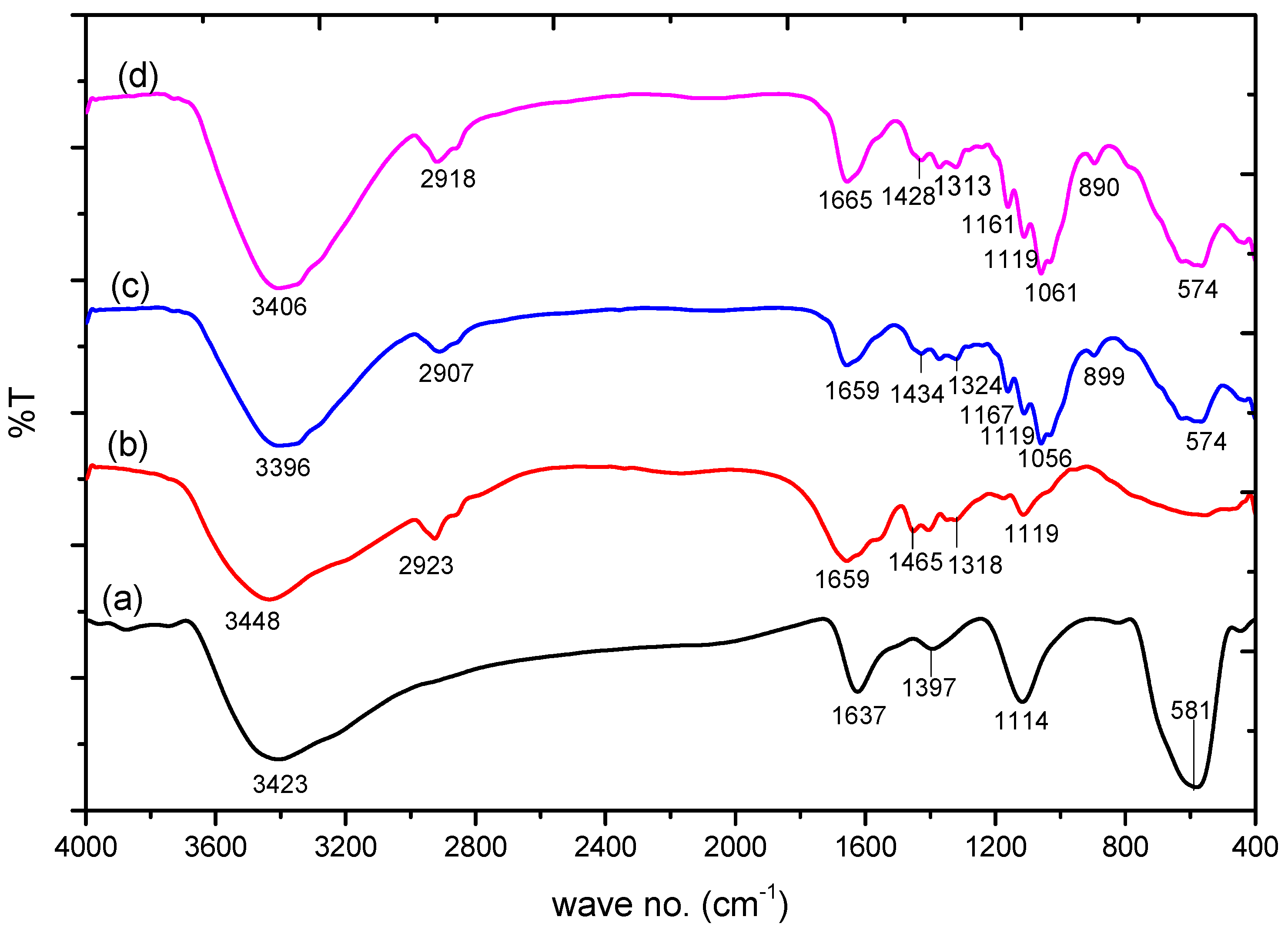
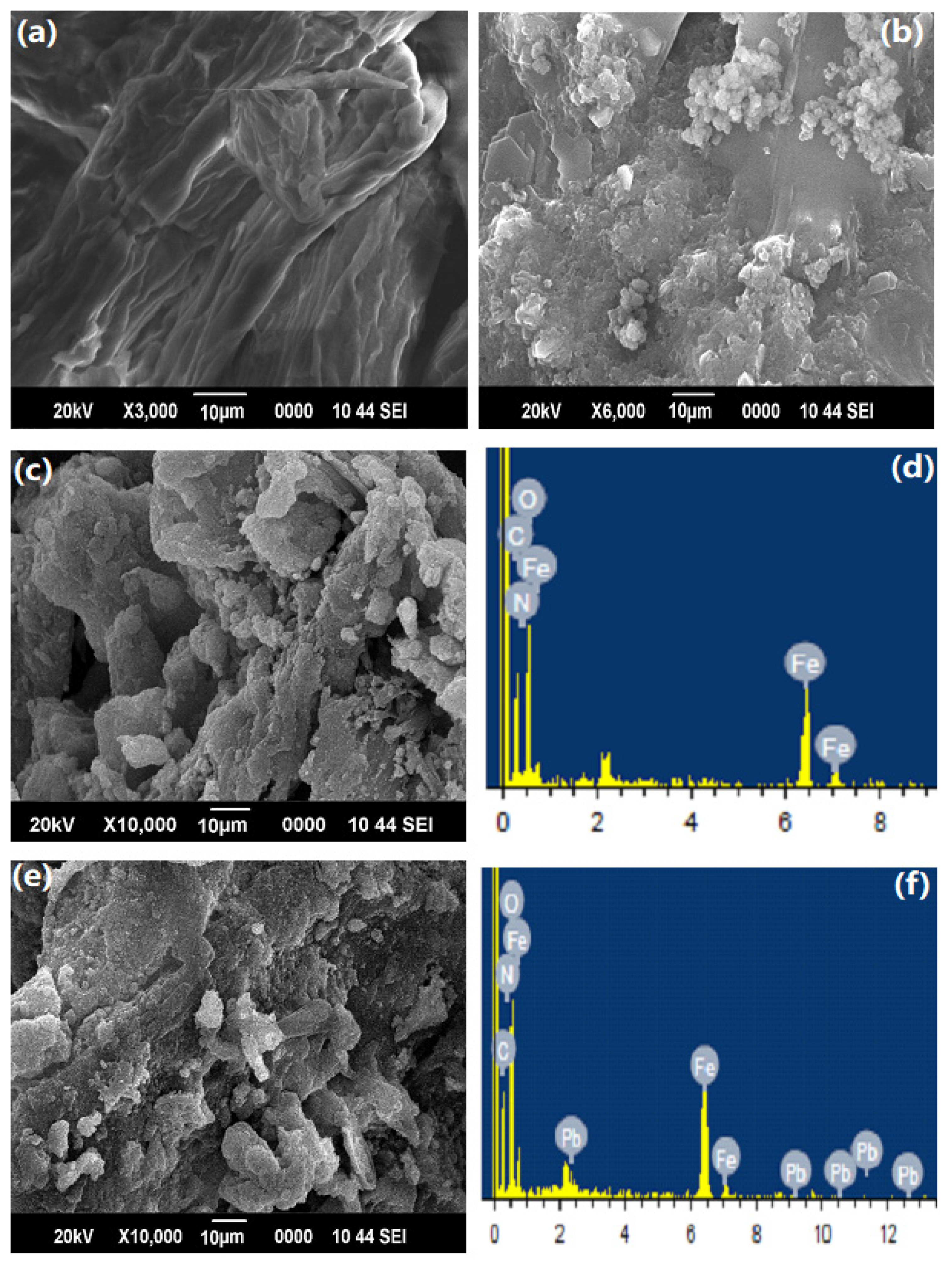
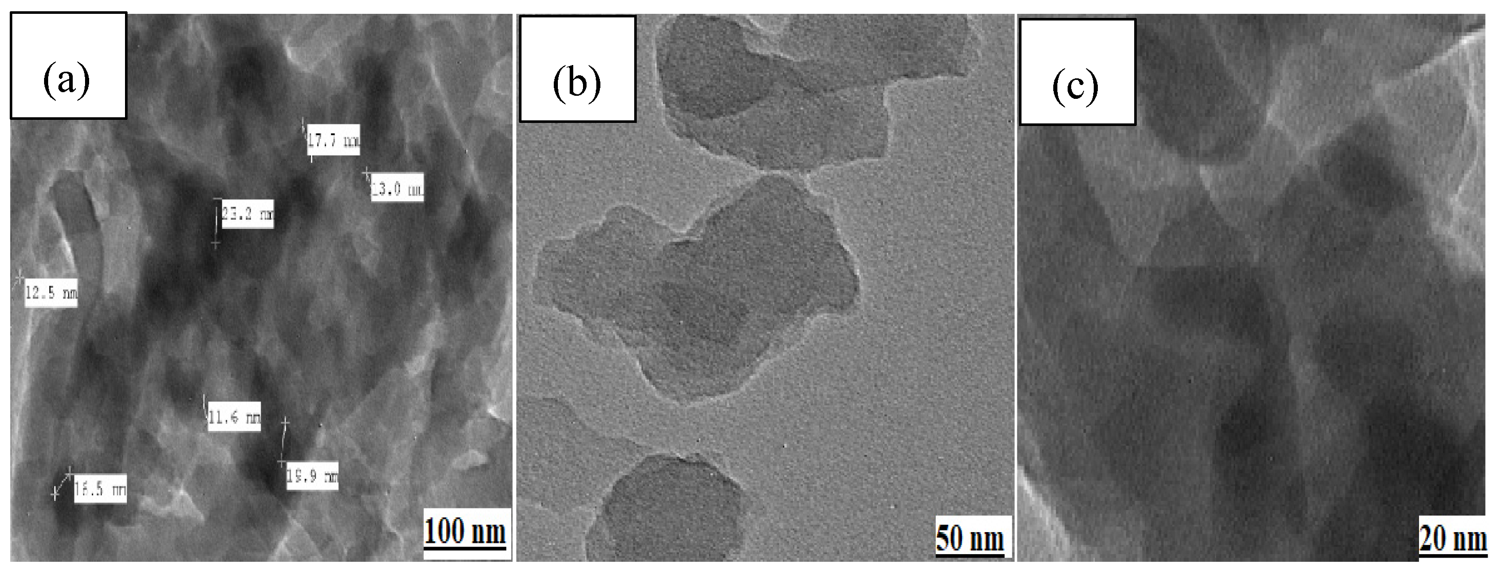
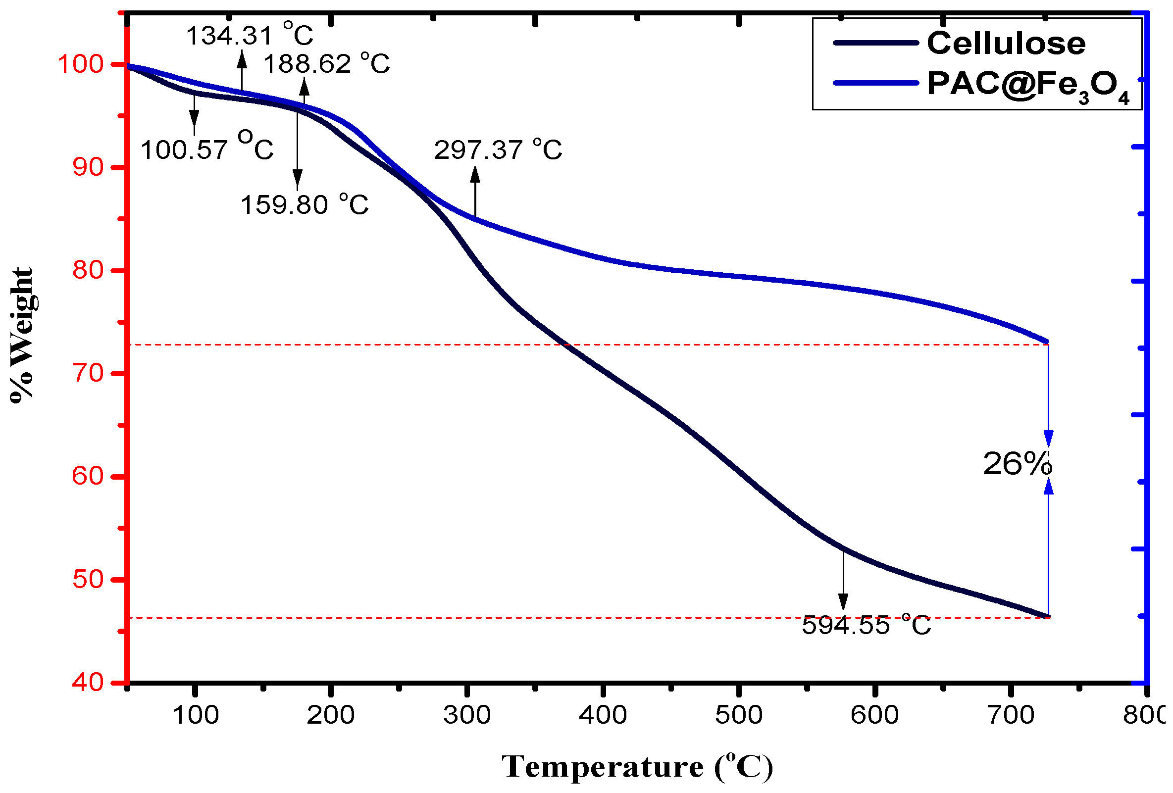
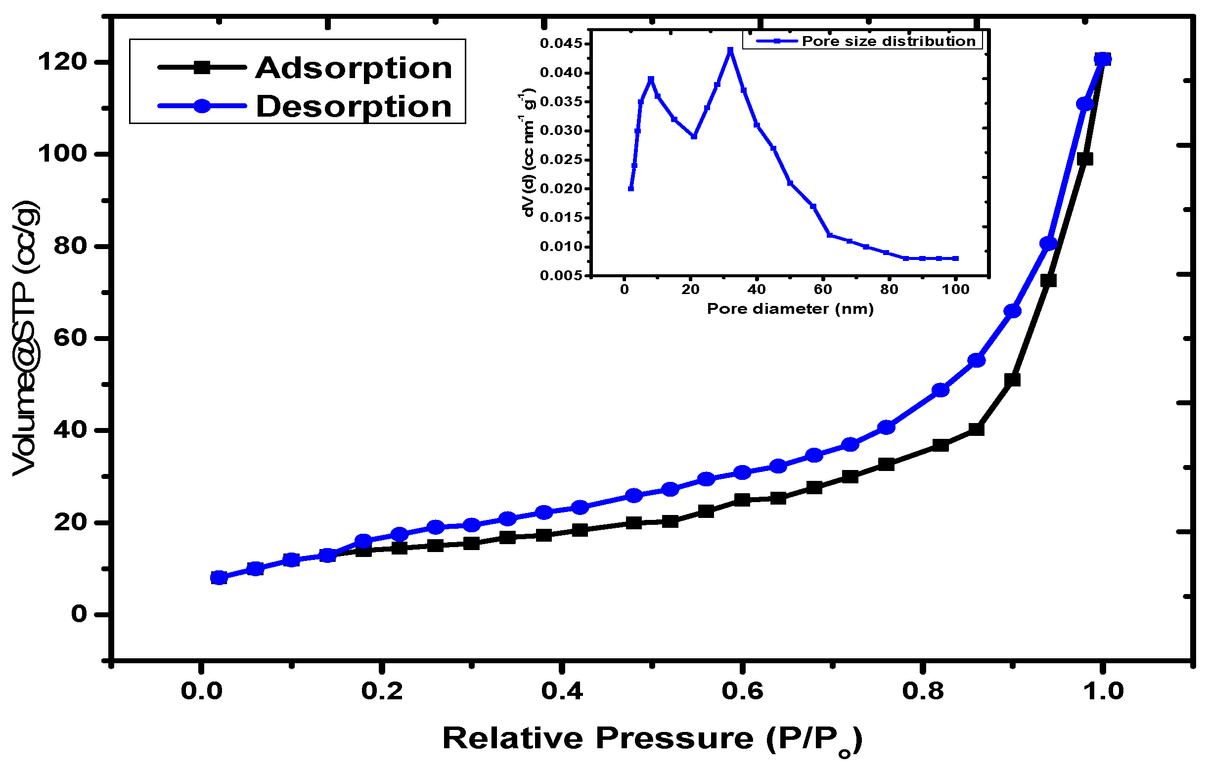
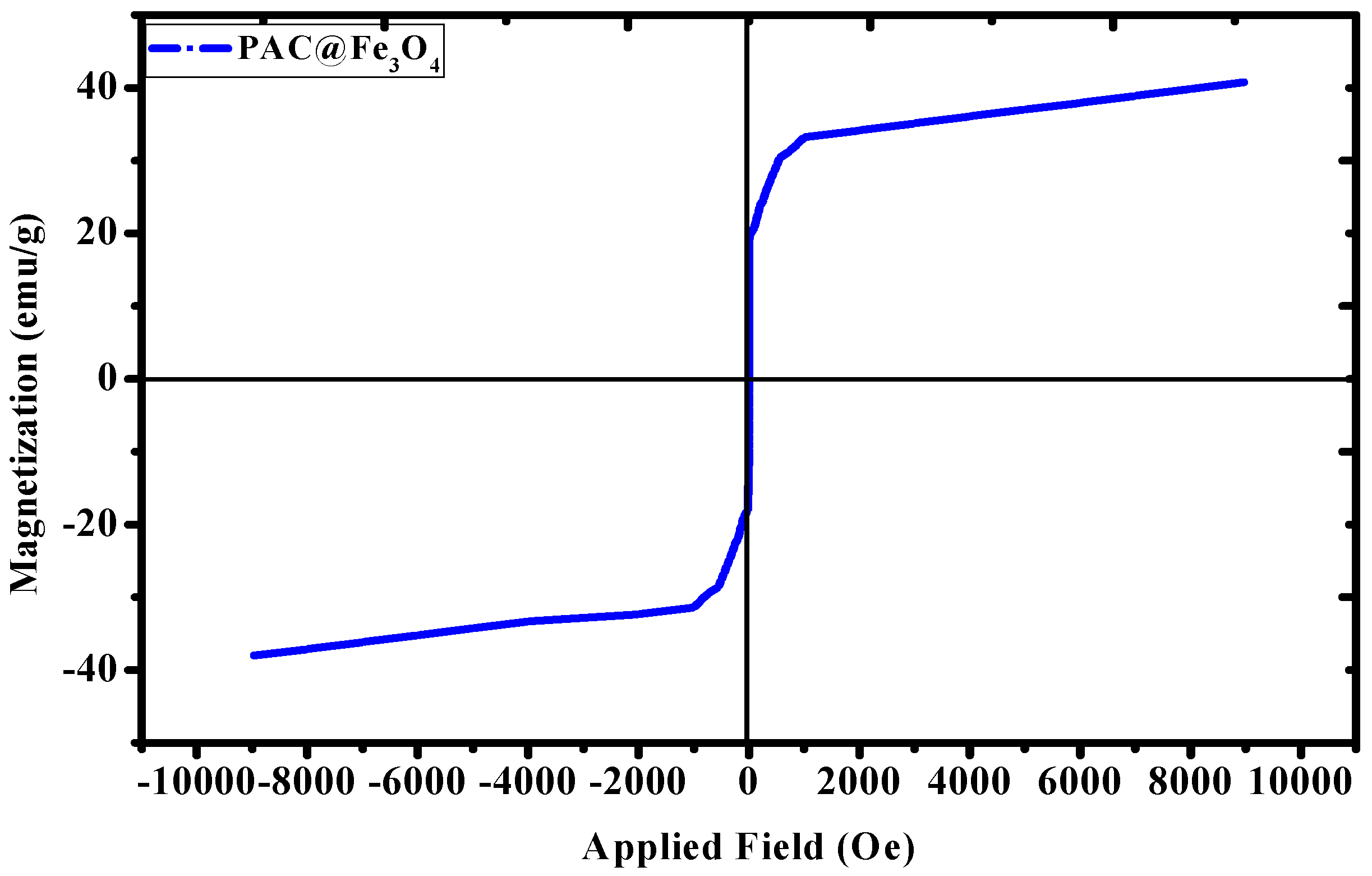
3.1. Characterization of the Material
3.2. Adsorption Behavior of PAC@Fe3O4 towards a Ternary System
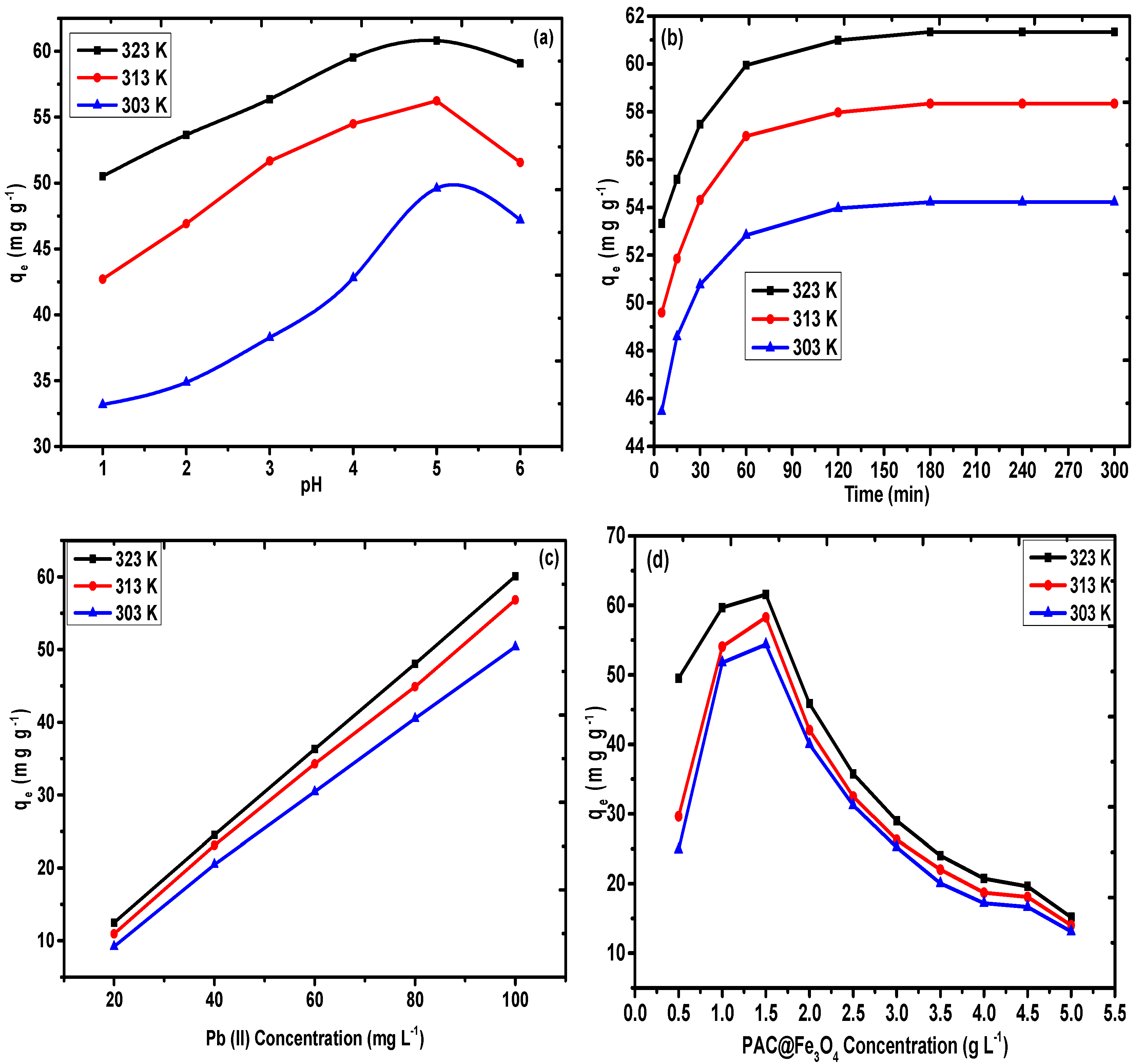
3.2.1. Impact of pH
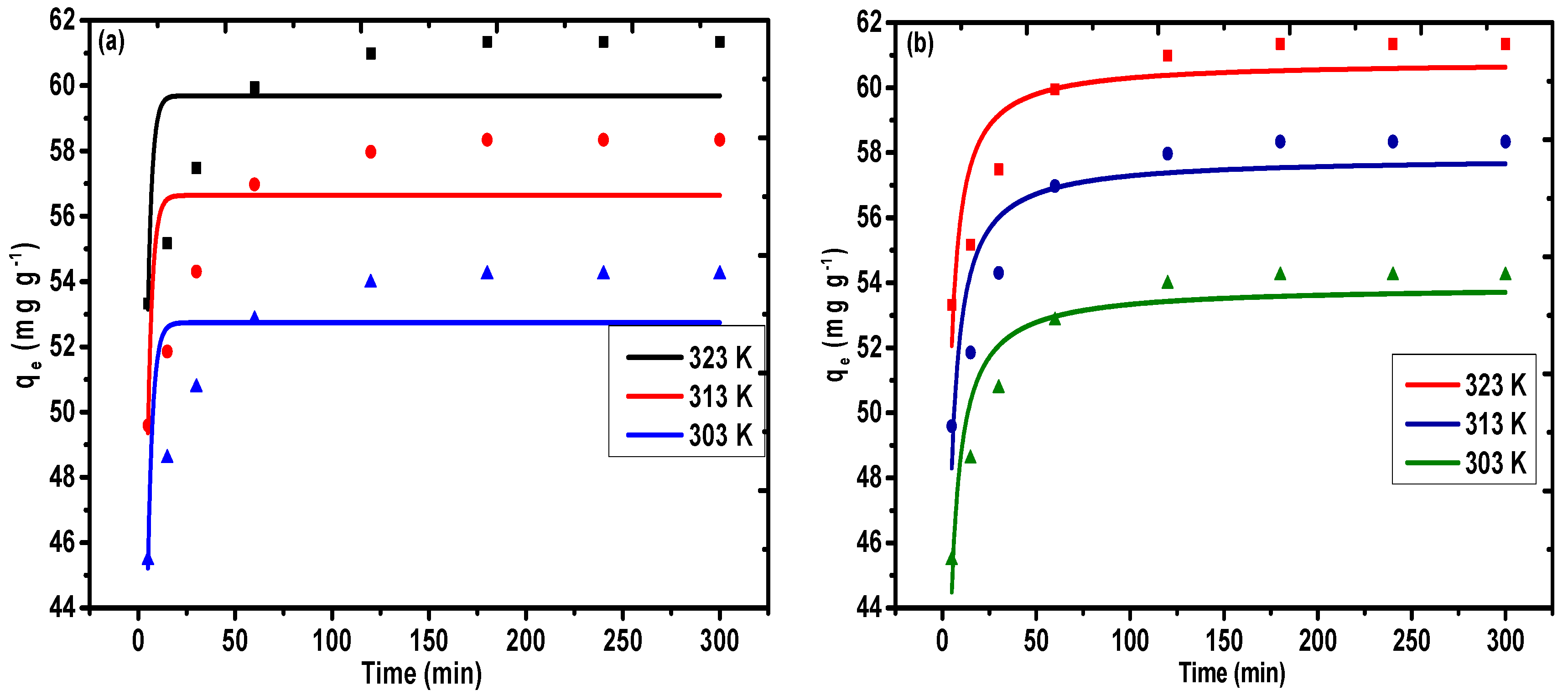
3.2.2. Impact of Contact Time
3.2.3. Impact of the Initial Metal Ion Concentration
3.2.4. Impact of Adsorbent Dose
3.2.5. Impact of Temperature
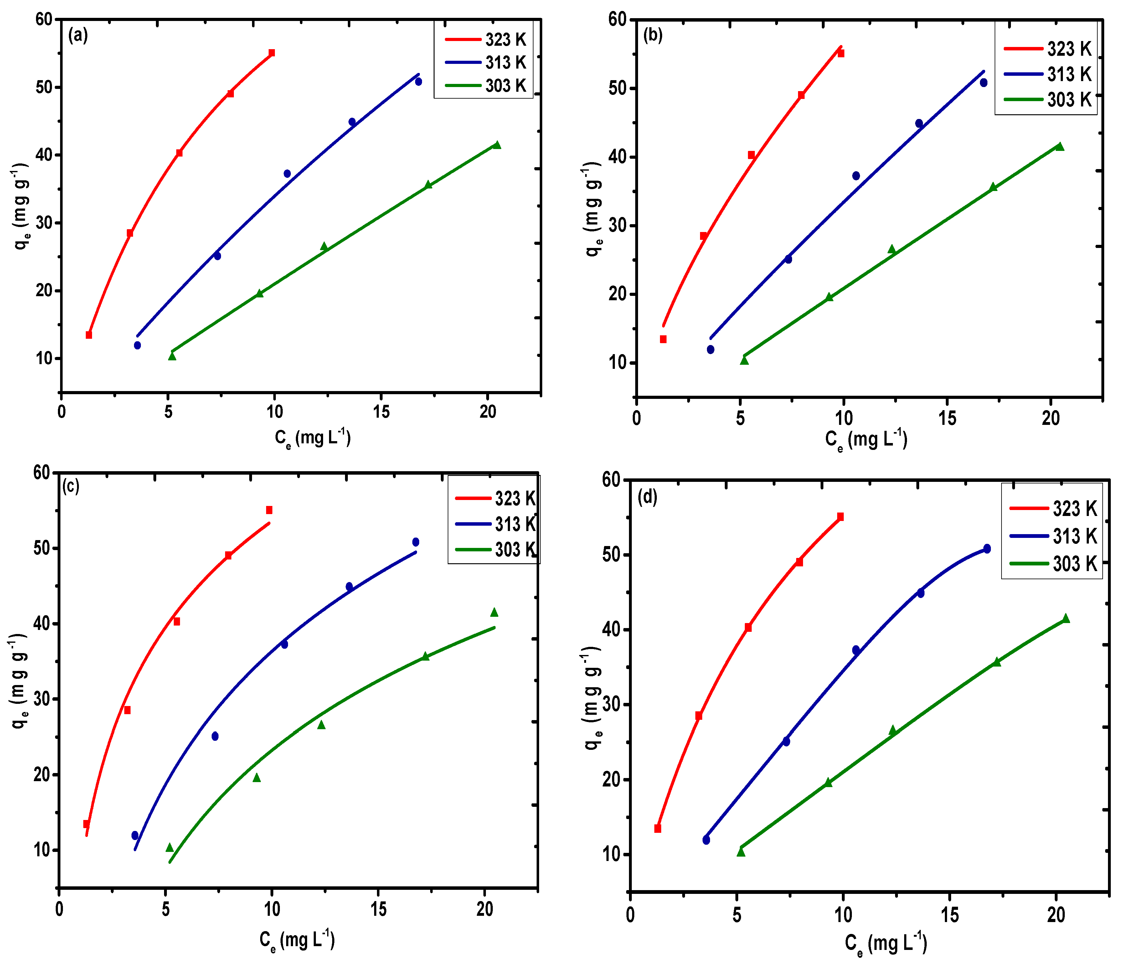
3.3. Adsorption Isotherms
3.4. Adsorption Kinetics
3.5. Adsorption Thermodynamics
3.6. Adsorption Mechanism
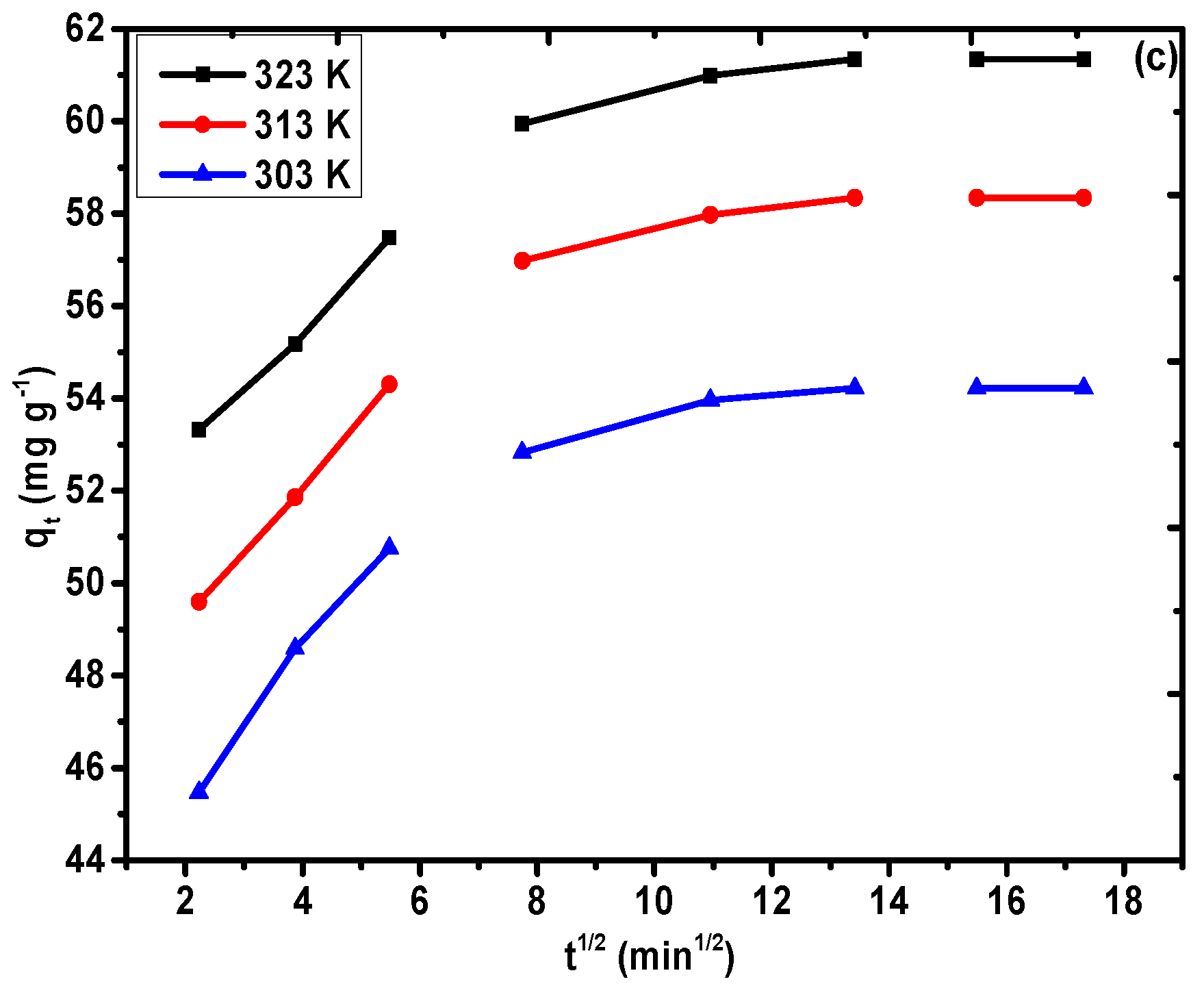
3.7. Impact of Other Competitive Ions
3.8. Desorption and Regeneration Tests
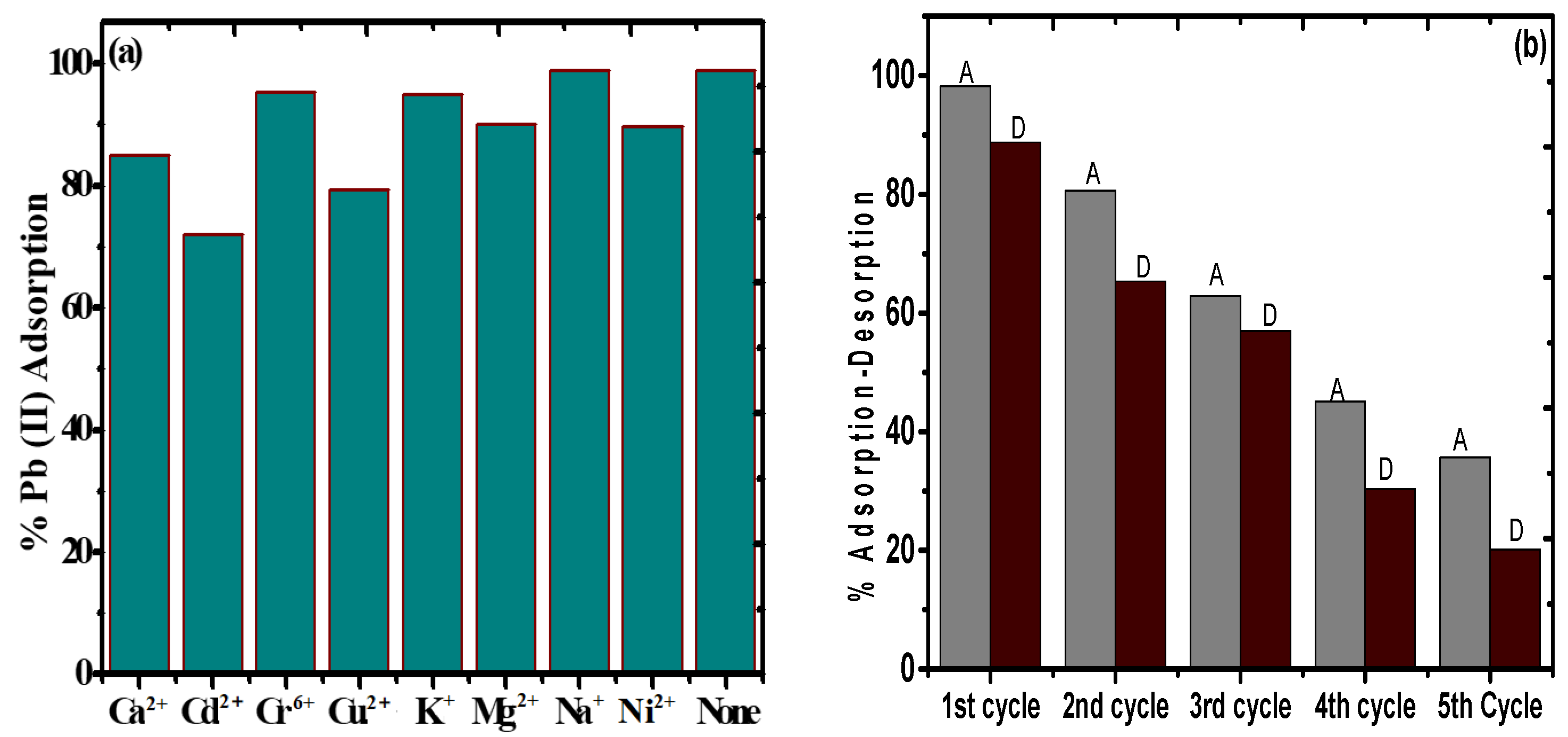
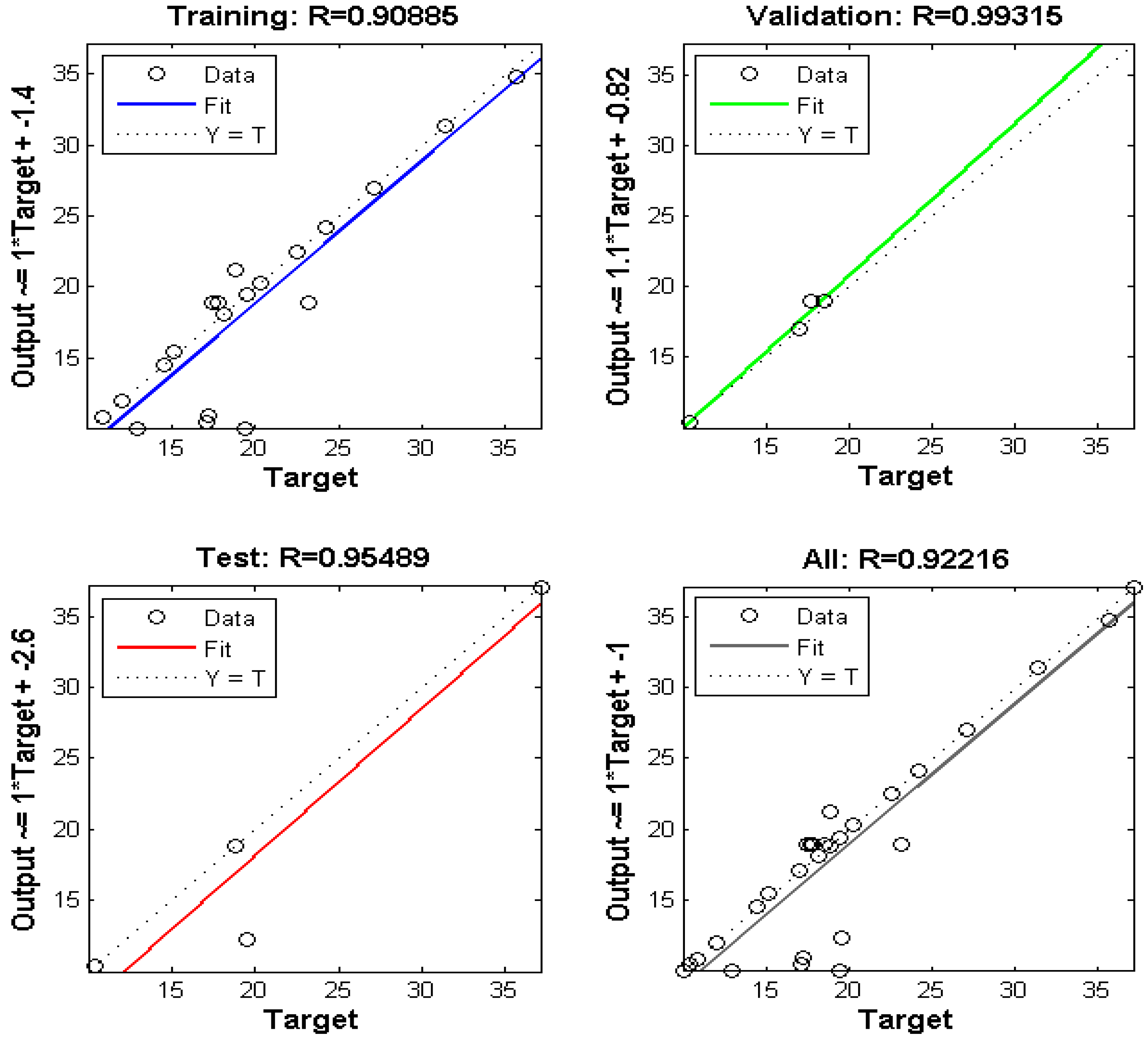
3.9. Optimization of the ANN Structure
4. Conclusions
Supplementary Materials
Author Contributions
Funding
Acknowledgments
Conflicts of Interest
Abbreviations
| TGA | thermogravimetric analysis |
| FTIR | Fourier transform-infrared |
| XRD | X-ray diffraction |
| SEM | scattering electron microscopy |
| TEM | transmission electron microscopy |
| BET | Brunauer–Emmett–Teller |
| MNPs | magnetic oxide nanoparticles |
| PAM | poly(acrylamide) |
| EDX | energy-dispersive X-ray |
| ANN | artificial neural network |
| BJH | Barett–Joyner–Halenda |
| MSE | mean square error |
References
- Khazaei, M.; Nasseri, S.; Ganjali, M.R.; Khoobi, M.; Nabizadeh, R.; Gholibegloo, E.; Nazmara, S. Selective removal of lead ions from aqueous solutions using 1,8-dihydroxyanthraquinone (DHAQ) functionalized graphene oxide; isotherm, kinetic and thermodynamic studies. RSC Adv. 2018, 8, 5685–5694. [Google Scholar] [CrossRef]
- Lingamdinne, L.P.; Koduru, J.R.; Chang, Y.-Y.; Karri, R.R. Process optimization and adsorption modeling of Pb(II) on nickel ferrite-reduced graphene oxide nano-composite. J. Mol. Liq. 2018, 250, 202–211. [Google Scholar] [CrossRef]
- Liu, J.; Mwamulima, T.; Wang, Y.; Fang, Y.; Song, S.; Peng, C. Removal of Pb(II) and Cr(VI) from aqueous solutions using the fly ash-based adsorbent material-supported zero-valent iron. J. Mol. Liq. 2017, 243, 205–211. [Google Scholar] [CrossRef]
- Pal, P.; Pal, A. Enhanced Pb2+ removal by anionic surfactant bilayer anchored on chitosan bead surface. J. Mol. Liq. 2017, 248, 713–724. [Google Scholar] [CrossRef]
- Guo, S.; Jiao, P.; Dan, Z.; Duan, N.; Zhang, J.; Chen, G.; Gao, W. Synthesis of magnetic bioadsorbent for adsorption of Zn(II), Cd(II) and Pb(II) ions from aqueous solution. Chem. Eng. Res. Des. 2017, 126, 217–231. [Google Scholar] [CrossRef]
- Zhang, X.; Li, Y.; Hou, Y. Preparation of magnetic polyethylenimine lignin and its adsorption of Pb(II). Int. J. Biol. Macromol. 2019, 141, 1102–1110. [Google Scholar] [CrossRef] [PubMed]
- Xu, W.; Song, Y.; Dai, K.; Sun, S.; Liu, G.; Yao, J. Novel ternary nanohybrids of tetraethylenepentamine and graphene oxide decorated with MnFe2O4 magnetic nanoparticles for the adsorption of Pb(II). J. Hazard. Mater. 2018, 358, 337–345. [Google Scholar] [CrossRef]
- Yang, K.; Lou, Z.; Fu, R.; Zhou, J.; Xu, J.; Baig, S.A.; Xu, X. Multiwalled carbon nanotubes incorporated with or without amino groups for aqueous Pb(II) removal: Comparison and mechanism study. J. Mol. Liq. 2018, 260, 149–158. [Google Scholar] [CrossRef]
- Lin, L.; Xu, X.; Papelis, C.; Xu, P. Innovative use of drinking water treatment solids for heavy metals removal from desalination concentrate: Synergistic effect of salts and natural organic matter. Chem. Eng. Res. Des. 2017, 120, 231–239. [Google Scholar] [CrossRef]
- Kong, D.; Qiao, N.; Wang, N.; Wang, Z.; Wang, Q.; Zhou, Z.; Ren, Z. Facile preparation of a nano-imprinted polymer on magnetite nanoparticles for the rapid separation of lead ions from aqueous solution. Phys. Chem. Chem. Phys. 2018, 20, 12870–12878. [Google Scholar] [CrossRef]
- Pawar, R.R.; Hong, S.M.; Jin, K.J.; Lee, S.M. Iron-oxide modified sericite alginate beads: A sustainable adsorbent for the removal of As(V) and Pb(II) from aqueous solutions. J. Mol. Liq. 2017, 240, 497–503. [Google Scholar]
- Liao, Q.; Yan, S.; Linghu, W.; Zhu, Y.; Shen, R.; Ye, F.; Feng, G.; Dong, L.; Asiri, A.M.; Xu, D.; et al. Impact of key geochemical parameters on the highly efficient sequestration of Pb(II) and Cd(II) in water using g-C3N4 nanosheets. J. Mol. Liq. 2018, 258, 40–47. [Google Scholar] [CrossRef]
- Husnain, S.M.; Um, W.; Woojin-Lee, W.-L.; Chang, Y.-S. Magnetite-based adsorbents for sequestration of radionuclides: A review. RSC Adv. 2018, 8, 2521–2540. [Google Scholar] [CrossRef]
- Kataria, N.; Garg, V.K. Optimization of Pb(II) and Cd(II) adsorption onto ZnO nanoflowers using central composite design: Isotherms and kinetics modelling. J. Mol. Liq. 2018, 271, 228–239. [Google Scholar] [CrossRef]
- Kumar, R.; Sharma, R.K.; Singh, A.P. Grafting of cellulose with N-isopropylacrylamide and glycidyl methacrylate for efficient removal of Ni(II), Cu(II) and Pd(II) ions from aqueous solution. Sep. Purif. Technol. 2019, 219, 249–259. [Google Scholar] [CrossRef]
- Li, Y.; Guo, C.; Shi, R.; Zhang, H.; Gong, L.; Dai, L. Chitosan/nanofibrillated cellulose aerogel with highly oriented microchannel structure for rapid removal of Pb(II) ions from aqueous solution. Carbohydr. Polym. 2019, 223, 115048. [Google Scholar] [CrossRef]
- Lu, J.; Jin, R.-N.; Liu, C.; Wang, Y.-F.; Ouyang, X. Magnetic carboxylated cellulose nanocrystals as adsorbent for the removal of Pb(II) from aqueous solution. Int. J. Biol. Macromol. 2016, 93, 547–556. [Google Scholar] [CrossRef]
- Vijayalakshmi, K.; Devi, B.M.; Latha, S.; Gomathi, T.; Sudha, P.N.; Venkatesan, J.; Anil, S. Batch adsorption and desorption studies on the removal of lead(II) from aqueous solution using nanochitosan/sodium alginate/microcrystalline cellulose beads. Int. J. Biol. Macromol. 2017, 104, 1483–1494. [Google Scholar] [CrossRef]
- Xie, Y.; Yuan, X.; Wu, Z.; Zeng, G.; Jiang, L.; Peng, X.; Li, H. Adsorption behavior and mechanism of Mg/Fe layered double hydroxide with Fe3O4-carbon spheres on the removal of Pb(II) and Cu(II). J. Colloid Interface Sci. 2019, 536, 440–455. [Google Scholar] [CrossRef]
- Lin, S.; Liu, L.; Yang, Y.; Zhang, W.; Lian, C.; Lin, K. Comparison of the adsorption preference using superparamagnetic Fe3O4-SH nanoparticles to remove aqueous heavy metal contaminants. Chem. Eng. Res. Des. 2017, 125, 319–327. [Google Scholar] [CrossRef]
- Guo, S.; Duan, N.; Dan, Z.; Chen, G.; Shi, F.; Gao, W. g-C3N4 modified magnetic Fe3O4 adsorbent: Preparation, characterization, and performance of Zn(II), Pb(II) and Cd(II) removal from aqueous solution. J. Mol. Liq. 2018, 258, 225–234. [Google Scholar] [CrossRef]
- El-Reash, Y.G.A.; Abdelghany, A.M.; Elrazak, A.A. Removal and separation of Cu(II) from aqueous solutions using nano-silver chitosan/polyacrylamide membranes. Int. J. Biol. Macromol. 2016, 86, 789–798. [Google Scholar] [CrossRef] [PubMed]
- Abdelwahab, N.A.; Helaly, F.M. Simulated visible light photocatalytic degradation of Congo red by TiO2 coated magnetic polyacrylamide grafted carboxymethylated chitosan. J. Ind. Eng. Chem. 2017, 50, 162–171. [Google Scholar] [CrossRef]
- Qiu, Z.; Niu, Y.; Fu, T.; Wang, K.; Mu, Q.; Wang, F. Removal of Ni(II) from fuel ethanol by PAMAM dendrimers/silica hybrid materials: Combined experimental and theoretical study. Chem. Eng. Res. Des. 2019, 144, 174–184. [Google Scholar] [CrossRef]
- Sun, X.; Huang, H.; Zhu, Y.; Du, Y.; Yao, L.; Jiang, X.; Gao, P. Adsorption of Pb2+ and Cd2+ onto Spirulina platensis harvested by polyacrylamide in single and binary solution systems. Colloids Surf. A Physicochem. Eng. Asp. 2019, 583, 123926. [Google Scholar] [CrossRef]
- Zhao, Y.; Zhao, Z.; Zhang, J.; Wei, M.; Jiang, X.; Hou, L. Gemini surfactant mediated HIPE template for the preparation of highly porous monolithic chitosan-g-polyacrylamide with promising adsorption performances. Eur. Polym. J. 2019, 112, 809–816. [Google Scholar] [CrossRef]
- Dil, E.A.; Ghaedi, M.; Asfaram, A.; Hajati, S.; Mehrabi, F.; Goudarzi, A. Preparation of nanomaterials for the ultrasound-enhanced removal of Pb2+ ions and malachite green dye: Chemometric optimization and modeling. Ultrason. Sonochem. 2017, 34, 677–691. [Google Scholar] [CrossRef]
- Javadian, H.; Asadollahpour, S.; Ruiz, M.; Sastre, A.M.; Ghasemi, M.; Asl SM, H.; Masomi, M. Using fuzzy inference system to predict Pb(II) removal from aqueous solutions by magnetic Fe3O4/H2SO4-activated Myrtus communis leaves carbon nanocomposite. J. Taiwan Inst. Chem. Eng. 2018, 91, 186–199. [Google Scholar] [CrossRef]
- Pan, S.; Li, J.; Wang, L.; Jafvert, C.T. Decomposition of complexed Pb(II) and subsequent adsorption of Pb(II) with yolk-shell Fe3O4@ hydrous zirconium oxide sphere. J. Colloid Interface Sci. 2019, 556, 65–73. [Google Scholar] [CrossRef]
- Sanaeishoar, H.; Sabbaghan, M.; Argyropoulos, D.S. Ultrasound assisted polyacrylamide grafting on nano-fibrillated cellulose. Carbohydr. Polym. 2018, 181, 1071–1077. [Google Scholar] [CrossRef]
- Langmuir, I. The adsorption of gases on plane surfaces of glass, mica and platinum. J. Am. Chem. Soc. 1918, 40, 1361–1403. [Google Scholar] [CrossRef]
- Freudlich, H.M.F. Unber die adsorption in losungen. Z. Phys. Chem. 1906, 57, 385–470. [Google Scholar]
- Temkin, M.J.; Pyzhev, V. Kinetics of ammonia synthesis on promoted iron catalysts. Acta Physiochem. 1940, 12, 217–222. [Google Scholar]
- Redlich, O.; Peterson, D.L. A useful adsorption isotherm. J. Phys. Chem. 1959, 63, 1024–1026. [Google Scholar] [CrossRef]
- Lagergren, S. About the theory of so-called adsorption of soluble substances. Handlingar 1898, 24, 1–39. [Google Scholar]
- Ho, Y.S. Review of second-order models for adsorption systems. J. Hazard. Mater. 2006, 136, 681–689. [Google Scholar] [CrossRef]
- Weber, W.J., Jr.; Morris, J.C. Kinetics of adsorption on carbon from solution. J. Sanit. Eng. Div. Am. Soc. Civil. Eng. 1963, 89, 31–59. [Google Scholar]
- Moradi, A.; Moghadam, P.N.; Hasanzadeh, R.; Sillanpää, M. Chelating magnetic nanocomposite for the rapid removal of Pb(II) ions from aqueous solutions: Characterization, kinetic, isotherm and thermodynamic studies. RSC Adv. 2017, 7, 433–448. [Google Scholar] [CrossRef]
- Guo, X.; Wang, J. A general kinetic model for adsorption: Theoretical analysis and modeling. J. Mol. Liq. 2019, 288, 111100. [Google Scholar] [CrossRef]
- Singha, B.; Bar, N.; Das, S.K. The use of artificial neural network (ANN) for modeling of Pb(II) adsorption in batch process. J. Mol. Liq. 2015, 211, 228–232. [Google Scholar] [CrossRef]
- Khandanlou, R.; Masoumi, H.R.F.; Ahmad, M.B.; Shameli, K.; Basri, M.; Kalantari, K. Enhancement of heavy metals sorption via nanocomposites of rice straw and Fe3O4 nanoparticles using artificial neural network (ANN). Ecol. Eng. 2016, 91, 249–256. [Google Scholar] [CrossRef]
- Subedi, N.; Lähde, A.; Abu-Danso, E.; Iqbal, J.; Bhatnagar, A. A comparative study of magnetic chitosan (Chi@Fe3O4) and graphene oxide modified magnetic chitosan (Chi@Fe3O4GO) nanocomposites for efficient removal of Cr(VI) from water. Int. J. Biol. Macromol. 2019, 137, 948–959. [Google Scholar] [CrossRef] [PubMed]
- Liu, T.; Ding, E.; Xue, F. Polyacrylamide and poly (N,N-dimethylacrylamide) grafted cellulose nanocrystals as efficient flocculants for kaolin suspension. Int. J. Biol. Macromol. 2017, 103, 1107–1112. [Google Scholar] [CrossRef] [PubMed]
- Kenawy, I.M.; Hafez, M.A.H.; Ismail, M.A.; Hashem, M.A. Adsorption of Cu(II), Cd(II), Hg(II), Pb(II) and Zn(II) from aqueous single metal solutions by guanyl-modified cellulose. Int. J. Biol. Macromol. 2018, 107, 1538–1549. [Google Scholar] [CrossRef]
- Thomas, B.; Raj, M.C.; Joy, J.; Moores, A.; Drisko, G.L.; Sanchez, C. Nanocellulose, a versatile green platform: From biosources to materials and their applications. Chem. Rev. 2018, 118, 11575–11625. [Google Scholar] [CrossRef]
- Kumawat, L.K.; Kumar, M.; Bhatt, P.; Sharma, A.; Asif, M.; Gupta, V.K. An easily accessible optical chemosensor for Cu2+ based on novel imidazoazine framework, its performance characteristics and potential applications. Sens. Actuators B Chem. 2017, 240, 365–375. [Google Scholar] [CrossRef]
- Mollahosseini, A.; Khadir, A.; Saeidian, J. Core–shell polypyrrole/Fe3O4 nanocomposite as sorbent for magnetic dispersive solid-phase extraction of Al+3 ions from solutions: Investigation of the operational parameters. J. Water Process Eng. 2019, 29, 100795. [Google Scholar] [CrossRef]
- Atta, A.M.; Gafer, A.K.; Al-Lohedan, H.A.; Abdullah, M.M.; Ezzat, A.O. Preparation of magnetite and silver poly (2-acrylamido-2-methyl propane sulfonic acid-co-acrylamide) nanocomposites for adsorption and catalytic degradation of methylene blue water pollutant. Polym. Int. 2019, 68, 1164–1177. [Google Scholar] [CrossRef]
- Song, T.; Yu, C.; He, X.; Lin, J.; Liu, Z.; Yang, X.; Zhang, Y.; Huang, Y.; Tang, C. Synthesis of magnetically separable porous BN microrods@Fe3O4 nanocomposites for Pb(II) adsorption. Colloids Surf. Physicochem. Eng. Asp. 2018, 537, 508–515. [Google Scholar] [CrossRef]
- Zhuang, L.; Zhang, W.; Zhao, Y.; Shen, H.; Lin, H.; Liang, J. Preparation and characterization of Fe3O4 particles with novel nanosheets morphology and magnetochromatic property by a modified solvothermal method. Sci. Rep. 2015, 5, 9320. [Google Scholar] [CrossRef]
- Manikandan, A.; Vijaya, J.J.; Mary, J.A.; Kennedy, L.J.; Dinesh, A. Structural, optical and magnetic properties of Fe3O4 nanoparticles prepared by a facile microwave combustion method. J. Ind. Eng. Chem. 2014, 20, 2077–2085. [Google Scholar] [CrossRef]
- Karimzadeh, I.; Aghazadeh, M.; Doroudi, T.; Ganjali, M.R.; Kolivand, P.H. Electrochemical preparation and characterization of chitosan-coated superparamagnetic iron oxide (Fe3O4) nanoparticles. Mater. Res. Innov. 2018, 22, 352–360. [Google Scholar] [CrossRef]
- Karimzadeh, I.; Aghazadeh, M.; Dalvand, A.; Doroudi, T.; Kolivand, P.H.; Ganjali, M.R.; Norouzi, P. Effective electrosynthesis and in situ surface coating of Fe3O4 nanoparticles with polyvinyl alcohol for biomedical applications. Mater. Res. Innov. 2019, 23, 1–8. [Google Scholar] [CrossRef]
- Golsefidi, M.A.; Sarkhosh, B. Preparation and characterization of rapid magnetic recyclable Fe3O4@SiO2@TiO2–Sn photocatalyst. J. Iran. Chem. Soc. 2017, 14, 1089–1098. [Google Scholar] [CrossRef]
- Saeedi, M.; Vahidi, O. Preparation and characterization of magnetic core–shell iron oxide@glycyrrhizic acid nanoparticles in ethanol–water mixed solvent. Appl. Phys. A Mater. Sci. Process. 2018, 124, 431. [Google Scholar] [CrossRef]
- Yousefzadeh, H.; Salarian, A.A.; Kala, H.S. Study of Pb(II) adsorption from aqueous solutions by TiO2 functionalized with hydroxide ethyl aniline (PHEA/n-TiO2). J. Mol. Liq. 2018, 263, 294–302. [Google Scholar] [CrossRef]
- Wang, N.; Jin, R.-N.; Omer, A.M.; Ouyang, X. Adsorption of Pb(II) from fish sauce using carboxylated cellulose nanocrystal: Isotherm, kinetics, and thermodynamic studies. Int. J. Biol. Macromol. 2017, 102, 232–240. [Google Scholar] [CrossRef]
- Wu, D.; Wang, Y.; Li, Y.; Wei, Q.; Hu, L.; Yan, T.; Feng, R.; Yan, L.; Du, B. Phosphorylated chitosan/CoFe2O4 composite for the efficient removal of Pb(II) and Cd(II) from aqueous solution: Adsorption performance and mechanism studies. J. Mol. Liq. 2019, 277, 181–188. [Google Scholar] [CrossRef]
- Koushkbaghi, S.; Zakialamdari, A.; Pishnamazi, M.; Ramandi, H.F.; Aliabadi, M.; Irani, M. Aminated-Fe3O4 nanoparticles filled chitosan/PVA/PES dual layers nanofibrous membrane for the removal of Cr(VI) and Pb(II) ions from aqueous solutions in adsorption and membrane processes. Chem. Eng. J. 2018, 337, 169–182. [Google Scholar] [CrossRef]
- Zong, P.; Cao, D.; Wang, S.; He, C.; Zhao, Y. Synthesis of Fe3O4/CD magnetic nanocomposite via low temperature plasma technique with high enrichment of Ni(II) from aqueous solution. J. Taiwan Inst. Chem. Eng. 2017, 70, 134–140. [Google Scholar] [CrossRef]
- Rakati, K.K.; Mirzaei, M.; Maghsoodi, S.; Shahbazi, A. Preparation and characterization of poly aniline modified chitosan embedded with ZnO-Fe3O4 for Cu(II) removal from aqueous solution. Int. J. Biol. Macromol. 2019, 130, 1025–1045. [Google Scholar] [CrossRef] [PubMed]
- Naushad, M.; Ahamad, T.; Al-Ghanim, K.A.; Al-Muhtaseb, A.H.; Eldesoky, G.E.; Khan, A.A. A highly porous nanocomposite (Fe3O4@BFR) for the removal of toxic Cd(II) ions from aqueous environment: Adsorption modelling and regeneration study. Compos. Part B Eng. 2019, 172, 179–185. [Google Scholar] [CrossRef]


| Elements | PAC@Fe3O4 | PAC@Fe3O4-Pb | ||
|---|---|---|---|---|
| Weight % | Atomic % | Weight % | Atomic % | |
| C | 32.07 | 45.64 | 25.78 | 43.28 |
| N | 3.44 | 4.20 | 0.00 | 0.00 |
| O | 39.92 | 42.64 | 34.63 | 43.64 |
| Fe | 24.57 | 7.52 | 34.97 | 12.62 |
| Pb | 0.00 | 0.00 | 4.61 | 0.45 |
| Model | Parameters | Pb (II) | ||
|---|---|---|---|---|
| Langmuir | 323 K | 313 K | 303 K | |
| qm (mg g−1) | 314.47 | 239.34 | 100.79 | |
| KL (L mg−1) | 0.121 | 0.016 | 0.003 | |
| R2 | 0.99 | 0.99 | 0.99 | |
| χ2 | 0.10 | 0.31 | 0.47 | |
| Freundlich | n | 1.58 | 1.14 | 1.02 |
| KF (mg g−1) (dm3/g)n | 13.14 | 4.48 | 2.22 | |
| R2 | 0.99 | 0.98 | 0.98 | |
| χ2 | 2.59 | 3.69 | 0.57 | |
| Temkin | AT (L mg−1) | 1.39 | 0.42 | 0.28 |
| bT (J mol−1) | 131.96 | 105.32 | 118.23 | |
| R2 | 0.98 | 0.97 | 0.97 | |
| χ2 | 3.72 | 5.64 | 4.53 | |
| Redlich‒Peterson | g | 0.96 | 5.31 | 5.81 |
| KR (L g−1) | 12.50 | 3.48 | 2.10 | |
| aR (L mg−1) | 0.14 | 4.80 | 1.02 | |
| R2 | 0.99 | 0.99 | 0.99 | |
| χ2 | 0.14 | 0.58 | 0.44 | |
| Model | Parameters | Pb(II) | ||
|---|---|---|---|---|
| Pseudo-First-Order | 323 K | 313 K | 303 K | |
| qe (exp) (mg g−1) | 61.34 | 58.34 | 54.22 | |
| qe (cal) (mg g−1) | 59.69 | 56.64 | 52.74 | |
| k1 (min−1) | 0.44 | 0.41 | 0.38 | |
| R2 | 0.43 | 0.46 | 0.56 | |
| χ2 | 5.75 | 6.30 | 4.67 | |
| Pseudo-Second-Order | qe (exp) (mg g−1) | 61.34 | 58.34 | 54.22 |
| qe (cal) (mg g−1) | 60.80 | 57.85 | 53.89 | |
| k2 (g mg−1 min−1) | 0.02 | 0.02 | 0.02 | |
| R2 | 0.79 | 0.82 | 0.89 | |
| χ2 | 2.04 | 2.06 | 1.17 | |
| Intra-particle Diffusion | Kid | 0.51 | 0.53 | 0.53 |
| C | 53.96 | 50.47 | 46.85 | |
| R2 | 0.78 | 0.76 | 0.73 | |
| χ2 | 1.09 | 1.21 | 1.23 | |
| No. of Neurons | R2 Value | MSE Value |
|---|---|---|
| 1 | 0.9241 | 0.0045 |
| 2 | 0.9572 | 0.0026 |
| 3 | 0.8901 | 0.0075 |
| 4 | 0.9323 | 0.0061 |
| 5 | 0.9845 | 0.0022 |
| 6 | 0.9744 | 0.0032 |
| 7 | 0.9648 | 0.0024 |
| 8 | 0.9778 | 0.0026 |
| 9 | 0.9799 | 0.001 |
| 10 | 0.9915 | 0.0028 |
| 11 | 0.9241 | 0.0035 |
| 12 | 0.9543 | 0.0025 |
| 13 | 0.9727 | 0.0041 |
| 14 | 0.9815 | 0.0021 |
| 15 | 0.9450 | 0.0016 |
| 16 | 0.9855 | 0.0029 |
| 17 | 0.9712 | 0.0038 |
| 18 | 0.9487 | 0.0015 |
| 19 | 0.9876 | 0.0027 |
| 20 | 0.9267 | 0.0043 |
| W1 | W2 | b1 | b2 |
|---|---|---|---|
| [−0.16147 0.85147 −1.1877 1.125 1.8233 0.12975 −0.72917 1.6183 −0.068404 −0.31243 1.8263 1.0474 0.33743 1.7101 −0.165 1.2238 0.85285 −0.41348 0.57248 −0.15122] | [5.1699; | [−2.7344] | |
| 3.2342; | |||
| 2.4921; | |||
| [2.3555 0.22624 1.4495 1.7728; | 3.3494; | ||
| −1.7946 1.2629 1.8741 1.1642; | −1.7066; | ||
| 1.6833 1.0471 0.20408 1.9717; | −2.0543; | ||
| 0.87665 1.9143 −2.0152 −0.58027; | −0.9055; | ||
| 0.21074 −0.44249 3.3956 −0.4547; | 1.3696; | ||
| 0.059627 −1.0689 −0.5686 0.8507; | −0.92683; | ||
| −1.6308 1.5808 2.2064 0.30565; | 0.6882; | ||
| 1.8589 0.58103 −2.6895 −1.0942; | 1.5333; | ||
| −2.1314 −1.6081 −1.0178 −1.4578; | −3.3323; | ||
| −2.9458 2.3503 −1.3511 0.86399; | −0.30565; | ||
| 0.63077 −1.5321 1.3034 −0.23255; | 0.36439; | ||
| 1.3319 0.64905 −0.51145 2.138; | −1.6027; | ||
| 1.3292 0.083706 −1.8689 1.8066; | −0.30928; | ||
| 1.3137 −0.96565 1.7822 −3.1317] | 3.1452; | ||
| 2.623; | |||
| 2.9375; | |||
| 2.176] |
© 2019 by the authors. Licensee MDPI, Basel, Switzerland. This article is an open access article distributed under the terms and conditions of the Creative Commons Attribution (CC BY) license (http://creativecommons.org/licenses/by/4.0/).
Share and Cite
Hasan, I.; Khan, R.A.; Alharbi, W.; Alharbi, K.H.; Alsalme, A. In Situ Copolymerized Polyacrylamide Cellulose Supported Fe3O4 Magnetic Nanocomposites for Adsorptive Removal of Pb(II): Artificial Neural Network Modeling and Experimental Studies. Nanomaterials 2019, 9, 1687. https://doi.org/10.3390/nano9121687
Hasan I, Khan RA, Alharbi W, Alharbi KH, Alsalme A. In Situ Copolymerized Polyacrylamide Cellulose Supported Fe3O4 Magnetic Nanocomposites for Adsorptive Removal of Pb(II): Artificial Neural Network Modeling and Experimental Studies. Nanomaterials. 2019; 9(12):1687. https://doi.org/10.3390/nano9121687
Chicago/Turabian StyleHasan, Imran, Rais Ahmad Khan, Walaa Alharbi, Khadijah H. Alharbi, and Ali Alsalme. 2019. "In Situ Copolymerized Polyacrylamide Cellulose Supported Fe3O4 Magnetic Nanocomposites for Adsorptive Removal of Pb(II): Artificial Neural Network Modeling and Experimental Studies" Nanomaterials 9, no. 12: 1687. https://doi.org/10.3390/nano9121687
APA StyleHasan, I., Khan, R. A., Alharbi, W., Alharbi, K. H., & Alsalme, A. (2019). In Situ Copolymerized Polyacrylamide Cellulose Supported Fe3O4 Magnetic Nanocomposites for Adsorptive Removal of Pb(II): Artificial Neural Network Modeling and Experimental Studies. Nanomaterials, 9(12), 1687. https://doi.org/10.3390/nano9121687