
Gold Nanoparticles Synthesis and Antimicrobial Effect on Fibrous Materials


Abstract
1. Introduction
2. Research Methods
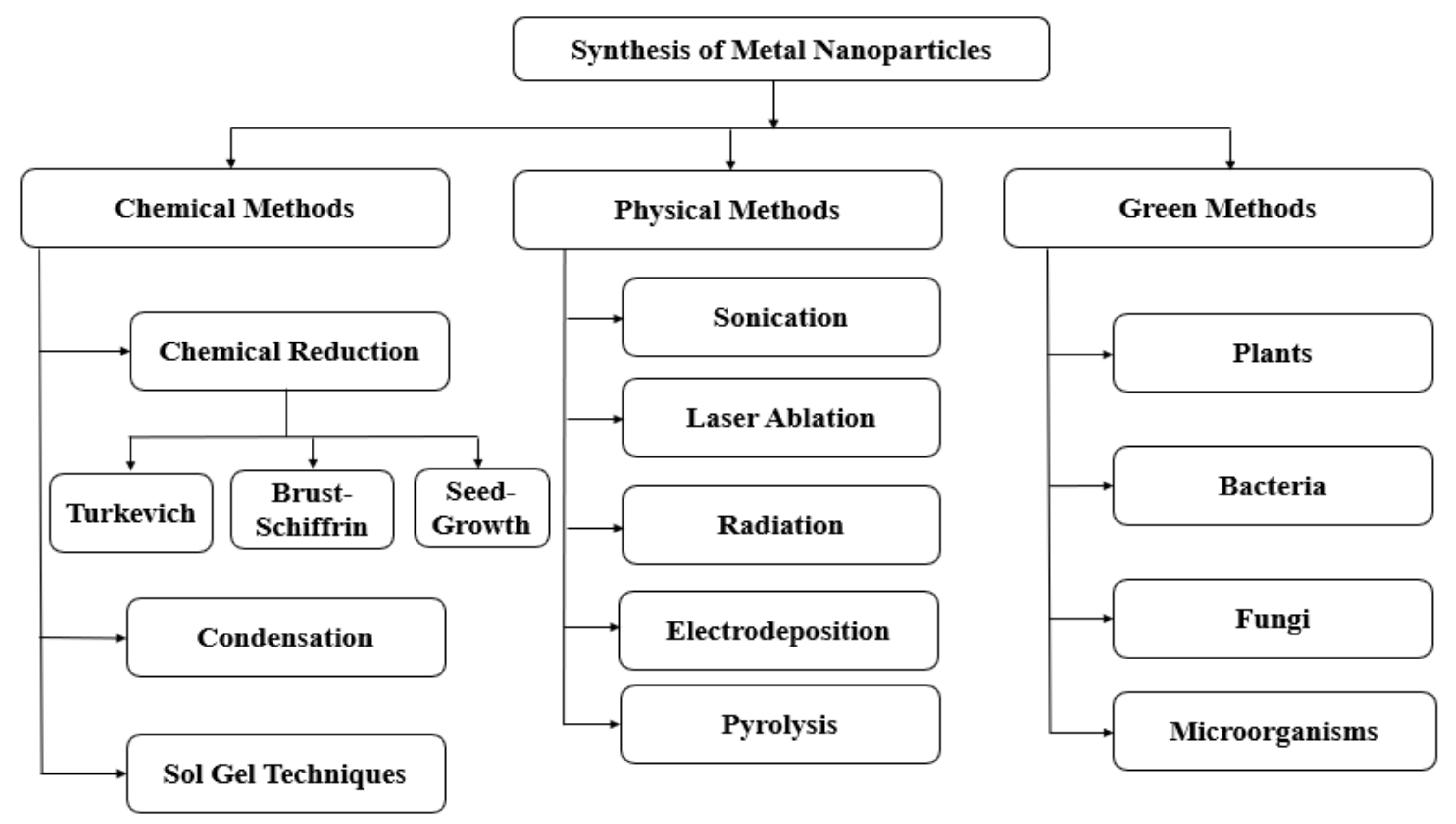
3. AuNPs Synthesis
4. Methods to Prepare Textile Materials Functionalized with AuNPs
4.1. Functionalization of Fabrics with AuNPs
4.1.1. Chemical Reduction Method without Pre-Treatments on Fabrics
4.1.2. Chemical Reduction Method with Pre-Treatments on Fabrics
4.1.3. Chemical Reduction Method Using Thermal Treatment
4.1.4. Green-Bio Synthesis
4.1.5. Electrochemical Synthesis
4.2. Functionalization of Fibers/Yarns/Threads with AuNPs
4.2.1. Chemical Reduction Method without Pre-Treatments on Fabrics
4.2.2. Chemical Reduction Method with Pre-Treatments
4.2.3. Reduction with Photochemical Treatments
4.2.4. Green Synthesis
4.3. Functionalization of Nanofibers/Scaffolds/Membranes with AuNPs
4.3.1. Chemical Reduction Method without Pre-Treatments
4.3.2. Chemical Reduction Method with Pre-Treatments
4.3.3. Chemical Reduction by Photo Reduction/UV Radiation/Photo Reduction
4.3.4. Green Synthesis
4.3.5. Reduction with Thermal Treatments
4.3.6. Reduction with Other Treatments
5. Antimicrobial Properties of AuNPs on Textiles
6. Conclusions with Future Perspectives
Author Contributions
Funding
Institutional Review Board Statement
Informed Consent Statement
Data Availability Statement
Acknowledgments
Conflicts of Interest
Abbreviations
References
- Zhao, P.; Li, N.; Astruc, D. State of the Art in Gold Nanoparticle Synthesis. Coord. Chem. Rev. 2013, 257, 638–665. [Google Scholar] [CrossRef]
- Motitswe, M.G.; Fayemi, O.E.; Drummond, H.P. Electrochemical and Spectroscopic Properties of Green Synthesized Gold Nanoparticles Doped in Polyacrylonitrile Nanofibers. J. Clust. Sci. 2020, 32, 683–692. [Google Scholar] [CrossRef]
- Stensberg, M.C.; Wei, Q.; McLamore, E.S.; Porterfield, D.M.; Wei, A.; Sepúlveda, M.S. Toxicological Studies on Silver Nanoparticles: Challenges and Opportunities in Assessment, Monitoring and Imaging. Nanomedicine 2011, 6, 879–898. [Google Scholar] [CrossRef] [PubMed]
- Okkeh, M.; Bloise, N.; Restivo, E.; De Vita, L.; Pallavicini, P.; Visai, L. Gold Nanoparticles: Can They Be the next Magic Bullet for Multidrug-Resistant Bacteria? Nanomaterials 2021, 11, 312. [Google Scholar] [CrossRef]
- Roy, S.; Das, T.K.; Maiti, G.P.; Basu, U. Microbial Biosynthesis of Nontoxic Gold Nanoparticles. Mater. Sci. Eng. B 2016, 203, 41–51. [Google Scholar] [CrossRef]
- Baruah, D.; Goswami, M.; Yadav, R.N.S.; Yadav, A.; Das, A.M. Biogenic Synthesis of Gold Nanoparticles and Their Application in Photocatalytic Degradation of Toxic Dyes. J. Photochem. Photobiol. B Biol. 2018, 186, 51–58. [Google Scholar] [CrossRef]
- Shamaila, S.; Zafar, N.; Riaz, S.; Sharif, R.; Nazir, J.; Naseem, S. Gold Nanoparticles: An Efficient Antimicrobial Agent against Enteric Bacterial Human Pathogen. Nanomaterials 2016, 6, 71. [Google Scholar] [CrossRef]
- Andra, S.; Balu, S.; Jeevanandam, J.; Muthalagu, M. Emerging Nanomaterials for Antibacterial Textile Fabrication. Naunyn Schmiedeberg’s Arch Pharmacol. Modifying 2021, 1–28. [Google Scholar] [CrossRef]
- Toma, H.E.; Zamarion, V.M.; Toma, S.H.; Araki, K. The Coordination Chemistry at Gold Nanoparticles. J. Braz. Chem. Soc. 2010, 21, 1158–1176. [Google Scholar] [CrossRef]
- Bakshi, M.S. How Surfactants Control Crystal Growth of Nanomaterials. Cryst. Growth Des. 2016, 16, 1104–1133. [Google Scholar] [CrossRef]
- Daruich De Souza, C.; Ribeiro Nogueira, B.; Rostelato, M.E.C.M. Review of the Methodologies Used in the Synthesis Gold Nanoparticles by Chemical Reduction. J. Alloys Compd. 2019, 798, 714–740. [Google Scholar] [CrossRef]
- Zhao, J.; Stenzel, M.H. Entry of Nanoparticles into Cells: The Importance of Nanoparticle Properties. Polym. Chem. 2018, 9, 259–272. [Google Scholar] [CrossRef]
- Kalimuthu, K.; Cha, B.S.; Kim, S.; Park, K.S. Eco-Friendly Synthesis and Biomedical Applications of Gold Nanoparticles: A Review. Microchem. J. 2020, 152, 104296. [Google Scholar] [CrossRef]
- Fan, J.; Cheng, Y.; Sun, M. Functionalized Gold Nanoparticles: Synthesis, Properties and Biomedical Applications. Chem. Rec. 2020, 20, 1474–1504. [Google Scholar] [CrossRef] [PubMed]
- Jadoun, S.; Verma, A.; Arif, R. Modification of Textiles via Nanomaterials and Their Applications. Front. Text. Mater. Polym. Nanomater. Enzym. Adv. Modif. Tech. 2020, 135–152. [Google Scholar] [CrossRef]
- Murugesh Babu, K.; Sahana, N.; Anitha, D.V.; Kavya, B.S. Silk Fibroin Coated Antimicrobial Textile Medical Products. J. Text. Inst. 2020, 1–9. [Google Scholar] [CrossRef]
- Fateixa, S.; Pinheiro, P.C.; Nogueira, H.I.S.; Trindade, T. Gold Loaded Textile Fibres as Substrates for SERS Detection. J. Mol. Struct. 2019, 1185, 333–340. [Google Scholar] [CrossRef]
- Aflori, M.; Serbezeanu, D.; Carja, I.D.; Fortunato, G. Gold Nanoparticles Incorporated into Electrospun Polyimide Fibers. Chem. Lett. 2015, 44, 1440–1442. [Google Scholar] [CrossRef]
- Hussain, M.H.; Abu Bakar, N.F.; Mustapa, A.N.; Low, K.F.; Othman, N.H.; Adam, F. Synthesis of Various Size Gold Nanoparticles by Chemical Reduction Method with Different Solvent Polarity. Nanoscale Res. Lett. 2020, 15, 140. [Google Scholar] [CrossRef] [PubMed]
- Govindrao, P.; Ghule, N.W.; Haque, A.; Kalaskar, M.G. Journal of Drug Delivery Science and Technology Metal Nanoparticles Synthesis: An Overview on Methods of Preparation, Advantages and Disadvantages, and Applications. J. Drug Deliv. Sci. Technol. 2019, 53, 101174. [Google Scholar] [CrossRef]
- Meyers, M.A.; Mishra, A.; Benson, D.J. Mechanical Properties of Nanocrystalline Materials. Prog. Mater. Sci. 2006, 51, 427–556. [Google Scholar] [CrossRef]
- Nadagouda, M.N.; Speth, T.F.; Varma, R.S. Microwave-Assisted Green Synthesis of Silver Nanostructures. Acc. Chem. Res. 2011, 44, 469–478. [Google Scholar] [CrossRef] [PubMed]
- Mukherjee, P.; Ahmad, A.; Mandal, D.; Senapati, S.; Sainkar, S.R.; Khan, M.I.; Parishcha, R.; Ajaykumar, P.V.; Alam, M.; Kumar, R.; et al. Fungus-Mediated Synthesis of Silver Nanoparticles and Their Immobilization in the Mycelial Matrix: A Novel Biological Approach to Nanoparticle Synthesis. Nano Lett. 2001, 1, 515–519. [Google Scholar] [CrossRef]
- Baker, S.; Prasad, M.N.N.; Satish, S. Endogenic Mediated Synthesis of Gold Nanoparticles Bearing Bactericidal Activity. J. Microsc. Ultrastruct. 2016, 4, 162–166. [Google Scholar] [CrossRef]
- Das, S.K.; Das, A.R.; Guha, A.K. Microbial Synthesis of Multishaped Gold Nanostructures. Gold Nanostruct. 2010, 6, 1012–1021. [Google Scholar] [CrossRef]
- Schröfel, A.; Kratošová, G. Biosynthesis of Metallic Nanoparticles and Their Applications. In Intracellular Delivery. Fundamental Biomedical Technologies; Prokop, A., Ed.; Springer: Dordrecht, The Netherlands, 2011; pp. 373–409. [Google Scholar] [CrossRef]
- De Freitas, L.F.; Varca, G.H.C.; Batista, J.G.D.S.; Lugão, A.B. An Overview of the Synthesis of Gold Nanoparticles Using Radiation Technologies. Nanomaterials 2018, 8, 939. [Google Scholar] [CrossRef]
- Herizchi, R.; Abbasi, E.; Milani, M.; Akbarzadeh, A.; Herizchi, R.; Abbasi, E.; Milani, M.; Akbarzadeh, A. Current Methods for Synthesis of Gold Nanoparticles Current Methods for Synthesis of Gold Nanoparticles. Artif. Cells Nanomed. Biotechnol. 2016, 1401, 596–602. [Google Scholar] [CrossRef] [PubMed]
- Lee, K.X.; Shameli, K.; Yew, Y.P.; Teow, S.Y.; Jahangirian, H.; Rafiee-Moghaddam, R.; Webster, T.J. Recent Developments in the Facile Bio-Synthesis of Gold Nanoparticles (AuNPs) and Their Biomedical Applications. Int. J. Nanomed. 2020, 15, 275–300. [Google Scholar] [CrossRef]
- Rana, A.; Yadav, K.; Jagadevan, S. A Comprehensive Review on Green Synthesis of Nature-Inspired Metal Nanoparticles: Mechanism, Application and Toxicity. J. Clean. Prod. 2020, 272, 122880. [Google Scholar] [CrossRef]
- Dauthal, P.; Mukhopadhyay, M. Noble Metal Nanoparticles: Plant-Mediated Synthesis, Mechanistic Aspects of Synthesis, and Applications. Ind. Eng. Chem. Res. 2016, 55, 9557–9577. [Google Scholar] [CrossRef]
- Kunoh, T.; Takeda, M.; Matsumoto, S.; Suzuki, I.; Takano, M.; Kunoh, H.; Takada, J. Green Synthesis of Gold Nanoparticles Coupled with Nucleic Acid Oxidation. ACS Sustain. Chem. Eng. 2018, 6, 364–373. [Google Scholar] [CrossRef]
- Pereira, C.; Pereira, A.M.; Freire, C.; Pinto, T.V.; Costa, R.S.; Teixeira, J.S. Nanoengineered Textiles: From Advanced Functional Nanomaterials to Groundbreaking High-Performance Clothing. In Handbook of Functionalized Nanomaterials for Industrial Applications; Elsevier: Amsterdam, The Netherlands, 2020. [Google Scholar] [CrossRef]
- Massella, D.; Argenziano, M.; Ferri, A.; Guan, J.; Giraud, S.; Cavalli, R.; Barresi, A.A.; Salaün, F. Bio-Functional Textiles: Combining Pharmaceutical Nanocarriers with Fibrous Materials for Innovative Dermatological Therapies. Pharmaceutics 2019, 11, 403. [Google Scholar] [CrossRef]
- Sanchez-Herencia, A.J. Water Based Colloidal Processing of Ceramic Laminates. Key Eng. Mater. 2007, 333, 39–48. [Google Scholar] [CrossRef]
- Engates, K.E.; Shipley, H.J. Adsorption of Pb, Cd, Cu, Zn, and Ni to Titanium Dioxide Nanoparticles: Effect of Particle Size, Solid Concentration, and Exhaustion. Environ. Sci. Pollut. Res. 2011, 18, 386–395. [Google Scholar] [CrossRef] [PubMed]
- Ribeiro, A.I.; Senturk, D.; Silva, K.S.; Modic, M.; Cvelbar, U.; Dinescu, G.; Mitu, B.; Nikiforov, A.; Leys, C.; Kuchakova, I.; et al. Efficient Silver Nanoparticles Deposition Method on DBD Plasma-Treated Polyamide 6,6 for Antimicrobial Textiles. In IOP Conference Series: Materials Science and Engineering; IOP Publishing: Bristol, UK, 2018; Volume 460, p. 0120070. [Google Scholar] [CrossRef]
- Boomi, P.; Ganesan, R.; Prabu Poorani, G.; Jegatheeswaran, S.; Balakumar, C.; Gurumallesh Prabu, H.; Anand, K.; Marimuthu Prabhu, N.; Jeyakanthan, J.; Saravanan, M. Phyto-Engineered Gold Nanoparticles (AuNPs) with Potential Antibacterial, Antioxidant, and Wound Healing Activities Under in Vitro and in Vivo Conditions. Int. J. Nanomed. 2020, 15, 7553–7568. [Google Scholar] [CrossRef]
- Magura, J.; Zeleňáková, A.; Zeleňák, V.; Kaňuchová, M. Thiol-Modified Gold Nanoparticles Deposited on Silica Support Using Dip Coating. Appl. Surf. Sci. 2014, 315, 392–399. [Google Scholar] [CrossRef]
- Tang, J.; Ou, Q.; Zhou, H.; Qi, L.; Man, S. Seed-Mediated Electroless Deposition of Gold Nanoparticles for Highly Uniform and Efficient SERS Enhancement. Nanomaterials 2019, 9, 185. [Google Scholar] [CrossRef]
- Preda, N.; Enculescu, M.; Zgura, I.; Socol, M.; Matei, E.; Vasilache, V.; Enculescu, I. Superhydrophobic Properties of Cotton Fabrics Functionalized with ZnO by Electroless Deposition. Mater. Chem. Phys. 2013, 138, 253–261. [Google Scholar] [CrossRef]
- Elmaaty, T.A.; El-Nagare, K.; Raouf, S.; Abdelfattah, K.; El-Kadi, S.; Abdelaziz, E. One-Step Green Approach for Functional Printing and Finishing of Textiles Using Silver and Gold NPs. RSC Adv. 2018, 8, 25546–25557. [Google Scholar] [CrossRef]
- Dietzel, M.; Bieri, N.R.; Poulikakos, D. Dropwise Deposition and Wetting of Nanoparticle Suspensions. Int. J. Heat Fluid Flow 2008, 29, 250–262. [Google Scholar] [CrossRef]
- Ranzoni, A.; Cooper, M.A. The Growing Influence of Nanotechnology in Our Lives. In Micro- and Nanotechnology in Vaccine Development; Skwarczynski, M., Toth, I., Eds.; Elsevier Inc.: Amsterdam, The Netherlands, 2017; pp. 1–20. [Google Scholar]
- Basarir, F.; Yoon, T.H. Sonication-Assisted Layer-by-Layer Deposition of Gold Nanoparticles for Highly Conductive Gold Patterns. Ultrason. Sonochem. 2012, 19, 621–626. [Google Scholar] [CrossRef] [PubMed]
- Zhong, W. Nanofibres for Medical Textiles. In Advances in Smart Medical Textiles: Treatments and Health Monitoring; van Langenhove, L., Ed.; Woodhead Publishing, Elsevier Inc.: Amsterdam, The Netherlands, 2016; pp. 57–70. [Google Scholar] [CrossRef]
- Jose Varghese, R.; Sakho, E.H.M.; Parani, S.; Thomas, S.; Oluwafemi, O.S.; Wu, J. Introduction to Nanomaterials: Synthesis and Applications. In Nanomaterials for Solar Cell Applications; Thomas, S., Sakho, E.H.M., Kalarikkal, N., Oluwafemi, S.O., Wu, J., Eds.; Elsevier Inc.: Amsterdam, The Netherlands, 2019; pp. 75–95. [Google Scholar] [CrossRef]
- Chan, K.L.; Fawcett, D.; Poinern, G.E.J. Gold Nanoparticle Treated Textile-Based Materials for Potential Use as Wearable Sensors. Int. J. Sci. 2016, 2, 82–89. [Google Scholar] [CrossRef]
- Shanmugasundaram, O.L.; Ramkumar, M. Characterization and Study of Physical Properties and Antibacterial Activities of Human Hair Keratin–Silver Nanoparticles and Keratin–Gold Nanoparticles Coated Cotton Gauze Fabric. J. Ind. Text. 2018, 47, 798–814. [Google Scholar] [CrossRef]
- Zheng, Y.; Xiao, M.; Jiang, S.; Ding, F.; Wang, J. Coating Fabrics with Gold Nanorods for Colouring, UV-Protection, and Antibacterial Functions. Nanoscale 2013, 5, 788–795. [Google Scholar] [CrossRef] [PubMed]
- Lin, X.; Zou, F.; Chen, X.; Tang, B. Functional Modification of Nylon Fabrics Based on Noble Metal Nanoparticles. IOP Conf. Ser. Mater. Sci. Eng. 2017, 231, 012175. [Google Scholar] [CrossRef]
- Silva, I.O.; Ladchumananandasivam, R.; Nascimento, J.H.O.; Silva, K.K.O.S.; Oliveira, F.R.; Souto, A.P.; Felgueiras, H.P.; Zille, A. Multifunctional Chitosan/Gold Nanoparticles Coatings for Biomedical Textiles. Nanomaterials 2019, 9, 1064. [Google Scholar] [CrossRef] [PubMed]
- Gao, L.; Feng, J.; Xu, S.; Shi, M.; Yao, L.; Wang, L.; Yang, Z. General Strategy to Prepare Single-Layered Ag–Au–Pt Nanocrystal Ternary-Coated Biomass Textiles through Polymer-Driven Self-Assembly. Nanomaterials 2020, 10, 495. [Google Scholar] [CrossRef]
- Ahmed, H.B.; El-Hawary, N.S.; Emam, H.E. Self-Assembled AuNPs for Ingrain Pigmentation of Silk Fabrics with Antibacterial Potency. Int. J. Biol. Macromol. 2017, 105, 720–729. [Google Scholar] [CrossRef] [PubMed]
- Zhou, L.; Yu, K.; Lu, F.; Lan, G.; Dai, F.; Shang, S.; Hu, E. Minimizing Antibiotic Dosage through in Situ Formation of Gold Nanoparticles across Antibacterial Wound Dressings: A Facile Approach Using Silk Fabric as the Base Substrate. J. Clean. Prod. 2020, 243, 118604. [Google Scholar] [CrossRef]
- Radić, N.; Obradović, B.M.; Kostić, M.; Dojčinović, B.; Hudcová, M.; Kuraica, M.M.; Černák, M. Deposition of Gold Nanoparticles on Polypropylene Nonwoven Pretreated by Dielectric Barrier Discharge and Diffuse Coplanar Surface Barrier Discharge. Plasma Chem. Plasma Process. 2013, 33, 201–218. [Google Scholar] [CrossRef]
- Ikegami, M.; Matsumoto, T.; Kobayashi, Y.; Jikihara, Y.; Nakayama, T.; Ohashi, H.; Honma, T.; Takei, T.; Haruta, M. Air Purification by Gold Catalysts Supported on PET Nonwoven Fabric. Appl. Catal. B Environ. 2013, 134–135, 130–135. [Google Scholar] [CrossRef]
- Cheng, D.; Bai, X.; He, M.; Wu, J.; Yang, H.; Ran, J.; Cai, G.; Wang, X. Polydopamine-Assisted Immobilization of Ag@AuNPs on Cotton Fabrics for Sensitive and Responsive SERS Detection. Cellulose 2019, 26, 4191–4204. [Google Scholar] [CrossRef]
- Liu, H.; Goh, W.; Norsten, T.B. Aqueous-Based Formation of Gold Nanoparticles on Surface-Modified Cotton Textiles. J. Mol. Eng. Mater. 2013, 1, 1250001. [Google Scholar] [CrossRef]
- Baruah, B.; Downer, L.; Agyeman, D. Fabric-Based Composite Materials Containing ZnO-NRs and ZnO-NRs-AuNPs and Their Application in Photocatalysis. Mater. Chem. Phys. 2019, 231, 252–259. [Google Scholar] [CrossRef]
- Ma, H.; Chi, H.; Wu, J.; Wang, M.; Li, J.; Hoshina, H.; Saiki, S.; Seko, N. A Novel Avenue to Gold Nanostructured Microtubes Using Functionalized Fiber as the Ligand, the Reductant, and the Template. ACS Appl. Mater. Interfaces 2013, 5, 8761–8765. [Google Scholar] [CrossRef] [PubMed]
- Padbury, R.P.; Halbur, J.C.; Krommenhoek, P.J.; Tracy, J.B.; Jur, J.S. Thermal Stability of Gold Nanoparticles Embedded within Metal Oxide Frameworks Fabricated by Hybrid Modifications onto Sacrificial Textile Templates. Langmuir 2015, 31, 1135–1141. [Google Scholar] [CrossRef]
- Tang, B.; Sun, L.; Kaur, J.; Yu, Y.; Wang, X. In-Situ Synthesis of Gold Nanoparticles for Multifunctionalization of Silk Fabrics. Dye. Pigment. 2014, 103, 183–190. [Google Scholar] [CrossRef]
- Liu, J.; Zhou, J.; Tang, B.; Zeng, T.; Li, Y.; Li, J.; Ye, Y.; Wang, X. Surface Enhanced Raman Scattering (SERS) Fabrics for Trace Analysis. Appl. Surf. Sci. 2016, 386, 296–302. [Google Scholar] [CrossRef]
- Zhang, Z.; Lv, X.; Chen, Q.; An, J. Complex Coloration and Antibacterial Functionalization of Silk Fabrics Based on Noble Metal Nanoparticles. J. Eng. Fiber. Fabr. 2019, 14. [Google Scholar] [CrossRef]
- Ganesan, R.M.; Gurumallesh Prabu, H. Synthesis of Gold Nanoparticles Using Herbal Acorus Calamus Rhizome Extract and Coating on Cotton Fabric for Antibacterial and UV Blocking Applications. Arab. J. Chem. 2019, 12, 2166–2174. [Google Scholar] [CrossRef]
- Ibrahim, N.A.; Eid, B.M.; Abdel-Aziz, M.S. Green Synthesis of AuNPs for Eco-Friendly Functionalization of Cellulosic Substrates. Appl. Surf. Sci. 2016, 389, 118–125. [Google Scholar] [CrossRef]
- Tang, B.; Lin, X.; Zou, F.; Fan, Y.; Li, D.; Zhou, J.; Chen, W.; Wang, X. In Situ Synthesis of Gold Nanoparticles on Cotton Fabric for Multifunctional Applications. Cellulose 2017, 24, 4547–4560. [Google Scholar] [CrossRef]
- Tao, J.; Tang, B.; Li, P.; He, D.; Liao, L.; Peng, Z.; Wang, X. Natural Rubber Particle Modified Fabrics with Catalytic Activity and Hydrophobicity. Compos. Sci. Technol. 2018, 162, 123–130. [Google Scholar] [CrossRef]
- Boomi, P.; Ganesan, R.M.; Poorani, G.; Gurumallesh Prabu, H.; Ravikumar, S.; Jeyakanthan, J. Biological Synergy of Greener Gold Nanoparticles by Using Coleus Aromaticus Leaf Extract. Mater. Sci. Eng. C 2019, 99, 202–210. [Google Scholar] [CrossRef]
- Boomi, P.; Poorani, G.P.; Selvam, S.; Palanisamy, S.; Jegatheeswaran, S.; Anand, K.; Balakumar, C.; Premkumar, K.; Prabu, H.G. Green Biosynthesis of Gold Nanoparticles Using Croton Sparsiflorus Leaves Extract and Evaluation of UV Protection, Antibacterial and Anticancer Applications. Appl. Organomet. Chem. 2020, 34, 1–13. [Google Scholar] [CrossRef]
- Haslinger, S.; Ye, Y.; Rissanen, M.; Hummel, M.; Sixta, H. Cellulose Fibers for High-Performance Textiles Functionalized with Incorporated Gold and Silver Nanoparticles. ACS Sustain. Chem. Eng. 2020, 8, 649–658. [Google Scholar] [CrossRef]
- Velmurugan, P.; Shim, J.; Bang, K.S.; Oh, B.T. Gold Nanoparticles Mediated Coloring of Fabrics and Leather for Antibacterial Activity. J. Photochem. Photobiol. B Biol. 2016, 160, 102–109. [Google Scholar] [CrossRef]
- Yao, Y.; Tang, B.; Chen, W.; Sun, L.; Wang, X. Sunlight-Induced Coloration of Silk. Nanoscale Res. Lett. 2016, 11, 293. [Google Scholar] [CrossRef] [PubMed]
- Tang, B.; Tao, J.; Xu, S.; Wang, J.; Hurren, C.; Xu, W.; Sun, L.; Wang, X. Using Hydroxy Carboxylate to Synthesize Gold Nanoparticles in Heating and Photochemical Reactions and Their Application in Textile Colouration. Chem. Eng. J. 2011, 172, 601–607. [Google Scholar] [CrossRef]
- Pasta, M.; Hu, L.; La Mantia, F.; Cui, Y. Electrodeposited Gold Nanoparticles on Carbon Nanotube-Textile: Anode Material for Glucose Alkaline Fuel Cells. Electrochem. Commun. 2012, 19, 81–84. [Google Scholar] [CrossRef]
- Ballerini, D.R.; Ngo, Y.H.; Garnier, G.; Ladeeig, B.P.; Wei, S. Gold Nanoparticle-Functionalized Thread as a Substrate for SERS Study of Analytes Both Bound and Unbound to Gold. AIChE J. 2012, 59, 215–228. [Google Scholar] [CrossRef]
- Adamo, C.B.; Junger, A.S.; Bressan, L.P.; da Silva, J.A.F.; Poppi, R.J.; de Jesus, D.P. Fast and Straightforward In-Situ Synthesis of Gold Nanoparticles on a Thread-Based Microfluidic Device for Application in Surface-Enhanced Raman Scattering Detection. Microchem. J. 2020, 156, 104985. [Google Scholar] [CrossRef]
- Islam, M.T.; Padilla, J.E.; Dominguez, N.; Alvarado, D.C.; Alam, M.S.; Cooke, P.; Tecklenburg, M.M.J.; Noveron, J.C. Green Synthesis of Gold Nanoparticles Reduced and Stabilized by Squaric Acid and Supported on Cellulose Fibers for the Catalytic Reduction of 4-Nitrophenol in Water. RSC Adv. 2016, 6, 91185–91191. [Google Scholar] [CrossRef]
- Park, H.J.; Kim, W.J.; Ah, C.S.; Jun, Y.; Yun, Y.J. Solution-Processed Au-Ag Core-Shell Nanoparticle-Decorated Yarns for Human Motion Monitoring. RSC Adv. 2017, 7, 10539–10544. [Google Scholar] [CrossRef]
- Xia, Y.; Wan, J.; Gu, Q. Silk Fibroin Fibers Supported with High Density of Gold Nanoparticles: Fabrication and Application as Catalyst. Gold Bull. 2011, 44, 171–176. [Google Scholar] [CrossRef][Green Version]
- Tang, B.; Yao, Y.; Li, J.; Qin, S.; Zhu, H.; Kaur, J.; Chen, W.; Sun, L.; Wang, X. Functional Application of Noble Metal Nanoparticles In Situ Synthesized on Ramie Fibers. Nanoscale Res. Lett. 2015, 10, 366. [Google Scholar] [CrossRef] [PubMed]
- Islam, M.T.; Dominguez, N.; Ahsan, M.A.; Dominguez-Cisneros, H.; Zuniga, P.; Alvarez, P.J.J.; Noveron, J.C. Sodium Rhodizonate Induced Formation of Gold Nanoparticles Supported on Cellulose Fibers for Catalytic Reduction of 4-Nitrophenol and Organic Dyes. J. Environ. Chem. Eng. 2017, 5, 4185–4193. [Google Scholar] [CrossRef]
- Emam, H.E.; El-Zawahry, M.M.; Ahmed, H.B. One-Pot Fabrication of AgNPs, AuNPs and Ag-Au Nano-Alloy Using Cellulosic Solid Support for Catalytic Reduction Application; Elsevier Inc.: Amsterdam, The Netherlands, 2017; Volume 166. [Google Scholar] [CrossRef]
- Yun, Y.J.; Ah, C.S.; Hong, W.G.; Kim, H.J.; Shin, J.H.; Jun, Y. Highly Conductive and Environmentally Stable Gold/Graphene Yarns for Flexible and Wearable Electronics. Nanoscale 2017, 9, 11439–11445. [Google Scholar] [CrossRef]
- Yu, Q.; Kong, X.; Ma, Y.; Wang, R.; Liu, Q.; Hinestroza, J.P.; Wang, A.X.; Vuorinen, T. Multi-Functional Regenerated Cellulose Fibers Decorated with Plasmonic Au Nanoparticles for Colorimetry and SERS Assays. Cellulose 2018, 25, 6041–6053. [Google Scholar] [CrossRef]
- Liu, Y.; Song, T.; Jia, X.; Meng, L.; Mao, X. Gold Nanoparticles Decorated Carbon Nanotube Probe Based Immunochromatographic Assay on Cotton Thread. Sens. Actuators B Chem. 2017, 251, 1112–1118. [Google Scholar] [CrossRef]
- Jia, X.; Song, T.; Liu, Y.; Meng, L.; Mao, X. An Immunochromatographic Assay for Carcinoembryonic Antigen on Cotton Thread Using a Composite of Carbon Nanotubes and Gold Nanoparticles as Reporters. Anal. Chim. Acta 2017, 969, 57–62. [Google Scholar] [CrossRef] [PubMed]
- Ullah, N.; Odda, A.H.; Li, D.; Wang, Q.; Wei, Q. One-Pot Green Synthesis of Gold Nanoparticles and Its Supportive Role in Surface Activation of Non-Woven Fibers as Heterogeneous Catalyst. Colloids Surfaces A Physicochem. Eng. Asp. 2019, 571, 101–109. [Google Scholar] [CrossRef]
- Nolasco-Arizmendi, V.; Morales-Luckie, R.; Sánchez-Mendieta, V.; Hinestroza, J.P.; Castro-Longoria, E.; Vilchis-Nestor, A.R. Formation of Silk–Gold Nanocomposite Fabric Using Grapefruit Aqueous Extract. Text. Res. J. 2013, 83, 1229–1235. [Google Scholar] [CrossRef]
- Koga, H.; Tokunaga, E.; Hidaka, M.; Umemura, Y.; Saito, T.; Isogai, A.; Kitaoka, T. Topochemical Synthesis and Catalysis of Metal Nanoparticles Exposed on Crystalline Cellulose Nanofibers. Chem. Commun. 2010, 46, 8567–8569. [Google Scholar] [CrossRef]
- Zhang, T.; Wang, W.; Zhang, D.; Zhang, X.; Yurong, M.; Zhou, Y.; Qi, L. Biotemplated Synthesis of Cold Nanoparticle-Bacteria Cellulose Nanofiber Nanocomposites and Their Application in Biosensing. Adv. Funct. Mater. 2010, 20, 1152–1160. [Google Scholar] [CrossRef]
- Wang, W.; Zhang, T.J.; Zhang, D.W.; Li, H.Y.; Ma, Y.R.; Qi, L.M.; Zhou, Y.L.; Zhang, X.X. Amperometric Hydrogen Peroxide Biosensor Based on the Immobilization of Heme Proteins on Gold Nanoparticles-Bacteria Cellulose Nanofibers Nanocomposite. Talanta 2011, 84, 71–77. [Google Scholar] [CrossRef]
- Zhou, B.; Li, Y.; Deng, H.; Hu, Y.; Li, B. Antibacterial Multilayer Films Fabricated by Layer-by-Layer Immobilizing Lysozyme and Gold Nanoparticles on Nanofibers. Colloids Surfaces B Biointerfaces 2014, 116, 432–438. [Google Scholar] [CrossRef]
- Cheng, H.H.; Chen, F.; Yu, J.; Guo, Z.X. Gold-Nanoparticle-Decorated Thermoplastic Polyurethane Electrospun Fibers Prepared through a Chitosan Linkage for Catalytic Applications. J. Appl. Polym. Sci. 2016, 134, 1–9. [Google Scholar] [CrossRef]
- Yang, X.; Yang, J.; Wang, L.; Ran, B.; Jia, Y.; Zhang, L.; Yang, G.; Shao, H.; Jiang, X. Pharmaceutical Intermediate-Modified Gold Nanoparticles: Against Multidrug-Resistant Bacteria and Wound-Healing Application via an Electrospun Scaffold. ACS Nano 2017, 11, 5737–5745. [Google Scholar] [CrossRef]
- Li, Y.; Tian, Y.; Zheng, W.; Feng, Y.; Huang, R.; Shao, J.; Tang, R.; Wang, P.; Jia, Y.; Zhang, J.; et al. Composites of Bacterial Cellulose and Small Molecule-Decorated Gold Nanoparticles for Treating Gram-Negative Bacteria-Infected Wounds. Small 2017, 13, 1–10. [Google Scholar] [CrossRef] [PubMed]
- Fang, X.; Ma, H.; Xiao, S.; Shen, M.; Guo, R.; Cao, X.; Shi, X. Facile Immobilization of Gold Nanoparticles into Electrospun Polyethyleneimine/Polyvinyl Alcohol Nanofibers for Catalytic Applications. J. Mater. Chem. 2011, 21, 4493–4501. [Google Scholar] [CrossRef]
- Reyes-López, S.Y.; Cornejo-Monroy, D.; González-García, G. A Novel Route for the Preparation of Gold Nanoparticles in Polycaprolactone Nanofibers. J. Nanomater. 2015, 2015. [Google Scholar] [CrossRef]
- Subair, R.; Tripathi, B.P.; Formanek, P.; Simon, F.; Uhlmann, P.; Stamm, M. Polydopamine Modified Membranes with in Situ Synthesized Gold Nanoparticles for Catalytic and Environmental Applications. Chem. Eng. J. 2016, 295, 358–369. [Google Scholar] [CrossRef]
- Abedalwafa, M.A.; Li, Y.; Ni, C.; Yang, G.; Wang, L. Non-Enzymatic Colorimetric Sensor Strip Based on Melamine-Functionalized Gold Nanoparticles Assembled on Polyamide Nanofiber Membranes for the Detection of Metronidazole. Anal. Methods 2019, 11, 3706–3713. [Google Scholar] [CrossRef]
- Chen, Y.; Chen, S.; Wang, B.; Yao, J.; Wang, H. TEMPO-Oxidized Bacterial Cellulose Nanofibers-Supported Gold Nanoparticles with Superior Catalytic Properties. Carbohydr. Polym. 2016, 160, 34–42. [Google Scholar] [CrossRef]
- Wu, X.; Lu, C.; Zhou, Z.; Yuan, G.; Xiong, R.; Zhang, X. Green Synthesis and Formation Mechanism of Cellulose Nanocrystal-Supported Gold Nanoparticles with Enhanced Catalytic Performance. Environ. Sci. Nano 2014, 1, 71–79. [Google Scholar] [CrossRef]
- Liu, Z.; Zhou, C.; Zheng, B.; Qian, L.; Mo, Y.; Luo, F.; Shi, Y.; Choi, M.M.F.; Xiao, D. In Situ Synthesis of Gold Nanoparticles on Porous Polyacrylonitrile Nanofibers for Sensing Applications. Analyst 2011, 136, 4545–4551. [Google Scholar] [CrossRef] [PubMed]
- Senthamizhan, A.; Celebioglu, A.; Balusamy, B.; Uyar, T. Immobilization of Gold Nanoclusters inside Porous Electrospun Fibers for Selective Detection of Cu(II): A Strategic Approach to Shielding Pristine Performance. Sci. Rep. 2015, 5, 15608. [Google Scholar] [CrossRef] [PubMed]
- Chen, X.; Li, D.; Li, G.; Luo, L.; Ullah, N.; Wei, Q.; Huang, F. Facile Fabrication of Gold Nanoparticle on Zein Ultrafine Fibers and Their Application for Catechol Biosensor. Appl. Surf. Sci. 2014, 328, 444–452. [Google Scholar] [CrossRef]
- Zhu, H.; Du, M.; Zhang, M.; Zou, M.; Yang, T.; Wang, L.; Yao, J.; Guo, B. Probing the Unexpected Behavior of AuNPs Migrating through Nanofibers: A New Strategy for the Fabrication of Carbon Nanofiber-Noble Metal Nanocrystal Hybrid Nanostructures. J. Mater. Chem. A 2014, 2, 11728–11741. [Google Scholar] [CrossRef]
- Serbezeanu, D.; Popa, A.M.; Sava, I.; Carja, I.D.; Amberg, M.; Rossi, R.M.; Fortunato, G. Design and Synthesis of Polyimide—Gold Nanofibers with Tunable Optical Properties. Eur. Polym. J. 2015, 64, 10–20. [Google Scholar] [CrossRef]
- Niu, T.; Xu, J.; Xiao, W.; Huang, J. Cellulose-Based Catalytic Membranes Fabricated by Deposition of Gold Nanoparticles on Natural Cellulose Nanofibres. RSC Adv. 2014, 4, 4901–4904. [Google Scholar] [CrossRef]
- Sakai, S.; Kawa, S.; Sawada, K.; Taya, M. Electrospun Polystyrene Fiber-Templating Ultrafine Gold Hollow Fiber Production. Gold Bull. 2013, 46, 97–101. [Google Scholar] [CrossRef][Green Version]
- Anka, F.H.; Perera, S.D.; Ratanatawanate, C.; Balkus, K.J. Polyacrylonitrile Gold Nanoparticle Composite Electrospun Fibers Prepared by in Situ Photoreduction. Mater. Lett. 2012, 75, 12–15. [Google Scholar] [CrossRef]
- Sawada, K.; Sakai, S.; Taya, M. Polyacrylonitrile-Based Electrospun Nanofibers Carrying Gold Nanoparticles in Situ Formed by Photochemical Assembly. J. Mater. Sci. 2014, 49, 4595–4600. [Google Scholar] [CrossRef]
- Zhou, X.; Zhao, Z.; He, Y.; Ye, Y.; Zhou, J.; Zhang, J.; Ouyang, Q.; Tang, B.; Wang, X. Photoinduced Synthesis of Gold Nanoparticle–Bacterial Cellulose Nanocomposite and Its Application for in-Situ Detection of Trace Concentration of Dyes in Textile and Paper. Cellulose 2018, 25, 3941–3953. [Google Scholar] [CrossRef]
- Shamsabadi, A.S.; Ranjbar, M.; Tavanai, H.; Farnood, A. Electrospinning of Gold Nanoparticles Incorporated PAN Nanofibers via In-Situ Laser Ablation of Gold in Electrospinning Solution. Mater. Res. Express 2019, 6, 055051. [Google Scholar] [CrossRef]
- Allahverdiyev, A.M.; Kon, K.V.; Abamor, E.S.; Bagirova, M.; Rafailovich, M. Coping with Antibiotic Resistance: Combining Nanoparticles with Antibiotics and Other Antimicrobial Agents. Expert Rev. Anti Infect. Ther. 2011, 9, 1035–1052. [Google Scholar] [CrossRef] [PubMed]
- Dizaj, S.M.; Lotfipour, F.; Barzegar-Jalali, M.; Zarrintan, M.H.; Adibkia, K. Antimicrobial Activity of the Metals and Metal Oxide Nanoparticles. Mater. Sci. Eng. C 2014, 44, 278–284. [Google Scholar] [CrossRef] [PubMed]
- Cui, Y.; Zhao, Y.; Tian, Y.; Zhang, W.; Lü, X.; Jiang, X. The Molecular Mechanism of Action of Bactericidal Gold Nanoparticles on Escherichia Coli. Biomaterials 2012, 33, 2327–2333. [Google Scholar] [CrossRef] [PubMed]
- Sánchez-López, E.; Gomes, D.; Esteruelas, G.; Bonilla, L.; Lopez-Machado, A.L.; Galindo, R.; Cano, A.; Espina, M.; Ettcheto, M.; Camins, A.; et al. Metal-Based Nanoparticles as Antimicrobial Agents: An Overview. Nanomaterials 2020, 10, 292. [Google Scholar] [CrossRef] [PubMed]
- Feng, Z.V.; Gunsolus, I.L.; Qiu, T.A.; Hurley, K.R.; Nyberg, L.H.; Frew, H.; Johnson, K.P.; Vartanian, A.M.; Jacob, L.M.; Lohse, S.E.; et al. Impacts of Gold Nanoparticle Charge and Ligand Type on Surface Binding and Toxicity to Gram-Negative and Gram-Positive Bacteria. Chem. Sci. 2015, 6, 5186–5196. [Google Scholar] [CrossRef] [PubMed]


| Method for Synthesis of AuNPs | Deposition Method | Fabric/Textile | Precursor Salt | Reducing Agent | Stabilizing Agent | Additional Information | Size of NPs | Application | Reference |
|---|---|---|---|---|---|---|---|---|---|
| Chemical reduction | Drop-wise deposition | Cotton, silk, wool, polyester and nylon—F | Chloroauric acid (0.001 M) | Sodium borohydride solution of (0.1 M, 3 mL) | Sodium citrate 3 mL solution of 0.001 M | Sodium citrate also act as capping agent | n.a. | Wearable sensors | [48] |
| Chemical reduction | Padding | Cotton—F | Chloroauric acid (0.01 Wt %, 50 mL) | Trisodium citrate (1 wt%) | No stabilizing agent | Keratin coating (360 mL of keratin solution (10 mg/mL concentration). | 71.8 nm | Antimicrobial textiles | [49] |
| Chemical reduction | Printing and paste method | Polyester—F | Gold (III) chloride hydrate | Sodium citrate | No stabilizing agent | One step green procedure | 13–20 nm | Coloration, UV protection | [42] |
| Chemical reduction, seed-mediated growth | Immersion | Silk and cotton—F | Tetrachloroauric acid (0.01 M, 0.25 mL) Tetrachloroauric acid (0.01 M, 20 mL) | Sodium borohydride (0.01 M, 0.6 mL) Ascorbic acid (0.1 M, 3.2 mL) | CTAB (0.1 M) 9.75 mL CTAB (0.1 M) 400 mL | Au nanorods with spherical shape. | 19 nm | Textile’s collation, UV protection, and antibacterial | [50] |
| In situ chemical reduction | Immersion and heating | Nylon—F | Tetrachloroauric (III) acid (0.05, 0.10, 0.15 and 0.20 mM) | Trisodium citrate | Trisodium citrate | pH value is in the range of 5.0–6.5 as per the concentration. | n.a. | UV blocking textiles | [51] |
| Chemical reduction | Exhaustion | Soybean—KF | Tetrachloroauric acid 0.01% W/V | Sodium citrate dihydrate (1% W/V, 2 mL) | Chitosan | Treatment with chitosan; spherical shape. | 34.6 ± 0.5 nm | UV blocking and antimicrobial textiles | [52] |
| In situ chemical reduction | Impregnation | Cotton, silk, and wool—F | Hydrogen tetrachloroaurate (III) trihydrate | Sodium borohydride (1.3 g/L) | n.a. | AuNPs are mixed with other nanoparticles such as Ag and Pt; AuNPs have spherical shape. | 6.64 nm | Antimicrobial textiles | [53] |
| In situ synthesis, groups on silk | Immersion and constant shaking at 85 °C | Silk—F | Gold (III) chloride (0.5–2 mM) | Silk macro molecular chains | Silk fabric | Hydrogen peroxide used for activation of silk macro molecules. | 22–66 and 18–49 nm | Fabric coloration and antimicrobial properties | [54] |
| In situ synthesis, sericin from silk | Soaking and sonication | Silk—F | Tetrachloroauric (III) acid (10 mg/mL, 500 mL) | Reduction by sericin from silk | n.a. | Spherical and ellipsoidal; pH = 12. | 11 ± 4 nm | Antimicrobial textiles | [55] |
| Chemical reduction | Immersion | Polypropylene—NW | Tetrachloroauric Acid (1 mM) | Gallic acid (0.5 mM) | Without stabilizer | Surface activation by dielectric barrier discharge (DBD) and diffuse coplanar surface barrier discharge (DCSBD). | 20 nm | Antimicrobial textiles | [56] |
| n.a. | Deposition-precipitation | Poly(ethylene terephthalate—NW | Tetrachloroauric acid (5 mmol/L) | n.a. | n.a. | Fabric coated with ZrO2 fine particles before deposition of AuNPs at pH = 7 | n.a. | Air filter | [57] |
| In situ chemical reduction | Immersion | Cotton—F | Tetrachloroauric acid (10 mM) | Polydopamine (2 mg/mL) | n.a. | Treated with polydopamine before depositing nanoparticles; AgNPs were deposited prior to AuNPs at pH = 8.5. | n.a. | Catalysis | [58] |
| In situ chemical reduction | Immersion and stirring | Cotton—F | Hydrogen tetra-chloroaurate (III) trihydrate HAuCl4 (1 mL, 20 mM) | N-vinyl pyrrolidone (0.1 mL) | 1-Hexadecylamine | Surface modification by ATS is crucial for the formation of gold nano particles; spherical shaped. | 2–7 nm | Textile coloration | [59] |
| Chemical reduction | Dip coating | Cotton—F | Sodium tetrachlorocuprate (III) dihydrate (1%, 90 µL) | Trisodium citrate (1%, 2.7 mL) | n.a. | Cotton fabric precoated with Zn nanorods before deposition of AuNPs. | 18.5 ± 2.8 nm | Photocatalysis | [60] |
| In situ chemical reduction | Soaked in solution | Polyethylene-coated polypropylene—NW | Chloroauric acid | Amine groups grafted in textile surface | Amine groups grafted in textile surface | PE-coated PP fabric was used as a ligand and template; fabric is treated by the electron beam; spherical shape. | 5–20 nm | n.a. | [61] |
| Chemical reduction | Soaked in solution of AuNPs solution | Nylon-6—F | Tetrachloroauric acid (0.0863 g) | Oleylamine | n.a. | – | 14.6 ± 1.4 nm | Catalytic systems | [62] |
| In situ synthesis, tyrosine groups on silk fiber | Immersed in solution and heating | Silk—F | Tetrachloroauric (III) acid Various concentrations | Tyrosine groups on silk fiber | Tyrosine groups on silk fiber | Different shapes were observed according to the Wt % of the precursor solution. | 21.3 ± 3.4 nm | Textile colorations and antimicrobial effect | [63] |
| In situ synthesis | Immersion and heating | Silk—F | Tetrachloroauric acid (0.1–0.6 mM, 50 mL) | n.a. | n.a. | Spherical, triangular nanoplates, truncated nanoprisms, and polygonal; depend on concentration of the precursor solution. | n.a. | Fabric coloration | [64] |
| In situ synthesis, sericin from silk | Immersion and heating | Silk—F | Tetrachloroauric (III) acid (0.3 mM) | n.a. | n.a. | The pH value of solutions was adjusted to 3; spherical and platelike shape. | n.a. | Fabric coloration and antimicrobial properties | [65] |
| Biological reduction | Pad-dry-cure | Cotton—F | Chloroauric acid (0.001 M, 2.5 mL) | Acorus calamus rhizome extract (2.5 mL) | Plant extract | Small spherical ball and bigger spherical ball, and it depends on the concentration at pH = 4, 7 and 9.2. | (0.001 M) below 100 nm (0.01 M) 100–500 nm | Antibacterial and UV blocking | [66] |
| Biological reduction | Sonication | Cotton and viscose—KF | Tetrachloroauric acid (3 mM, 100 mL) | Bacterial isolates (Streptomyces Sp) | Streptomyces Sp | Plasma treatment along with the combination of TiO2NPs and ZnONPs spherical shape. | 4–13 nm | Antimicrobial and UV- blocking textiles | [67] |
| In situ synthesis, hydroxyl groups on cellulose | In situ synthesis | Cotton—KF | Tetrachloroauric acid (0.025, 0.05, 0.075, 0.10, and 0.125 mM) | Hydroxyl groups from cellulose | n.a. | Cotton also acted as a reducing agent; spherical and triangular nanoplates. | 8.7 ± 1.2, 8.6 ± 1.3, 14.1 ± 3.0, 17.4 ± 3.0, and 20.5 ± 3.8 nm | Catalytic, UV blocking, and antibacterial textiles | [68] |
| In situ synthesis, hydroxyl groups on cellulose | Dip and dry | Cotton and polyester—F | Tetrachloroauric (III) acid (5.88 × 10⁻⁴ M, 198 mL,) | Hydroxyl groups on cellulose | Natural rubber latex | – | 31 nm | Catalytic textiles | [69] |
| Biological reduction | Pad dry cure | Cotton—F | Tetrachloroauric acid (0.001 M and 0.1 M, 2.5 mL) | Coleus aromaticus leaf extract (2.5 mL) | Coleus aromaticus leaf extract | Spherical, rod, and triangular shapes, pH = 7. | Different sizes (<20 nm) | Antimicrobial textiles | [70] |
| Biological reduction | Pad-dry-cure | Cotton—F | Chloroauric acid (0.001 M) | Croton sparsiflorus leaves Extract | Croton Sparsiflorus leaves extract | Low concentration: bulbous shape high concentration: spherical shape; the fabric was pre-treated through scouring and bleaching. | 12.2–12.7 High concertation 16.6 nm. | UV protection, antibacterial, and anticancer | [71] |
| Biological reduction | Dry-jet wet spinning process | Cellulose—NW | Tetrachloroauric acid (0.03 mL/g, 50 mM) | Bleached birch pre-hydrolyzed kraft pulp | n.a. | – | n.a. | UV blocking | [72] |
| In situ biological reduction | Immersion | Silk and cotton—F | Tetrachloroauric acid (2.00 × 10⁻⁴ M, 80 mL) | Ginkgo biloba Linn leaf powder extract | n.a. | Rectangular, spherical, hexagonal with smooth edges, or roughly circular in shape. | 10–75 nm | Textile colorations and antimicrobial effect | [73] |
| In situ chemical reduction | Immersion | Silk and cotton—F | Tetrachloroauric acid (2.00 × 10⁻⁴ M, 80 mL) | Potassium borohydride | n.a. | – | n.a. | ||
| In situ biological and chemical reduction | Immersion | Silk and cotton—F | Tetrachloroauric acid (2.00 × 10⁻⁴ M, 80 mL) | Ginkgo biloba Linn leaf powder extract and potassium borohydride | n.a. | – | n.a. | ||
| In situ photoreduction | Immersion | Silk—F | Tetrachloroauric acid (0.2, 0.3, 0.4, 0.5, 0.6, and 0.7 mM) | n.a. | n.a. | In situ synthesized AuNPs on through the induction of sunlight; spherical shape. | 16.9 ± 1.2, 24.1 ± 1.7, 23.0 ± 2.1, 20.6 ± 1.2, 19.9 ± 1.3, and 28.4 ± 1.6 nm | Fabric coloration | [74] |
| Heating and photochemical | Exhaustion | Wool—F | Tetrachloroauric acid (0.2 mM) | Trisodium citrate, D-Malic acid disodium salt or disodium tartrate (1 mM) | Trisodium citrate | Spherical and egg shapes were observed for heating method and photochemical synthesis at pH 4, respectively. | Various sizes | Textile coloration | [75] |
| n.a. | Electrodeposition | Polyester—F | Gold (III) chloride trihydrate (20 mM) | n.a. | n.a. | Coating single-walled carbon nanotubes on the polyester textile substrate before AuNPs deposition. | 50 nm | Fuel cells—conductive fabrics | [76] |
| Method for Synthesis of AuNPs | Deposition Method | Fabric/Textile | Precursor Salt | Reducing Agent | Stabilizing Agent | Additional Information | Size of NPs | Application | Reference |
|---|---|---|---|---|---|---|---|---|---|
| Photochemical reduction | Exhaustion | Silk and nylon—Fb | Tetrachloroauric acid (0.2 mM) | Trisodium citrate, D-malic acid disodium slat, and disodium tartrate (1 mM) | Trisodium citrate | Spherical and egg shapes were observed for heating method and photo chemical synthesis at pH = 4, respectively | Various sizes | Textile coloration | [75] |
| Chemical reduction | Soaked in solution | Cotton—T | Hydrogen tetrachloroaurate (0.65 mM) | Sodium citrate tribasic dihydrate | n.a. | SERS technique was used to detect and analyze adsorbed gold nanoparticles. | 20 and 60 nm | Diagnostics for surface-enhanced Raman scattering (SERS) spectroscopy | [77] |
| Chemical reduction | Immersion and capillary action | Cotton—T | Tetrachloroauric acid solution 2% (V/V) | Sodium citrate (2% (M/V)) | n.a. | Spherical shape; used HCl and NaOH for pH. | 20–40 nm | Surface-enhanced Raman scattering detection | [78] |
| Chemical reduction | Sonication | Cellulose—Fb | Tetrachloroauric (III) acid (0.5 mM, 20 mL) | Sodium squarate in water | Sodium squarate in water | AuNPs synthesized in water; Spherical in shape. | 21.01 nm | Catalysis | [79] |
| Chemical reduction | Immersion and stirring | Cotton—Y | Tetrachloroauric (III) acid (1 mM) | Trisodium citrate dihydrate (4 mM) | Citrate | pH 3–4. | 13 nm | Human motion sensor/wearable sensor | [80] |
| In situ chemical reduction | Soaking in solution | Silk fibroin—Fb | Tetrachloroauric (III) acid (10 mmol L−1) | Sulfonated polyaniline (20 mL of 5 wt %) | n.a. | Sulfonated polyaniline modified fibers catalytic reduction reaction of p-nitrophenol by NaBH4 | 50–100 nm | Catalysis | [81] |
| In situ chemical reduction | Immersion | Ramie—Fb | Tetrachloroauric (III) acid with different concentrations | Sodium borohydride | n.a. | AuNPs were synthesized in acidic condition, pH = 2–6. | n.a. | Textile coloration and antimicrobial textiles | [82] |
| In situ chemical reduction | Immersion and stirring | Cellulose—Fb | Tetracholoroaurate (0.5 mM) | Sodium rhodizonate | Sodium rhodizonate | Size depends on temperature; spherical shape. | 11 nm at 23 °C and 7 nm at 80 °C | Catalysis | [83] |
| Chemical reduction | Immersion and stirring | Cellulose—Fb | Gold chloride (AuCl3) | p-nitro-aniline (2 mM) Sodium borohydride (150 mM) | Cellulosic macromolecules | n.a. | 26.1 nm | Catalysis | [84] |
| Chemical reduction | Electroless deposition | Gold/graphene—Y | Tetrachloroauric (III) acid (1.6 mM) | Hydroxylamine | n.a. | Spherical to plate; dependent on reaction time. | 40 nm | Wearable electronics | [85] |
| Chemical reduction | Immersion | Regenerated cellulose—Fb | Gold (III) chloride triydrate (1 mM) | Trisodium citrate (1%, 2.2 mL) | Trisodium citrate | Fibers were grafted with positive charge; spherical shape | 40–50 nm | colorimetry and surface-enhanced Raman scattering (SERS) assays | [86] |
| Chemical reduction | Sonication | Cotton—T | Tetrachloroauric (III) acid (0.01%, W/V) | Trisodium citrate | n.a. | AuNPs coated on CNTs CNTs were functionalized with PDDA; homogenous surface. | 15 nm | Immunological chromatographic sensor | [87] |
| Chemical reduction | Centrifugation | Cotton—T | Tetrachloroauric (III) acid (0.01%, W/V) | Trisodium citrate | n.a. | AuNPs coated on CNTs CNTs were functionalized with PDDA; homogenous surface. | 15 ± 3 nm | Immunological chromatographic sensor | [88] |
| In situ green synthesis | Immersion | Cotton—Fb | Hydrogen tetrachloroaurate (III) hydrate (0.05 mM) | Osmanthus fragrans 10% (m/v) | Osmanthus fragrans | Spherical and hexagonal shape. | 40 and 60 nm | Heterogeneous catalyst | [89] |
| Biological reduction | Soaking | Silk—Fb | Tetrachloroauric acid (10−3 M) | Citrus paradisi extract | n.a. | Quasi-spherical | 30 nm | Textile coloration | [90] |
| Deposition Method | Fabric/Textile | Precursor Salt | Reducing Agent | Stabilizing Agent | Additional Information | Size of NPs | Application | Reference |
|---|---|---|---|---|---|---|---|---|
| Method for Synthesis: Biological reduction | ||||||||
| Electrospinning solution | Polyacrylonitrile—NF | Tetrachloroauric (III) acid (0.001 M) | Banana peel extract, phenolic compounds and flavonoids | n.a. | Spherical shape. | 9 nm | Electrochemical sensors | [2] |
| Method for Synthesis: Chemical reduction | ||||||||
| Electrospinning solution | Polyimide—NF | Gold (III) chloride hydrate | Thermal treatment at 200 °C in polyimide nanofibers | n.a. | Thermally reduced Au3+ to Au0. | n.a. | n.a. | [18] |
| Immersion | Crystalline cellulose—NF | Tetrachloroauric acid | Sodium borohydride | n.a. | - | <5 nm | Catalysis | [91] |
| Immersion | Bacterial cellulose—NF | Tetrachloroauric acid | Poly (ethyleneimine) | Poly(ethyleneimine) | Spherical shape. | ≈9 nm | Biosensors | [92] |
| Immersion | Bacterial cellulose—NF | Tetrachloroauric acid (20 mM) | Poly (ethyleneimine) | n.a. | BC nanofibers are produced by ultrasonic cell disruption system at room temperature. | ≈9 nm | Biosensors | [93] |
| Immersion | Cellulose acetate (mats)—NF | Tetrachloroauric (III) acid (1 Wt %, 3 mL) | Trisodium citrate | Trisodium citrate | LBS self-assembly technique. | – | Antimicrobial textiles | [94] |
| Immersion | Thermoplastic polyurethane—NF | Hydrogen tetrachloroaurate | Trisodium citrate | Chitosan | Precoated with chitosan reduction in 4-nitrophenol by sodium borohydride, pH value 3–11. | 16 nm | Catalysis | [95] |
| Electrospinning solution | PCL/Gelatin—NF/S | Tetrachloroauric (III) acid | Sodium borohydride | n.a. | Process is done in the presence of antibiotic intermediates. | 3 nm | Antimicrobial textiles, wound treatment | [96] |
| Soaking | Bacterial cellulose—M | Tetrachloroauric (III) acid (40 mg mL−1) | Sodium borohydride | n.a. | Gold nanoparticles modified with 4,6-Diamino-2-Pyrimidinethiol (DAPT). | ≈3 nm | Antimicrobial textiles, wound treatment | [97] |
| Electrospinning solution | PEI/PVA—NF | Chloroauric acid (0.5 mM) | Sodium borohydride | n.a. | The fibrous were cross-linked via glutaraldehyde (GA) vapor to produce water-stable fibrous mats; round shaped. | 11.8 nm | Catalysis | [98] |
| Electrospinning solution | Polycaprolactone—NF | Chloroauric acid (0.5 mM) | Sodium borohydride (1.25 mM) | n.a. | Round shape. | 5–6 nm | Heterogeneous catalysis and SERS | [99] |
| Immersion and shaking | PET track-etched micro-porous—M | Tetrachloroauric (III) acid (1 mg mL−1) | Dopamine on membranes | n.a. | Coated with dopamine. | n.a. | Catalysis | [100] |
| Dropping | Polyamide—NF/M | Hydrogen tetrachloroaurate (III) (1.00 mM) | Trisodium citrate dihydrate (0.30 M) | Trisodium citrate dihydrate | Spherical | 20.1 ± 1.76 nm | Colorimetric sensor | [101] |
| Immersion | Bacterial cellulose—NF | Chloroauric acid (0.4 mM) | Sodium borohydride (6 mM) | n.a. | 2,2,3,3-tetramethylpiperidine-l-oxyl (TEMPO)-oxidized (TOBCNS); spherical shape. | 4.30 ± 0.97 nm | Catalysis | [102] |
| Immersion | Cellulose—NF | Tetrachloroauric (III) acid solution (0.2 mM) | Hydrazine hydrate | n.a. | Unsupported AuNPs. | 18.3 ± 3.5 nm | Catalysis | [103] |
| Method for Synthesis: In situ chemical reduction | ||||||||
| Electrospinning solution | Polyacrylonitrile—NF | Hydrogen tetrachloroaurate (III) trihydrate | 4-(Dimethylamino) benzaldehyde | n.a. | – | 6 nm | Biosensors | [104] |
| Soaking | Cellulose Acetate—M | Tetrachloroauric (III) acid | Dithiothreitol (DTT) | Porous fiber network | DTT capped AuNPs. | 2.5 ± 0.5 nm | Sensors | [105] |
| Electrospinning solution | Zein—NF | Tetrachloroauric (III) acid (20 mM, 0.25 mL) | Poly(ethyleneimine) | n.a. | Spherical shape; value of pH 3.0–7.0. | 90.9 nm | Biosensors | [106] |
| Electrospinning solution | Polyacrylonitrile—NF | Tetrachloroauric (III) acid (0.30 mmol) | n.a. | n.a. | Au-PANF was prepared and electrospun to form mats; spherical shape. | 2.3 ± 0.5 nm | Sensors | [107] |
| Electrospinning solution | Polyimide—NF | Tetrachloroauric (III) acid (0.5, 1, and 3 Wt %) | Polyimide and high temperature (200 °C) | Polyimide | Reduction takes place due to thermal reactions. | 9–22 nm | High temperature end-of-service indicators | [108] |
| Immersion | Cellulose—M | Potassium gold (III) chloride | Sodium borohydride | Poly(diallyl-dimethylammonium chloride) poly(sodium-p-styrenesulfonate) | Membranes previously coated with titania gel. | 3.5 ± 0.7 nm | n.a. | [109] |
| Immersion and continuous shaking | PET track-etched microporous—M | Tetrachloroauric (III) acid (1 mg mL−1) | Sodium borohydride | n.a. | Coated with dopamine. | n.a. | Catalysis | [100] |
| Method for Synthesis: Photoreduction | ||||||||
| Electrospinning solution | Polystyrene (mats)—NF | Tetrachloroauric (III) acid (3% (W/W) | Ultraviolet irradiation | n.a. | Undergone electrospinning and then mats exposed to UV light. | n.a. | n.a. | [110] |
| Method for Synthesis: In situ photoreduction | ||||||||
| Electrospinning solution | Polyacrylonitrile—NF | Gold (III) chloride hydrate(0.044 M, 0.022 M) | Sodium alginate | n.a. | UV light has been used during electrospinning; spherical shape. | 21.4 nm (0.044 M) 5.8 nm (0.022 M) | n.a. | [111] |
| Electrospinning solution | Polyacrylonitrile—NF | Tetrachloroauric (III) acid | N,N-dimethylformamide | n.a. | Undergone electrospinning and then mats exposed to UV light. | 4.7–5.4 nm (5 days of UV radiation) | n.a. | [112] |
| Immersion | Cellulose—NF | Tetrachloroauric (III) acid solution (0.2 mM, 20 mL) | Cellulose nanocrystals | Cellulose nanocrystals | CNs were produced from microcrystalline cellulose. | 30.5 ± 13.4 nm | Catalysis | [103] |
| Immersion | Bacterial cellulose—M | Tetrachloroauric acid (0.2, 0.4, 0.6 mM) | n.a. | n.a. | Xenon lamp was used in the process of synthesis of AuNPs; spherical shape. | n.a. | Sensors | [113] |
| Method for Synthesis: Laser ablation | ||||||||
| Electrospinning solution | Polyacrylonitrile—NF | Gold plate | n.a. | n.a. | Spherical shape; face centered cubic crystal structure with crystallite size of 8 nm. | 17 nm | Glucose sensors | [114] |
| Fabric | Test Standards | Test Method | Bacterial Used | Results (Synthesis/Antimicrobial) | Reference |
|---|---|---|---|---|---|
| Cotton—F | n.a. | Kirby–Bauer | K. pneumoniae P. aeruginosa, S. aureus, and E. coli | AuNPs-keratin, 71.8 nm, sodium citrate/ Zone diameters ranging from 3.5 to 3.6 cm. | [49] |
| Polyester—F Cotton—F | AATCC 147-1988 | Parallel streak method | S. aureus, B. cereus, E. coli, and C. utilis | AuNPs, 13–20 nm, sodium citrate/ Zone diameters ranging from 16 to 22 mm Polyester and cotton fabrics printed with AuNPs showed excellent antimicrobial activity in all tested strains. | [42] |
| Soybean—KF | ASTM-E2149-01 | Shake flask method | S. aureus and E. coli | AuNPs-chitosan, 34.6 ± 0.5 nm, sodium citrate/ 99.94% against S. aureus (3.23 of log reduction) and 96.26% (1.43 of log reduction) against E. coli. | [52] |
| Cotton, silk, and wool—F | n.a. | Shake flask method | E. coli (ATCC 8099), S. aureus (ATCC 6538) | (HBPAA)-encapsulated Au-Ag-PtNPs, 6.64 nm/ Coating of nanoparticles have improved the antibacterial efficacy, and it enhanced linearly with the concentration. | [53] |
| Silk—F | n.a. | Turbidimetric method | S. aureus (ATCC 47077) and E. coli (ATCC 25922) | AuNPs, 22–66 and 18–49 nm, hydrogen peroxide/ Optical density reduced from 2.5 to 1.11–1.23 for E. coli and 2.12 to 0.93–1.11 for S. aureus after 24 h incubation. | [54] |
| Silk—F | Broth microdilution method | Broth microdilution method, inhibition zone | S. aureus and E. coli | AuNPs-gentamicin, 11±4nm/ Inhibition zone against E. Coli 25.8 mm, S. aureus 26 mm. | [55] |
| Polypropylene—F | n.a. | Agar diffusion test | S. aureus (ATCC 6538) E. coli (ATCC 11229) | AuNPs, 20 nm, gallic acid/ Activity only in contact; S. aureus was more sensitive, and the best antibacterial activity was shown for PP nonwoven fabrics treated with DCSBD (12 s (24 J/cm2) and DBD 120 s (14.4 J/cm2). | [56] |
| Silk—F | AATCC 100-2004 (Clause 10.2) test standard with slight modification | Agar plate method | E. coli (ATCC 11229) | AuNPs, 21.3 ± 3.4 nm, tyrosine on silk/ No bacteria colonies found on agar medium of silk fabrics treated with gold NPs; AuNPs on silk inhibited growth of bacteria. | [63] |
| Silk—F | AATCC 100-2012 (Clause 10.2) standard with slight modifications | Shake flask method | E. coli (ATCC 25922) | AuNPs, no size determination/ CFU reduction values of 99.96%. | [65] |
| Cotton—F | AATCC 100 | Agar plate method | S. aureus (MTCC 96) and E. coli (MTCC1671) | AuNPs, 10 nm, A. calamus rhizome extract/ Percentage reduction 50.05% and 63.9% against S. aureus after 24 and 48 h culture, respectively; 58.0% and 80.3% against E. coli after 24 and 48 h culture, respectively. | [66] |
| Cotton and viscose—KF | n.a. | Agar plate method and expressed as zone inhibition method | S. aureus and E. coli | AuNPs-ZnONPs, 4–13 nm, Streptomyces sp. isolates/ Plasma-treated AuNPs/ZnONPs showed the highest values. For cotton inhibition zone against E. Coli was 27.5 mm, and for S. aureus was 30 mm. For viscose inhibition zone against E. Coli was 25 mm, and against S. aureus was 27 mm. | [67] |
| Cotton—KF | AATCC 100-2004 (clause 10.2) test standard with slight modifications | Agar plate method | E. coli (ATCC 11229) | AuNPs, 8.7–20.5 nm, cotton hydroxyl groups/ No bacteria colonies were found on the agar medium of the AuNP-treated cotton fabric; inhibit the growth of bacteria. | [68] |
| Cotton—F | n.a. | Disc diffusion susceptibility assay using MHA medium, inhibition zone | S. epidermidis (ATCC 35984) and E. coli (ATCC 10546) | AuNPs, <20 nm, Coleus aromaticus leaf extract/ Inhibition zone against E. Coli was 27 mm and S. Epidermidis was 22 mm. | [70] |
| Cotton—F | n.a. | Disc diffusion susceptibility assay, inhibition zone | E. coli (MTCC 10546) and S. epidermidis (MTCC 35984) | AuNPs, 12.2–12.7, Croton sparsiflorus leaf extract/ 30 and 26 mm diameter against S. epidermidis and E. coli, respectively. | [71] |
| Fabric | Test Standards | Test Method | Bacterial Used | Results (Synthesis/Antimicrobial) | Reference |
|---|---|---|---|---|---|
| Cellulose acetate electrospun mats | n.a. | Disc diffusion susceptibility assay, inhibition zone | S. aureus and E. coli | AuNPs-lysozyme, 18.7 nm, sodium citrate/ Composite mats have revealed significant antibacterial activity. The inhibition activity was better against S. aureus than E. coli. | [94] |
| Bacterial cellulose membrane | n.a. | Shake flask test | E. coli (ATCC1175), P. aeruginosa (1.2387), MDR E. coli (Q1124), and MDR P. aeruginosa (N3966) | AuNPs-DAPT, 3 nm, sodium borohydride/ Au-DAPT NPs inhibited E. coli and MDR E. coli at a MIC of 4 µg/mL and inhibited P. aeruginosa and MDR P. aeruginosa at MIC of 8 and 16 µg/mL, respectively. Additionally, it promotes wound repair. | [97] |
Publisher’s Note: MDPI stays neutral with regard to jurisdictional claims in published maps and institutional affiliations. |
© 2021 by the authors. Licensee MDPI, Basel, Switzerland. This article is an open access article distributed under the terms and conditions of the Creative Commons Attribution (CC BY) license (https://creativecommons.org/licenses/by/4.0/).
Share and Cite
Mehravani, B.; Ribeiro, A.I.; Zille, A. Gold Nanoparticles Synthesis and Antimicrobial Effect on Fibrous Materials. Nanomaterials 2021, 11, 1067. https://doi.org/10.3390/nano11051067
Mehravani B, Ribeiro AI, Zille A. Gold Nanoparticles Synthesis and Antimicrobial Effect on Fibrous Materials. Nanomaterials. 2021; 11(5):1067. https://doi.org/10.3390/nano11051067
Chicago/Turabian StyleMehravani, Behnaz, Ana Isabel Ribeiro, and Andrea Zille. 2021. "Gold Nanoparticles Synthesis and Antimicrobial Effect on Fibrous Materials" Nanomaterials 11, no. 5: 1067. https://doi.org/10.3390/nano11051067
APA StyleMehravani, B., Ribeiro, A. I., & Zille, A. (2021). Gold Nanoparticles Synthesis and Antimicrobial Effect on Fibrous Materials. Nanomaterials, 11(5), 1067. https://doi.org/10.3390/nano11051067