Correction: Leone et al. In Situ Crosslinking Bionanocomposite Hydrogels with Potential for Wound Healing Applications. J. Funct. Biomater. 2019, 10, 50

 ,
,  ,
,  , and
, and 
Error in Figure and Figure Legend
Text Correction
- A correction has been made to Section 2, Paragraph 4:
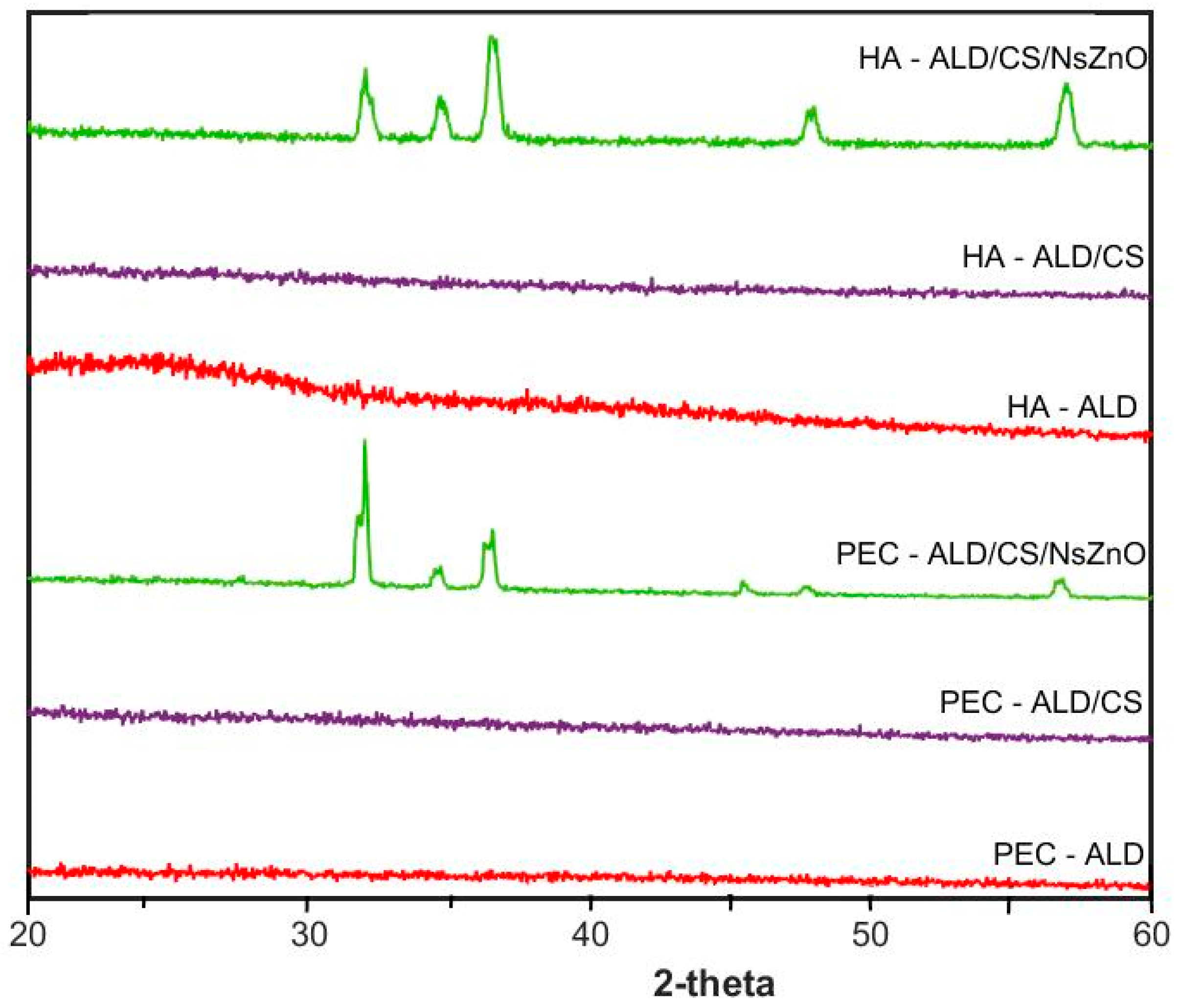
- The X-ray diffraction (XRD) patterns (Figure 3) of HA-ALD and PEC-ALD are amorphous, and the gels formed from these components in the absence of NsZnO are correspondingly amorphous [56]. The XRD patterns of the bionanocomposite hydrogels containing NsZnO have peaks corresponding to NsZnO that are in agreement with our previously reported data [57], with clear evidence of a crystalline hexagonal phase with a wurtzite structure: the five main reflection peaks (100), (002), (101), (102) and (110) are consistent with those of the standard card for the hexagonal phase ZnO (JCPDS ICDD 36e1451). The inclusion of the NsZnO nanoparticles in the gels is clearly evident from the presence of the wurtzitic NsZnO XRD patterns in the bionanocomposite hydrogels. The preservation of the wurtzitic ZnO crystalline structure after the dispersion of the nanoparticles in the gel matrix is significant because of the link between structure and function [41]. Importantly, scanning electron microscopy (SEM) and energy-dispersive X-ray spectroscopy (EDS) confirmed the homogeneous dispersion of the NsZnO particles inside the polymeric matrix (Figure A2). This is of interest for the antibacterial activity of these biomaterials, as the reproducibility of the diffusion of Zn2+ will be dependent upon the homogeneity of dispersion within the gel matrix.
- A correction has been made to Section 3.9:
- The pattern of the X-ray diffraction of the samples was obtained by using a PANalytical X’Pert Diffractometer (Cu Kα radiation, Almelo, The Netherlands). Data were collected with a 2D solid-state detector (PIXcel) from 20° to 60° (2θ) with a step size of 0.001° (2θ).
Reference
- Leone, F.; Firlak, M.; Challen, K.; Bonnefin, W.; Onida, B.; Wright, K.L.; Hardy, J.G. In Situ Crosslinking Bionanocomposite Hydrogels with Potential for Wound Healing Applications. J. Funct. Biomater. 2019, 10, 50. [Google Scholar] [CrossRef] [PubMed]
Disclaimer/Publisher’s Note: The statements, opinions and data contained in all publications are solely those of the individual author(s) and contributor(s) and not of MDPI and/or the editor(s). MDPI and/or the editor(s) disclaim responsibility for any injury to people or property resulting from any ideas, methods, instructions or products referred to in the content. |
© 2025 by the authors. Licensee MDPI, Basel, Switzerland. This article is an open access article distributed under the terms and conditions of the Creative Commons Attribution (CC BY) license (https://creativecommons.org/licenses/by/4.0/).
Share and Cite
Leone, F.; Firlak, M.; Challen, K.; Bonnefin, W.; Onida, B.; Wright, K.L.; Hardy, J.G. Correction: Leone et al. In Situ Crosslinking Bionanocomposite Hydrogels with Potential for Wound Healing Applications. J. Funct. Biomater. 2019, 10, 50. J. Funct. Biomater. 2025, 16, 212. https://doi.org/10.3390/jfb16060212
Leone F, Firlak M, Challen K, Bonnefin W, Onida B, Wright KL, Hardy JG. Correction: Leone et al. In Situ Crosslinking Bionanocomposite Hydrogels with Potential for Wound Healing Applications. J. Funct. Biomater. 2019, 10, 50. Journal of Functional Biomaterials. 2025; 16(6):212. https://doi.org/10.3390/jfb16060212
Chicago/Turabian StyleLeone, Federica, Melike Firlak, Kirsty Challen, Wayne Bonnefin, Barbara Onida, Karen L. Wright, and John G. Hardy. 2025. "Correction: Leone et al. In Situ Crosslinking Bionanocomposite Hydrogels with Potential for Wound Healing Applications. J. Funct. Biomater. 2019, 10, 50" Journal of Functional Biomaterials 16, no. 6: 212. https://doi.org/10.3390/jfb16060212
APA StyleLeone, F., Firlak, M., Challen, K., Bonnefin, W., Onida, B., Wright, K. L., & Hardy, J. G. (2025). Correction: Leone et al. In Situ Crosslinking Bionanocomposite Hydrogels with Potential for Wound Healing Applications. J. Funct. Biomater. 2019, 10, 50. Journal of Functional Biomaterials, 16(6), 212. https://doi.org/10.3390/jfb16060212