

Material-Driven Therapeutics: Functional Nanomaterial Design Paradigms Revolutionizing Osteosarcoma Treatment


Abstract
1. Introduction
2. Functional Properties of Nanomaterials
2.1. Drug Delivery
2.1.1. Controlled-Release
2.1.2. Target-Oriented
2.2. Immune Regulation
2.2.1. Targeted Modulation of Immune Cell Activity
2.2.2. Remodeling the Immunosuppressive Microenvironment
2.3. Catalytic Properties
2.3.1. Enzyme-Mimetic Catalytic Activity
2.3.2. Cascade Catalytic Reactions
2.3.3. Exogenous Field-Enhanced Catalysis
2.4. Thermal Effect
2.4.1. Photothermal Effects
2.4.2. Magnetothermal Effects
2.5. Tissue Engineering
2.5.1. Bioactivity Regulation
2.5.2. Tissue Regeneration and Biomimetic Support
3. Nanomaterials for Treatment of OS
3.1. Nanovesicle

| Nanomaterial | Cell Line | Functional Properties | Tumor Model | Inhibition Rate | Innovation Points | Ref. |
|---|---|---|---|---|---|---|
| EXO-RIF | 143B, MG63 | Rifampicin delivery | Xenograft nude mice | 67.3% | BMSC exosomes, Drp1 agonist | [62] |
| EM-Dox | 143B | DOX delivery | carcinoma in situ | 82.9% | Exosome mimetics (EMs) | [63] |
| Exo-Dox | MG63, 143B | DOX nanocarrier | Xenograft nude mouse | 83.3% | SDF1-CXCR4 targeted therapy | [64] |
| BT-Exo-CAP | MG63, 143B | Carrying CAP | carcinoma in situ | 75% | Bone targeting, ferroptosis | [65] |
| DAELNs | HOS, MG63 | OS-targeted agents | Xenograft model | 70% | Dipsacus asperoides-derived P38/JNK signaling pathway | [66] |
| YSA-SPION-MV/MTX | MG63, 143B | MTX delivery | Orthotopic model | significantly inhibited | EphA2 magnetic targeting | [67] |
| ALN-LWMH-DOX-Lip | K7M2 | DOX delivery | Orthotopic model | 79.2% | Bone targeting, anti-metastasis | [68] |
| HA-DOPE@Lips/HNK | 143B | Honokiol delivery | Xenograft model | 63.7% | Hyaluronic acid HNK | [69] |
| FA-Res/Lps | 143B | Resveratrol delivery | Metastasis models | 82.9% | Lung metastasis JAK2/STAT3 pathway | [70] |
3.1.1. Liposomes
3.1.2. Exosomes
3.1.3. Virus-Like Particles
3.2. Polymer
3.2.1. Poly (Lactic-c–o-Glycolic Acid)
3.2.2. Polydopamine
3.2.3. Hydrogel
3.3. Inorganic Nonmetal
3.3.1. Calcium-Based Nanomaterials
3.3.2. Carbon-Based Nanomaterials
3.3.3. Silicon-Based Nanomaterials
3.4. Metal Based
3.4.1. Noble Metal Nanoparticles
3.4.2. Metal Oxide
3.4.3. Metal–Organic Framework
4. Discussion
Author Contributions
Funding
Institutional Review Board Statement
Informed Consent Statement
Data Availability Statement
Acknowledgments
Conflicts of Interest
Abbreviations
| OS | Osteosarcoma |
| EPR | Enhanced Permeability and Retention |
| mTOR | Mammalian Target of Rapamycin |
| CAR-T | Chimeric Antigen Receptor T cell |
| PD-1 | Programmed Cell Death Protein 1 |
| DOX | Doxorubicin |
| miRNA | MicroRNA |
| HIF-1α | Hypoxia-Inducible Factor 1 alpha |
| pH | Power of Hydrogen |
| ROS | Reactive Oxygen Species |
| CDT | Chemodynamic Therapy |
| PDT | Photodynamic Therapy |
| PTT | Photothermal Therapy |
| MOFs | Metal–Organic Frameworks |
| PLGA | Poly(lactic-co-glycolic acid) |
| PDA | Polydopamine |
| TCP | Tricalcium Phosphate |
| MRI | Magnetic Resonance Imaging |
| CT | Computed Tomography |
| CD47 | Cluster of Differentiation 47 |
| PD-L1 | Programmed Death-Ligand 1 |
| siRNA | Small Interfering RNA |
References
- Meltzer, P.S.; Helman, L.J. New Horizons in the Treatment of Osteosarcoma. N. Engl. J. Med. 2021, 385, 2066–2076. [Google Scholar] [CrossRef]
- Ritter, J.; Bielack, S.S. Osteosarcoma. Ann. Oncol. 2010, 21, vii320–vii325. [Google Scholar] [CrossRef] [PubMed]
- Rathore, R.; Van Tine, B.A. Pathogenesis and Current Treatment of Osteosarcoma: Perspectives for Future Therapies. J. Clin. Med. 2021, 10, 1182. [Google Scholar] [CrossRef] [PubMed]
- Zhao, X.; Wu, Q.; Gong, X.; Liu, J.; Ma, Y. Osteosarcoma: A review of current and future therapeutic approaches. Biomed. Eng. Online 2021, 20, 24. [Google Scholar] [CrossRef]
- Isakoff, M.S.; Bielack, S.S.; Meltzer, P.; Gorlick, R. Osteosarcoma: Current Treatment and a Collaborative Pathway to Success. J. Clin. Oncol. 2015, 33, 3029–3035. [Google Scholar] [CrossRef]
- Yu, S.; Yao, X. Advances on immunotherapy for osteosarcoma. Mol. Cancer 2024, 23, 192. [Google Scholar] [CrossRef]
- Kansara, M.; Teng, M.W.; Smyth, M.J.; Thomas, D.M. Translational biology of osteosarcoma. Nat. Rev. Cancer 2014, 14, 722–735. [Google Scholar] [CrossRef]
- Ji, T.; Zhao, Y.; Ding, Y.; Nie, G. Using functional nanomaterials to target and regulate the tumor microenvironment: Diagnostic and therapeutic applications. Adv. Mater. 2013, 25, 3508–3525. [Google Scholar] [CrossRef] [PubMed]
- Luan, X.; Kong, H.; He, P.; Yang, G.; Zhu, D.; Guo, L.; Wei, G. Self-Assembled Peptide-Based Nanodrugs: Molecular Design, Synthesis, Functionalization, and Targeted Tumor Bioimaging and Biotherapy. Small 2023, 19, e2205787. [Google Scholar] [CrossRef]
- Liu, Y.; Wang, Y.; Song, S.; Zhang, H. Tumor Diagnosis and Therapy Mediated by Metal Phosphorus-Based Nanomaterials. Adv. Mater. 2021, 33, e2103936. [Google Scholar] [CrossRef]
- Xie, J.; Gong, L.; Zhu, S.; Yong, Y.; Gu, Z.; Zhao, Y. Emerging Strategies of Nanomaterial-Mediated Tumor Radiosensitization. Adv. Mater. 2019, 31, e1802244. [Google Scholar] [CrossRef]
- Chen, C.; Wang, S.; Wang, J.; Yao, F.; Tang, X.; Guo, W. Nanosized drug delivery strategies in osteosarcoma chemotherapy. APL Bioeng. 2023, 7, 011501. [Google Scholar] [CrossRef] [PubMed]
- Fatema, K.N.; Li, L.L.; Ahmad, K.; Kim, J.; Lee, D.W. Development of Multifunctional PAA-Alginate-Carboxymethyl Cellulose Hydrogel-Loaded Fiber-Reinforced Biomimetic Scaffolds for Controlled Release of Curcumin. Int. J. Biol. Macromol. 2025, 301, 140449. [Google Scholar] [CrossRef] [PubMed]
- Xu, S.; Hu, Z.; Zheng, W.; Qin, C.; Bai, X.; Yang, Q.; Zeng, T.; Mo, D.; Zhou, B.; Lu, C.; et al. GSH-Responsive Pt-Based Nanomotor with Improved Doxorubicin Delivery for Synergistic Osteosarcoma Chemotherapy. Acta Biomater. 2025, 195, 390–405. [Google Scholar] [CrossRef] [PubMed]
- Lavrador, P.; Esteves, M.R.; Gaspar, V.M.; Mano, J.F. Stimuli-Responsive Nanocomposite Hydrogels for Biomedical Applications. Adv. Funct. Mater. 2021, 31, 2005941. [Google Scholar] [CrossRef]
- Huang, H.; Asghar, S.; Lin, L.; Chen, S.; Yuan, C.; Sang, M.; Xiao, Y. Design and evaluation of a multi-responsive dual-modality bone-targeted drug delivery vehicle for the treatment of osteosarcoma. Int. J. Pharm. 2025, 671, 125191. [Google Scholar] [CrossRef]
- Jiang, H.; Luo, Y.; Li, B.; Wu, C.; Wang, D.; Xin, Y.; Xu, W.; Xiao, J. IL-11-Engineered Macrophage Membrane-Coated Reactive Oxygen Species-Responsive Nanoparticles for Targeted Delivery of Doxorubicin to Osteosarcoma. ACS Appl. Mater. Interfaces 2024, 16, 55981–55995. [Google Scholar] [CrossRef]
- Kaniewska, K.; Mackiewicz, M.; Smutok, O.; Gonchar, M.; Katz, E.; Karbarz, M. Enzymatically Triggered Drug Release from Microgels Controlled by Glucose Concentration. ACS Biomater. Sci. Eng. 2024, 10, 6415–6424. [Google Scholar] [CrossRef]
- Murphy, J.N.; Kobti, J.L.; Dao, M.; Wear, D.; Okoko, M.; Pandey, S.; Vukotic, V.N. Therapeutic coordination polymers: Tailoring drug release through metal-ligand interactions. Chem. Sci. 2024, 15, 7041–7050. [Google Scholar] [CrossRef]
- Cao, J.; Zhu, C.; Cao, Z.; Ke, X. CPPs-modified chitosan as permeability-enhancing chemotherapeutic combined with gene therapy nanosystem by thermosensitive hydrogel for the treatment of osteosarcoma. Int. J. Biol. Macromol. 2024, 267 Pt 2, 130915. [Google Scholar] [CrossRef]
- Luo, S.; Zhao, C.; Wang, R.; Wu, D. Sequential drug release nanocomposites for synergistic therapy in disease treatment. J. Mater. Chem. B 2025, 13, 4313–4329. [Google Scholar] [CrossRef]
- Ju, Q.; Huang, R.; Hu, R.; Fan, J.; Zhang, D.; Ding, J.; Li, R. Phytic acid-modified manganese dioxide nanoparticles oligomer for magnetic resonance imaging and targeting therapy of osteosarcoma. Drug Deliv. 2023, 30, 2181743. [Google Scholar] [CrossRef]
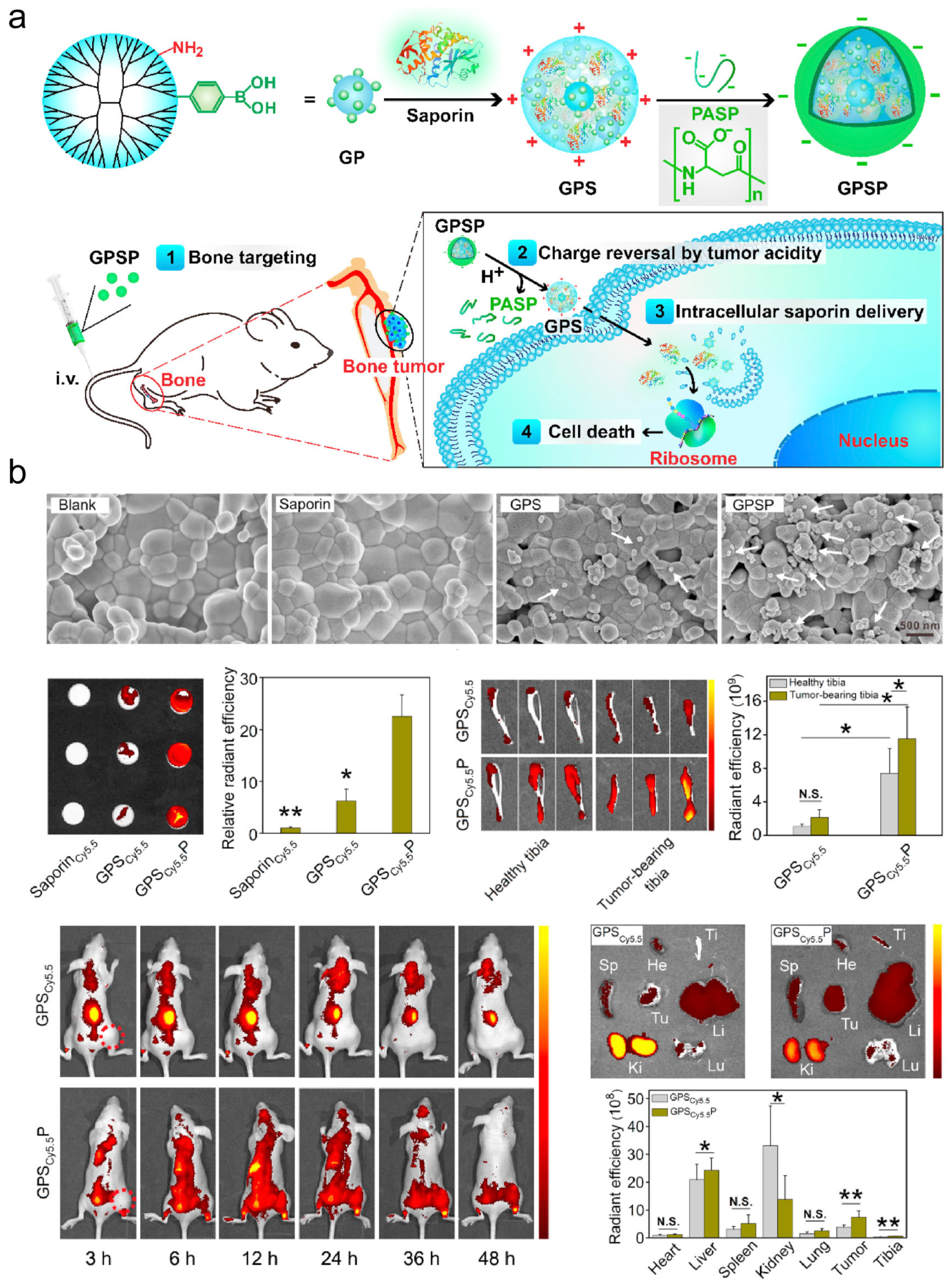
- Yan, Y.; Zhou, L.; Sun, Z.; Song, D.; Cheng, Y. Targeted and intracellular delivery of protein therapeutics by a boronated polymer for the treatment of bone tumors. Bioact. Mater. 2022, 7, 333–340. [Google Scholar] [CrossRef]
- Liu, T.; Liang, X.; Liu, W.; Yang, S.; Cui, T.; Yan, F.; Li, Z. iRGD-Targeted Biosynthetic Nanobubbles for Ultrasound Molecular Imaging of Osteosarcoma. Int. J. Nanomed. 2025, 20, 791–805. [Google Scholar] [CrossRef]
- Liu, Y.J.; Dong, S.H.; Hu, W.H.; Chen, Q.L.; Zhang, S.F.; Song, K.; Han, Z.C.; Li, M.M.; Han, Z.T.; Liu, W.B.; et al. A multifunctional biomimetic nanoplatform for image-guideded photothermal-ferroptotic synergistic osteosarcoma therapy. Bioact. Mater. 2024, 36, 157–167. [Google Scholar] [CrossRef]
- Wei, X.; Feng, J.; Chen, L.; Zhang, C.; Liu, Y.; Zhang, Y.; Xu, Y.; Zhang, J.; Wang, J.; Yang, H.; et al. METTL3-Mediated m6A Modification of LINC00520 Confers Glycolysis and Chemoresistance in Osteosarcoma via Suppressing Ubiquitination of ENO1. Cancer Lett. 2024, 217194. [Google Scholar] [CrossRef]
- Jin, W.; Zhang, T.; Zhou, W.; He, P.; Sun, Y.; Hu, S.; Chen, H.; Ma, X.; Peng, Y.; Yi, Z.; et al. Discovery of 2-Amino-3-Cyanothiophene Derivatives as Potent STAT3 Inhibitors for the Treatment of Osteosarcoma Growth and Metastasis. J. Med. Chem. 2022, 65, 6710–6728. [Google Scholar] [CrossRef]
- Liu, W.; Guo, M.; Hu, Y.; Chen, Y.; Wang, Y.; Nezamzadeh-Ejhieh, A.; Li, H.; Lu, C.; Liu, J. Recent advances in metal-based nanomaterials for malignant bone tumor therapy. Eur. J. Med. Chem. 2025, 288, 117427. [Google Scholar] [CrossRef]
- Yang, X.; Gao, S.; Yang, B.; Yang, Z.; Lou, F.; Huang, P.; Zhao, P.; Guo, J.; Fang, H.; Chu, B.; et al. Bioinspired Tumor-Targeting and Biomarker-Activatable Cell-Material Interfacing System Enhances Osteosarcoma Treatment via Biomineralization. Adv. Sci. 2023, 10, e2302272. [Google Scholar] [CrossRef]
- Cheng, M.; Jiang, Y.; Wang, Y.; Wu, Y.; Zhu, Y. Enhancing osteosarcoma therapy through aluminium hydroxide nanosheets-enabled macrophage modulation. Int. J. Pharm. 2024, 649, 123640. [Google Scholar] [CrossRef]
- Luo, T.; Fan, Z.; Zeng, A.; Wang, A.; Pan, Y.; Xu, Y.; Chen, H.; Chen, W.; Nie, D.; Lin, J.; et al. Biomimetic Targeted Co-Delivery System Engineered from Genomic Insights for Precision Treatment of Osteosarcoma. Adv. Sci. 2025, 12, e2410427. [Google Scholar] [CrossRef]
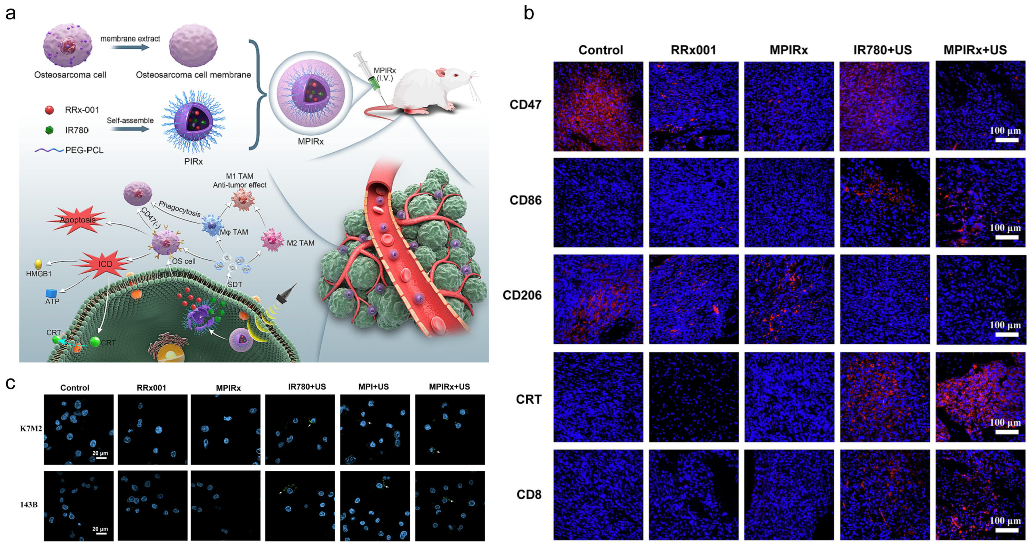
- Gong, M.; Huang, Y.; Feng, H.; Lin, J.; Huang, A.; Hu, J.; Tang, Q.; Zhu, X.; Han, S.; Lu, J.; et al. A Nanodrug Combining CD47 and Sonodynamic Therapy Efficiently Inhibits Osteosarcoma Deterioration. J. Control. Release 2023, 355, 68–84. [Google Scholar] [CrossRef]
- Wang, X.; Guo, X.; Ren, H.; Song, X.; Chen, L.; Yu, L.; Ren, J.; Chen, Y. An “Outer Piezoelectric and Inner Epigenetic” Logic-Gated PANoptosis for Osteosarcoma Sono-Immunotherapy and Bone Regeneration. Adv. Mater. 2025, 37, e2415814. [Google Scholar] [CrossRef]
- Lian, H.; Zhang, J.; Hou, S.; Ma, S.; Yu, J.; Zhao, W.; Zhao, D.; Zhang, Z. Immunotherapy of osteosarcoma based on immune microenvironment modulation. Front. Immunol. 2024, 15, 1498060. [Google Scholar] [CrossRef]
- Wei, Z.; Xia, K.; Liu, W.; Huang, X.; Wei, Z.; Guo, W. CGREF1 Modulates Osteosarcoma Proliferation by Regulating the Cell Cycle Through the Wnt/β-Catenin Signaling Pathway. Mol. Med. 2024, 30, 260. [Google Scholar] [CrossRef]
- Nie, J.J.; Zhang, B.; Luo, P.; Luo, M.; Luo, Y.; Cao, J.; Wang, H.; Mao, J.; Xing, Y.; Liu, W.; et al. Enhanced pyroptosis induction with pore-forming gene delivery for osteosarcoma microenvironment reshaping. Bioact. Mater. 2024, 38, 455–471. [Google Scholar] [CrossRef]
- Elzoghby, A.O.; Samir, O.; Emam, H.E.; Soliman, A.; Abdelgalil, R.M.; Elmorshedy, Y.M.; Elkhodairy, K.A.; Nasr, M.L. Engineering nanomedicines for immunogenic eradication of cancer cells: Recent trends and synergistic approaches. Acta Pharm. Sin. B 2024, 14, 2475–2504. [Google Scholar] [CrossRef]
- Xia, J.; Hu, C.; Ji, Y.; Wang, M.; Jin, Y.; Ye, L.; Xie, D.; Jiang, S.; Li, R.; Hu, Z.; et al. Copper-Loaded Nanoheterojunction Enables Superb Orthotopic Osteosarcoma Therapy via Oxidative Stress and Cell Cuproptosis. ACS Nano. 2023, 17, 21134–21152. [Google Scholar] [CrossRef]
- Li, M.; Wang, M.; Huang, J.; Tang, S.; Yang, J.; Xu, Z.; Xu, G.; Chen, X.; Liu, J.; Yang, C. High-performance pyrite nano-catalyst driven photothermal/chemodynamic synergistic therapy for Osteosarcoma. J. Nanobiotechnol. 2024, 22, 141. [Google Scholar] [CrossRef]
- Liang, Y.; Liao, C.; Guo, X.; Li, G.; Yang, X.; Yu, J.; Zhong, J.; Xie, Y.; Zheng, L.; Zhao, J. RhRu Alloy-Anchored MXene Nanozyme for Synergistic Osteosarcoma Therapy. Small 2023, 19, e2205511. [Google Scholar] [CrossRef]
- Liu, C.; Xu, X.; Chen, Y.; Yin, M.; Mäkilä, E.; Zhou, W.; Su, W.; Zhang, H. Metabolism-Regulating Nanozyme System for Advanced Nanocatalytic Cancer Therapy. Small 2024, 20, e2307794. [Google Scholar] [CrossRef] [PubMed]
- Tong, F.; Ye, Y.; Chen, B.; Gao, J.; Liu, L.; Ou, J.; van Hest, J.C.M.; Liu, S.; Peng, F.; Tu, Y. Bone-Targeting Prodrug Mesoporous Silica-Based Nanoreactor with Reactive Oxygen Species Burst for Enhanced Chemotherapy. ACS Appl. Mater. Interfaces 2020, 12, 34630–34642. [Google Scholar] [CrossRef] [PubMed]
- Geng, B.; Yang, X.; Li, P.; Shi, W.; Pan, D.; Shen, L. W-Doped TiO2 Nanorods for Multimode Tumor Eradication in Osteosarcoma Models Under Single Ultrasound Irradiation. ACS Appl. Mater. Interfaces 2021, 13, 45325–45334. [Google Scholar] [CrossRef] [PubMed]
- Qi, M.H.; Wang, D.D.; Qian, W.; Zhang, Z.L.; Ao, Y.W.; Li, J.M.; Huang, S.W. High-Efficiency Gold Nanoaggregates for NIR LED-Driven Sustained Mild Photothermal Therapy Achieving Complete Tumor Eradication and Immune Enhancement. Adv. Mater. 2025, 37, e2412191. [Google Scholar] [CrossRef]
- Liang, Y.; Wang, C.; Yu, S.; Fan, Y.; Jiang, Y.; Zhou, R.; Yan, W.; Sun, Y. IOX1 Epigenetically Enhanced Photothermal Therapy of 3D-Printing Silicene Scaffolds Against Osteosarcoma with Favorable Bone Regeneration. Mater. Today Bio 2023, 23, 100887. [Google Scholar] [CrossRef]
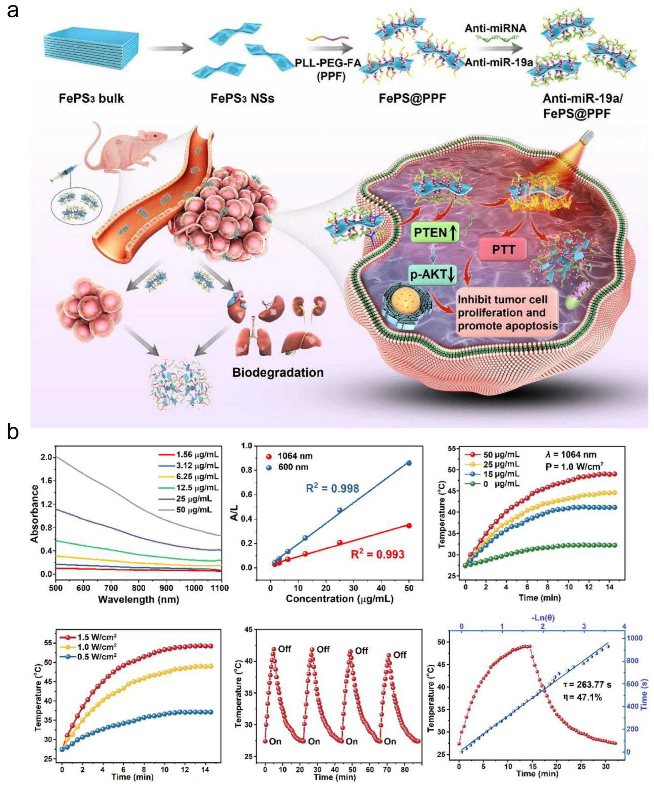
- Luo, T.; Jiang, M.; Cheng, Z.; Lin, Y.; Chen, Y.; Zhang, Z.; Zhou, J.; Zhou, W.; Yu, X.F.; Li, S.; et al. Biodegradable FePS3 nanoplatform for efficient treatment of osteosarcoma by combination of gene and NIR-II photothermal therapy. J. Nanobiotechnol. 2023, 21, 224. [Google Scholar] [CrossRef]
- Ge, J.; Yang, N.; Yang, Y.; Yu, H.; Yang, X.; Wang, Y.; Wang, T.; Cheng, S.; Wang, Y.; Han, Z.; et al. The combination of eddy thermal effect of biodegradable magnesium with immune checkpoint blockade shows enhanced efficacy against osteosarcoma. Bioact. Mater. 2023, 25, 73–85. [Google Scholar] [CrossRef]
- Yu, K.; Zhou, H.; Xu, Y.; Cao, Y.; Zheng, Y.; Liang, B. Engineering a triple-functional magnetic gel driving mutually-synergistic mild hyperthermia-starvation therapy for osteosarcoma treatment and augmented bone regeneration. J. Nanobiotechnol. 2023, 21, 201. [Google Scholar] [CrossRef]
- Wang, C.; Zhou, G.; Guo, X.; Zhang, W.; Wu, C. Electrical Stimulation Promotes Endocytosis of Magnetic Nanoparticles by Cancer Cells. Small 2024, 20, e2403381. [Google Scholar] [CrossRef]
- Lukin, I.; Erezuma, I.; Desimone, M.F.; Zhang, Y.S.A. Dolatshahi-Pirouz, and G. Orive, Nanomaterial-based drug delivery of immunomodulatory factors for bone and cartilage tissue engineering. Biomater. Adv. 2023, 154, 213637. [Google Scholar] [CrossRef]
- Shi, Q.; Lu, Y.; Zhang, G.; Yang, X.; Li, R.; Zhang, G.; Guo, X.; Song, J.; Ding, Q. Multifunctional Mesoporous Silica Nanoparticles for pH-Response and Photothermy Enhanced Osteosarcoma Therapy. Colloids Surf. B Biointerfaces 2022, 217, 112615. [Google Scholar] [CrossRef] [PubMed]
- Xu, C.; Xia, Y.; Zhuang, P.; Liu, W.; Mu, C.; Liu, Z.; Wang, J.; Chen, L.; Dai, H.; Luo, Z. FePSe3-Nanosheets-Integrated Cryogenic-3D-Printed Multifunctional Calcium Phosphate Scaffolds for Synergistic Therapy of Osteosarcoma. Small 2023, 19, e2303636. [Google Scholar] [CrossRef]
- Wang, L.; Dai, Z.; Bi, J.; Chen, Y.; Wang, Z.; Sun, Z.; Ji, Z.; Wang, H.; Zhang, Y.; Wang, L.; et al. Polydopamine-functionalized calcium-deficient hydroxyapatite 3D-printed scaffold with sustained doxorubicin release for synergistic chemo-photothermal therapy of osteosarcoma and accelerated bone regeneration. Mater. Today Bio 2024, 29, 101253. [Google Scholar] [CrossRef] [PubMed]
- Heng, C.; Zheng, X.; Hui, J.; Ma, X.; Fan, D. Neodymium and manganese ions co-doped whitlockite for temperature monitoring, photothermal therapy, and bone tissue repair in osteosarcoma. J. Colloid Interface Sci. 2024, 653 Pt B, 1488–1503. [Google Scholar] [CrossRef]
- Han, R.; Min, Y.; Li, G.; Chen, S.; Xie, M.; Zhao, Z. Supercritical CO2-Assisted Fabrication of CM-PDA/SF/nHA Nanofibrous Scaffolds for Bone Regeneration and Chemo-Photothermal Therapy Against Osteosarcoma. Biomater. Sci. 2023, 11, 5218–5231. [Google Scholar] [CrossRef] [PubMed]
- Cheng, Q.; Kang, Y.; Yao, B.; Dong, J.; Zhu, Y.; He, Y.; Ji, X. Genetically Engineered-Cell-Membrane Nanovesicles for Cancer Immunotherapy. Adv. Sci. 2023, 10, e2302131. [Google Scholar] [CrossRef] [PubMed]
- Gao, J.; Su, Y.; Wang, Z. Engineering bacterial membrane nanovesicles for improved therapies in infectious diseases and cancer. Adv. Drug Deliv. Rev. 2022, 186, 114340. [Google Scholar] [CrossRef]
- Liu, C.; Feng, Q.; Sun, J. Lipid Nanovesicles by Microfluidics: Manipulation, Synthesis, and Drug Delivery. Adv. Mater. 2019, 31, e1804788. [Google Scholar] [CrossRef]
- Sun, M.; Yang, J.; Fan, Y.; Zhang, Y.; Sun, J.; Hu, M.; Sun, K.; Zhang, J. Beyond Extracellular Vesicles: Hybrid Membrane Nanovesicles as Emerging Advanced Tools for Biomedical Applications. Adv. Sci. 2023, 10, e2303617. [Google Scholar] [CrossRef]
- Liu, J.; You, Q.; Liang, F.; Ma, L.; Zhu, L.; Wang, C.; Yang, Y. Ultrasound-nanovesicles interplay for theranostics. Adv. Drug Deliv. Rev. 2024, 205, 115176. [Google Scholar] [CrossRef] [PubMed]
- Du, S.; Guan, Y.; Xie, A.; Yan, Z.; Gao, S.; Li, W.; Rao, L.; Chen, X.; Chen, T. Extracellular vesicles: A rising star for therapeutics and drug delivery. J. Nanobiotechnol. 2023, 21, 231. [Google Scholar] [CrossRef] [PubMed]
- Chen, W.; Lin, W.; Yu, N.; Zhang, L.; Wu, Z.; Chen, Y.; Li, Z.; Gong, F.; Li, N.; Chen, X.; et al. Activation of Dynamin-Related Protein 1 and Induction of Mitochondrial Apoptosis by Exosome-Rifampicin Nanoparticles Exerts Anti-Osteosarcoma Effect. Int. J. Nanomed. 2022, 17, 5431–5446. [Google Scholar] [CrossRef]
- Wang, J.; Li, M.; Jin, L.; Guo, P.; Zhang, Z.; Zhanghuang, C.; Tan, X.; Mi, T.; Liu, J.; Wu, X.; et al. Exosome mimetics derived from bone marrow mesenchymal stem cells deliver doxorubicin to osteosarcoma in vitro and in vivo. Drug Deliv. 2022, 29, 3291–3303. [Google Scholar] [CrossRef]
- Wei, H.; Chen, F.; Chen, J.; Lin, H.; Wang, S.; Wang, Y.; Wu, C.; Lin, J.; Zhong, G. Mesenchymal Stem Cell Derived Exosomes as Nanodrug Carrier of Doxorubicin for Targeted Osteosarcoma Therapy via SDF1-CXCR4 Axis. Int. J. Nanomed. 2022, 17, 3483–3495. [Google Scholar] [CrossRef] [PubMed]
- Chen, W.; Li, Z.; Yu, N.; Zhang, L.; Li, H.; Chen, Y.; Gong, F.; Lin, W.; He, X.; Wang, S.; et al. Bone-Targeting Exosome Nanoparticles Activate Keap1/Nrf2/GPX4 Signaling Pathway to Induce Ferroptosis in Osteosarcoma Cells. J. Nanobiotechnol. 2023, 21, 355. [Google Scholar] [CrossRef]
- Lu, J.; Chen, J.; Ye, J.; Shi, Z.; Gao, X.; Chen, P.; Chang, Y.; Lin, H.; Li, P. Dipsacus Asperoides-Derived Exosomes-Like Nanoparticles Inhibit the Progression of Osteosarcoma via Activating P38/JNK Signaling Pathway. Int. J. Nanomed. 2024, 19, 1097–1108. [Google Scholar] [CrossRef]
- Wang, Z.; He, Z.; Wan, J.; Chen, A.; Cheng, P.; Zhu, W. EphA2-specific microvesicles derived from tumor cells facilitate the targeted delivery of chemotherapeutic drugs for osteosarcoma therapy. J. Nanobiotechnol. 2024, 22, 89. [Google Scholar] [CrossRef] [PubMed]
- Wu, H.; Luo, Y.; Xu, D.; Ke, X.; Ci, T. Low molecular weight heparin modified bone targeting liposomes for orthotopic osteosarcoma and breast cancer bone metastatic tumors. Int. J. Biol. Macromol. 2020, 164, 2583–2597. [Google Scholar] [CrossRef]
- Zhang, X.; Chen, H.; Zhang, Y.; Huang, Q.; Feng, J.; Xing, H.; Fu, X.; Yan, X.; Zhang, Y.; Xu, Q.; et al. HA-DOPE-Modified Honokiol-Loaded Liposomes Targeted Therapy for Osteosarcoma. Int. J. Nanomed. 2022, 17, 5137–5151. [Google Scholar] [CrossRef]
- Zhu, W.T.; Zeng, X.F.; Yang, H.; Jia, M.L.; Zhang, W.; Liu, W.; Liu, S.Y. Resveratrol Loaded by Folate-Modified Liposomes Inhibits Osteosarcoma Growth and Lung Metastasis via Regulating JAK2/STAT3 Pathway. Int. J. Nanomed. 2023, 18, 2677–2691. [Google Scholar] [CrossRef]
- Wang, Y.-F.; Zhang, C.; Yang, K.; Wang, Y.; Shan, S.; Yan, Y.; Dawson, K.A.; Wang, C.; Liang, X.-J. Transportation of AIE-visualized nanoliposomes is dominated by the protein corona. Natl. Sci. Rev. 2021, 8, nwab068. [Google Scholar] [CrossRef]
- Yan, J.; Wei, D.; Zhao, Z.; Sun, K.; Sun, Y. Osteosarcoma-targeting PtIV prodrug amphiphile for enhanced chemo-immunotherapy via Ca2+ trapping. Acta Biomater. 2025, 193, 474–483. [Google Scholar] [CrossRef]
- Turánek, J.; Kosztyu, P.; Turánek Knötigová, P.; Bartheldyová, E.; Hubatka, F.; Odehnalová, N.; Mikulík, R.; Vaškovicová, N.; Čelechovská, H.; Kratochvílová, I.; et al. Long circulating liposomal platform utilizing hydrophilic polymer-based surface modification: Preparation, characterisation, and biological evaluation. Int. J. Pharm. 2024, 661, 124465. [Google Scholar] [CrossRef] [PubMed]
- Ren, K.; Cao, X.; Zheng, L.; Liu, S.; Li, L.; Cheng, L.; Tian, T.; Tong, X.; Wang, H.; Jiang, L. Liposomes decorated with β-conglycinin and glycinin: Construction, structure and in vitro digestive stability. Int. J. Biol. Macromol. 2024, 269, 131900. [Google Scholar] [CrossRef] [PubMed]
- Zhao, L.; Liu, C.; Wang, T.; Sun, L.; Wu, F.; Yu, D. Combined multispectral analysis and molecular docking to research the interaction of soybean isolate protein with different kinds of phospholipid liposomes and its effect on liposome properties. Food Chem. 2025, 474, 143160. [Google Scholar] [CrossRef]
- Chen, X.-A.; Chuang, C.-C.; Chen, C.-C.; Lee, C.-Y.; Chin, C.-Y.; Young, J.-J.; Bai, M.-Y.; Chuang, C.-C. Polyelectrolyte-coated liposomes microfluidically assembled in one-step for enhancing cell endocytosis and in-vivo immune responses. Colloids Surf. B Biointerfaces 2024, 241, 114030. [Google Scholar] [CrossRef]
- Song, Y.; Li, H.; Yuan, Y.; Zhang, D.; Wang, Z.; Qi, B.; Jiang, P.; Yu, A. Synergistic photothermal-sonodynamic therapy for antibacterial and immune reprogramming in chronic osteomyelitis. J. Control. Release 2025, 381, 113612. [Google Scholar] [CrossRef] [PubMed]
- Zhang, W.; Liu, Y.; Luo, Y.; Xu, J.; Zhang, B.; Feng, P.; Guo, C.; Wang, Y.; Huang, Z.; Kong, Q.; et al. Reactivating P53 to treat osteosarcoma: A tetrahedral framework nucleic acids-based approach. Int. J. Biol. Macromol. 2025, 304, 140765. [Google Scholar] [CrossRef]
- Kodel, H.d.A.C.; Alizadeh, P.; Ebrahimi, S.N.; Machado, T.O.X.; Oliveira, M.B.P.P.; Fathi, F.; Souto, E.B. Liposomes and Niosomes: New trends and applications in the delivery of bioactive agents for cancer therapy. Int. J. Pharm. 2025, 668, 124994. [Google Scholar] [CrossRef]
- Liu, Y.; Wang, T.; Chi, X.; Yu, S.; He, W.; He, H.; Wang, G.; Hao, K.; Zhang, J. Modeling based dynamics mechanism and pathway of liposome penetration in multicellular tumor spheroid for liposome optimization. Int. J. Pharm. 2025, 671, 125237. [Google Scholar] [CrossRef]
- Han, Z.; Peng, X.; Yang, Y.; Yi, J.; Zhao, D.; Bao, Q.; Long, S.; Yu, S.-X.; Xu, X.-X.; Liu, B.; et al. Integrated microfluidic-SERS for exosome biomarker profiling and osteosarcoma diagnosis. Biosens. Bioelectron. 2022, 217, 114709. [Google Scholar] [CrossRef] [PubMed]
- Almeida, S.F.F.; Fonseca, A.; Sereno, J.; Ferreira, H.R.S.; Lapo-Pais, M.; Martins-Marques, T.; Rodrigues, T.; Oliveira, R.C.; Miranda, C.; Almeida, L.P.; et al. Osteosarcoma-Derived Exosomes as Potential PET Imaging Nanocarriers for Lung Metastasis. Small 2022, 18, e2203999. [Google Scholar] [CrossRef] [PubMed]
- Du, J.; Meng, X.; Yang, M.; Chen, G.; Li, J.; Zhu, Z.; Wu, X.; Hu, W.; Tian, M.; Li, T.; et al. NGR-Modified CAF-Derived exos Targeting Tumor Vasculature to Induce Ferroptosis and Overcome Chemoresistance in Osteosarcoma. Adv. Sci. 2025, 12, 2410918. [Google Scholar] [CrossRef] [PubMed]
- Jiang, M.; Jike, Y.; Liu, K.; Gan, F.; Zhang, K.; Xie, M.; Zhang, J.; Chen, C.; Zou, X.; Jiang, X.; et al. Exosome-mediated miR-144-3p promotes ferroptosis to inhibit osteosarcoma proliferation, migration, and invasion through regulating ZEB1. Mol. Cancer 2023, 22, 113. [Google Scholar] [CrossRef] [PubMed]
- Liu, W.; Li, L.; Bai, X.; Zhang, M.; Lv, W.; Ma, Y.; Sun, Y.; Zhang, H.; Jiang, Q.; Yao, Q.; et al. Osteosarcoma Cell-Derived Migrasomes Promote Macrophage M2 Polarization to Aggravate Osteosarcoma Proliferation and Metastasis. Adv. Sci. 2025, 12, 2409870. [Google Scholar] [CrossRef]
- Huang, X.; Wu, W.; Jing, D.; Yang, L.; Guo, H.; Wang, L.; Zhang, W.; Pu, F.; Shao, Z. Engineered exosome as targeted lncRNA MEG3 delivery vehicles for osteosarcoma therapy. J. Control. Release Off. J. Control. Release Soc. 2022, 343, 107–117. [Google Scholar] [CrossRef]
- Sun, Y.; Guo, J.; Yu, L.; Guo, T.; Wang, J.; Wang, X.; Chen, Y. PD-L1+ exosomes from bone marrow-derived cells of tumor-bearing mice inhibit antitumor immunity. Cell. Mol. Immunol. 2021, 18, 2402–2409. [Google Scholar] [CrossRef]
- Wang, J.; Zhang, H.; Sun, X.; Wang, X.; Ren, T.; Huang, Y.; Zhang, R.; Zheng, B.; Guo, W. Exosomal PD-L1 and N-cadherin predict pulmonary metastasis progression for osteosarcoma patients. J. Nanobiotechnology 2020, 18, 151. [Google Scholar] [CrossRef]
- Suffian, I.F.B.M.; Al-Jamal, K.T. Bioengineering of virus-like particles as dynamic nanocarriers for in vivo delivery and targeting to solid tumours. Adv. Drug Deliv. Rev. 2022, 180, 114030. [Google Scholar] [CrossRef]
- Yin, D.; Zhong, Y.; Ling, S.; Lu, S.; Wang, X.; Jiang, Z.; Wang, J.; Dai, Y.; Tian, X.; Huang, Q.; et al. Dendritic-cell-targeting virus-like particles as potent mRNA vaccine carriers. Nat. Biomed. Eng. 2025, 9, 185–200. [Google Scholar] [CrossRef]
- Morales-Molina, A.; Gambera, S.; Leo, A.; García-Castro, J. Combination immunotherapy using G-CSF and oncolytic virotherapy reduces tumor growth in osteosarcoma. J. Immunother. Cancer 2021, 9, e001703. [Google Scholar] [CrossRef] [PubMed]
- Cai, J.X.; Liu, J.H.; Wu, J.Y.; Li, Y.J.; Qiu, X.H.; Xu, W.J.; Xu, P.; Xiang, D.-X. Hybrid Cell Membrane-Functionalized Biomimetic Nanoparticles for Targeted Therapy of Osteosarcoma. Int. J. Nanomedicine 2022, 17, 837–854. [Google Scholar] [CrossRef]
- Alsulays, B.B.; Aodah, A.H.; Ahmed, M.M.; Anwer, M.K. Preparation and Evaluation of Chitosan Coated PLGA Nanoparticles Encapsulating Ivosidenib with Enhanced Cytotoxicity Against Human Liver Cancer Cells. Int. J. Nanomedicine 2024, 19, 3461–3473. [Google Scholar] [CrossRef] [PubMed]
- Wang, J.; Liang, S.; Chen, S.; Ma, T.; Chen, M.; Niu, C.; Leng, Y.; Wang, L. Bacterial outer membrane vesicle-cancer cell hybrid membrane-coated nanoparticles for sonodynamic therapy in the treatment of breast cancer bone metastasis. J. Nanobiotechnology 2024, 22, 328. [Google Scholar] [CrossRef]
- Crisafulli, E.; Scalzone, A.; Tonda-Turo, C.; Girón-Hernández, J.; Gentile, P. Multimodal layer-by-layer nanoparticles: A breakthrough in gene and drug delivery for osteosarcoma. J. Mater. Chem. B 2024, 12, 12540–12552. [Google Scholar] [CrossRef] [PubMed]
- Li, Y.; Hou, H.; Zhang, P.; Zhang, Z. Co-delivery of doxorubicin and paclitaxel by reduction/pH dual responsive nanocarriers for osteosarcoma therapy. Drug Deliv. 2020, 27, 1044–1053. [Google Scholar] [CrossRef]
- Wang, Y.; Zhang, L.; Zhao, G.; Zhang, Y.; Zhan, F.; Chen, Z.; He, T.; Cao, Y.; Hao, L.; Wang, Z.; et al. Homologous targeting nanoparticles for enhanced PDT against osteosarcoma HOS cells and the related molecular mechanisms. J. Nanobiotechnology 2022, 20, 83. [Google Scholar] [CrossRef]
- Li, X.; Wang, L.; Wang, L.; Yu, J.; Lu, G.; Zhao, W.; Miao, C.; Zou, C.; Wu, J. Overcoming therapeutic failure in osteosarcoma via Apatinib-encapsulated hydrophobic poly(ester amide) nanoparticles. Biomater. Sci. 2020, 8, 5888–5899. [Google Scholar] [CrossRef]
- Qiu, R.; Sun, D.; Bai, Y.; Li, J.; Wang, L. Application of tumor-targeting peptide-decorated polypeptide nanoparticles with doxorubicin to treat osteosarcoma. Drug Deliv. 2020, 27, 1704–1717. [Google Scholar] [CrossRef]
- Jiang, J.; Wang, R.; Yang, L.; Sha, Y.; Zhao, S.; Guo, J.; Chen, D.; Zhong, Z.; Meng, F. IL-11Rα-targeted nanostrategy empowers chemotherapy of relapsed and patient-derived osteosarcoma. J. Control. Release Off. J. Control. Release Soc. 2022, 350, 460–470. [Google Scholar] [CrossRef]
- Li, M.; Lin, Z.-I.; Yang, J.; Huang, H.; Liu, G.-L.; Liu, Q.; Zhang, X.; Zhang, Y.; Xu, Z.; Lin, H.; et al. Biodegradable Carbon Dioxide-Derived Non-Viral Gene Vectors for Osteosarcoma Gene Therapy. Adv. Healthc. Mater. 2023, 12, 2201306. [Google Scholar] [CrossRef] [PubMed]
- Mpekris, F.; Panagi, M.; Michael, C.; Voutouri, C.; Tsuchiya, M.; Wagatsuma, C.; Kinoh, H.; Osada, A.; Akinaga, S.; Yoshida, S.; et al. Translational nanomedicine potentiates immunotherapy in sarcoma by normalizing the microenvironment. J. Control. Release Off. J. Control. Release Soc. 2023, 353, 956–964. [Google Scholar] [CrossRef]
- Lou, H.; Ji, A.; Qu, C.; Liu, H.; Jiang, L.; Chen, H.; Cheng, Z. A Small-Molecule Based Organic Nanoparticle for Photothermal Therapy and Near-Infrared-IIb Imaging. ACS Appl. Mater. Interfaces 2022, 14, 35454–35465. [Google Scholar] [CrossRef] [PubMed]
- Yuan, Y.; Diao, S.; Ni, X.; Zhang, D.; Yi, W.; Jian, C.; Hu, X.; Li, D.; Yu, A.; Zhou, W.; et al. Peptide-based semiconducting polymer nanoparticles for osteosarcoma-targeted NIR-II fluorescence/NIR-I photoacoustic dual-model imaging and photothermal/photodynamic therapies. J. Nanobiotechnology 2022, 20, 44. [Google Scholar] [CrossRef]
- Liu, Y.; Jiang, Z.; Tong, S.; Sun, Y.; Zhang, Y.; Zhang, J.; Zhao, D.; Su, Y.; Ding, J.; Chen, X. Acidity-Triggered Transformable Polypeptide Self-Assembly to Initiate Tumor-Specific Biomineralization. Adv. Mater. 2023, 35, 2203291. [Google Scholar] [CrossRef] [PubMed]
- Zhang, F.; Chen, J.; Luo, W.; Wen, C.; Mao, W.; Yang, Y.; Liu, C.; Xu, Y.; Chen, W.; Wen, L. Mitochondria targeted biomimetic platform for chemo/photodynamic combination therapy against osteosarcoma. Int. J. Pharm. 2024, 652, 123865. [Google Scholar] [CrossRef]
- Si, M.; Xia, Y.; Cong, M.; Wang, D.; Hou, Y.; Ma, H. In situ Co-Delivery of Doxorubicin and Cisplatin by Injectable Thermosensitive Hydrogels for Enhanced Osteosarcoma Treatment. Int. J. Nanomedicine 2022, 17, 1309–1322. [Google Scholar] [CrossRef]
- Freeman, F.E.; Dosta, P.; Shanley, L.C.; Ramirez Tamez, N.; Riojas Javelly, C.J.; Mahon, O.R.; Kelly, D.J.; Artzi, N. Localized Nanoparticle-Mediated Delivery of miR-29b Normalizes the Dysregulation of Bone Homeostasis Caused by Osteosarcoma whilst Simultaneously Inhibiting Tumor Growth. Adv. Mater. 2023, 35, e2207877. [Google Scholar] [CrossRef]
- Wang, H.; Chen, Y.; Wei, R.; Zhang, J.; Zhu, J.; Wang, W.; Wang, Z.; Wupur, Z.; Li, Y.; Meng, H. Synergistic Chemoimmunotherapy Augmentation via Sequential Nanocomposite Hydrogel-Mediated Reprogramming of Cancer-Associated Fibroblasts in Osteosarcoma. Adv. Mater. 2024, 36, e2309591. [Google Scholar] [CrossRef]
- Ma, G.; Zhang, X.; Zhao, K.; Zhang, S.; Ren, K.; Mu, M.; Wang, C.; Wang, X.; Liu, H.; Dong, J.; et al. Polydopamine Nanostructure-Enhanced Water Interaction with pH-Responsive Manganese Sulfide Nanoclusters for Tumor Magnetic Resonance Contrast Enhancement and Synergistic Ferroptosis-Photothermal Therapy. ACS Nano 2024, 18, 3369–3381. [Google Scholar] [CrossRef]
- Zhou, Z.; Zhou, C.; Liu, J.; Yuan, Y.; Yao, C.; Liu, M.; Deng, L.; Sun, J.; Chen, Z.; Wang, L.; et al. Tumor specific in situ synthesis of therapeutic agent for precision cancer therapy. J. Nanobiotechnology 2024, 22, 612. [Google Scholar] [CrossRef]
- Li, B.; Liu, F.; Ye, J.; Cai, X.; Qian, R.; Zhang, K.; Zheng, Y.; Wu, S.; Han, Y. Regulation of Macrophage Polarization Through Periodic Photo-Thermal Treatment to Facilitate Osteogenesis. Small 2022, 18, e2202691. [Google Scholar] [CrossRef] [PubMed]
- Zhang, W.; Li, L.; Wang, Z.; Nie, Y.; Yang, Y.; Li, C.; Zhang, Y.; Jiang, Y.; Kou, Y.; Zhang, W.; et al. Injectable and adhesive MgO2-potentiated hydrogel with sequential tumor synergistic therapy and osteogenesis for challenging postsurgical osteosarcoma treatment. Biomaterials 2025, 315, 122959. [Google Scholar] [CrossRef] [PubMed]
- Xiao, C.; Wang, R.; Fu, R.; Yu, P.; Guo, J.; Li, G.; Wang, Z.; Wang, H.; Nie, J.; Liu, W.; et al. Piezo-enhanced near infrared photocatalytic nanoheterojunction integrated injectable biopolymer hydrogel for anti-osteosarcoma and osteogenesis combination therapy. Bioact. Mater. 2024, 34, 381–400. [Google Scholar] [CrossRef]
- Xie, D.; Hu, C.; Zhu, Y.; Yao, J.; Li, J.; Xia, J.; Ye, L.; Jin, Y.; Jiang, S.; Hu, T.; et al. Sequential Therapy for Osteosarcoma and Bone Regeneration via Chemodynamic Effect and Cuproptosis Using a 3D-Printed Scaffold with TME-Responsive Hydrogel. Small 2025, 21, 2406639. [Google Scholar] [CrossRef] [PubMed]
- Li, C.; Zhang, W.; Nie, Y.; Du, X.; Huang, C.; Li, L.; Long, J.; Wang, X.; Tong, W.; Qin, L.; et al. Time-Sequential and Multi-Functional 3D Printed MgO2/PLGA Scaffold Developed as a Novel Biodegradable and Bioactive Bone Substitute for Challenging Postsurgical Osteosarcoma Treatment. Adv. Mater. 2024, 36, e2308875. [Google Scholar] [CrossRef]
- He, H.; Li, H.; Pu, A.; Li, W.; Ban, K.; Xu, L. Hybrid assembly of polymeric nanofiber network for robust and electronically conductive hydrogels. Nat. Commun. 2023, 14, 759. [Google Scholar] [CrossRef]
- Ma, Y.; Lai, P.; Sha, Z.; Li, B.; Wu, J.; Zhou, X.; He, C.; Ma, X. TME-responsive nanocomposite hydrogel with targeted capacity for enhanced synergistic chemoimmunotherapy of MYC-amplified osteosarcoma. Bioact. Mater. 2025, 47, 83–99. [Google Scholar] [CrossRef]
- Wu, H.; Chen, C.; Li, J.; Yu, D.; Wu, X.; Huang, H.; Tang, Z.; Wu, Q.; Yan, S.; Wang, N.; et al. Engineered Magneto-Piezoelectric Nanoparticles-Enhanced Scaffolds Disrupt Biofilms and Activate Oxidative Phosphorylation in Icam1+ Macrophages for Infectious Bone Defect Regeneration. ACS Nano 2024, 18, 35575–35594. [Google Scholar] [CrossRef]
- Chu, X.; Mi, B.; Xiong, Y.; Wang, R.; Liu, T.; Hu, L.; Yan, C.; Zeng, R.; Lin, J.; Fu, H.; et al. Bioactive nanocomposite hydrogel enhances postoperative immunotherapy and bone reconstruction for osteosarcoma treatment. Biomaterials 2025, 312, 122714. [Google Scholar] [CrossRef]
- Costa, S.; Rodrigues, J.; Vieira, C.; Dias, S.; Viegas, J.; Castro, F.; Sarmento, B.; Leite Pereira, C. Advancing osteosarcoma 3D modeling in vitro for novel tumor microenvironment-targeted therapies development. J. Control. Release 2024, 376, 1068–1085. [Google Scholar] [CrossRef] [PubMed]
- Chen, C.; Pang, X.; Li, Y.; Yu, X. Ultrafast Self-Healing, Superstretchable, and Ultra-Strong Polymer Cluster-Based Adhesive Based on Aromatic Acid Cross-Linkers for Excellent Hydrogel Strain Sensors. Small 2024, 20, 2305875. [Google Scholar] [CrossRef] [PubMed]
- Yang, X.; Xu, L.; Wang, C.; Wu, J.; Zhu, B.; Meng, X.; Qiu, D. Reinforcing Hydrogel by Nonsolvent-Quenching-Facilitated In Situ Nanofibrosis. Adv. Mater. 2023, 35, 2303728. [Google Scholar] [CrossRef]
- Zhang, W.; Yin, C.; Qi, L.; Liu, Z.; Xu, R.; Tu, C.; Li, Z. RFWD3 Reprograms Nucleotide Metabolism Through PHGDH to Induce Chemoresistance In Osteosarcoma. Adv. Sci. 2025, 12, 2410937. [Google Scholar] [CrossRef]
- Li, Y.; Han, Y.; Li, H.; Niu, X.; Zhang, D.; Wang, K. Antimicrobial Hydrogels: Potential Materials for Medical Application. Small 2024, 20, 2304047. [Google Scholar] [CrossRef]
- Wang, R.; Liu, W.; Wang, Q.; Li, G.; Wan, B.; Sun, Y.; Niu, X.; Chen, D.; Tian, W. Anti-osteosarcoma effect of hydroxyapatite nanoparticles both in vitro and in vivo by downregulating the FAK/PI3K/Akt signaling pathway. Biomater. Sci. 2020, 8, 4426–4437. [Google Scholar] [CrossRef] [PubMed]
- Liu, Y.; Qiao, Z.; Gao, J.; Wu, F.; Sun, B.; Lian, M.; Qian, J.; Su, Y.; Zhu, X.; Zhu, B. Hydroxyapatite-Bovine Serum Albumin-Paclitaxel Nanoparticles for Locoregional Treatment of Osteosarcoma. Adv. Healthc. Mater. 2021, 10, e2000573. [Google Scholar] [CrossRef]
- Liu, Y.; Nadeem, A.; Sebastian, S.; Olsson, M.A.; Wai, S.N.; Styring, E.; Engellau, J.; Isaksson, H.; Tägil, M.; Lidgren, L.; et al. Bone mineral: A trojan horse for bone cancers. Efficient mitochondria targeted delivery and tumor eradication with nano hydroxyapatite containing doxorubicin. Mater. Today Bio 2022, 14, 100227. [Google Scholar] [CrossRef]
- Wu, H.; Liu, S.; Chen, S.; Hua, Y.; Li, X.; Zeng, Q.; Zhou, Y.; Yang, X.; Zhu, X.; Tu, C.; et al. A Selective Reduction of Osteosarcoma by Mitochondrial Apoptosis Using Hydroxyapatite Nanoparticles. Int. J. Nanomedicine 2022, 17, 3691–3710. [Google Scholar] [CrossRef]
- Wu, H.; Wang, R.; Li, S.; Chen, S.; Liu, S.; Li, X.; Yang, X.; Zeng, Q.; Zhou, Y.; Zhu, X.; et al. Aspect ratio-dependent dual-regulation of the tumor immune microenvironment against osteosarcoma by hydroxyapatite nanoparticles. Acta Biomater. 2023, 170, 427–441. [Google Scholar] [CrossRef]
- Hu, J.; Jiang, Y.; Tan, S.; Zhu, K.; Cai, T.; Zhan, T.; He, S.; Chen, F.; Zhang, C. Selenium-doped calcium phosphate biomineral reverses multidrug resistance to enhance bone tumor chemotherapy. Nanomedicine Nanotechnol. Biol. Med. 2021, 32, 102322. [Google Scholar] [CrossRef] [PubMed]
- Wang, Y.-C.; Tsai, S.; Chen, M.-H.; Hsieh, F.-Y.; Chang, Y.-C.; Tung, F.-I.; Liu, T.-Y. Synthesis and Functionalization of Graphene Materials for Biomedical Applications: Recent Advances, rcoma. ACS Appl. Mater. Interfaces 2022, 14, 5586–5597. [Google Scholar] [CrossRef] [PubMed]
- AbouAitah, K.; Soliman, A.A.F.; Swiderska-Sroda, A.; Nassrallah, A.; Smalc-Koziorowska, J.; Gierlotka, S.; Lojkowski, W. Co-Delivery System of Curcumin and Colchicine Using Functionalized Mesoporous Silica Nanoparticles Promotes Anticancer and Apoptosis Effects. Pharmaceutics 2022, 14, 2770. [Google Scholar] [CrossRef] [PubMed]
- He, L.; Habibovic, P.; Rijt, S. van Selenium-incorporated mesoporous silica nanoparticles for osteosarcoma therapy. Biomater. Sci. 2023, 11, 3828–3839. [Google Scholar] [CrossRef]
- Ye, L.; Yu, C.; Xia, J.; Ni, K.; Zhang, Y.; Ying, X.; Xie, D.; Jin, Y.; Sun, R.; Tang, R.; et al. Multifunctional nanomaterials via cell cuproptosis and oxidative stress for treating osteosarcoma and OS-induced bone destruction. Mater. Today Bio 2024, 25, 100996. [Google Scholar] [CrossRef]
- Hu, M.; Fang, J.; Zhang, Y.; Wang, X.; Zhong, W.; Zhou, Z. Design and evaluation a kind of functional biomaterial for bone tissue engineering: Selenium/mesoporous bioactive glass nanospheres. J. Colloid Interface Sci. 2020, 579, 654–666. [Google Scholar] [CrossRef]
- Irani, M.; Mir Mohamad Sadeghi, G.; Haririan, I. A novel biocompatible drug delivery system of chitosan/temozolomide nanoparticles loaded PCL-PU nanofibers for sustained delivery of temozolomide. Int. J. Biol. Macromol. 2017, 97, 744–751. [Google Scholar] [CrossRef]
- Gisbert-Garzarán, M.; Berkmann, J.C.; Giasafaki, D.; Lozano, D.; Spyrou, K.; Manzano, M.; Steriotis, T.; Duda, G.N.; Schmidt-Bleek, K.; Charalambopoulou, G.; et al. Engineered pH-Responsive Mesoporous Carbon Nanoparticles for Drug Delivery. ACS Appl. Mater. Interfaces 2020, 12, 14946–14957. [Google Scholar] [CrossRef]
- Huang, X.; Chen, J.; Wu, W.; Yang, W.; Zhong, B.; Qing, X.; Shao, Z. Delivery of MutT homolog 1 inhibitor by functionalized graphene oxide nanoparticles for enhanced chemo-photodynamic therapy triggers cell death in osteosarcoma. Acta Biomater. 2020, 109, 229–243. [Google Scholar] [CrossRef]
- Giusto, E.; Žárská, L.; Beirne, D.F.; Rossi, A.; Bassi, G.; Ruffini, A.; Montesi, M.; Montagner, D.; Ranc, V.; Panseri, S. Graphene Oxide Nanoplatforms to Enhance Cisplatin-Based Drug Delivery in Anticancer Therapy. Nanomaterials 2022, 12, 2372. [Google Scholar] [CrossRef]
- Xu, Y.; Du, L.; Han, B.; Wang, Y.; Fei, J.; Xia, K.; Zhai, Y.; Yu, Z. Black phosphorus quantum dots camouflaged with platelet-osteosarcoma hybrid membrane and doxorubicin for combined therapy of osteosarcoma. J. Nanobiotechnology 2023, 21, 243. [Google Scholar] [CrossRef] [PubMed]
- Consoli, G.M.L.; Maugeri, L.; Musso, N.; Gulino, A.; D’Urso, L.; Bonacci, P.; Buscarino, G.; Forte, G.; Petralia, S. One-Pot Synthesis of Luminescent and Photothermal Carbon Boron-Nitride Quantum Dots Exhibiting Cell Damage Protective Effects. Adv. Healthc. Mater. 2024, 13, e2303692. [Google Scholar] [CrossRef] [PubMed]
- Bigham, A.; Fasolino, I.; Borsacchi, S.; Valente, C.; Calucci, L.; Turacchio, G.; Pannico, M.; Serrano-Ruiz, M.; Ambrosio, L.; Raucci, M.G. A theragenerative bio-nanocomposite consisting of black phosphorus quantum dots for bone cancer therapy and regeneration. Bioact. Mater. 2024, 35, 99–121. [Google Scholar] [CrossRef]
- Zhang, M.; Huang, M.; Rui, L.; Huan, X.; Li, Y.; Huang, Y.; Wei, W. Polystyrene microplastics as carriers for nano-hydroxyapatite particles: Impact of surface functionalization and mechanistic insights. J. Hazard. Mater. 2024, 479, 135680. [Google Scholar] [CrossRef]
- Verma, R.; Mishra, S.R.; Gadore, V.; Ahmaruzzaman, M. Hydroxyapatite-based composites: Excellent materials for environmental remediation and biomedical applications. Adv. Colloid Interface Sci. 2023, 315, 102890. [Google Scholar] [CrossRef]
- Huang, H.; Qiang, L.; Fan, M.; Liu, Y.; Yang, A.; Chang, D.; Li, J.; Sun, T.; Wang, Y.; Guo, R.; et al. 3D-printed tri-element-doped hydroxyapatite/polycaprolactone composite scaffolds with antibacterial potential for osteosarcoma therapy and bone regeneration. Bioact. Mater. 2024, 31, 18–37. [Google Scholar] [CrossRef] [PubMed]
- Zhang, D.; Tan, J.; Xu, R.; Du, H.; Xie, J.; Peng, F.; Liu, X. Collaborative Design of MgO/FeOx Nanosheets on Titanium: Combining Therapy with Regeneration. Small 2023, 19, 2204852. [Google Scholar] [CrossRef] [PubMed]
- Xu, Y.; Xu, C.; Song, H.; Feng, X.; Ma, L.; Zhang, X.; Li, G.; Mu, C.; Tan, L.; Zhang, Z.; et al. Biomimetic bone-periosteum scaffold for spatiotemporal regulated innervated bone regeneration and therapy of osteosarcoma. J. Nanobiotechnology 2024, 22, 250. [Google Scholar] [CrossRef]
- Wang, Y.; Wu, Y.; Li, L.; Ma, C.; Zhang, S.; Lin, S.; Zhang, L.W.; Wang, Y.; Gao, M. Chemotherapy-Sensitized In Situ Vaccination for Malignant Osteosarcoma Enabled by Bioinspired Calcium Phosphonate Nanoagents. ACS Nano 2023, 17, 6247–6260. [Google Scholar] [CrossRef]
- Parra-Torrejón, B.; Cáceres, A.; Sánchez, M.; Sainz, L.; Guzmán, M.; Bermúdez-Perez, F.J.; Ramírez-Rodríguez, G.B.; Delgado-López, J.M. Multifunctional Nanomaterials for Biofortification and Protection of Tomato Plants. Environ. Sci. Technol. 2023, 57, 14950–14960. [Google Scholar] [CrossRef]
- Wu, Y.; Cheng, M.; Jiang, Y.; Zhang, X.; Li, J.; Zhu, Y.; Yao, Q. Calcium-based biomaterials: Unveiling features and expanding applications in osteosarcoma treatment. Bioact. Mater. 2024, 32, 385–399. [Google Scholar] [CrossRef] [PubMed]
- Liu, H.; Wen, Z.; Liu, Z.; Yang, Y.; Wang, H.; Xia, X.; Ye, J.; Liu, Y. Unlocking the potential of amorphous calcium carbonate: A star ascending in the realm of biomedical application. Acta Pharm. Sin. B 2024, 14, 602–622. [Google Scholar] [CrossRef] [PubMed]
- Dong, L.; Xu, Y.-J.; Sui, C.; Zhao, Y.; Mao, L.-B.; Gebauer, D.; Rosenberg, R.; Avaro, J.; Wu, Y.-D.; Gao, H.-L.; et al. Highly hydrated paramagnetic amorphous calcium carbonate nanoclusters as an MRI contrast agent. Nat. Commun. 2022, 13, 5088. [Google Scholar] [CrossRef]
- Wang, X.; Wang, Y.; Zhang, W.; Zhu, X.; Liu, Z.; Liu, M.; Liu, S.; Li, B.; Chen, Y.; Wang, Z.; et al. Biomimetic-gasdermin-protein-expressing nanoplatform mediates tumor-specific pyroptosis for cancer immunotherapy. J. Control. Release Off. J. Control. Release Soc. 2024, 367, 61–75. [Google Scholar] [CrossRef]
- Nicholas, T.C.; Stones, A.E.; Patel, A.; Michel, F.M.; Reeder, R.J.; Aarts, D.G.A.L.; Deringer, V.L.; Goodwin, A.L. Geometrically frustrated interactions drive structural complexity in amorphous calcium carbonate. Nat. Chem. 2024, 16, 36–41. [Google Scholar] [CrossRef]
- Ju, Y.-M.; Zhao, Y.; Guan, Q.-F.; Yang, S.-Y.; Wang, W.; Yan, B.-B.; Meng, Y.-F.; Li, S.-C.; Tang, P.-P.; Mao, L.-B.; et al. Amorphous Calcium Carbonate Cluster Nanospheres in Water-Deficient Organic Solvents. Angew. Chem. Int. Ed. 2022, 61, e202211254. [Google Scholar] [CrossRef] [PubMed]
- Taheriazam, A.; Abad, G.G.Y.; Hajimazdarany, S.; Imani, M.H.; Ziaolhagh, S.; Zandieh, M.A.; Bayanzadeh, S.D.; Mirzaei, S.; Hamblin, M.R.; Entezari, M.; et al. Graphene oxide nanoarchitectures in cancer biology: Nano-modulators of autophagy and apoptosis. J. Control. Release Off. J. Control. Release Soc. 2023, 354, 503–522. [Google Scholar] [CrossRef]
- Rahimi, S.; Chen, Y.; Zareian, M.; Pandit, S.; Mijakovic, I. Cellular and subcellular interactions of graphene-based materials with cancerous and non-cancerous cells. Adv. Drug Deliv. Rev. 2022, 189, 114467. [Google Scholar] [CrossRef]
- Itoo, A.M.; Vemula, S.L.; Gupta, M.T.; Giram, M.V.; Kumar, S.A.; Ghosh, B.; Biswas, S. Multifunctional graphene oxide nanoparticles for drug delivery in cancer. J. Control. Release 2022, 350, 26–59. [Google Scholar] [CrossRef]
- Liu, W.; Wang, Q.; Luo, H.; Luo, B.; Zhao, F.; Kang, Y.; Zhang, Y.; Shao, L. Nanographene Oxide Promotes Angiogenesis by Regulating Osteoclast Differentiation and Platelet-Derived Growth Factor Secretion. ACS Nano 2024, 18, 22390–22403. [Google Scholar] [CrossRef]
- An, N.; Yan, X.; Qiu, Q.; Zhang, Z.; Zhang, X.; Zheng, B.; Zhao, Z.; Guo, J.; Liu, Y. Human periodontal ligament stem cell sheets activated by graphene oxide quantum dots repair periodontal bone defects by promoting mitochondrial dynamics dependent osteogenic differentiation. J. Nanobiotechnology 2024, 22, 133. [Google Scholar] [CrossRef] [PubMed]
- Xiao, Y.; Pang, Y.X.; Yan, Y.; Qian, P.; Zhao, H.; Manickam, S.; Wu, T.; Pang, C.H. Synthesis and Functionalization of Graphene Materials for Biomedical Applications: Recent Advances, Challenges, and Perspectives. Adv. Sci. 2023, 10, 2205292. [Google Scholar] [CrossRef] [PubMed]
- Chu, D.; Qu, H.; Huang, X.; Shi, Y.; Li, K.; Lin, W.; Xu, Z.; Li, D.; Chen, H.; Gao, L.; et al. Manganese Amplifies Photoinduced ROS in Toluidine Blue Carbon Dots to Boost MRI Guided Chemo/Photodynamic Therapy. Small 2024, 20, 2304968. [Google Scholar] [CrossRef] [PubMed]
- Cillari, R.; Acúrcio, R.C.; Barateiro, A.; Florindo, H.F.; Mauro, N.; Cavallaro, G. Harnessing sulfur-doped carbon nanodots conjugated with IDO inhibitors act as a dual-mode breast cancer immunotherapy. J. Controlled Release 2025, 381, 113575. [Google Scholar] [CrossRef]
- Zhong, T.; Yang, Y.; Pang, M.; Pan, Y.; Jing, S.; Qi, Y.; Huang, Y. Human Serum Albumin-Coated 10B Enriched Carbon Dots as Targeted “Pilot Light” for Boron Neutron Capture Therapy. Adv. Sci. 2024, 11, 2406577. [Google Scholar] [CrossRef]
- Tang, J.; Hu, J.; Bai, X.; Wang, Y.; Cai, J.; Zhang, Z.; Geng, B.; Pan, D.; Shen, L. Near-Infrared Carbon Dots With Antibacterial and Osteogenic Activities for Sonodynamic Therapy of Infected Bone Defects. Small 2024, 20, e2404900. [Google Scholar] [CrossRef]
- Li, J.; Zhao, X.; Gong, X. The Emerging Star of Carbon Luminescent Materials: Exploring the Mysteries of the Nanolight of Carbon Dots for Optoelectronic Applications. Small 2024, 20, 2400107. [Google Scholar] [CrossRef]
- Oladzadabbasabadi, N.; Dheyab, M.A.; Nafchi, A.M.; Ghasemlou, M.; Ivanova, E.P.; Adhikari, B. Turning food waste into value-added carbon dots for sustainable food packaging application: A review. Adv. Colloid Interface Sci. 2023, 321, 103020. [Google Scholar] [CrossRef]
- García-Hevia, L.; Soltani, R.; González, J.; Chaloin, O.; Ménard-Moyon, C.; Bianco, A.; Fanarraga, M.L. Carbon nanotubes targeted to the tumor microenvironment inhibit metastasis in a preclinical model of melanoma. Bioact. Mater. 2024, 34, 237–247. [Google Scholar] [CrossRef]
- Wang, X.; Gong, Z.; Wang, T.; Law, J.; Chen, X.; Wanggou, S.; Wang, J.; Ying, B.; Francisco, M.; Dong, W.; et al. Mechanical nanosurgery of chemoresistant glioblastoma using magnetically controlled carbon nanotubes. Sci. Adv. 2023, 9, eade5321. [Google Scholar] [CrossRef]
- Cheng, M.; Kong, Q.; Tian, Q.; Cai, W.; Wang, C.; Yuan, M.; Wang, W.; Wang, P.; Yan, W. Osteosarcoma-targeted Cu and Ce based oxide nanoplatform for NIR II fluorescence/magnetic resonance dual-mode imaging and ros cascade amplification along with immunotherapy. J. Nanobiotechnology 2024, 22, 151. [Google Scholar] [CrossRef] [PubMed]
- Luo, J.-Q.; Liu, R.; Chen, F.-M.; Zhang, J.-Y.; Zheng, S.-J.; Shao, D.; Du, J.-Z. Nanoparticle-Mediated CD47-SIRPα Blockade and Calreticulin Exposure for Improved Cancer Chemo-Immunotherapy. ACS Nano 2023, 17, 8966–8979. [Google Scholar] [CrossRef] [PubMed]
- Fei, H.; Jin, Y.; Jiang, N.; Zhou, Y.; Wei, N.; Liu, Y.; Miao, J.; Zhang, L.; Li, R.; Zhang, A.; et al. Gint4.T-siHDGF chimera-capped mesoporous silica nanoparticles encapsulating temozolomide for synergistic glioblastoma therapy. Biomaterials 2024, 306, 122479. [Google Scholar] [CrossRef] [PubMed]
- Tan, J.; Ding, B.; Chen, H.; Meng, Q.; Li, J.; Yang, C.; Zhang, W.; Li, X.; Han, D.; Zheng, P.; et al. Effects of Skeleton Structure of Mesoporous Silica Nanoadjuvants on Cancer Immunotherapy. Small 2024, 20, 2305567. [Google Scholar] [CrossRef]
- Lee, Z.-H.; Lee, M.-F.; Chen, J.-H.; Tsou, M.-H.; Wu, Z.-Y.; Lee, C.-Z.; Huang, Y.-Y.; Lin, S.-M.; Lin, H.-M. Fucoidan with three functions extracted from Sargassum aquifolium integrated rice-husk synthesis dual-imaging mesoporous silica nanoparticle. J. Nanobiotechnology 2022, 20, 298. [Google Scholar] [CrossRef]
- Chen, Z.-A.; Wu, C.-H.; Wu, S.-H.; Huang, C.-Y.; Mou, C.-Y.; Wei, K.-C.; Yen, Y.; Chien, I.-T.; Runa, S.; Chen, Y.-P.; et al. Receptor Ligand-Free Mesoporous Silica Nanoparticles: A Streamlined Strategy for Targeted Drug Delivery across the Blood–Brain Barrier. ACS Nano 2024, 18, 12716–12736. [Google Scholar] [CrossRef]
- Wang, Z.; Chen, F.; Cao, Y.; Zhang, F.; Sun, L.; Yang, C.; Xie, X.; Wu, Z.; Sun, M.; Ma, F.; et al. An Engineered Nanoplatform with Tropism Toward Irradiated Glioblastoma Augments Its Radioimmunotherapy Efficacy. Adv. Mater. 2024, 36, 2314197. [Google Scholar] [CrossRef]
- Liu, G.; Xia, R.; Gui, M.; Zhang, L.; Zhou, X.; Xue, J.; Cai, Y.; Cao, Y.; Xiao, Y.; Chen, Z. Turn Hood into Good: Recycling Silicon from Mesoporous Silica Nanoparticles through Magnesium Modification to Lower Toxicity and Promote Tissue Regeneration. ACS Nano 2024, 18, 32932–32949. [Google Scholar] [CrossRef]
- Lefaix, L.; Navarro, M.; Nogués, C.; Blanquer, A.; Murillo, G. Tailoring Piezoelectric Nanogenerators and Microdevices for Cellular Excitation: Impact of Size and Morphology. Adv. Sci. 2025, 2415028. [Google Scholar] [CrossRef]
- Kaasalainen, M.; Zhang, R.; Vashisth, P.; Birjandi, A.A.; S’Ari, M.; Martella, D.A.; Isaacs, M.; Mäkilä, E.; Wang, C.; Moldenhauer, E.; et al. Lithiated porous silicon nanowires stimulate periodontal regeneration. Nat. Commun. 2024, 15, 487. [Google Scholar] [CrossRef]
- Li, R.; Zhu, Z.; Zhang, B.; Jiang, T.; Zhu, C.; Mei, P.; Jin, Y.; Wang, R.; Li, Y.; Guo, W.; et al. Manganese Enhances the Osteogenic Effect of Silicon-Hydroxyapatite Nanowires by Targeting T Lymphocyte Polarization. Adv. Sci. 2024, 11, 2305890. [Google Scholar] [CrossRef] [PubMed]
- Chen, Y.-X.; Luo, Y.-P.; Hou, X.-D.; Zhang, L.; Wang, T.-L.; Li, X.-F.; Liu, Z.-Q.; Zhao, J.-H.; Aierken, A.; Cai, Z.-Y.; et al. Natural Affinity Driven Modification by Silicene to Construct a “Thermal Switch” for Tumorous Bone Loss. Adv. Sci. 2024, 11, 2404534. [Google Scholar] [CrossRef] [PubMed]
- Bian, Y.; Zhao, K.; Hu, T.; Tan, C.; Liang, R.; Weng, X. A Se Nanoparticle/MgFe-LDH Composite Nanosheet as a Multifunctional Platform for Osteosarcoma Eradication, Antibacterial and Bone Reconstruction. Adv. Sci. 2024, 11, 2403791. [Google Scholar] [CrossRef]
- Moses, J.C.; Sapkota, A.; Wu, Y.; Martinez, I.; Handa, H.; Brisbois, E.J. In Situ Nitric Oxide Generating Nano-Bioactive Glass-Based Coatings and Its Therapeutic Ion Release toward Attenuating Implant-Associated Fibrosis and Infection. Small 2025, 21, e2411984. [Google Scholar] [CrossRef]
- Li, L.; Qin, W.; Ye, T.; Wang, C.; Qin, Z.; Ma, Y.; Mu, Z.; Jiao, K.; Tay, F.R.; Niu, W.; et al. Bioactive Zn-V-Si-Ca Glass Nanoparticle Hydrogel Microneedles with Antimicrobial and Antioxidant Properties for Bone Regeneration in Diabetic Periodontitis. ACS Nano 2025, 19, 7981–7995. [Google Scholar] [CrossRef]
- Zhang, M.; Zhai, X.; Ma, T.; Huang, Y.; Jin, M.; Yang, H.; Fu, H.; Zhang, S.; Sun, T.; Jin, X.; et al. Sequential Therapy for Bone Regeneration by Cerium Oxide-Reinforced 3D-Printed Bioactive Glass Scaffolds. ACS Nano 2023, 17, 4433–4444. [Google Scholar] [CrossRef]
- Xu, L.; Xu, M.; Sun, X.; Feliu, N.; Feng, L.; Parak, W.J.; Liu, S. Quantitative Comparison of Gold Nanoparticle Delivery via the Enhanced Permeation and Retention (EPR) Effect and Mesenchymal Stem Cell (MSC)-Based Targeting. ACS Nano 2023, 17, 2039–2052. [Google Scholar] [CrossRef]
- Fu, R.; Xianyu, Y. Gold Nanomaterials-Implemented CRISPR-Cas Systems for Biosensing. Small 2023, 19, 2300057. [Google Scholar] [CrossRef] [PubMed]
- Yu, J.H.; Jeong, M.S.; Cruz, E.O.; Alam, I.S.; Tumbale, S.K.; Zlitni, A.; Lee, S.Y.; Park, Y.I.; Ferrara, K.; Kwon, S.-H.; et al. Highly Excretable Gold Supraclusters for Translatable In Vivo Raman Imaging of Tumors. ACS Nano 2023, 17, 2554–2567. [Google Scholar] [CrossRef]
- Zhang, Y.; Ge, X.; Gao, S.; Song, J. Glutathione and transglutaminase responsive janus gold nanorods for photoacoustic imaging-guided radiotherapy and chemodynamic therapy of tumors. J. Control. Release Off. J. Control. Release Soc. 2025, 380, 751–759. [Google Scholar] [CrossRef]
- Zhao, Y.; Pan, Y.; Zou, K.; Lan, Z.; Cheng, G.; Mai, Q.; Cui, H.; Meng, Q.; Chen, T.; Rao, L.; et al. Biomimetic manganese-based theranostic nanoplatform for cancer multimodal imaging and twofold immunotherapy. Bioact. Mater. 2023, 19, 237–250. [Google Scholar] [CrossRef] [PubMed]
- Chakraborty, A.; Das, A.; Raha, S.; Barui, A. Size-dependent apoptotic activity of gold nanoparticles on osteosarcoma cells correlated with SERS signal. J. Photochem. Photobiol. B 2020, 203, 111778. [Google Scholar] [CrossRef]
- Yang, Y.-Y.; Zheng, Y.; Liu, J.-J.; Chang, Z.-P.; Wang, Y.-H.; Shao, Y.-Y.; Hou, R.-G.; Zhang, X. Natural Chlorogenic Acid Planted Nanohybrids with Steerable Hyperthermia for Osteosarcoma Suppression and Bone Regeneration. Adv. Healthc. Mater. 2023, 12, e2300325. [Google Scholar] [CrossRef]
- Bu, Y.; Huang, R.; Li, Z.; Zhang, P.; Zhang, L.; Yang, Y.; Liu, Z.; Guo, K.; Gao, F. Anisotropic Truncated Octahedral Au with Pt Deposition on Arris for Localized Surface Plasmon Resonance-Enhanced Photothermal and Photodynamic Therapy of Osteosarcoma. ACS Appl. Mater. Interfaces 2021, 13, 35328–35341. [Google Scholar] [CrossRef] [PubMed]
- Zhang, Y.; Hu, H.; Deng, X.; Song, Q.; Xing, X.; Liu, W.; Zhang, Y. Cascade-Enhanced Catalytic Nanocomposite with Glutathione Depletion and Respiration Inhibition for Effective Starving-Chemodynamic Therapy Against Hypoxic Tumor. Int. J. Nanomedicine 2022, 17, 5491–5510. [Google Scholar] [CrossRef] [PubMed]
- Hu, X.-K.; Rao, S.-S.; Tan, Y.-J.; Yin, H.; Luo, M.-J.; Wang, Z.-X.; Zhou, J.-H.; Hong, C.-G.; Luo, Z.-W.; Du, W.; et al. Fructose-coated Angstrom silver inhibits osteosarcoma growth and metastasis via promoting ROS-dependent apoptosis through the alteration of glucose metabolism by inhibiting PDK. Theranostics 2020, 10, 7710–7729. [Google Scholar] [CrossRef]
- Wen, X.; Wang, Q.; Dai, T.; Shao, J.; Wu, X.; Jiang, Z.; Jacob, J.A.; Jiang, C. Identification of possible reductants in the aqueous leaf extract of mangrove plant Rhizophora apiculata for the fabrication and cytotoxicity of silver nanoparticles against human osteosarcoma MG-63 cells. Mater. Sci. Eng. C Mater. Biol. Appl. 2020, 116, 111252. [Google Scholar] [CrossRef]
- Cheng, J.; Wang, W.; Xu, X.; Lin, Z.; Xie, C.; Zhang, Y.; Zhang, T.; Li, L.; Lu, Y.; Li, Q. AgBiS2 nanoparticles with synergistic photodynamic and bioimaging properties for enhanced malignant tumor phototherapy. Mater. Sci. Eng. C 2020, 107, 110324. [Google Scholar] [CrossRef]
- Chu, X.; Zhang, L.; Li, Y.; He, Y.; Zhang, Y.; Du, C. NIR Responsive Doxorubicin-Loaded Hollow Copper Ferrite @ Polydopamine for Synergistic Chemodynamic/Photothermal/Chemo-Therapy. Small 2023, 19, 2205414. [Google Scholar] [CrossRef]
- Cheng, J.; Wang, X.; Qiu, L.; Li, Y.; Marraiki, N.; Elgorban, A.M.; Xue, L. Green synthesized zinc oxide nanoparticles regulates the apoptotic expression in bone cancer cells MG-63 cells. J. Photochem. Photobiol. B 2020, 202, 111644. [Google Scholar] [CrossRef]
- Seshadri, V.D. Zinc oxide nanoparticles from Cassia auriculata flowers showed the potent antimicrobial and in vitro anticancer activity against the osteosarcoma MG-63 cells. Saudi J. Biol. Sci. 2021, 28, 4046–4054. [Google Scholar] [CrossRef]
- Zhang, Y.; Song, Q.; Zhang, Y.; Xiao, J.; Deng, X.; Xing, X.; Hu, H.; Zhang, Y. Iron-Based Nanovehicle Delivering Fin56 for Hyperthermia-Boosted Ferroptosis Therapy Against Osteosarcoma. Int. J. Nanomedicine 2024, 19, 91–107. [Google Scholar] [CrossRef]
- Liang, B.; Zuo, D.; Yu, K.; Cai, X.; Qiao, B.; Deng, R.; Yang, J.; Chu, L.; Deng, Z.; Zheng, Y.; et al. Multifunctional bone cement for synergistic magnetic hyperthermia ablation and chemotherapy of osteosarcoma. Mater. Sci. Eng. C Mater. Biol. Appl. 2020, 108, 110460. [Google Scholar] [CrossRef]
- Popov, A.L.; Han, B.; Ermakov, A.M.; Savintseva, I.V.; Ermakova, O.N.; Popova, N.R.; Shcherbakov, A.B.; Shekunova, T.O.; Ivanova, O.S.; Kozlov, D.A.; et al. PVP-stabilized tungsten oxide nanoparticles: pH sensitive anti-cancer platform with high cytotoxicity. Mater. Sci. Eng. C 2020, 108, 110494. [Google Scholar] [CrossRef]
- Du, C.; Zhou, M.; Jia, F.; Ruan, L.; Lu, H.; Zhang, J.; Zhu, B.; Liu, X.; Chen, J.; Chai, Z.; et al. D-arginine-loaded metal-organic frameworks nanoparticles sensitize osteosarcoma to radiotherapy. Biomaterials 2021, 269, 120642. [Google Scholar] [CrossRef]
- Wang, Y.; Williams, G.R.; Zheng, Y.; Guo, H.; Chen, S.; Ren, R.; Wang, T.; Xia, J.; Zhu, L.-M. Polydopamine-cloaked Fe-based metal organic frameworks enable synergistic multidimensional treatment of osteosarcoma. J. Colloid Interface Sci. 2023, 651, 76–92. [Google Scholar] [CrossRef]
- Ma, L.; Zhou, J.; Wu, Q.; Luo, G.; Zhao, M.; Zhong, G.; Zheng, Y.; Meng, X.; Cheng, S.; Zhang, Y. Multifunctional 3D-printed scaffolds eradiate orthotopic osteosarcoma and promote osteogenesis via microwave thermo-chemotherapy combined with immunotherapy. Biomaterials 2023, 301, 122236. [Google Scholar] [CrossRef]
- Wu, W.; Yu, X.; Sun, J.; Han, Y.; Ma, Y.; Zhang, G.; Ma, Q.; Li, Q.; Xiang, H. Zeolitic Imidazolate Framework (ZIF-8) Decorated Iron Oxide Nanoparticles Loaded Doxorubicin Hydrochloride for Osteosarcoma Treatment—In vitro and in vivo Preclinical Studies. Int. J. Nanomedicine 2023, 18, 7985–7999. [Google Scholar] [CrossRef]
- Deng, X.; Zhao, R.; Song, Q.; Zhang, Y.; Zhao, H.; Hu, H.; Zhang, Z.; Liu, W.; Lin, W.; Wang, G. Synthesis of dual-stimuli responsive metal organic framework-coated iridium oxide nanocomposite functionalized with tumor targeting albumin-folate for synergistic photodynamic/photothermal cancer therapy. Drug Deliv. 2022, 29, 3142–3154. [Google Scholar] [CrossRef] [PubMed]
- Li, T.; Gao, M.; Wu, Z.; Yang, J.; Mo, B.; Yu, S.; Gong, X.; Liu, J.; Wang, W.; Luo, S.; et al. Tantalum-Zirconium Co-Doped Metal-Organic Frameworks Sequentially Sensitize Radio-Radiodynamic-Immunotherapy for Metastatic Osteosarcoma. Adv. Sci. Weinh. Baden-Wurtt. Ger. 2023, 10, e2206779. [Google Scholar] [CrossRef] [PubMed]
- Chen, H.; Fu, Y.; Feng, K.; Zhou, Y.; Wang, X.; Huang, H.; Chen, Y.; Wang, W.; Xu, Y.; Tian, H.; et al. Polydopamine-coated UiO-66 nanoparticles loaded with perfluorotributylamine/tirapazamine for hypoxia-activated osteosarcoma therapy. J. Nanobiotechnology 2021, 19, 298. [Google Scholar] [CrossRef]
- Fu, X.; Rehman, U.; Wei, L.; Chen, Z.-S.; Abourehab, M.A.S.; Kesharwani, P.; Cheng, Z.-H. Silver-dendrimer nanocomposite as emerging therapeutics in anti-bacteria and beyond. Drug Resist. Updates 2023, 68, 100935. [Google Scholar] [CrossRef]
- Iqbal, Y.; Amin, F.; Aziz, M.H.; Wahab, R. In-situ fabrication of resveratrol loaded sodium alginate coated silver nanoparticles for in vitro studies of mitochondrial-targeted anticancer treatment against MCF-7 cell lines. Int. J. Biol. Macromol. 2024, 280, 135656. [Google Scholar] [CrossRef]
- Zhang, Y.; Zhang, Y.; Jian, M.; Pei, Y.; Liu, J.; Zheng, X.; Tang, K. Sustained-release, antibacterial, adhesive gelatin composite hydrogel with AgNPs double-capped with curdlan derivatives. Int. J. Biol. Macromol. 2024, 277, 134222. [Google Scholar] [CrossRef]
- Han, M.; Xia, Z.; Zou, Y.; Hu, P.; Zhang, M.; Yang, X.; Ma, M.-G.; Yang, R. Comparative Study and Transcriptomic Analysis on the Antifungal Mechanism of Ag Nanoparticles and Nanowires Against Trichosporon asahii. Int. J. Nanomedicine 2024, 19, 11789–11804. [Google Scholar] [CrossRef]
- Tan, Z.; Zhao, W.; Yin, Y.; Xu, M.; Pan, W.; Liu, Y.; Zhang, Q.; Gale, B.K.; Rui, Y.; Liu, J. Insight into the formation and biological effects of natural organic matter corona on silver nanoparticles in water environment using biased cyclical electrical field-flow fractionation. Water Res. 2023, 228, 119355. [Google Scholar] [CrossRef]
- Nõlvak, H.; Truu, M.; Tiirik, K.; Devarajan, A.K.; Peeb, A.; Truu, J. The effect of synthetic silver nanoparticles on the antibiotic resistome and the removal efficiency of antibiotic resistance genes in a hybrid filter system treating municipal wastewater. Water Res. 2023, 237, 119986. [Google Scholar] [CrossRef]
- Xiang, J.; Li, Y.; Zhang, Y.; Wang, G.; Xu, H.; Zhou, Z.; Tang, J.; Shen, Y. Polyphenol-cisplatin complexation forming core-shell nanoparticles with improved tumor accumulation and dual-responsive drug release for enhanced cancer chemotherapy. J. Control. Release Off. J. Control. Release Soc. 2021, 330, 992–1003. [Google Scholar] [CrossRef]
- Zhang, R.; You, X.; Luo, M.; Zhang, X.; Fang, Y.; Huang, H.; Kang, Y.; Wu, J. Poly(β-cyclodextrin)/platinum prodrug supramolecular nano system for enhanced cancer therapy: Synthesis and in vivo study. Carbohydr. Polym. 2022, 292, 119695. [Google Scholar] [CrossRef]
- Sancho-Albero, M.; Martín-Pardillos, A.; Lujan, L.; Sebastian, V.; Santamaria, J.; Martín-Duque, P. Exosomes loaded with ultrasmall Pt nanoparticles: A novel low-toxicity alternative to cisplatin. J. Nanobiotechnology 2022, 20, 473. [Google Scholar] [CrossRef]
- Zhou, T.; Zhu, K.; Yang, Z.; Qian, Z.; Zong, S.; Cui, Y.; Wang, Z. Chemically Powered Nanomotors with Magnetically Responsive Function for Targeted Delivery of Exosomes. Small 2024, 20, 2311207. [Google Scholar] [CrossRef] [PubMed]
- Xie, P.; Jin, Q.; Zhang, L.; Zhang, H.; Montesdeoca, N.; Karges, J.; Xiao, H.; Mao, X.; Song, H.; Shang, K. Endowing Pt(IV) with Perfluorocarbon Chains and Human Serum Albumin Encapsulation for Highly Effective Antitumor Chemoimmunotherapy. ACS Nano 2024, 18, 13683–13695. [Google Scholar] [CrossRef]
- Jie, Z.; Xiong, B.; Shi, J. Allicin-Decorated FeO1-xOH Nanocatalytic Medicine for Fe2+/Fe3+ Cycling-Promoted Efficient and Sustained Tumor Regression. Adv. Sci. 2024, 11, 2402801. [Google Scholar] [CrossRef]
- Wang, L.; Yang, Q.; Huo, M.; Lu, D.; Gao, Y.; Chen, Y.; Xu, H. Engineering Single-Atomic Iron-Catalyst-Integrated 3D-Printed Bioscaffolds for Osteosarcoma Destruction with Antibacterial and Bone Defect Regeneration Bioactivity. Adv. Mater. 2021, 33, 2100150. [Google Scholar] [CrossRef] [PubMed]
- Bai, S.; Yang, N.; Wang, X.; Gong, F.; Dong, Z.; Gong, Y.; Liu, Z.; Cheng, L. Ultrasmall Iron-Doped Titanium Oxide Nanodots for Enhanced Sonodynamic and Chemodynamic Cancer Therapy. ACS Nano 2020, 14, 15119–15130. [Google Scholar] [CrossRef] [PubMed]
- He, G.; Pan, Y.; Zeng, F.; Qin, S.; Luan, X.; Lu, Q.; Xie, C.; Hu, P.; Gao, Y.; Yang, J.; et al. Microfluidic Synthesis of CuH Nanoparticles for Antitumor Therapy through Hydrogen-Enhanced Apoptosis and Cuproptosis. ACS Nano 2024, 18, 9031–9042. [Google Scholar] [CrossRef]
- Xie, L.; Gong, J.; He, Z.; Zhang, W.; Wang, H.; Wu, S.; Wang, X.; Sun, P.; Cai, L.; Wu, Z.; et al. A Copper-Manganese Based Nanocomposite Induces Cuproptosis and Potentiates Anti-Tumor Immune Responses. Small 2025, 21, 2412174. [Google Scholar] [CrossRef]
- Gu, L.; Sun, Y.; Bai, T.; Shao, S.; Tang, S.; Xue, P.; Cai, W.; Qin, X.; Zeng, X.; Yan, S. Functional nanozyme system for synergistic tumor immunotherapy via cuproptosis and ferroptosis activation. J. Nanobiotechnology 2025, 23, 212. [Google Scholar] [CrossRef]
- He, G.; Nie, J.-J.; Liu, X.; Ding, Z.; Luo, P.; Liu, Y.; Zhang, B.-W.; Wang, R.; Liu, X.; Hai, Y.; et al. Zinc oxide nanoparticles inhibit osteosarcoma metastasis by downregulating β-catenin via HIF-1α/BNIP3/LC3B-mediated mitophagy pathway. Bioact. Mater. 2023, 19, 690–702. [Google Scholar] [CrossRef]
- Lu, Y.; Liu, A.; Jin, S.; Dai, J.; Yu, Y.; Wen, P.; Zheng, Y.; Xia, D. Additively Manufactured Biodegradable Zn-Based Porous Scaffolds to Suppress Osteosarcoma and Promote Osteogenesis. Adv. Mater. 2025, 37, 2410589. [Google Scholar] [CrossRef]
- Zlotver, I.; Sosnik, A. Glucosylated Hybrid TiO2/Polymer Nanomaterials for Actively Targeted Sonodynamic Therapy of Cancer. Small 2024, 20, e2305475. [Google Scholar] [CrossRef] [PubMed]
- Fu, L.; Zhang, W.; Zhou, X.; Fu, J.; He, C. Tumor cell membrane-camouflaged responsive nanoparticles enable MRI-guided immuno-chemodynamic therapy of orthotopic osteosarcoma. Bioact. Mater. 2022, 17, 221–233. [Google Scholar] [CrossRef]
- Ding, B.; Zheng, P.; Jiang, F.; Zhao, Y.; Wang, M.; Chang, M.; Ma, P.; Lin, J. MnOx Nanospikes as Nanoadjuvants and Immunogenic Cell Death Drugs with Enhanced Antitumor Immunity and Antimetastatic Effect. Angew. Chem. Int. Ed. 2020, 59, 16381–16384. [Google Scholar] [CrossRef]
- Deng, Z.; Xi, M.; Zhang, C.; Wu, X.; Li, Q.; Wang, C.; Fang, H.; Sun, G.; Zhang, Y.; Yang, G.; et al. Biomineralized MnO2 Nanoplatforms Mediated Delivery of Immune Checkpoint Inhibitors with STING Pathway Activation to Potentiate Cancer Radio-Immunotherapy. ACS Nano 2023, 17, 4495–4506. [Google Scholar] [CrossRef] [PubMed]
- Ke, Q.; Jiang, K.; Li, H.; Zhang, L.; Chen, B. Hierarchically Micro-, Meso-, and Macro-Porous MOF Nanosystems for Localized Cross-Scale Dual-Biomolecule Loading and Guest-Carrier Cooperative Anticancer Therapy. ACS Nano 2024, 18, 21911–21924. [Google Scholar] [CrossRef]
- Fang, C.; Cen, D.; Wang, Y.; Wu, Y.; Cai, X.; Li, X.; Han, G. ZnS@ZIF-8 core-shell nanoparticles incorporated with ICG and TPZ to enable H2S-amplified synergistic therapy. Theranostics 2020, 10, 7671–7682. [Google Scholar] [CrossRef] [PubMed]
- Chen, Z.; Yang, W.; Tang, Y.; Dong, Q.; Huang, K.; Tan, J.; Zhang, J.; Cai, J.; Yu, Q.; Dai, Q.; et al. Trifunctional Sialylation-Based SF-ZIF@NA Hydrogel for Selective Osteoclast Inhibition and Enhanced Bone-Vessel Regeneration in Osteoporotic Bone Defects. Adv. Sci. 2025, 12, 2415895. [Google Scholar] [CrossRef]
- Tang, H.; Yu, Y.; Zhan, X.; Chai, Y.; Zheng, Y.; Liu, Y.; Xia, D.; Lin, H. Zeolite imidazolate framework-8 in bone regeneration: A systematic review. J. Control. Release Off. J. Control. Release Soc. 2024, 365, 558–582. [Google Scholar] [CrossRef]
- Wang, B.; Zeng, Y.; Liu, S.; Zhou, M.; Fang, H.; Wang, Z.; Sun, J. ZIF-8 induced hydroxyapatite-like crystals enabled superior osteogenic ability of MEW printing PCL scaffolds. J. Nanobiotechnology 2023, 21, 264. [Google Scholar] [CrossRef]
- Wu, J.; Wang, Z.; Jin, X.; Zhang, S.; Li, T.; Zhang, Y.; Xing, H.; Yu, Y.; Zhang, H.; Gao, X.; et al. Hammett Relationship in Oxidase-Mimicking Metal–Organic Frameworks Revealed through a Protein-Engineering-Inspired Strategy. Adv. Mater. 2021, 33, 2005024. [Google Scholar] [CrossRef]
- Liang, H.; Liu, R.; Hu, C.; An, X.; Zhang, X.; Liu, H.; Qu, J. Synergistic effect of dual sites on bimetal-organic frameworks for highly efficient peroxide activation. J. Hazard. Mater. 2021, 406, 124692. [Google Scholar] [CrossRef]
- Zhang, Y.; Yang, S.-H.; Xin, Y.; Cai, B.; Hu, P.-F.; Dai, H.-Y.; Liang, C.-M.; Meng, Y.-T.; Su, J.-H.; Zhang, X.-J.; et al. Designing Symmetric Gradient Honeycomb Structures with Carbon-Coated Iron-Based Composites for High-Efficiency Microwave Absorption. Nano-Micro Lett. 2024, 16, 234. [Google Scholar] [CrossRef] [PubMed]
- Chen, B.; Fan, D.; Pinto, R.V.; Dovgaliuk, I.; Nandi, S.; Chakraborty, D.; García-Moncada, N.; Vimont, A.; McMonagle, C.J.; Bordonhos, M.; et al. A Scalable Robust Microporous Al-MOF for Post-Combustion Carbon Capture. Adv. Sci. 2024, 11, 2401070. [Google Scholar] [CrossRef] [PubMed]
- Zhou, J.; Guo, M.; Wu, D.; Shen, M.; Liu, D.; Ding, T. Synthesis of UiO-66 loaded-caffeic acid and study of its antibacterial mechanism. Food Chem. 2023, 402, 134248. [Google Scholar] [CrossRef]
- Eldin, Z.E.; Dishisha, T.; Sayed, O.M.; Salama, H.M.; Farghali, A. A novel synergistic enzyme-antibiotic therapy with immobilization of mycobacteriophage Lysin B enzyme onto Rif@UiO-66 nanocomposite for enhanced inhaled anti-TB therapy; Nanoenzybiotics approach. Int. J. Biol. Macromol. 2024, 262, 129675. [Google Scholar] [CrossRef]
- Huang, H.; Heng, Y.; Yu, Z.; Zhang, X.; Zhu, X.; Fang, Z.; Li, J.; Guo, X. Solvent-free synthesis of defective Zr-based metal-organic framework from waste plastic bottles for highly efficient lomefloxacin removal. J. Colloid Interface Sci. 2024, 670, 509–518. [Google Scholar] [CrossRef] [PubMed]
- Cheng, S.; Xie, P.; Yu, Z.; Gu, R.; Su, Y. Enhanced adsorption performance of UiO-66 via modification with functional groups and integration into hydrogels. Environ. Res. 2022, 212, 113354. [Google Scholar] [CrossRef]
- Wang, J.; Yang, C.; Fu, M.; Ye, D.; Fan, L.; Hu, Y. Derivatives of Br-doped metal-organic framework for improved acetaldehyde adsorption-photocatalytic oxidation. Sci. Total Environ. 2024, 932, 172941. [Google Scholar] [CrossRef]
- Liu, T.; Wang, P.; Wang, Z.-L. A high-efficient and recyclable aged nanoscale zero-valent iron compound for V5+ removal from wastewater: Characterization, performance and mechanism. Chemosphere 2022, 302, 134833. [Google Scholar] [CrossRef]
- Li, R.; Zhuang, W.; Gao, Y.; Bai, Y.; Zhang, K.; Wang, Z. Interfacial modulation of hierarchically porous UIO-66 for the immobilization of Rhizomucor miehei lipase towards the efficient synthesis of 1,3-dioleic acid glycerol. Int. J. Biol. Macromol. 2025, 291, 138993. [Google Scholar] [CrossRef]
- Wang, S.; Zhang, L.; Yang, H.; Li, C.; Wang, Z.; Xiong, J.; Xv, Y.; Wang, Z.; Shen, J.; Jiang, H. The effects of UiO-66 ultrafine particles on the rapid detection of sulfonamides in milk: Adsorption performance and mechanism. Food Chem. 2023, 417, 135878. [Google Scholar] [CrossRef]
- Wang, S.; Ai, Z.; Niu, X.; Yang, W.; Kang, R.; Lin, Z.; Waseem, A.; Jiao, L.; Jiang, H.L. Linker Engineering of Sandwich-Structured Metal-Organic Framework Composites for Optimized Photocatalytic H2 Production. Adv. Mater. 2023, 35, e2302512. [Google Scholar] [CrossRef]
- Emanet, M.; Lefevre, M.C.; Ceccarelli, M.C.; Battaglini, M.; Carmignani, A.; Schiavone, F.; Marino, A.; De Pasquale, D.; Prato, M.; De Boni, F.; et al. Polydopamine Nanoparticle-Based Combined Chemotherapy and Photothermal Therapy for the Treatment of Liver Cancer. ACS Appl. Mater. Interfaces 2024, 16, 40695–40713. [Google Scholar] [CrossRef] [PubMed]
- Tan, M.; Cao, G.; Wang, R.; Cheng, L.; Huang, W.; Yin, Y.; Ma, H.; Ho, S.-H.; Wang, Z.; Zhu, M.; et al. Metal-ion-chelating phenylalanine nanostructures reverse immune dysfunction and sensitize breast tumour to immune checkpoint blockade. Nat. Nanotechnol. 2024, 19, 1903–1913. [Google Scholar] [CrossRef] [PubMed]
- Chen, Y.; Wang, C.; Wu, Y.; Wang, Y.; Meng, Y.; Wu, F.; Zhang, H.; Cheng, Y.Y.; Jiang, X.; Shi, J.; et al. Nutrient-delivery and metabolism reactivation therapy for melanoma. Nat. Nanotechnol. 2024, 19, 1399–1408. [Google Scholar] [CrossRef]
- Smith, T.T.; Stephan, S.B.; Moffett, H.F.; McKnight, L.E.; Ji, W.; Reiman, D.; Bonagofski, E.; Wohlfahrt, M.E.; Pillai, S.P.S.; Stephan, M.T. In situ programming of leukaemia-specific T cells using synthetic DNA nanocarriers. Nat. Nanotechnol. 2017, 12, 813–820. [Google Scholar] [CrossRef]
- Dutta, S.D.; Hexiu, J.; Moniruzzaman, M.; Patil, T.V.; Acharya, R.; Kim, J.S.; Lim, K.-T. Tailoring osteoimmunity and hemostasis using 3D-Printed nano-photocatalytic bactericidal scaffold for augmented bone regeneration. Biomaterials 2025, 316, 122991. [Google Scholar] [CrossRef] [PubMed]
- Shah, S.; Dhawan, V.; Holm, R.; Nagarsenker, M.S.; Perrie, Y. Liposomes: Advancements and innovation in the manufacturing process. Adv. Drug Deliv. Rev. 2020, 154–155, 102–122. [Google Scholar] [CrossRef]
- Tan, F.; Li, X.; Wang, Z.; Li, J.; Shahzad, K.; Zheng, J. Clinical applications of stem cell-derived exosomes. Signal Transduct. Target. Ther. 2024, 9, 1–31. [Google Scholar] [CrossRef]
- Jin, S.; Xia, X.; Huang, J.; Yuan, C.; Zuo, Y.; Li, Y.; Li, J. Recent advances in PLGA-based biomaterials for bone tissue regeneration. Acta Biomater. 2021, 127, 56–79. [Google Scholar] [CrossRef]
- Bertsch, P.; Diba, M.; Mooney, D.J.; Leeuwenburgh, S.C.G. Self-Healing Injectable Hydrogels for Tissue Regeneration. Chem. Rev. 2023, 123, 834–873. [Google Scholar] [CrossRef] [PubMed]
- Gu, M.; Li, W.; Jiang, L.; Li, X. Recent progress of rare earth doped hydroxyapatite nanoparticles: Luminescence properties, synthesis and biomedical applications. Acta Biomater. 2022, 148, 22–43. [Google Scholar] [CrossRef] [PubMed]
- Kankala, R.K.; Han, Y.-H.; Na, J.; Lee, C.-H.; Sun, Z.; Wang, S.-B.; Kimura, T.; Ok, Y.S.; Yamauchi, Y.; Chen, A.-Z.; et al. Nanoarchitectured Structure and Surface Biofunctionality of Mesoporous Silica Nanoparticles. Adv. Mater. 2020, 32, 1907035. [Google Scholar] [CrossRef] [PubMed]
- Dong, L.; Yang, J.; Chhowalla, M.; Loh, K.P. Synthesis and reduction of large sized graphene oxide sheets. Chem. Soc. Rev. 2017, 46, 7306–7316. [Google Scholar] [CrossRef]
- Jahangirian, H.; Kalantari, K.; Izadiyan, Z.; Rafiee-Moghaddam, R.; Shameli, K.; Webster, T.J. A review of small molecules and drug delivery applications using gold and iron nanoparticles. Int. J. Nanomedicine 2019, 14, 1633–1657. [Google Scholar] [CrossRef]
- Yuan, S.; Feng, L.; Wang, K.; Pang, J.; Bosch, M.; Lollar, C.; Sun, Y.; Qin, J.; Yang, X.; Zhang, P.; et al. Stable Metal–Organic Frameworks: Design, Synthesis, and Applications. Adv. Mater. 2018, 30, 1704303. [Google Scholar] [CrossRef]


| Nanomaterial | Cell Line | Functional Properties | Tumor Model | Inhibition Rate | Innovation Points | Ref. |
|---|---|---|---|---|---|---|
| PTX-PLGA@[143B-RAW] NPs | 143B | Paclitaxel delivery | Xenograft mice | 60% | Hybrid cell membrane biomimetic delivery | [92] |
| mPEG-PaLA NP-PTX-DOX | K7M2 | Drug delivery | Subcutaneous model | 60% | Co-delivery, pH responsiveness | [96] |
| Nano-Apatinib 8P4-Apa NPs | 143B SJSA1 | Apatinib delivery | Carcinoma in situ | 60% | solve the issue of TKI therapeutic resistance | [98] |
| STP-NPs/DOX | 143B | DOX delivery | Carcinoma in situ | 75.26 ± 4.28% | Tumor-targeting peptide, redox-responsive | [99] |
| GPSP nanoparticles | 143B | Saporin delivery | Orthotopic model | significantly inhibited | Intracellularprotein delivery, boronated polymer | [23] |
| MH-PLGA- IR780 NPs | MG63 143B K7M2 | PDT | Xenograft model | significantly inhibited | Homologous targeting Ferroptosis PA imaging | [97] |
| IL11-PDox | 143B | DOX delivery | Orthotopic model | 83.3% | Adjuvant chemotherapy IL-11Rα-targeting | [100] |
| CPCHCs/ siPLK1 NPs | MNNGHOS U2-OS | siRNA delivery | CDX tumor model | 74.73% | Carbon dioxide-derived Non-viral gene vectors | [101] |
| MPIRx nanodrugs | K7M2 143B | SDT | Subcutaneous model | completely eradicated | TAM modulation CD47 immune checkpoint | [32] |
| NC-6300+aPDL1 | K7M2 | Combined aPD-L1 | Subcutaneous model | significantly inhibited | Nano-immuno-oncology | [102] |
| IT-TQF NPs | 143B | PTT | Metastatic models | surgical excision | imaging-guided surgery NIR-IIb fluorophore | [103] |
| SPN-PT | 143B MG63 | PTT PDT | Xenograft model | significantly inhibited | Dual-modal imaging | [104] |
| BINP | K7M2 143B | Biomineralization | Subcutaneous models | 83.8% | Acidity-triggered bone-targeting | [105] |
| D@SLNP@ OSM-IR780 | K7M2 | Chemo- PDT | Subcutaneous model | 98.9% | Mitochondria targeted | [106] |
| PLGA-PEG-PLGA hydrogels | Saos-2 MG-63 | Localized delivery | Xenograft model | significantly inhibited | Co-delivery of DOX and CDDP | [107] |
| miR-29b:pBAE nanoparticles | SaOS2 K7M2 | miR-29b delivery | Orthotopic model | 45% | bone regenerations MicroRNAs | [108] |
| FSD-CHR @PPP | K7M2-Luc | siRad18 delivery | Orthotopic model | significantly inhibited | Chemotherapy resistance Cell-penetrating peptide | [20] |
| L-Dox/Nox4i @Gel | MNNGHOS K7M2 NIH3T3 | Chemo- immunotherapy | Stroma-rich OS mouse model | significantly inhibited | Cancer-associated fibroblasts Sequential hydrogels | [109] |
| Nanomaterial | Cell Line | Functional Properties | Tumor Model | Inhibition Rate | Innovation Points | Ref. |
|---|---|---|---|---|---|---|
| nano-HAPs | OS-732 | Downregulates the signaling pathway | Xenograft model | 75.4% | FAK/PI3K/Akt signaling pathway | [126] |
| HA–BSA–PTX NPs | 143B | PTX delivery | In situ OS model | significantly inhibited | Sustained drug release Osteogenesis effects | [127] |
| DOX-HA complex | 143B | DOX delivery | Xenograft model | significantly inhibited | Nano-hydroxyapatite nHA micro-hydroxyapatite mHA | [128] |
| HANPs | 143B UMR106 | Mitochondrial Apoptosis | Xenograft model | significantly inhibited | hydroxyapatite nanoparticles Different morphologies | [129] |
| HANPs | UMR106 | Tumor immune microenvironment | Xenograft model | significantly inhibited | Different aspect ratios Macrophage polarization | [130] |
| DOX@ Se-CaP | MG63/ DXR | DOX delivery | MDR-OS model | significantly inhibited | Multidrug resistance Calcium phosphate | [131] |
| CaF2:Eu NPs | 143B | Adjuvant radiotherapy | Orthotopic model | significantly inhibited | Migration inhibition Lung metastasis | [132] |
| Ce6@ZA/MSN/DOX-TK-DOXY | OS-732 | PDT ROS waterfall flow | Orthotopic model | significantly inhibited | Chemotherapy sensitivity Bone targeting | [42] |
| MSNPCLCR/ CSCEFA | HOS | CL CR co-delivery | —— | —— | Active cancer targeting Natural agent | [133] |
| SeNP-MSNs | SaoS-2 | Se delivery | —— | —— | induces ROS cell apoptosis | [134] |
| MCD nanoparticles | 143B U2OS | PTT Cuproptosis | Orthotopic model | 69.7% | Mild photo-thermal therapy Neoplastic bone destruction | [135] |
| DOX-Se/MBGnanospheres | MG63 | DOX-Se delivery | —— | —— | Filling biomaterial Bone tissue engineering | [136] |
| Cs-g-PCL/MBGs/Cisplatin | MG-63 | Cisplatin delivery | —— | —— | Magnetic bioactive glasses Nanofiber | [137] |
| Mesoporous Carbon Nps | HOS | Drug delivery | —— | —— | Self-immolative coating Controlled release | [138] |
| PEG-GO-FA/ICG NPs | U2 MG63 SaOS-2 | MTH1 inhibitor | Xenograft model | significantly inhibited | ER stress Chemo-PDT | [139] |
| GO@PEG-Pt | MG63 SAOS-2 | Cisplatin-based drug delivery | —— | —— | Platinum-based drug Nanomedicine | [140] |
| BPQDs-DOX @OPM | Saos-2 | PTT DOX delivery | Xenograft model | significantly inhibited | Hybrid membrane | [141] |
| Carbon- BNQDs | MG-63 | Photothermal | —— | —— | One-pot synthesis Anti-inflammatory Antioxidant | [142] |
| F127-BG -BPQDs | Saos-2 | Tissue regeneration Microwave treatment | —— | —— | Bioactive glass Theragenerative | [143] |
| Nanomaterial | Cell Line | Functional Properties | Tumor Model | Inhibition Rate | Innovation Points | Ref. |
|---|---|---|---|---|---|---|
| anti-miR-19a/FePS@PPF | HOS MG63 | PTT miRNA delivery | Xenograft model | completely eradicated | FePS3 nanoplatform Gene therapy | [46] |
| Au NPs | MG63 | Apoptosis | —— | 50% | Size-dependent cytotoxicity | [192] |
| AuNR@CA nanohybrids | Saos-2 | Steerable hyperthermia | Xenograft model | 73.5% | Chlorogenic acid Bone regeneration | [193] |
| F-AgÅPs | 143B SJSA-1 | Inhibiting PDK | Subcutaneous models | 49.19% | Glucose metabolism ROS-dependent apoptosis | [196] |
| Silver nanoparticles | MG-63 | Cytotoxic | —— | significantly inhibited | Rhizophora apiculata Tannin | [197] |
| AgBiS2 NPs | UMR-106 | CT imaging phototherapy | Subcutaneous model | completely inhibited | synergistic photodynamic bioimaging properties | [198] |
| DOX@ HCFP NPs | 143b | DOX delivery CDT PTT | Xenograft model | completely inhibited | hollow copper ferrite nanospheres | [199] |
| FeS2@ CP NPs | U2 HOS | PTT CDT | Xenograft model | 92.44% | Pyrite nanoparticle Fenton catalyst | [39] |
| W-TiO2 nanorods | 143B | SDT CDT TME-regulating | Xenograft model | completely eradicated | GSH depletion Tumor microenvironment | [43] |
| ZnONPs | MG-63 | ROS apoptosis | —— | significantly inhibited | Rehmanniae radix green synthesized | [200] |
| AS-ZnONPs | MG-63 | Apoptosis | —— | significantly inhibited | Green synthesized | [201] |
| FSR-Fin56 | MNNG/ HOS | Ferroptosis PTT CDT | Subcutaneous models | significantly inhibited | iron-based nanovehicle LPOs | [202] |
| DOX/Fe3O4@PMMA | 143B | MH ablation DOX delivery | Xenograft model | 91.8% | PMMA bone cement Magnetic hyperthermia | [203] |
| WO3 NPs | MNNGHOS | High cytotoxicity | —— | 50% | Ph-sensitive PVP-stabilized | [204] |
| MnO2@ PA NPs | U2OS 143B | MRI Bone targeting | Xenograft model | significantly inhibited | Phytic acid targeting therapy | [22] |
| mCu&Ce@ ICG/RGD | 143B | PTT-CDT-ICD | Subcutaneous model | significantly inhibited | NIR II fluorescent/ MR bio-imaging | [171] |
| HA@MOF/ D-Arg | K7M2 | Radiotherapy | Subcutaneous model | significantly inhibited | Metal–organic frameworks Hypoxia | [205] |
| DArg/GOX/TPZ@MOF(Fe)PDA/Fe3+/FA-BSA | 143B | RT ST GT CDT MRI Chemotherapy | Subcutaneous model | significantly inhibited | Starvation therapy Gas therapy | [206] |
| Ti-ZIF-8@ DOX-IDO (TZDI) | K7M2 | Immunotherapy Microwave ablation | Orthotopic model | significantly inhibited | Microwave thermo-chemotherapy | [207] |
| DOX-Fe3O4 @ZIF-8(DFZ) | K7M2 | DOX delivery | Subcutaneous model | 83.22% | PH-responsive magnetic nanoparticles | [208] |
| IrO2@ZIF-8/BSAFA Ce6 | MNNGHOS | PTT PDT | Subcutaneous model | significantly inhibited | Iridium oxide dual-stimulus-responsive | [209] |
| Porous TZM | K7M2 | RT–RDT immunotherapy | Metastasis model | completely eradicated | X-ray radiosensitizer anti-PD-L1 treatments | [210] |
| TPZ/PFA@UiO-66@PDA | 143B | PTT TPZ/PFA delivery | Subcutaneous model | 71% | Hypoxia-activated Metal–organic framework | [211] |
| TOhAu@Pt- PEG-Ce6/HA | LM-8 | PTT PDT oxygen production | Subcutaneous model | significantly inhibited | Anisotropic plasmon resonance | [194] |
| GOxCuCaP@MNSsMET@PEG-RGD | MNNG/ HOS | starvation therapy CDT | Subcutaneous model | 85.64% | Biomineralization Glutathione depletion | [195] |
| Representative Nanomaterials | Advantages | Limitations |
|---|---|---|
| Liposome [258] | Structural flexibility | Poor stability |
| Biosafety | Short circulating half-life | |
| Biphasic drug-carrying capacity | Limited drug encapsulation | |
| Exosome [259] | Crossing the biological barrier | Difficult to purify and low output |
| Natural targeting tendency | Biological function unknown | |
| Endogenous signaling properties | Significant heterogeneity | |
| Poly(lactic-co-glycolicacid) (PLGA) [260] | Degradability | Uneven particle size distribution |
| Scale-up potential | Solvent residue problems | |
| High in vitro stability | Limitations of hydrophobic matrices | |
| Controlled circulation in vivo | Differences in cellular uptake efficiency | |
| Hydrogel [261] | High water absorption and retention | Insufficient mechanical properties |
| Tunable mechanical properties | Functionalization Limitations | |
| Injectability and moldability | Poor degradation controllability | |
| Hydroxyapatite (HA) [262] | Osteoconductivity and osteoinductivity | Brittleness and low toughness |
| Bionic properties and highly processable | Poor fatigue resistance and pyrolysis risk | |
| Morphological diversity | High machining difficulty | |
| Mesoporous silica (MSN) [263] | Tunable pore size and pore structure | Non-specific adsorption problems |
| Inert silica matrix | Brittleness and susceptibility to collapse | |
| Proven preparation process | Problems with clogged orifices | |
| Cost-controllable | Activity of surface silica hydroxyl groups | |
| Graphene oxide (GO) [264] | Highly chemically modified simple process | Poor dispersion and mechanical anisotropy |
| Good thermal and electrical conductivity | Limited water solubility | |
| Easily available raw materials, low cost | Limited thermal stability | |
| Gold nanoparticles (Au NPs) [265] | Surface plasmon resonance effect | Aggregation and stability issues |
| Scattering catalytic activity | Restricted excretory pathways | |
| High efficiency of photothermal conversion | High cost of precious metals | |
| Metal–organic frameworks (MOFs) [266] | Recyclability and recycling potential | Production costs and process complexity |
| Optical and magnetic properties | Mass transfer resistance and functional | |
| Multi-field coupling function | metal ion toxicity and low impurity tolerance |
Disclaimer/Publisher’s Note: The statements, opinions and data contained in all publications are solely those of the individual author(s) and contributor(s) and not of MDPI and/or the editor(s). MDPI and/or the editor(s) disclaim responsibility for any injury to people or property resulting from any ideas, methods, instructions or products referred to in the content. |
© 2025 by the authors. Licensee MDPI, Basel, Switzerland. This article is an open access article distributed under the terms and conditions of the Creative Commons Attribution (CC BY) license (https://creativecommons.org/licenses/by/4.0/).
Share and Cite
Zhang, Z.; He, F.; Li, W.; Liu, B.; Deng, C.; Qin, X. Material-Driven Therapeutics: Functional Nanomaterial Design Paradigms Revolutionizing Osteosarcoma Treatment. J. Funct. Biomater. 2025, 16, 213. https://doi.org/10.3390/jfb16060213
Zhang Z, He F, Li W, Liu B, Deng C, Qin X. Material-Driven Therapeutics: Functional Nanomaterial Design Paradigms Revolutionizing Osteosarcoma Treatment. Journal of Functional Biomaterials. 2025; 16(6):213. https://doi.org/10.3390/jfb16060213
Chicago/Turabian StyleZhang, Zewei, Fang He, Wenqu Li, Beibei Liu, Cheng Deng, and Xiaojuan Qin. 2025. "Material-Driven Therapeutics: Functional Nanomaterial Design Paradigms Revolutionizing Osteosarcoma Treatment" Journal of Functional Biomaterials 16, no. 6: 213. https://doi.org/10.3390/jfb16060213
APA StyleZhang, Z., He, F., Li, W., Liu, B., Deng, C., & Qin, X. (2025). Material-Driven Therapeutics: Functional Nanomaterial Design Paradigms Revolutionizing Osteosarcoma Treatment. Journal of Functional Biomaterials, 16(6), 213. https://doi.org/10.3390/jfb16060213