Article

Bacterial Diversity Associated with Millepora alcicornis, Phyllogorgia dilatata and Mussismilia harttii Collected from Two Distinct Corals Reefs on the Brazilian Coast

by
Rosiane Andrade da Costa
1,
Maria Wanna Figueiredo
1,
Henrique Fragoso dos Santos
2,
Otávio Henrique Bezerra Pinto
3,
Cristine Chaves Barreto
1,4,
Sérgio Amorim de Alencar
1,* and
Simoni Campos Dias
1,*
1
Programa de Pós-Graduação em Ciências Genômicas e Biotecnologia, Universidade Católica de Brasília—UCB, Brasilia 71966-700, DF, Brazil
2
Departamento de Biologia Marinha, Universidade Federal Fluminense, Reitoria da UFF Rua Miguel de Frias, 9 Icaraí, Niterói 24220-900, RJ, Brazil
3
Departamento de Biologia Celular, Instituto de Ciências Biológicas, Universidade de Brasília—UnB, Brasilia 70910-900, DF, Brazil
4
Graduate Program in Biotechnology, Universidade Federal de Uberlândia (UFU), Patos de Minas. R. Padre Pavoni, 290, Patos de Minas 38701-002, MG, Brazil
*
Authors to whom correspondence should be addressed.
J. Mar. Sci. Eng. 2025, 13(2), 358; https://doi.org/10.3390/jmse13020358
Submission received: 16 November 2024 / Revised: 24 December 2024 / Accepted: 7 February 2025 / Published: 15 February 2025
(This article belongs to the Special Issue Marine Biota Distribution and Biodiversity)

Abstract

:
Corals can be considered holobiont organisms, since they have an important symbiotic relationship with microbial communities such as zooxanthellae, bacteria, Archaea, fungi and viruses. It is important to understand how those microbial communities influence the health of the corals and how environmental conditions could affect them. The present study aimed to describe the bacterial communities associated with three Brazilian coral species, Millepora alcicornis, Mussismilia harttii and Phyllogorgia dilatata, by a culture-independent method, using 16S rRNA gene sequencing. The corals were collected from two distinct coral reefs: Recife de Fora, in Bahia (BA) and Búzios, in Rio de Janeiro (RJ). The phylum Proteobacteria showed the highest relative abundance in most corals and sites. The bacterial compositions of these three corals from the two sample sites were very distinct from each other, not presenting similarities in coral species or related to sampling site. In M. alcicornes/RJ, the most abundant class was Gammaproteobacteria, order Piscirickettsiales, while the same species collected in BA showed unassigned Gammaproteobacteria, and Vibrionaceae was the second most abundant family. M. harttii/BA presented the most distinct bacterial phylum composition with 16 phyla (26% Proteobacteria, 16% Chloroflexi, 12% Acidobacteriota).

1. Introduction

Coral reefs are complex environments formed by sessile cnidarians, and are responsible for the diversity and productivity of marine ecosystems [1]. The structure of reefs is formed by the association between skeletons of calcareous algae, corals and bryozoans together with structures that contain calcium carbonate of organic origin [2]. Reef ecosystems occur throughout the world, being found in environments with extreme or highly variable temperatures, close to volcanic areas and in turbid regions, among others. These ecosystems are sources of research regarding the response of reef organisms to environmental stress [3].
Corals are invertebrate animals that maintain a symbiotic relationship with dinoflagellate algae, belonging to the Symbiodiniaceae family. In this relationship, the coral benefits from the products photosynthetically fixed by the algae, while offering them protection, in addition to nitrogenous compounds important for autotrophic metabolism [4,5,6]. In addition to dinoflagellates, coral establishes mutualistic relationships with other organisms that make up its microbiota, present in mucus and tissues. These relationships define a coral holobiont, characterized by the functional interaction between the cnidarian host and the microscopic organisms associated with it, through various metabolic pathways [7]. In this aspect, the microbiota present in the holobiont performs different functions, which include protection against pathogens, nutrient acquisition, stress tolerance and regulation of the immune system [8]. When undergoing different types of environmental stress, the coral microbiota undergoes changes that can lead to diseases or the adaptation of corals to new conditions [9,10].
The coral holobiont is believed to be capable of hosting distinct bacterial subcommunities, namely: (i) the core microbiome, composed of a few symbiotic bacteria selected by the coral and present in practically all coral species; (ii) the microbiome formed by spatially selected microorganisms that occupy functional niches; and (iii) the microbiome capable of responding to abiotic and biotic processes, composed of a highly variable bacterial community [11]. Several factors such as thermal stress [12], pH changes [13], diseases [14] and bleaching [15] are reported as responsible for temporal changes in microbial communities. In addition, geographic location is also associated with changes in the microbiome, and the proximity of reefs to regions of greater human activity may indirectly influence coral health through the microbial assemblage [16].
In Brazil, coral reefs extend along a large part of the country’s coast. They have different structures compared to other reefs, as they have mushroom-shaped growths, called chapeirões. Despite having a low diversity of coral fauna, Brazilian reefs are rich in endemic species, with the main building forms originating from the coral fauna of the Tertiary [17]. Endemic species represent 49% of the coral species on Brazilian reefs. There are 20 species distributed among the groups of zooxanthellate stony corals (Favia leptophylla, Mussismilia braziliensis, M. harttii, M. hispida and Siderastrea stellata), hydrocorals (Millepora braziliensis, Millepora laboreli and Millepora nitida) [18], octacorals (Leptogorgia pseudogracilis, Leptogorgia violacea, Muricea flamma, Muriceopsis metaclados, Neospongodes atlantica, Olindagorgia gracilis, Phyllogorgia dilatata, Plexaurella grandiflora, Plexaurella regia, Stephanogorgia rattoi and Trichogorgia brasiliensis) and black corals (Cirrhipathes secchini) [19]. Among these species, three are on the list of endangered species, with Millepora laborei and Mussismilia brasiliensis in the vulnerable category and Mussismilia harttii in the endangered category.
The reefs of the South Atlantic have characteristics that can contribute to the resilience of corals in stressful events. For example, the high turbidity found along the Brazilian coast can act as a potential protection for corals in situations of rising water temperatures [20]. Despite this, Brazilian reefs are increasingly suffering due to major heat waves causing mass bleaching of corals. In 2019, an unprecedented heat event in the South Atlantic caused a mass death of the Brazilian reef-building coral Millepora alcicornis, with mortality rates between 43 and 89% in the studied sites. Mussismilia harttii and Mussismilia braziliensis were also affected by the same bleaching event, exceeding a mortality rate of 50% [21]. A recent study found that the corals that suffered bleaching in 2019, approximately 70% of the reef cover in southern Bahia, Brazil, showed no signs of recovery, with no gain in coral cover. The slow recovery and growth of corals may be related to characteristics such as the predominance of massive corals and high turbidity, the same factors that can make corals more resistant to bleaching [22].
Studying the microbiota of marine corals is crucial to understanding and preserving the delicate balance of coral reef ecosystems. Since the microbiota associated with corals performs several essential functions, such as protection against pathogens, facilitating the acquisition of nutrients, increasing tolerance to stress and regulating the immune system, it is critical in determining the health and adaptability of coral ecosystems [9,23,24].
Despite this, there are still few studies on the microbiota of corals, especially those from the Brazilian coast. A study carried out with the coral M. alcicornis collected in Porto Seguro, Bahia, Brazil, using molecular methods, showed the predominance of bacteria from the phylum Firmicutes, followed by Proteobacteria. At the genus level, the genus Thalassospira was observed specifically for this coral, with other genera such as Acinetobacter, Bacillus, Mycobacterium, Pseudomonas, Staphylococcus and Synechococcus being common genera among the coral species evaluated in the study [25]. The bacterial community of Mussismilia species (M. braziliensis, M. harttii and M. hispida) was analyzed by Castro et al. The results showed that in the three species, sequences associated with Gamaproteobacteria predominated (60–85%), with the genera Vibrio, Neptuniibacter, Marinomonas and Alteromonas being the most commonly observed among the sequences [26].
As coral reefs are a vital component of marine biodiversity, the study of their microbiota is necessary for planning conservation strategies, especially in countries such as Brazil, where coral reefs have unique structures and are home to a significant number of endemic species. Therefore, the aim of the present study was to investigate the bacterial composition and diversity of three coral species, Millepora alcicornis, Phyllogorgia dilatata and Mussismilia harttii, collected from two distinct Brazilian coral reefs.
Millepora is significantly evolutionarily distant from both Phyllogorgia and Mussismilia, as it belongs to a different class (Hydrozoa) within Cnidaria. Phyllogorgia and Mussismilia are both in the class Anthozoa, but they belong to different subclasses: Octocorallia and Hexacorallia, respectively. Within Anthozoa, Mussismilia (Hexacorallia) and Phyllogorgia (Octocorallia) are also quite evolutionarily distant, diverging early in the history of Anthozoa [27]. Millepora diverged from the other two during the split of Hydrozoa and Anthozoa, which occurred over 500 million years ago. Phyllogorgia and Mussismilia likely diverged around 400 million years ago, during the early evolution of Anthozoa. Therefore Millepora is the most distantly related, being a hydrozoan. Phyllogorgia and Mussismilia are closer to each other but still represent different evolutionary paths within the anthozoan corals [27].
Regarding physiology, the coral genera Millepora, Phyllogorgia and Mussismilia exhibit distinct characteristics due to differences in their taxonomy, morphology and ecological roles. While Millepora and Mussismilia both have calcareous skeletons and symbiotic relationships with zooxanthellae, they differ taxonomically and in their mechanisms of prey capture and defense. Phyllogorgia, on the other hand, lacks a calcareous skeleton and symbiotic algae, relying on filter feeding for nutrition. These physiological differences reflect their adaptations to distinct ecological niches within coral reef ecosystems [24,28,29].

2. Materials and Methods

2.1. Site Description and Sampling

The first sampling site was in Armação dos Búzios, Rio de Janeiro, Brazil (22°45.3′ S, 041°54.1′ N). Sampling took place in April 2018. The second sampling site was Recife de Fora which is located a few kilometers off the coast of Porto Seguro, Bahia, Brazil, between latitudes 16°23′30″ S and 16°25′06″ S and longitudes 38°58′30″ W and 38° 59’18″ W. This sampling took place in February 2020 (Figure 1).
The species Millepora alcicornis and Phyllogorgia dilatata were sampled from both sites; Mussismilia harttii was sampled only from Bahia, because it did not occur at the other site. Fragments from three individual corals of each species were collected in 50 mL centrifuge sterile tubes and transported to the laboratory on ice (Figure 2).
The collection was supported by the license number 72795-1 for collection of bacterial and zoological material, issued by the Sistema de autorização e informação da biodiversidade—SISBIO of the Ministry of the Environment—MMA, Brazil.

2.2. DNA Isolation, PCR Amplification and 16S Sequencing

Coral fragments were washed with 3% sterile saline to remove loosely attached bacteria. Each sample of fragment was macerated in liquid nitrogen using a pestle and mortar. Then, 0.25 g of the macerate was submitted to DNA extraction using the DNeasy PowerSoil Kit (Qiagen, Hilden, Germany) following the manufacturer’s instructions. PCR reactions were performed with a final volume of 20 μL, containing 10 μL of GoTaq® Green Master Mix 2x (Promega, Madison, WI, USA), 0.3 μM of forward primer 16S region V4 515F (5′ TCGTCGGCAGCGTCAGATGTGTATAAGAGACAGGTGCCAGCMGCCGCGGTAA 3′) and 0.3 μM of reverse primer 16S V4 806R (5′ GTCTCGTGGGCTCGGAGATGTGTATAAGAGACAGGGACTACHVGGGTWTCTAAT 3′), 5 μL of genomic DNA and enough sterile ultrapure water to reach 20 μL. The amplification consisted of an initial denaturation at 94 °C for 3 min, followed by 30 cycles of denaturation at 94 °C for 45 s, annealing at 55 °C for 1 min, extension at 72 °C for 1 min and a final extension at 72 °C for 10 min. The amplification reactions were conducted in a VeritiTM Thermal Cycler (Applied Biosystems). After the amplification reaction of each sample, amplified DNA was confirmed through 2% agarose gel electrophoresis stained with 0.03% (v/v) UniSafe Dye. The indexing reaction was performed according to the Nextera XT Index kit protocol (Illumina).

2.3. Illumina 16S Sequencing

The generated libraries were subjected to purification steps using Agencourt AMPure XP magnetic beads (Beckman Coulter) to remove very small fragments from the total population of molecules and primer remnants. Quantification was then performed using real-time PCR methodology with the KAPA-KK4824 Kit (Library Quantification Kit–Illumina/Universal) on the QuantStudio 3 equipment (Applied Biosystems), all according to the manufacturer’s protocol. An equimolar pool of DNA was generated by normalizing all samples to 5 nM for sequencing, which was carried out using the Illumina MiSeq next-generation sequencing system (Illumina® Sequencing) and the MiSeq Reagent Micro 300-cycle kit—with 2 × 150 bp reads—by the company BPI Biotecnologia, Pesquisa e Inovação (Brazil).

2.4. Bioinformatics Analysis and Statistics

Raw reads were filtered using Sickle version 1.33 -q 30, to trim 3′ or 5′ ends with a low Phred quality score. The remaining sequences were imported to QIIME 2-2019.10 software for bioinformatics analyses [30]. The qiime2-dada2 plugin was applied for filtering, removal of artifacts, dereplication, turning paired-end into merged reads, and to remove chimeras [31]. Taxonomic assignments were determined for amplicon sequence variants (ASVs) using the qiime2-feature-classifier [32] classify-sklearn against Silva 138 Ref NR 99 database pre-trained with naive Bayes classifier (mitochondria and eukaryotic sequences were removed).

3. Results

3.1. Bacterial Sequencing and Sample Coverage

In this study, the bacterial microbiota associated with three coral species collected from the states of Rio de Janeiro and Bahia, in Brazil, was identified using Illumina Miseq sequencing approach of the V4 region of the 16S rRNA gene. A total of 1,267,268 Illumina paired-end reads were obtained from five samples, with three replicates from each sample. The average percentage loss of reads after the trimming stage was 4.1 (Table 1).
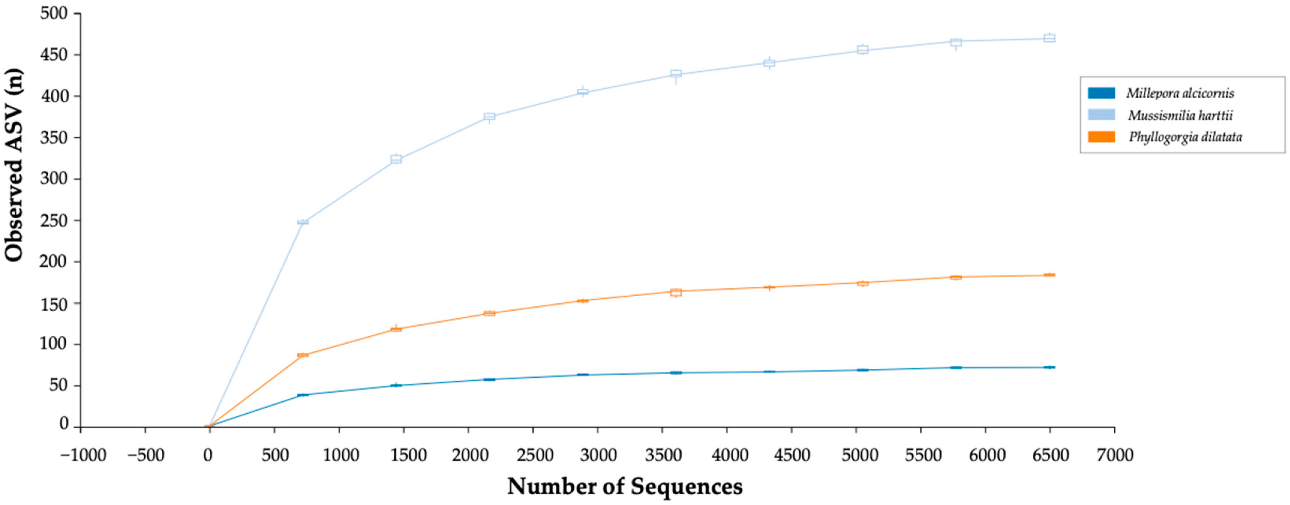
The rarefaction curves of the three microbiotas using observed ASVs reached a plateau and showed a good depth of sample coverage, indicating that the number of reads obtained from sequencing was sufficient to capture most of the species diversity present in these samples (Figure 3).

3.2. Bacterial Composition Analysis

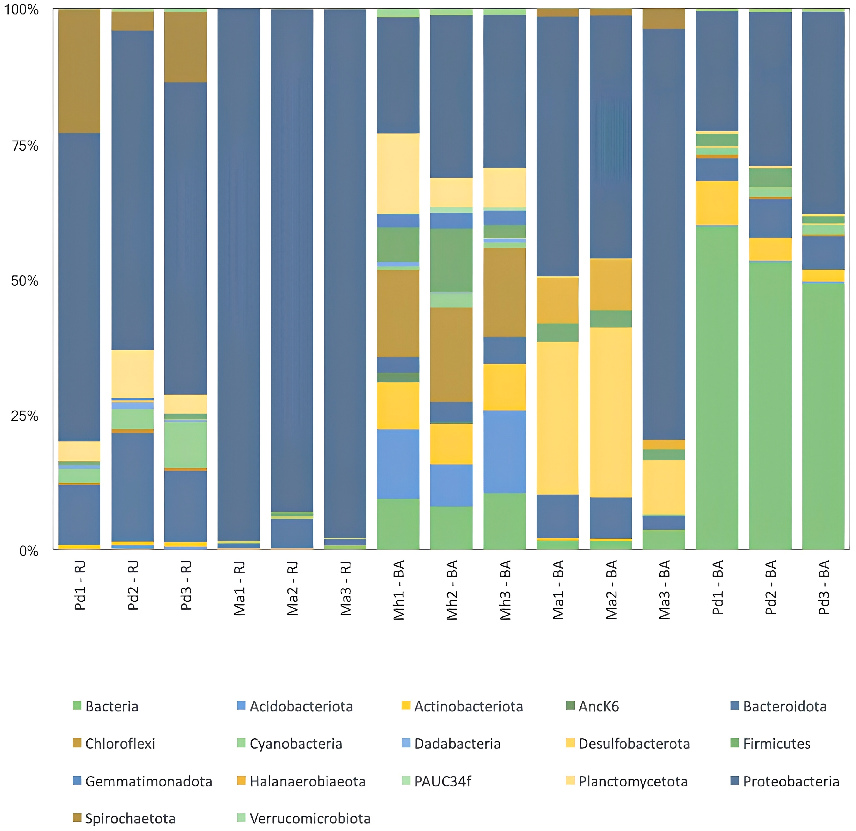
The phylum Proteobacteria presented the highest relative abundance in most corals, except in Phyllogorgia dilatata/BA (Figure 4). This sample presented a high number of sequences that could not be assigned to any phylum, and Proteobacteria presented the second highest relative abundance. However, most of the reads from these samples could not be assigned to either bacteria or Archaea, suggesting that many sequences belong to phyla not yet present in the database used. In contrast, P. dilatata from another sampling site, Búzios, RJ, presented less than 1% of non-assigned sequences. In P. dilatata/RJ Proteobacteria presented the highest relative abundance (55% of sequences), followed by Bacteroidota and Spirochaetota. The Proteobacteria phylum was almost equally divided into Alpha and Gammaproteobacteria classes, and most of the sequences in each class were unassigned to further taxonomic levels. The classified sequence ASVs with higher than 1% of relative abundance belonged to Alphaproteobacteria and were assigned to orders Rhizobiales and Thalassobaculales, families Parvularculaceae and Rhodobacteraceae and genera Pelagibius, Tistlia, Pyruvatibacter, Methyloceanibacter and Roseitalea.
Fragments of Mussismilia hartti/BA presented the most distinct bacterial phylum composition. A total of 16 phyla presented relative abundance over 1% and there was no clear predominance of one phylum. This sample presented relative abundances of 26% Proteobacteria and 16% Chloroflexi, followed by 12% Acidobacteriota. Among the Proteobacteria, the Alphaproteobacteria ASVs with higher that 1% relative abundance were those from the order Rhizobiales, the family Rhodobacteraceae and the genus Pseudovibrio. Only the genus Pseudomonas in the class Gammaprotebacteria fulfilled this criterion. The Cloroflexi phylum presented the classes Anaerolineae, Dehalococcoidia, KD4-96 and TK17, with relative abundances higher than 1%. This phylum also presented the ASV with the highest relative abundance: genus A4b (7.1%). Among the Acidobacteriota, the family Thermoanaerobaculaceae presented the highest ASV with second highest relative abundance (6.3%). M. harttii/BA samples presented Archaea ASVs with higher than 1% of relative abundance. They belonged to the phylum Crenarchaeota, class Nitrososphaeria, a group of ammonia-oxidizing Archaea. These ASVs were also observed in P. dilatata/RJ, P. dilatata/BA and M. alcicornis/BA, but they presented relative abundances below 1%.
Among the samples of M. alcicornis collected in Rio de Janeiro and Bahia, the highest relative abundance of Proteobacteria was also observed. However, the composition of phyla among the samples of this coral from different regions was different. In M. alcicornis/BA, sequences classified as Desulfobacterota, Bacteroidota, Halanaerobiaeota, Firmicutes and Spirochaetota were observed. In contrast, the fragment of M. alcicornis/RJ presented a higher relative abundance of Proteobacteria, followed by Bacteroidota and Firmicutes.
The microbiota associated with the three coral species studied (M. alcicornis, P. dilatata and M. harttii) exhibited both similarities and distinct differences, reflecting the unique environments and physiological characteristics of their hosts. Our studies showed that the bacterial communities associated with M. alcicornis were predominantly composed of the phyla Proteobacteria, both in Rio de Janeiro (over 90%) and in Bahia (between 45 and 76%). In the latter, Desulfobacterota and Halanaerobiaeota phyla were also detected, in contrast to samples from Rio de Janeiro. Similarly, the microbiota of P. dilatata samples collected from Rio de Janeiro and Bahia also included a significant presence of Proteobacteria (57%), followed by Spirochaetota and Bacteroidota. However, samples from Bahia also showed the presence of Actinobacteriota. In contrast, although the microbiota of M. harttii was also dominated by Proteobacteria (between 21% and 29.3%), it showed a more diverse presence of phyla than the other two, including Acidobacteriota (between 7.6% and 15%), Chloroflexi (between 15.7% and 17.1%), Firmicutes (between 2.3% and 11.4%) and Planctomycetota (between 7.1% and 14.2%).

3.3. Diversity Analysis of the Microbiota

Principal coordinates analysis (PCoA) revealed that the bacterial compositions among each coral species replica were highly similar to each other, suggesting that the bacterial composition of each individual coral was stable, likely influenced by the coral’s specific conditions or physiological characteristics (Figure 5). However, there was no evident grouping by species or sampling site. At the phyla level, the relative abundances of bacteria associated with the three coral species showed a significant variation.

4. Discussion

The bacterial microbiome has emerged as a crucial component for the health and adaptation of reef-building corals. Recent studies have highlighted the diversity and complexity of bacterial communities associated with cnidaria species [25,33,34], revealing that these microorganisms play a multifaceted role in the health and resilience of cnidarians [35]. They assist in protection against pathogens, reduce coral bleaching and mortality [33], contribute to coral nutrition through nitrogen fixation [36], aid in the decomposition of organic matter and nutrient cycling, and are involved in the production of secondary metabolites [37,38,39]. Furthermore, the symbiotic relationship between corals and their bacterial microbiome may play a crucial role in coral resistance to thermal stress [40,41]. Therefore, the preservation and continued study of these interactions between corals and their bacterial microbiome are essential for the conservation of these marine ecosystems.
P. dilatata is endemic to the Brazilian coast, and M. harttii has also been categorized as endangered. P. dilatata is recognized for its production of sterols, mono- to tetra-terpenes, conjugated polyenals and peptides [42,43]. While the bacterial composition of M. harttii has been extensively investigated in a few studies, this is the first report of the characterization of the bacterial community associated with M. alcicornis and P. dilatata.
Studies of the coral microbiome point to the existence of a central microbiome among corals of different taxonomic groups. The phylum Proteobacteria was observed to be the most abundant in the bacterial community of Octocorals [44] and Scleractinian corals, although differences in diversity were observed at lower levels (class or order) [45]. It is suggested that the coral microbiome is composed of ubiquitous symbiotic bacteria selected by the host, microorganisms that occupy functional niches and bacteria with a high level of variability between species capable of responding to abiotic and biotic stimuli [11]. The results found for M. alcicornis, M. harttii and P. dilatata also point to Proteobacteria as the most abundant phylum in the analyzed samples.
The samples of M. alcicornis and P. dilatata corals collected in different regions, Bahia and Rio de Janeiro, showed a prevalence of Proteobacteria, although in the samples of P. dilatata collected in Bahia, this phylum was the second most abundant. The same samples collected in Rio de Janeiro and used in this study were also studied regarding the composition of the cultivable bacterial community in our research group. The results found in the cultivable bacterial community of M. alcicornis corroborate the results of the present study, with microorganisms of the Proteobacteria phylum also found in greater numbers among the microorganisms capable of cultivation. Unlike the results found for M. alcicornis, the cultivable bacterial community of P. dilatata presented an abundance of microorganisms of the Firmicutes phylum, with the Bacillus genus being the most distributed among the cultivated microorganisms [38], contrasting with the result found in this study. These findings are related to the cultivation methods used in the laboratory which may not always be able to represent the bacterial community of an environmental sample. This is because certain bacteria require specific nutrients and growth conditions, such as specific temperatures, pH, and oxygen levels. In addition, competition for nutrients, toxin production, or the generation time of certain bacteria can impair the successful growth of some bacterial phylotypes in mixed cultures [46]. This demonstrates the importance of using independent microorganism cultivation techniques to better understand the microbiota composition of these cnidarians.
The composition of the microbiota of corals may be related to several factors, such as changes in water temperature [12], diseases [47], human activity or geographic location. A study carried out with the Caribbean corals Porites astreoides and Montrastrea faveolata collected in three different regions demonstrated the difference in the composition of the bacterial community between the species and between the geographic locations. The results showed that the phylum Proteobacteria was the most abundant in all samples analyzed. However, the distribution of microorganisms belonging to Alphaproteobacteria, Betaproteobacteria and Gammaproteobacteria was different among the coral samples collected in different regions [16]. A similar result can be observed in the composition of the bacterial community of M. alcicornis collected in Bahia and Rio de Janeiro. Although samples collected in both regions showed a predominance of Proteobacteria in their microbiome, the presence of microorganisms from the phyla Desulfobacterota, Spirochaetota and Halanaerobiaeota was observed only in samples from M. alcicornis/BA.
Organisms belonging to the phylum Desulfobacterota have been identified in deep sulfidic habitats in the Black Sea, and have been shown to be terminal oxidizing microorganisms that can consume hydrogen produced during the fermentation of organic matter [48]. Similarly, new species of Acidobacteriota of the class Thermoanaerobaculia, isolated from sediments of sulfidic zones, have demonstrated important participation in the biogeochemical cycles of sulfur [49]. Organisms of this phylum are present in marine sediments worldwide and were found in the microbiome of M. harttii/BA in the present study.
The phylum Proteobacteria was the most abundant in most corals sampled in this study, and the same occurred with M. harttii and other Mussismilia spp. [26,50,51,52]. Members of the phylum Proteobacteria are frequently identified as a significant part of the coral microbiome and play important roles in the health and functionality of coral reefs [35,40,53]. Fernando et al. (2015) analyzed three distinct Mussismilia species of healthy and diseased individuals. They observed that while the diseased Mussismilia showed a predominance of Bacteroidetes (31%) and Gammaproteobacteria (21%) (particularly Vibrio and Thalassomonas), the healthy individuals presented mainly green algal chloroplasts (Ostreobium), followed by Bacterioidetes, Firmicutes and Proteobacteria [54].
When comparing the composition of the bacterial community between M. hartti collected in Recife de Fora and the results described by Fernando et al. (2015), from healthy M. harttii samples in Abrolhos, it is possible to observe a difference in the phyla with greater abundance between samples from the different locations [54]. While the healthy samples from Abrolhos showed a higher abundance of organisms from the phyla Proteobacteria and Bacterioidetes, the samples from Recife de Fora showed a greater diversity of bacterial phyla, with the most abundant sequences related to the phyla Proteobacteria and Chloroflexi, followed by the phylum Acidobacteriota. In addition to these, the phyla Firmicutes, Actnobacteriota and Gemmatimonadota were present in the samples from Recife de Fora. These results may indicate that the bacterial community may present different compositions among corals of the same species, but in different geographic locations, as seen in a study with other coral species [16].
Similar to the bacterial community of Millepora cf. platyphylla (Dubé et al. 2021), M. alcicornis exhibited a microbiome mainly dominated by Proteobacteria, particularly in the samples collected in Búzios, RJ, where over 95% of the sequences belonged to this phylum. In contrast, Millepora cf. platyphylla demonstrated an approximate 51% abundance of Proteobacteria [55].
The genus Candidatus Endoecteinascidia is a putative endosymbiont of the tunicate Ecteinascidia turbinata found in the Caribbean [56] and Mediterranean [57], and known as the source of the anti-cancer agent ET-743 [58]. This candidate genus is reported only in ascidians [59], and currently there is no report of this Endoecteinascidia in any coral microbiota studies or Ecteinascidia turbinata in Brazil.
This study showed an extensive diversity of bacterial phyla hosted by corals, surpassing the number observed in marine water [60]. However there was no indication of a species-specific relationship with respect to bacterial composition. Moreover, the bacterial community is a small piece of the complex microbial community that forms the holobiont. The relationship between the other groups like Symbiodiniaceae, fungi, virus, Archaea, algae and bacteria, in addition to the interaction with the coral host, must be considered [60].
It has been observed that bleaching events can alter the composition of the bacterial community. A study conducted by Bourne et al. (2008) reported changes in the microbiota in colonies of the coral Acropora millepora that were analyzed before, during and after bleaching. A change in the microbial community was observed as the colonies underwent bleaching [61]. In this study, it was possible to correlate the increase in temperature with the emergence of sequences associated with microorganisms of the genus Vibrio, a fact that can be observed even before visual signs of bleaching in the colonies. The study corroborates the idea that corals select their microbiota, since after the bleaching process the microbial community returned to being similar to that observed before this process [61]. Thus, the microbiome of corals can be used as biomarkers of environmental changes and diseases even before the physical changes that these organisms undergo when subjected to environmental stress.

5. Conclusions

Our results give an insight into the growing understanding of bacterial communities of three Brazilian coral species: Millepora alcicornis, Mussismilia harttii and Phyllogorgia dilatata, by a culture-independent method, using 16S rRNA gene sequencing. Samples taken in Bahia and Rio de Janeiro were used to define the bacterial population, and the most prevalent phylum was Proteobacteria. Nonetheless, the phylum Firmicutes was abundant in the cultivable bacterial community of M. alcicornis, with Bacillus being the most prevalent. The majority of the corals that were sampled had the highest abundance of the phylum Proteobacteria, which is important for the wellbeing and operation of coral reefs. Corals were discovered to have a more varied bacterial community than those found in sea water. Our findings underscore the need for continued research on coral–bacterial interactions, emphasizing their importance of the preservation and conservation of marine ecosystems, especially in regions like Brazil, where such studies remain limited.

Author Contributions

R.A.d.C., S.C.D. and C.C.B. conceived, designed and performed the experiments; O.H.B.P. analyzed the data; H.F.d.S., M.W.F., S.A.d.A. and R.A.d.C. wrote and edited the manuscript. We confirm that the manuscript and data are original and have not been previously published or considered elsewhere. All authors have read and agreed to the published version of the manuscript.

Funding

This work was supported by FAP-DF Agro Learning (Project Number: 478/2023; Nota de empenho: 2023NE01498).

Institutional Review Board Statement

Not applicable.

Informed Consent Statement

Not applicable.

Data Availability Statement

Data are contained within the article.

Conflicts of Interest

The authors declare no conflicts of interest.

References

  1. Garcia, T.M.; Matthews-Cascon, H.; Franklin-Junior, W. Millepora Alcicornis (Cnidaria: Hydrozoa) as Substrate for Benthic Fauna. Braz. J. Oceanogr. 2009, 57, 153–155. [Google Scholar] [CrossRef]
  2. Gates, R.D.; Ainsworth, T.D. The Nature and Taxonomic Composition of Coral Symbiomes as Drivers of Performance Limits in Scleractinian Corals. J. Exp. Mar. Biol. Ecol. 2011, 408, 94–101. [Google Scholar] [CrossRef]
  3. Burt, J.A.; Camp, E.F.; Enochs, I.C.; Johansen, J.L.; Morgan, K.M.; Riegl, B.; Hoey, A.S. Insights from Extreme Coral Reefs in a Changing World. Coral Reefs 2020, 39, 495–507. [Google Scholar] [CrossRef]
  4. Blackall, L.L.; Wilson, B.; Van Oppen, M.J.H. Coral—The World’s Most Diverse Symbiotic Ecosystem. Mol. Ecol. 2015, 24, 5330–5347. [Google Scholar] [CrossRef]
  5. Davy, S.K.; Allemand, D.; Weis, V.M. Cell Biology of Cnidarian-Dinoflagellate Symbiosis. Microbiol. Mol. Biol. Rev. 2012, 76, 229–261. [Google Scholar] [CrossRef]
  6. Muller-Parker, G.; D’Elia, C.F.; Cook, C.B. Interactions Between Corals and Their Symbiotic Algae. In Coral Reefs in the Anthropocene; Springer: Dordrecht, The Netherlands, 2015; pp. 99–116. [Google Scholar] [CrossRef]
  7. Goulet, T.L.; Erill, I.; Ascunce, M.S.; Finley, S.J.; Javan, G.T. Conceptualization of the Holobiont Paradigm as It Pertains to Corals. Front. Physiol. 2020, 11, 566968. [Google Scholar] [CrossRef]
  8. Boilard, A.; Dubé, C.E.; Gruet, C.; Mercière, A.; Hernandez-Agreda, A.; Derome, N. Defining Coral Bleaching as a Microbial Dysbiosis within the Coral Holobiont. Microorganisms 2020, 8, 1682. [Google Scholar] [CrossRef]
  9. Stabili, L.; Parisi, M.G.; Parrinello, D.; Cammarata, M. Cnidarian Interaction with Microbial Communities: From Aid to Animal’s Health to Rejection Responses. Mar. Drugs 2018, 16, 296. [Google Scholar] [CrossRef]
  10. Rosenberg, E.; Koren, O.; Reshef, L.; Efrony, R.; Zilber-Rosenberg, I. The Role of Microorganisms in Coral Health, Disease and Evolution. Nat. Rev. Microbiol. 2007, 5, 355–362. [Google Scholar] [CrossRef]
  11. Hernandez-Agreda, A.; Leggat, W.; Bongaerts, P.; Ainsworth, T.D. The Microbial Signature Provides Insight into the Mechanistic Basis of Coral Success across Reef Habitats. mBio 2016, 7, e00560-16. [Google Scholar] [CrossRef]
  12. Littman, R.A.; Bourne, D.G.; Willis, B.L. Responses of Coral-Associated Bacterial Communities to Heat Stress Differ with Symbiodinium Type on the Same Coral Host. Mol. Ecol. 2010, 19, 1978–1990. [Google Scholar] [CrossRef] [PubMed]
  13. Meron, D.; Atias, E.; Iasur Kruh, L.; Elifantz, H.; Minz, D.; Fine, M.; Banin, E. The Impact of Reduced PH on the Microbial Community of the Coral Acropora Eurystoma. ISME J. 2011, 5, 51–60. [Google Scholar] [CrossRef] [PubMed]
  14. Meyer, J.L.; Castellanos-Gell, J.; Aeby, G.S.; Häse, C.C.; Ushijima, B.; Paul, V.J. Microbial Community Shifts Associated With the Ongoing Stony Coral Tissue Loss Disease Outbreak on the Florida Reef Tract. Front. Microbiol. 2019, 10, 2244. [Google Scholar] [CrossRef] [PubMed]
  15. Qin, Z.; Yu, K.; Liang, J.; Yao, Q.; Chen, B. Significant Changes in Microbial Communities Associated With Reef Corals in the Southern South China Sea During the 2015/2016 Global-Scale Coral Bleaching Event. J. Geophys. Res. Oceans 2020, 125, e2019JC015579. [Google Scholar] [CrossRef]
  16. Morrow, K.M.; Moss, A.G.; Chadwick, N.E.; Liles, M.R. Bacterial Associates of Two Caribbean Coral Species Reveal Species-Specific Distribution and Geographic Variability. Appl. Environ. Microbiol. 2012, 78, 6438–6449. [Google Scholar] [CrossRef]
  17. Leão, Z.M.A.N.; Kikuchi, R.K.P.; Ferreira, B.P.; Neves, E.G.; Sovierzoski, H.H.; Oliveira, M.D.M.; Maida, M.; Correia, M.D.; Johnsson, R. Brazilian Coral Reefs in a Period of Global Change: A Synthesis. Braz. J. Oceanogr. 2016, 64, 97–116. [Google Scholar] [CrossRef]
  18. Leão, Z.M.A.N.; Kikuchi, R.K.P.; Testa, V. Corals and Coral Reefs of Brazil. In Latin American Coral Reefs; Elsevier: Amsterdam, The Netherlands, 2003; pp. 9–52. [Google Scholar] [CrossRef]
  19. Leão, Z.M.A.N.; Kikuchi, R.K.P. The Abrolhos Reefs of Brazil. In Coastal Marine Ecosystems of Latin America; Springer: Berlin/Heidelberg, Germany, 2001; pp. 83–96. [Google Scholar] [CrossRef]
  20. Teixeira, C.D.; Leitão, R.L.L.; Ribeiro, F.V.; Moraes, F.C.; Neves, L.M.; Bastos, A.C.; Pereira-Filho, G.H.; Kampel, M.; Salomon, P.S.; Sá, J.A.; et al. Sustained Mass Coral Bleaching (2016–2017) in Brazilian Turbid-Zone Reefs: Taxonomic, Cross-Shelf and Habitat-Related Trends. Coral Reefs 2019, 38, 801–813. [Google Scholar] [CrossRef]
  21. Duarte, G.A.S.; Villela, H.D.M.; Deocleciano, M.; Silva, D.; Barno, A.; Cardoso, P.M.; Vilela, C.L.S.; Rosado, P.; Messias, C.S.M.A.; Chacon, M.A.; et al. Heat Waves Are a Major Threat to Turbid Coral Reefs in Brazil. Front. Mar. Sci. 2020, 7, 515393. [Google Scholar] [CrossRef]
  22. Corazza, B.M.; Lacerda, C.H.F.; Güth, A.Z.; Marcançoli, R.K.M.; Bianchini, A.; Calderon, E.N.; Capel, K.C.C.; Conceição, E.; Faria, S.C.; Francini-Filho, R.B.; et al. No Coral Recovery Three Years after a Major Bleaching Event in Reefs in the Southwestern Atlantic Refugium. Mar. Biol. 2024, 171, 114. [Google Scholar] [CrossRef]
  23. Li, J.; Yang, Q.; Dong, J.; Sweet, M.; Zhang, Y.; Liu, C.; Zhang, Y.; Tang, X.; Zhang, W.; Zhang, S. Microbiome Engineering: A Promising Approach to Improve Coral Health. Engineering 2023, 28, 105–116. [Google Scholar] [CrossRef]
  24. Thompson, J.R.; Rivera, H.E.; Closek, C.J.; Medina, M. Microbes in the Coral Holobiont: Partners through Evolution, Development, and Ecological Interactions. Front. Cell Infect. Microbiol. 2015, 4, 176. [Google Scholar] [CrossRef] [PubMed]
  25. Vilela, C.L.S.; Villela, H.D.M.; da Costa Rachid, C.T.C.; Do Carmo, F.L.; Vermelho, A.B.; Peixoto, R.S. Exploring the Diversity and Biotechnological Potential of Cultured and Uncultured Coral-Associated Bacteria. Microorganisms 2021, 9, 2235. [Google Scholar] [CrossRef] [PubMed]
  26. De Castro, A.P.; Araújo, S.D.; Reis, A.M.M.; Pompeu, M.; Hatay, M.; de Moura, R.L.; Francini-Filho, R.B.; Thompson, F.L.; Krüger, R.H. Bacterial Communities Associated with Three Brazilian Endemic Reef Corals (Mussismilia spp.) in a Coastal Reef of the Abrolhos Shelf. Cont. Shelf Res. 2013, 70, 135–139. [Google Scholar] [CrossRef]
  27. Rosser, N.L.; Thomas, L.; Stankowski, S.; Richards, Z.T.; Kennington, W.J.; Johnson, M.S. Phylogenomics Provides New Insight into Evolutionary Relationships and Genealogical Discordance in the Reef-Building Coral Genus Acropora. Proc. R. Soc. B Biol. Sci. 2017, 284, 20162182. [Google Scholar] [CrossRef]
  28. Leite, D.C.A.; Leão, P.; Garrido, A.G.; Lins, U.; Santos, H.F.; Pires, D.O.; Castro, C.B.; van Elsas, J.D.; Zilberberg, C.; Rosado, A.S.; et al. Broadcast Spawning Coral Mussismilia Hispida Can Vertically Transfer Its Associated Bacterial Core. Front. Microbiol. 2017, 8, 239169. [Google Scholar] [CrossRef]
  29. Andrade Galvão de Medeiros, T.; Seoane, J.C.S.; Macedo de Mello Baptista, G.; Leal, P.R.; Dekker, A. Effect of Temperature and PH on the Millepora Alcicornis and Mussismilia Harttii Corals in Light of a Spectral Reflectance Response. Int. J. Remote Sens. 2022, 43, 2475–2502. [Google Scholar] [CrossRef]
  30. Bolyen, E.; Rideout, J.R.; Dillon, M.R.; Bokulich, N.A.; Abnet, C.C.; Al-Ghalith, G.A.; Alexander, H.; Alm, E.J.; Arumugam, M.; Asnicar, F.; et al. Reproducible, Interactive, Scalable and Extensible Microbiome Data Science Using QIIME 2. Nat. Biotechnol. 2019, 37, 852–857. [Google Scholar] [CrossRef]
  31. Callahan, B.J.; McMurdie, P.J.; Rosen, M.J.; Han, A.W.; Johnson, A.J.A.; Holmes, S.P. DADA2: High-Resolution Sample Inference from Illumina Amplicon Data. Nat. Methods 2016, 13, 581–583. [Google Scholar] [CrossRef]
  32. Bokulich, N.A.; Kaehler, B.D.; Rideout, J.R.; Dillon, M.; Bolyen, E.; Knight, R.; Huttley, G.A.; Gregory Caporaso, J. Optimizing Taxonomic Classification of Marker-Gene Amplicon Sequences with QIIME 2’s Q2-Feature-Classifier Plugin. Microbiome 2018, 6, 90. [Google Scholar] [CrossRef]
  33. Doering, T.; Tandon, K.; Topa, S.H.; Pidot, S.J.; Blackall, L.L.; van Oppen, M.J.H. Genomic Exploration of Coral-Associated Bacteria: Identifying Probiotic Candidates to Increase Coral Bleaching Resilience in Galaxea Fascicularis. Microbiome 2023, 11, 185. [Google Scholar] [CrossRef]
  34. Ziegler, M.; Seneca, F.O.; Yum, L.K.; Palumbi, S.R.; Voolstra, C.R. Bacterial Community Dynamics Are Linked to Patterns of Coral Heat Tolerance. Nat. Commun. 2017, 8, 14213. [Google Scholar] [CrossRef] [PubMed]
  35. McCauley, M.; Goulet, T.L.; Jackson, C.R.; Loesgen, S. Systematic Review of Cnidarian Microbiomes Reveals Insights into the Structure, Specificity, and Fidelity of Marine Associations. Nat. Commun. 2023, 14, 4899. [Google Scholar] [CrossRef]
  36. Raina, J.B.; Tapiolas, D.; Willis, B.L.; Bourne, D.G. Coral-Associated Bacteria and Their Role in the Biogeochemical Cycling of Sulfur. Appl. Environ. Microbiol. 2009, 75, 3492–3501. [Google Scholar] [CrossRef] [PubMed]
  37. Macedo, M.W.F.S.; Cunha, N.B.; Carneiro, J.A.; da Costa, R.A.; de Alencar, S.A.; Cardoso, M.H.; Franco, O.L.; Dias, S.C. Marine Organisms as a Rich Source of Biologically Active Peptides. Front. Mar. Sci. 2021, 8, 667764. [Google Scholar] [CrossRef]
  38. Macedo, M.W.F.S.; Costa, R.A.; de Lima, R.G.; Gomes, J.E.M.; Rocha, G.I.Y.; dos Santos, H.F.; Dias, S.C.; da Cunha, N.B. Bacterial Diversity Associated with Millepora Alcicornis and Phyllogorgia Dilatata Corals and Prospection for Genes Encoding Bioactive Molecules. Reg. Stud. Mar. Sci. 2023, 59, 102811. [Google Scholar] [CrossRef]
  39. Ritchie, K.B. Bacterial Symbionts of Corals and Symbiodinium. In Beneficial Microorganisms in Multicellular Life Forms; Springer: Berlin/Heidelberg, Germany, 2012; pp. 139–150. [Google Scholar] [CrossRef]
  40. Glasl, B.; Webster, N.S.; Bourne, D.G. Microbial Indicators as a Diagnostic Tool for Assessing Water Quality and Climate Stress in Coral Reef Ecosystems. Mar. Biol. 2017, 164, 91. [Google Scholar] [CrossRef]
  41. Tignat-Perrier, R.; van de Water, J.A.J.M.; Allemand, D.; Ferrier-Pagès, C. Holobiont Responses of Mesophotic Precious Red Coral Corallium Rubrum to Thermal Anomalies. Environ. Microbiome 2023, 18, 70. [Google Scholar] [CrossRef]
  42. Almeidaa, M.T.R.; Moritz, M.I.G.; Capel, K.C.C.; Pérez, C.D.; Schenkel, E.P. Chemical and Biological Aspects of Octocorals from the Brazilian Coast. Rev. Bras. Farmacogn. 2014, 24, 446–467. [Google Scholar] [CrossRef]
  43. Maia, L.F.; Fernandes, R.F.; Almeida, M.R.; de Oliveira, L.F.C. Rapid Assessment of Chemical Compounds from Phyllogorgia Dilatata Using Raman Spectroscopy. Rev. Bras. Farmacogn. 2015, 25, 619–626. [Google Scholar] [CrossRef]
  44. Van De Water, J.A.J.M.; Allemand, D.; Ferrier-Pagès, C. Host-Microbe Interactions in Octocoral Holobionts—Recent Advances and Perspectives. Microbiome 2018, 6, 64. [Google Scholar] [CrossRef]
  45. Xu, M.; Cheng, K.; Xiao, B.; Tong, M.; Cai, Z.; Jong, M.-C.; Chen, G.; Zhou, J. Bacterial Communities Vary from Different Scleractinian Coral Species and between Bleached and Non-Bleached Corals. Microbiol. Spectr. 2023, 11, e0491022. [Google Scholar] [CrossRef] [PubMed]
  46. Vartoukian, S.R.; Palmer, R.M.; Wade, W.G. Strategies for Culture of “unculturable” Bacteria. FEMS Microbiol. Lett. 2010, 309, 1–7. [Google Scholar] [CrossRef] [PubMed]
  47. Pantos, O.; Cooney, R.P.; Le Tissier, M.D.A.; Barer, M.R.; O’Donnell, A.G.; Bythell, J.C. The Bacterial Ecology of a Plague-like Disease Affecting the Caribbean Coral Montastrea Annularis. Environ. Microbiol. 2003, 5, 370–382. [Google Scholar] [CrossRef] [PubMed]
  48. Yadav, S.; Koenen, M.; Bale, N.J.; Reitsma, W.; Engelmann, J.C.; Stefanova, K.; Damsté, J.S.S.; Villanueva, L. Organic Matter Degradation in the Deep, Sulfidic Waters of the Black Sea: Insights into the Ecophysiology of Novel Anaerobic Bacteria. Microbiome 2024, 12, 98. [Google Scholar] [CrossRef]
  49. Flieder, M.; Buongiorno, J.; Herbold, C.W.; Hausmann, B.; Rattei, T.; Lloyd, K.G.; Loy, A.; Wasmund, K. Novel Taxa of Acidobacteriota Implicated in Seafloor Sulfur Cycling. ISME J. 2021, 15, 3159–3180. [Google Scholar] [CrossRef]
  50. de Castro, A.P.; Araújo, S.D.; Reis, A.M.M.; Moura, R.L.; Francini-Filho, R.B.; Pappas, G.; Rodrigues, T.B.; Thompson, F.L.; Krüger, R.H. Bacterial Community Associated with Healthy and Diseased Reef Coral Mussismilia Hispida from Eastern Brazil. Microb. Ecol. 2010, 59, 658–667. [Google Scholar] [CrossRef]
  51. Reis, A.M.M.; Araújo, S.D.; Moura, R.L.; Francini-Filho, R.B.; Pappas, G.; Coelho, A.M.A.; Krüger, R.H.; Thompson, F.L. Bacterial Diversity Associated with the Brazilian Endemic Reef Coral Mussismilia Braziliensis. J. Appl. Microbiol. 2009, 106, 1378–1387. [Google Scholar] [CrossRef]
  52. Vilela, C.L.S.; Villela, H.D.M.; Duarte, G.A.S.; Santoro, E.P.; Rachid, C.T.C.C.; Peixoto, R.S. Estrogen Induces Shift in Abundances of Specific Groups of the Coral Microbiome. Sci. Rep. 2021, 11, 2767. [Google Scholar] [CrossRef]
  53. Bayer, T.; Neave, M.J.; Alsheikh-Hussain, A.; Aranda, M.; Yum, L.K.; Mincer, T.; Hughen, K.; Apprill, A.; Voolstra, C.R. The Microbiome of the Red Sea Coral Stylophora Pistillata Is Dominated by Tissue-Associated Endozoicomonas Bacteria. Appl. Environ. Microbiol. 2013, 79, 4759–4762. [Google Scholar] [CrossRef]
  54. Fernando, S.C.; Wang, J.; Sparling, K.; Garcia, G.D.; Francini-Filho, R.B.; de Moura, R.L.; Paranhos, R.; Thompson, F.L.; Thompson, J.R. Microbiota of the Major South Atlantic Reef Building Coral Mussismilia. Microb. Ecol. 2015, 69, 267–280. [Google Scholar] [CrossRef]
  55. Dubé, C.E.; Ziegler, M.; Mercière, A.; Boissin, E.; Planes, S.; Bourmaud, C.A.F.; Voolstra, C.R. Naturally Occurring Fire Coral Clones Demonstrate a Genetic and Environmental Basis of Microbiome Composition. Nat. Commun. 2021, 12, 6402. [Google Scholar] [CrossRef] [PubMed]
  56. Pérez-Matos, A.E.; Rosado, W.; Govind, N.S. Bacterial Diversity Associated with the Caribbean Tunicate Ecteinascidia Turbinata. Antonie Leeuwenhoek 2007, 92, 155–164. [Google Scholar] [CrossRef]
  57. Moss, C.; Green, D.H.; Pérez, B.; Velasco, A.; Henríquez, R.; McKenzie, J.D. Intracellular Bacteria Associated with the Ascidian Ecteinascidia Turbinata: Phylogenetic and in Situ Hybridisation Analysis. Mar. Biol. 2003, 143, 99–110. [Google Scholar] [CrossRef]
  58. Rath, C.M.; Janto, B.; Earl, J.; Ahmed, A.; Hu, F.Z.; Hiller, L.; Dahlgren, M.; Kreft, R.; Yu, F.; Wolff, J.J.; et al. Meta-Omic Characterization of the Marine Invertebrate Microbial Consortium That Produces the Chemotherapeutic Natural Product ET-743. ACS Chem. Biol. 2011, 6, 1244–1256. [Google Scholar] [CrossRef] [PubMed]
  59. Erwin, P.M.; Pineda, M.C.; Webster, N.; Turon, X.; López-Legentil, S. Down under the Tunic: Bacterial Biodiversity Hotspots and Widespread Ammonia-Oxidizing Archaea in Coral Reef Ascidians. ISME J. 2014, 8, 575–588. [Google Scholar] [CrossRef]
  60. Mohamed, A.R.; Ochsenkühn, M.A.; Kazlak, A.M.; Moustafa, A.; Amin, S.A. The Coral Microbiome: Towards an Understanding of the Molecular Mechanisms of Coral-Microbiota Interactions. FEMS Microbiol. Rev. 2023, 47, fuad005. [Google Scholar] [CrossRef]
  61. Bourne, D.; Iida, Y.; Uthicke, S.; Smith-Keune, C. Changes in Coral-Associated Microbial Communities during a Bleaching Event. ISME J. 2008, 2, 350–363. [Google Scholar] [CrossRef]
Figure 1. A geographical map showing the sampling sites for the three corals in the Brazilian states of Rio de Janeiro (Armação dos Búzios) on the left, and Bahia (Recife de Fora) on the right. The static map of both locations displayed was requested from Google Maps.
Jmse 13 00358 g001
Figure 2. Corals collected from both sites (Phyllogorgia dilatata and Millepora alcicornis) and only from Recife de Fora (Mussismilia harttii).
Jmse 13 00358 g002
Figure 3. Rarefaction curve for the three coral microbiomes studied using observed amplicon sequence variants (ASVs).
Jmse 13 00358 g003
Figure 4. Bacterial communities showing phyla with relative abundance above 1% in at least one sample. Pd-J: Phyllogorgia dilatata/Rio de Janeiro; Ma-RJ: Millepora alcicornis/Rio de Janeiro; Mh-Mussismilia harttii/Bahia; Ma-BA: Millepora alcicornis/Bahia; Pd-BA: Phyllogorgia dilatata/Bahia.
Jmse 13 00358 g004
Figure 5. Principal coordinates analysis (PCoA) showing bacterial community dissimilarity among coral samples: Phyllogorgia dilatata/Rio de Janeiro; Millepora alcicornis/Rio de Janeiro; Mussismilia harttii/Bahia; Millepora alcicornis/Bahia and Phyllogorgia dilatata/Bahia.
Jmse 13 00358 g005
Table 1. Number of sequence reads obtained from Illumina sequencing of five coral samples and percentage loss after the removal of low quality bases.
SpeciesTotal Num. of ReadsTotal Num. Processed Reads% Data Filtered
Phyllogorgia dilatata (Búzios)274,442263,3724.0
Millepora alcicornis (Rio de Janeiro)24,475226,5417.4
Phyllogorgia dilatata (Bahia)27,493266,1443.2
Millepora alcicornis (Bahia)24,33723,5253.3
Mussismilia harttii (Bahia)229,776223,5612.7
Disclaimer/Publisher’s Note: The statements, opinions and data contained in all publications are solely those of the individual author(s) and contributor(s) and not of MDPI and/or the editor(s). MDPI and/or the editor(s) disclaim responsibility for any injury to people or property resulting from any ideas, methods, instructions or products referred to in the content.