Article

Lipopeptides Produced by Bacillus mojavensis P1709 as an Efficient Tool to Maintain Postharvest Cherry Tomato Quality and Quantity

Department of Applied Ecology, Institute of Environmental Sciences, Kazan Federal University, Kazan 420008, Russia
*
Author to whom correspondence should be addressed.
Agriculture 2022, 12(5), 609; https://doi.org/10.3390/agriculture12050609
Submission received: 11 February 2022 / Revised: 10 April 2022 / Accepted: 22 April 2022 / Published: 26 April 2022
(This article belongs to the Section Crop Protection, Diseases, Pests and Weeds)

Abstract

:
Because of significant yield losses caused by fungal pathogens, new efficient and environmentally safe methods of pest control are needed, and amphiphilic compounds (biosurfactants) produced by many microbes are considered a good alternative. In the present study, biosurfactants produced by the rhizosphere isolate Bacillus mojavensis P1709 were assessed for their ability to protect postharvest cherry tomatoes from decay and mycotoxin contamination caused by Fusarium oxysporum f. sp. lycopersici. It was demonstrated that the genome of B. mojavensis P1709 contained the fenD and srfAA genes, which are responsible for the synthesis of compounds of the fengycin and surfactin families. APF (acid-precipitated fraction) of B. mojavensis P1709 culture medium at a concentration of 20 g L−1 inhibited pathogen radial growth on agar plates by 93%, and T-2 and HT-2 mycotoxin production by 98%, after 5 days of cultivation. APF also suppressed fungal growth in the in vivo test on cherry tomato fruits by 93% and 25% on the 2nd and 7th days of incubation, respectively. The results obtained demonstrate that biosurfactants produced by B. mojavensis P1709 are an efficient tool for protecting postharvest cherry tomatoes from fungal mold decay and mycotoxin contamination.

1. Introduction

Between 10% and 40% of the yield before and after harvesting is estimated to be lost because of plant pathogens, especially those of fungal origin [1,2]. To inhibit their growth and active development, synthetic fungicides are widely used [3]. These have many negative consequences, such as soil and food contamination and a decrease in soil biodiversity. In addition, fungal pathogens develop mechanisms to protect themselves from fungicides, and therefore, higher amounts of currently used fungicides or the production of new fungicides are required [3,4].
Many studies have been dedicated to alternative methods of controlling plant pests, and biological control methods are considered promising. Biological control agents include biopesticides, in particular fungicides, bactericides and other compounds, produced by microorganisms or the microorganisms themselves [5]. They are reported to be efficient but also safe for future consumers; unlike chemical fungicides, biofungicides do not degrade to toxins and persistent compounds in food or the environment [6]. Moreover, biosurfactant molecules are now widely used in the pharmaceutical industry as antioxidants, antimicrobial and anticancer agents, etc. [7]. Therefore, interest in biocontrol agents is especially high for crop treatment at the end of the vegetation season and for food and feed treatment after harvesting [8]. In addition, biocontrol agents allow the quality of harvested fruits and vegetables to be maintained since they prevent the formation of toxic fungal metabolites during storage [9].
Biosurfactants are amphiphilic compounds produced by many microbes, bacteria, yeast, and fungi. In agriculture, biosurfactants may be used to control plant diseases and to promote plant growth. Biosurfactants include compounds of various chemical structures and lengths, and the most well studied are classes of rhamnolipids and lipopeptides produced by bacteria of the Pseudomonas and Bacillus genera, respectively [10,11,12]. Typically, microbes produce a complex mixture of different biosurfactants, and the individual structural variances of those surfactants cause different surface-active and bioactive characteristics of the whole mixture [13]. Thus, lipopeptides produced by Bacillus spp. include the surfactin, fengycin, and iturin families and other compounds in lesser amounts and frequency. Bacteria produce those compounds for various purposes that have not yet been fully described. Surfactins stimulate biofilm formation of rhizosphere bacteria and thereby enhance beneficial microbial colonization of plant roots; in addition, they promote plant growth and elicit the production of plant defense proteins. Furthermore, surfactins possess direct antibacterial toxicity. Fengycins and iturins are produced by Bacilli to directly suppress their predators and competitors, and they are highly toxic toward both fungi and bacteria. They also play a role in plant–microbial interactions, including plant defense induction [14,15,16,17,18]. Recently, lipopeptides have been of great practical interest because they are efficient and can be produced in large-scale bioreactors in amounts of 1 to 10–12 g L−1 [19].
Tomato is considered one of the most important crops worldwide; it can be consumed fresh and in various prepared meals [20,21]. A significant ratio of tomato yields is lost due to fungal pathogens, both in the process of harvesting and in the process of postharvest fruit storage. Botrytis cinerea, Alternaria alternata, and Fusarium oxysporum are reported to be the main pathogens causing postharvest losses of cherry tomatoes [20,21,22]. In addition to tomato fruit wilting and decay from mold, pathogenic fungi cause contamination of the fruit with their toxic secondary metabolites, mycotoxins, making the final product dangerous for consumer health [22,23]. Fusarium species are reported to produce the three most important classes of mycotoxins, namely, trichothecenes, zearalenone, and fumonisins [24]. Trichothecenes are a large class that includes important and highly dangerous compounds such as deoxynivalenol, and T-2 and HT-2 toxins. Trichothecenes inhibit protein synthesis and cause oxidative damage to cells, followed by the disruption of nucleic acid synthesis and ensuing apoptosis. They cause growth retardation, neuroendocrine changes, immunotoxicity, anorexia and other severe diseases [25]. Previously, T-2 toxin, its metabolite HT-2 toxin, and neosolaniol, belonging to the trichothecenes class, were revealed on decaying tomato fruit infected with Fusarium sp. [26]. Zearalenone is predominantly produced by Fusarium graminearum and, to a lesser extent, by other species of Fusarium. Zearalenone is able to bioaccumulate in animal and human bodies by binding to the estrogen receptors. Zearalenone causes hormonal imbalances and disorders of the reproductive system. Fumonisins are typical mycotoxins produced by Fusarium moniliforme and Fusarium proliferatum. The mechanisms of their toxicity in humans and animals have not been revealed; however, it was demonstrated that these compounds interfere with sphingolipid metabolism [25].
When attacked by a phytopathogen, plants, including harvested fruits, overcome several physiological and biochemical changes that activate the multicomponent complicated system of the plant immune defense system [27]. Among other components, this system includes the synthesis of defense-related enzymes such as chitinase, catalase, peroxidase, phenylalanine ammonialyase, and polyphenoloxidase. The defense enzymes are involved in the production and polymerization of phenolics, and in lignification, which prevent disease spread, the degradation of pathogen cell walls, and various other processes that inhibit pathogen growth or activity [28,29]. It was reported that the levels of defense-related enzymes play a crucial role in host resistance [30]. The enhancement of corresponding gene expression occurs due to signaling compounds such as salicylic acid, xylanase, ethylene, and polypeptide systems [31]. These compounds may be endogenic and exogenic, e.g., produced by plant symbiotic bacteria. The treatment of plants and harvested crops, fruits, and vegetables by these compounds or compound-producing microbes may be an efficient strategy for pathogen biocontrol. Such a treatment was demonstrated to be successful for tomato plants and harvested fruits attacked by fungal pathogens. Thus, Asanka Indunil Kumari and Vengadaramana (2017) reported that salicylic, ascorbic, and jasmonic acids, and H2O2 and ethanol, induce chitinase, peroxidase, phenylalanine ammonialyase, and polyphenol oxidase production in tomato plant leaf tissues [28], whereas Raynaldo and coauthors (2021) revealed the induction of polyphenol oxidase, peroxidase, catalase, and phenylalanine ammonialyase in postharvest cherry tomatoes with gray mold decay caused by Botrytis cinerea when the fruits were treated with Wickerhamomyces anomalus [21]. In their review, Crouzet and coauthors (2020) described several studies demonstrating that biosurfactants of lipopeptide and rhamnolipid classes are able to stimulate the plant immune system. However, the mechanisms of such stimulation are unclear. Most likely, biosurfactants activate defense enzyme synthesis not directly but rather through plant plasma membrane disturbances by initiating a cascade of molecular events leading to that outcome [4]. It is important to emphasize that the ability of the biosurfactant to induce the plant immune response strongly depends on its specific chemical structure, including carbon chain length and number of rings. Therefore, this ability should be individually assessed for each specific biosurfactant and each biosurfactant producer [32,33].
To solve the problem of postharvest tomato losses and mycotoxin contamination, different chemical and physical treatments in the process of storage are used. Treatment of tomatoes with biosurfactants is a good alternative since they can not only efficiently suppress phytopathogens or activate the plant immune system, but are also safe for consumption. However, the information concerning biosurfactant usage for postharvest tomatoes is still poor in the scientific literature. The objective of the present study was to obtain and characterize the biosurfactants produced by the B. mojavensis P1709 strain previously isolated from the plant rhizosphere, to check the antifungal activity of biosurfactants produced by this strain, and to assess defense-related enzyme production in cherry tomatoes after treatment with biosurfactants.

2. Materials and Methods

2.1. Biosurfactant Producing Bacteria

Biosurfactant-producing strain Bacillus mojavensis P1709 was obtained from the collection of the Institute of Environmental Sciences of Kazan Federal University (Russia). The strain was previously isolated from the rhizosphere soil of Lactuca sativa.

2.2. Emulsification Index Assessment

The emulsification index (E24) of the isolate was estimated according to methods previously outlined [34]. Briefly, the isolate was cultivated on Bushnell Hass (BH) medium with 1% glucose for 72 h at 30 °C and 180 rpm. The BH medium contained (g L−1): KH2PO4 (1.0), K2HPO4 (1.0), NH4NO3 (1.0), MgSO4·7H2O (0.2), FeCl3 (0.05), and CaCl2 (0.02). After cultivation, the culture was centrifuged at 8000 rpm for 20 min to obtain cell-free supernatant. Cell-free supernatant was mixed with crude oil (1:1 v/v) in a 15 mL experimental tube by vortexing, and the height of the emulsified layer was evaluated in relation to the total height of the liquid column in the tube.

2.3. Identification of Genes Responsible for Lipopeptide Synthesis

The ability of the bacterial strain to synthesize fengycin, iturin and surfactin was analyzed using specific primers for the fenD, ituC, and srfAA genes (Table 1).
DNA was extracted from the pellet of the overnight culture after centrifugation using a FastDNA™ SPIN Kit for Soil (MP Biomedicals, Santa Ana, CA, USA) according to the manufacturer’s instructions. PCR was carried out in a total volume of 20 μL that contained 1× PCR buffer, 0.2 mM dNTPs, 1 μM of each primer, 2 mM MgCl2, 1.25 U Taq DNA polymerase, and 1 μM of DNA. The amplification was carried out in a thermal MyCycler (Bio-Rad Laboratories, Inc., Hercules, CA, USA) using the following program: denaturation at 94 °C for 2 min; 35 cycles of 94 °C for 30 s 58 °C for 1 min, and extension at 72 °C for 1.5 min; and a final extension at 72 °C for 7 min [15,36,37]. The presence and approximate length of the amplicons were estimated using agarose gel electrophoresis with a 1000 bp ladder.

2.4. Biosurfactant Production, Extraction and Purification

For increased biosurfactant production, strain P1709 was cultivated in glycerol nitrate medium at 35 °C and 180 rpm for 6 days. The concentration of crude glycerol in the medium was 40 g L−1. The medium except crude glycerol contained (g L−1): NaNO3 (4.0), K2HPO4·3H2O (4.0), KH2PO4 (3.0), MgSO4·7H2O (0.5), KCl (0.5), NaCl (0.5), CaCl2·2H2O (0.2). Then, the mixture of crude biosurfactants produced by the strain was extracted using acid precipitation and purified as described below. As a result, the acid precipitated fraction (APF) was obtained and used for further analyses.
The APF was obtained according to the literature [38,39,40]. Briefly, after cultivation, the culture was centrifuged at 8000 rpm for 20 min. The cell free supernatant was adjusted to pH 2 using 2N HCl, incubated overnight at 4 °C, and centrifuged at 10,000 rpm and 4 °C for 20 min. The precipitate was purified by dissolving in a CHCl3:CH3OH (2:1, v/v) mixture followed by rotary evaporating under vacuum. The resulting yield of the APF was 5.3 g from 1 L of cell-free supernatant.

2.5. Chemical Characterization of the APF

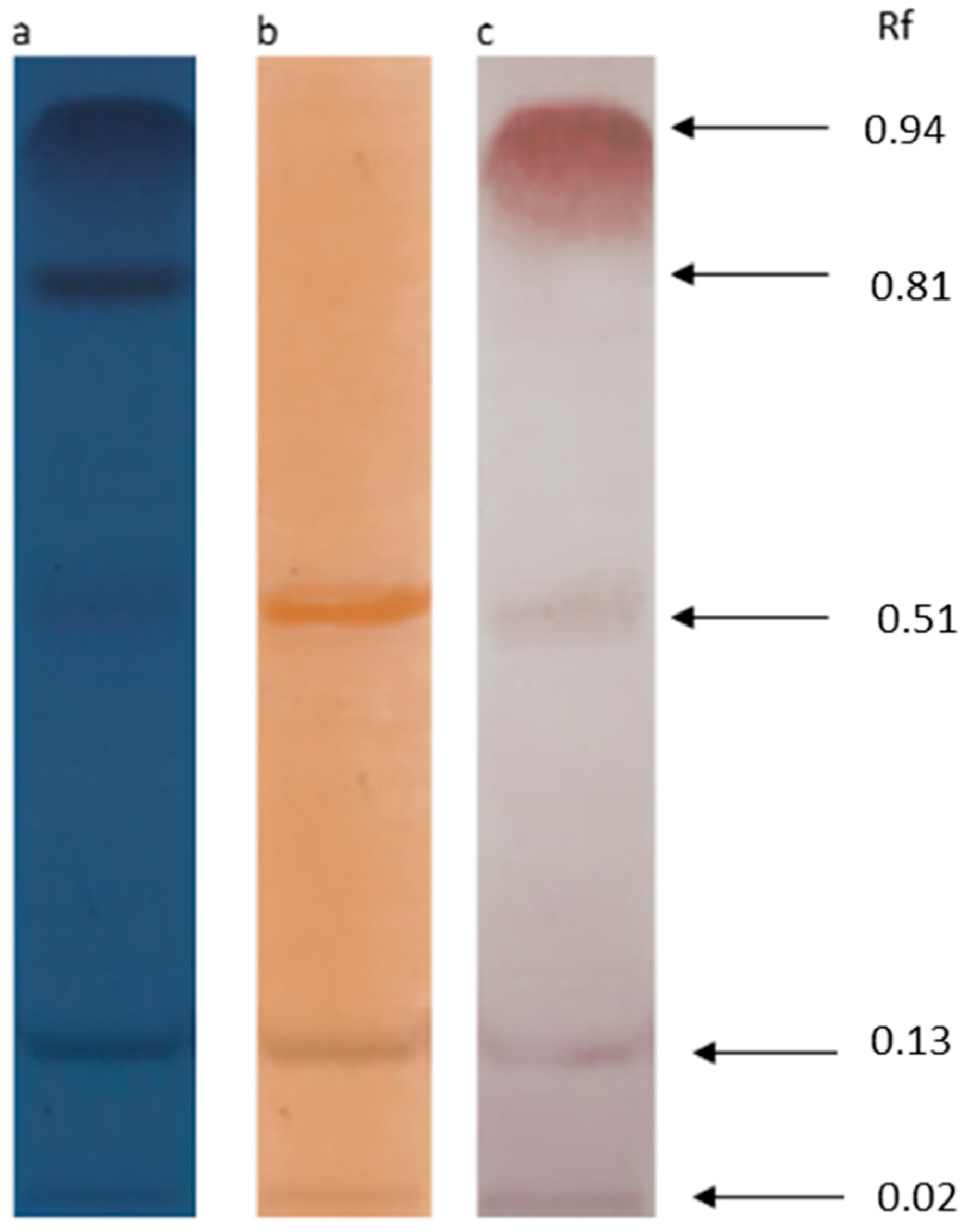
2.5.1. Thin Layer Chromatography (TLC) with Following Staining

To assess the composition of the biosurfactants obtained, the APF was dissolved in CHCl3:CH3OH (1:1 v/v) and spotted on a silica gel plate (G60, Merck, Darmstadt, Germany). Thin layer chromatography (TLC) was conducted on the CAMAG system (Switzerland). A mixture of chloroform:methanol:water (65:25:4, v/v/v) was used as the mobile phase. After separation in the silica gel, the spots on the TLC plate were revealed under UV light, and the Rf values of the spots were calculated. Detection of the main components was achieved by exposure to iodine vapor, p-anisaldehyde, and 1% ninhydrin solution, for staining of lipids, sugars, and free amino groups, respectively. The reagents were sprayed followed by heating at 110 °C until the detection of the definite spots [21,41]. Pure surfactin (≥98%), fengycin (≥90%), and iturin A (≥95%) (Sigma Aldrich, Burlington, MA, USA) were used as standards on the TLC plate.

2.5.2. Fourier Transform Infrared Spectroscopy (FTIR)

To determine the type and structure of APF, FTIR was performed using a Lumos I instrument (Bruker, Billerica, MA, USA). The spectra were collected from wavenumbers 600 to 4000 (cm−1).

2.6. Effects of Biosurfactants on Radial Fungal Growth and Mycotoxin Production In Vitro

Fusarium oxysporum f. sp. lycopersici obtained from the collection of the Department of Biochemistry and Biotechnology of Kazan Federal University (Russia) was used as a test fungal pathogen in this study. To collect the spores, the fungus was cultivated on potato dextrose agar (0.4% w/v potato extract, 2% w/v glucose, 1.5% w/v agar) for 120 h at 28 ± 2 °C [42]. After incubation, the plates were washed with sterile water, and the last was thereafter filtered through four layers of cheesecloth. Spore concentration was measured using a Scepter Handheld Automated Cell Counter (Millipore, Burlington, MA, USA). For further experiments, a spore concentration was adjusted to 1 × 10⁶ mL−1.
The antifungal activity was estimated as described by Caulier, et al. (2019) [11]. Briefly, malt extract agar (2% w/v malt extract, 0.1% w/v peptone, 2% w/v glucose) was pooled into Petri dishes (90 mm) and supplemented with APF to adjust the concentrations of 0.5, 2, 5, 10, and 20 g L−1 [43]. Ten microliters of spore suspension (1 × 10⁶ spores mL−1) were added to the center of the agar plates. The plates were incubated for 5 days at 25 °C. The diameter of the growth zone was measured and compared with that of the control Petri dishes (with no addition of biosurfactants).
To assess the presence and concentration of T-2 and HT-2 mycotoxins, the content of each Petri dish covered by the fungus was visually estimated and cut with a medical scalpel. The resulting substance containing the fungus and the agar medium was homogenized using a mixer mill MM 400 (Retsch, Haan, Germany) for 2 min at maximum speed. The homogenous substance obtained was analyzed using the enzyme immunoassay kit manufactured by RIDASCREEN® (R-Biopharm, Darmstadt, Germany) according to the provided instruction. The immunoassay results were estimated using a Multiskan FC microplate photometer (Thermo Fisher Scientific, Waltham, MA, USA). The total concentration of the two mycotoxins was expressed in µg per gram of homogenized substance. Further, the inhibition of the mycotoxins’ production was calculated comparing the concentration of the toxins in the Petri dishes treated with APF with that in the control dishes (without APF).

2.7. Effects of Biosurfactants on Fungal Growth on Cherry Tomatoes In Vivo

Fusarium oxysporum f. sp. lycopersici spores were obtained as described above. Cherry tomatoes (Solanum lycopersicum var. cerasiforme) that were 2–3 cm long and 1.0–1.5 cm in diameter, grown in the greenhouse of Kazan Federal University with no apparent diseases or visible wounds, were picked for the experiment. Fruits were surface-sterilized with 75% ethanol for 30 s, rinsed three times with sterile distilled water for 60 s, and then air-dried under sterile conditions [20]. Uniform wounds (3 mm diameter and 2 mm depth) were inflicted on each cherry tomato fruit with a sterile metal tube. Ten microliters of APF at different concentrations (0.5, 2, 5, 10, 20 g L−1) was added to each wound under sterile conditions. After 2 h, 10 µL of spore suspension of F. oxysporum (106 spores mL−1) (experimental variants) or 10 µL of sterile water (positive control variants) was added to each wound. For the negative control variants, 10 µL of sterile water was added to each wound twice. Six tomatoes were used for each variant (different APF concentrations, negative and positive controls). Six tomatoes of each variant were stored together in a closed paper box at 28 °C in the dark for 2 and 7 days. After incubation, the disease incidence was estimated as described by Khaliq et al. (2015) with slight modifications [44]. In particular, the fungal growth on each fruit was assessed visually using a scale from 0 to 5, where 0 = no signs of decay, 1 = small colonies of the pathogen are visible, 2 = the pathogen colonies occupy 30–50% of the wound, 3 = the pathogen colonies occupy 50–100% of the wound, 4 = decay diameter slightly exceeds the wound diameter (10–50%), 5 = decay diameter significantly exceeds the wound diameter (>50%). To calculate the disease incidence for each treatment variant, the following formula was used:
Disease   incidence   ( % ) = 1 5 ( DI   level )   ×   Number   of   tomato   fruit   at   the   DI   level   Total   number   of   tomato   fruit   in   the   treatment   ×   The   highest   score   ( 5 ) × 100

2.8. Effects of Biosurfactants on Defense-Related Enzymes of Cherry Tomatoes

Cherry tomatoes were wounded and spiked with fungal spores as described above. Ten microliters of APF (20 g L−1) or sterile water (for control) was added to each wound under sterile conditions. The fruits were incubated at 20 ± 2 °C under sterile conditions. Each six tomato fruits used for each enzyme assay and for each day of measurement (all together 144 fruits) were incubated separately. Defense-related enzymes polyphenoloxidase and peroxidase were assessed according to previously outlined methods [21].
Briefly, 1 g of fruit tissue was collected around each fruit wound at 2 h (initial level) and at 2–6 days after the beginning of the experiment. The plant tissue obtained was mixed with 10 mL of phosphate buffer (50 mmol L−1, pH 7.8, precooled at 4 ± 2 °C) containing 1.33 mmol L−1 ethylenediaminetetraacetic acid and 1% polyvinyl pyrrolidone, ground and centrifuged at 12,000 rpm at 4 ± 2 °C for 10 min. The supernatant was used for further investigations.
For polyphenoloxidase activity estimation, 0.2 mL of supernatant was mixed with 2.8 mL of catechol preheated for 10 min at 30 ± 2 °C. The absorbance was measured every 3 min at 398 nm. For peroxidase activity estimation, 0.2 mL of supernatant was mixed with 2.2 mL of 0.3% guaiacol (50 mmol L−1, pH 6.4) and incubated for 10 min at 30 ± 2 °C. Then, 0.6 mL of 0.3% H2O2 (50 mmol L−1, pH 6.4) was added, and the mixture was incubated for 10 min at 30 ± 2 °C. The absorbance was measured every 3 min at 470 nm. For both enzymes, 1 unit (U per gram fresh weight) was defined as the increase in absorbance of 0.01 per minute.
The activities of polyphenoloxidase and peroxidase were expressed in U g−1 fresh weight for each day of measurement for both control and APF treated variants. Then, the percentage of the two enzyme activities from the corresponding initial levels was calculated.

2.9. Statistical Analysis

In vivo and in vitro experiments with F. oxysporum were conducted in six replicates. The E24 index, TLC profile, and FTIR spectra were measured in triplicate. Error bars in the figures represent the standard error of means from the replicates. The Wilcoxon signed rank test was used to determine statistically significant differences (p < 0.05). Statistical analysis was performed in Statistica 10.0 software (StatSoft Inc., Tulsa, OK, USA). Graphs were prepared using Microsoft Excel 2016 MSO (Microsoft, Redmond, WA, USA).

3. Results and Discussion

3.1. Emulsifying Ability of B. mojavensis P1709

To confirm that the culture medium of the B. mojavensis P1709 isolate contained biosurfactants, the emulsifying index E24 was measured. This index reflects the process of contact enhancement between oil (or other nonpolar liquids) and water caused by biosurfactants [45]. It was revealed that the E24 index was equal to 61 ± 5%, which is relatively high compared with results presented in the literature [46,47,48].
According to the published studies, bacteria belonging to Bacillus genera typically produce biosurfactants of the lipopeptide class such as surfactin, iturin, or fengycin [6]. Therefore, we assumed that high emulsifying activity revealed before was due to the presence of one or several of those compounds in the cultural medium of the B. mojavensis P1709. To check the assumption, the isolate genome was investigated to reveal the presence of genes responsible for lipopeptide synthesis, and the composition of crude biosurfactants produced by the isolate was analyzed using TLC and FTIR methods.

3.2. Antimicrobial Peptide Synthesis Genes

The ability of B. mojavensis P1709 to synthesize fengycins, iturins, and surfactins was analyzed using PCR with the specific primers for the corresponding genes. According to the gel electrophoresis results (Figure 1), only two of three genes were present in the genome of the strain investigated—fenD and srfAA. It should be added that the approximate lengths of the amplicons corresponded to the expected ones (600–700 bp and 200–300 bp for fenD and srfAA genes, respectively) [6]. It can be assumed that the cultural medium of B. mojavensis P1709 contained two types of biosurfactants—fengycins and surfactins. Cawoy et al. (2019) demonstrated that Bacillus species isolated from the phyllosphere, when able to produce lipopeptides, produced either a three-component mixture of all three types of compounds or a two-component mixture consisting of fengycin and surfactin but not iturin. Of the two lipopeptides produced, fengycin possessed a significantly higher antifungal activity [49].

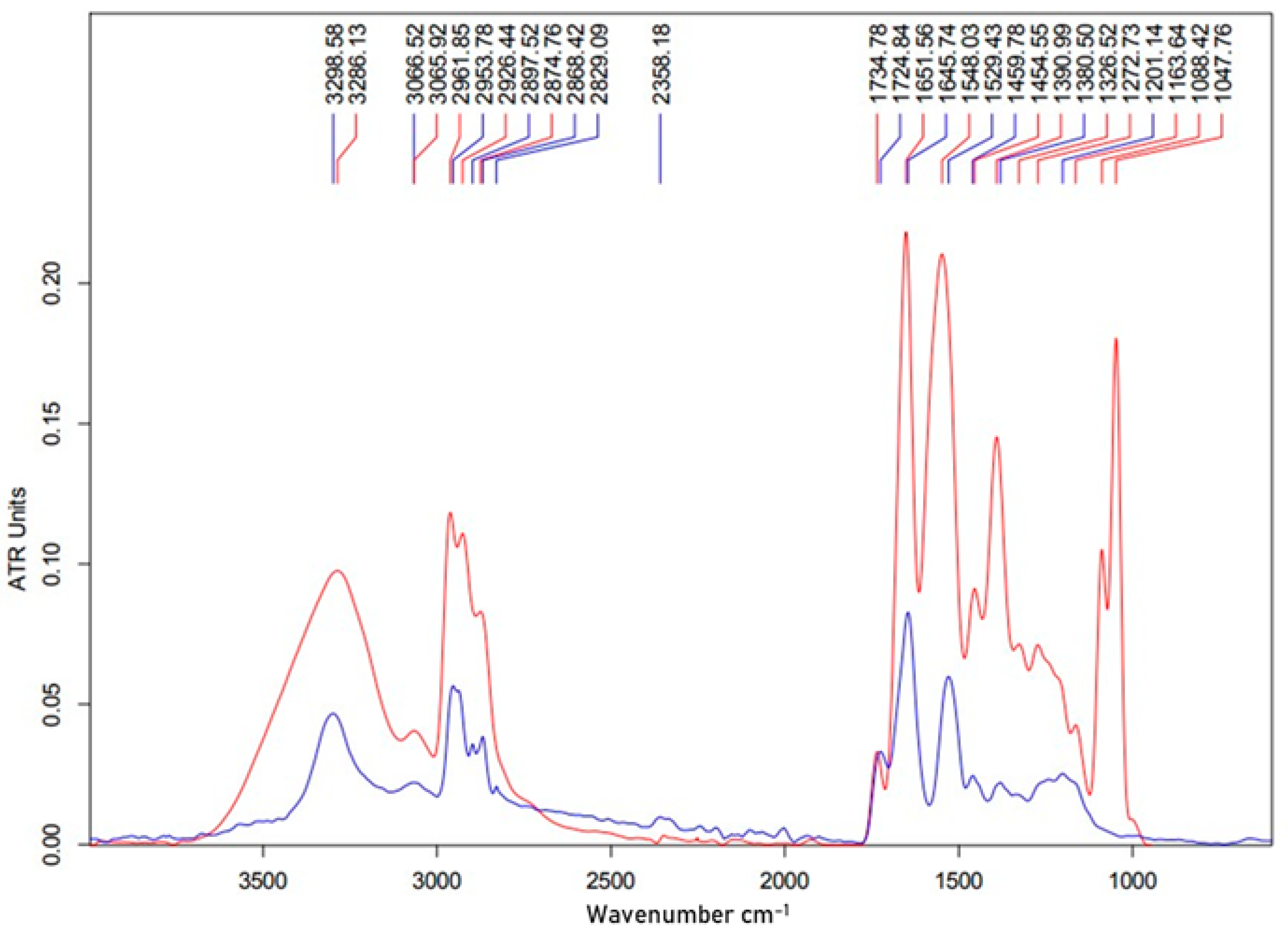
3.3. TLC and FTIR Analysis

TLC and FTIR analyses were used to partly reveal the chemical structure of the biosurfactants produced by B. mojavensis P1709. As presented in Figure 2a, exposure of the TLC plate to UV revealed five main spots with retention factors (Rf) of 0.02, 0.13, 0.51, 0.81, and 0.94. The TLC plate was treated with iodine vapor, ninhydrin, and anisaldehyde to reveal the basic chemical nature of the biosurfactants. Two of the spots with Rf values of 0.13 and 0.51 were colored both yellow and red, indicating the presence of lipids and peptides in the compounds. None of the two spots were colored by ninhydrin (Figure 2b,c).
By comparison with the Rf of the pure substances, these spots could be related to fengycin and surfactin. In the FTIR spectrum obtained for APF, peaks that are characteristic of peptides (wavelengths 3298–3285 and 3066–3050 for NH groups, 1651–1645 for CO groups, and 1548–1529 for CN groups) and of aliphatic chains (wavelength 2961–2829) can be observed (Figure 3). The FTIR spectrum is typical for biosurfactants of a lipopeptide nature described in other investigations, and coincides with the spectrum of the surfactin standard [50,51]. It can be concluded that the results of the chemical analysis confirmed those of the PCR analysis, which show that APF produced by B. mojavensis P1709 contained compounds of the two families of lipopeptides—fengycins and surfactins.

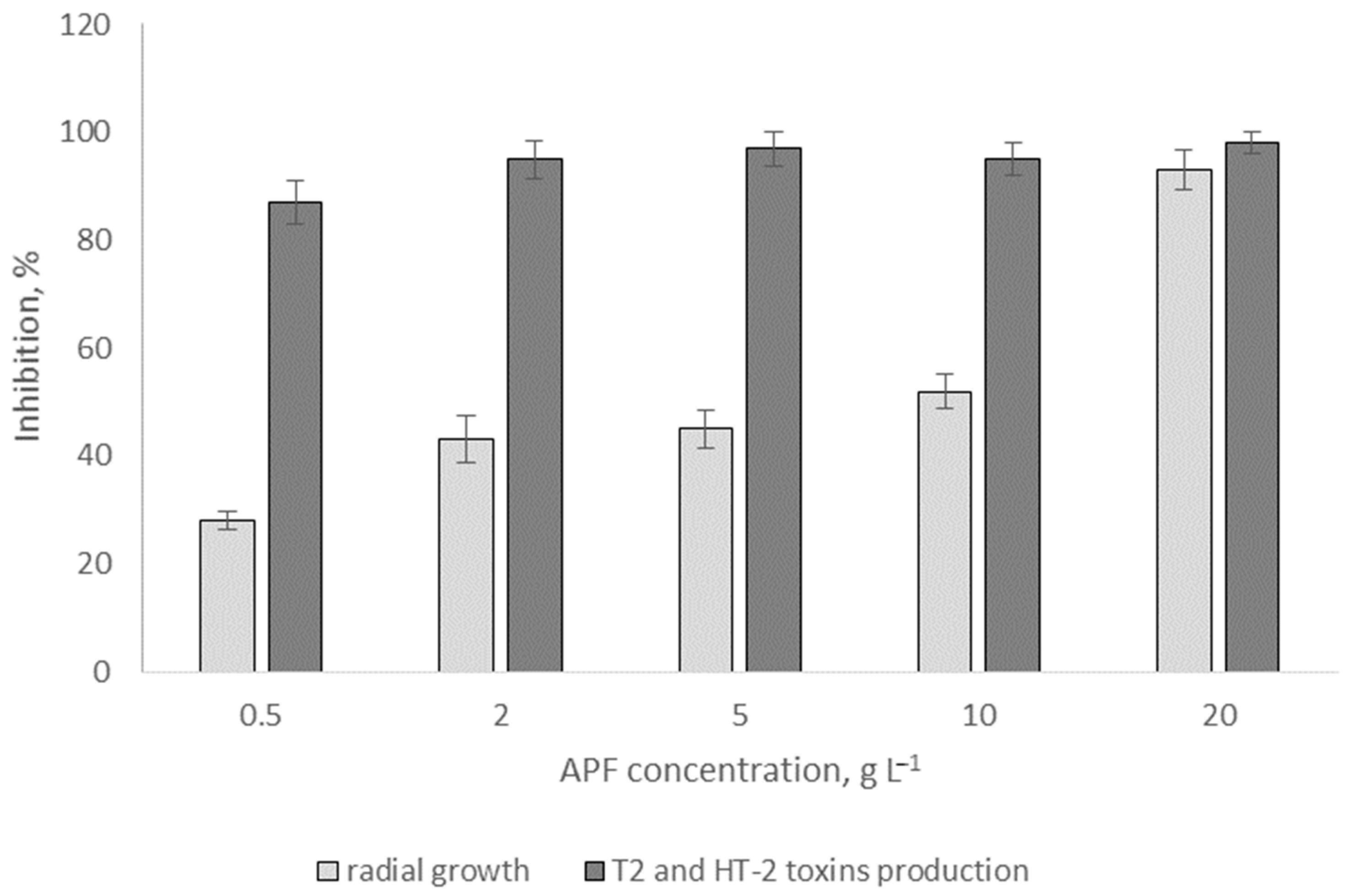
3.4. Effects of Biosurfactants on Radial Fungal Growth and Mycotoxin Production In Vitro

The APF of lipopeptides produced by B. mojavensis P1709 at concentrations of 0.5–20 g L−1 was assessed for its ability to inhibit F. oxysporum radial growth and mycotoxin production (Figure 4). It was revealed that fungal growth was inhibited even at relatively low concentrations of APF (28 ± 3% at 0.5 g L−1). The radial growth of F. oxysporum was inversely correlated with APF concentration in the agar medium; thus, the radial growth of the pathogen in the medium containing 20 g L−1 was inhibited by 93 ± 11% as compared with that in control medium. Previously, secondary metabolites of Bacilli were demonstrated to be efficient against F. oxysporum f. lycopersici specific for tomato plants and fruits [52]. On the control plates without the addition of lipopeptides, T-2 and HT-2 toxins were revealed at a concentration of 76 µg g−1. It should be noted that the acute toxic effect of T-2 toxin was registered for mammals that received orally a single dose above 2 mg per kg body weight, whereas chronic toxic effects were observed at a T-2 toxin dose of 25 µg per kg body weight consumed for 32–40 days [53]. Interestingly, the agar zones with a fungal presence even at low APF concentrations contained significantly lower amounts of mycotoxins than the control. This result suggests that fungal metabolism was affected even though mycelial growth was possible.

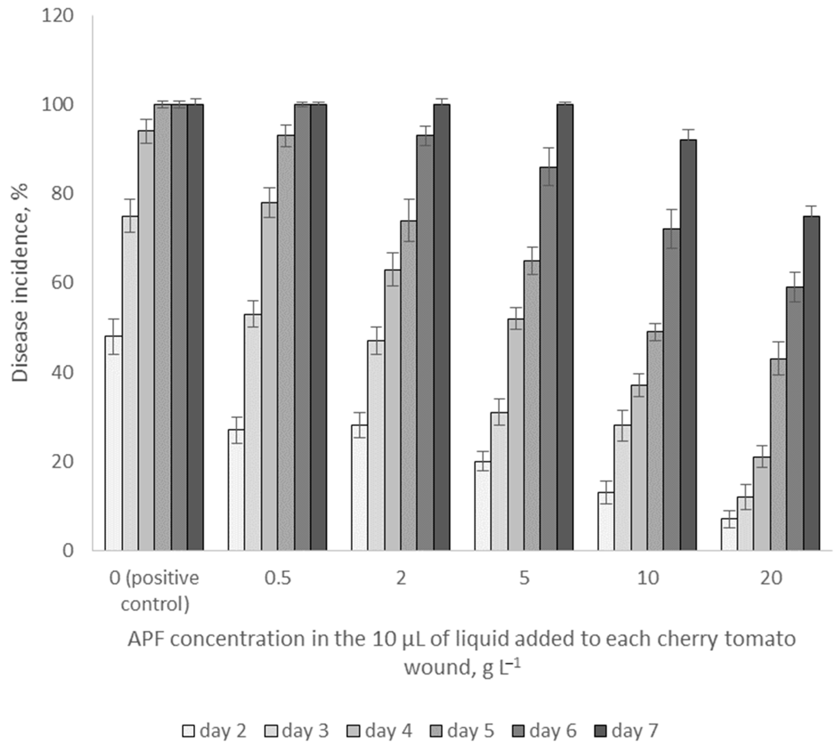
3.5. Effects of APF on Fungal Growth on Cherry Tomatoes In Vivo

To assess the influence of biosurfactants produced by B. mojavensis P1709 in vivo, cherry tomatoes were wounded, treated with APF at different concentrations, and thereafter infected by the pathogen. The results reflecting fusariosis disease incidence of tomato fruits after 2, 3, 4, 5, 6, and 7 days of incubation are presented in Figure 5.
The results showed that APF suppressed F. oxysporum growth at all concentrations used (from 0.5 to 20 g L−1) and that the effect was more pronounced at higher concentrations. Thus, after two days of incubation, the disease incidence was estimated to be 48% in the control variant, and 27% and 7% in the variants treated with 0.5 g L−1 at 20 g L−1 APF, respectively. The disease incidence increased over time at all concentrations; this parameter reached 100% on the 5th day after inoculation in the control variant and on the 6th, 7th, and 7th days in the variants treated with 0.5, 2, and 5 g L−1 biosurfactants, respectively, but did not reach 100% in the variants treated with 10 and 20 g-l biosurfactants over the duration of the experiment. The antagonistic activity of B. mojavensis P1709-produced biosurfactants revealed in this study is comparable with that of the B. subtilis strain, which was used to suppress another fungal plant pathogen. Kumar and coauthors (2021) [6] demonstrated that 20 g L−l lipopeptide biosurfactant caused a 44% disease incidence reduction in chili fruits infected by Colletotrichum capsisi after 5 days of incubation. Interestingly, these authors demonstrated an even higher efficacy of the biosurfactants when the fruits were treated with them after a certain time interval after infection. Yan and coauthors (2014) reported that the use of biosurfactants of another class—rhamnolipids—at a concentration of 0.5 g L−1 caused an approximately 20% reduction in disease incidence 3 days after the infection of cherry tomatoes with Alternaria alternata [54].

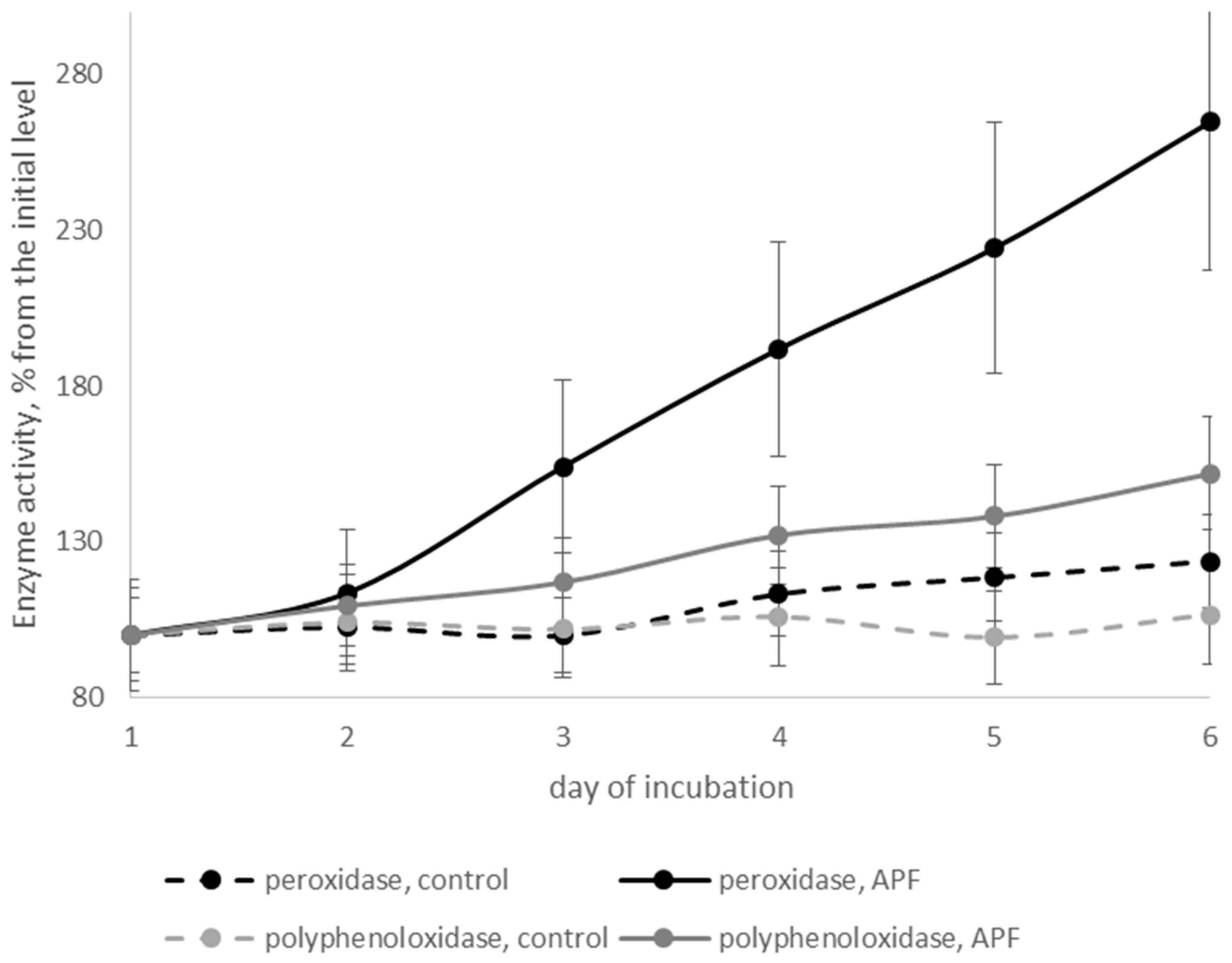
3.6. Effects of Biosurfactants on Defense-Related Enzymes of Cherry Tomatoes

Defense-related enzymes are produced in plants when they are attacked by pathogens. The chemical elicitors that are produced in plants as a response to stress induce the expression of genes encoding those enzymes directly or through a cascade of reactions. The latter, in turn, are involved in pathogen toxic metabolite deactivation, pathogen membrane disturbances, and many other processes preventing disease spread [27]. There are reports that biosurfactants may also play a role in inducing plant resistance systems [4,5,15,46]. In our study, we assessed the ability of APF obtained from B. mojavensis P1709 culture medium to increase the production of two defense-related enzymes—polyphenoloxidase and peroxidase—in cherry tomato fruits infected with F. oxysporum. Several potential mechanisms have been suggested to explain the antipathogenic activity of polyphenoloxidase, including the direct toxicity of quinones that are generated upon this enzyme involvement, the reduced availability of cellular proteins for the pathogen due to their alkylation, the formation of the physical barrier for the pathogen in the cell wall due to the interaction of quinones with proteins, and the generation of hydrogen peroxide or other reactive oxygen species [55]. Peroxidases are reported to be involved in the formation of lignin and therefore cell wall reinforcement and in the production of reactive oxygen species [56].
The initial level of the enzyme activities in the cherry tomato tissue was estimated to be 27.1 and 4.2 U g−1 fresh weight, respectively. Comparable levels of polyphenoloxidase and peroxidase activity in cherry tomatoes were observed by other authors [21]. In the control cherry tomatoes that were artificially infected by the pathogen but were not treated with APF, the level of polyphenoloxidase activity did not change significantly during the 6 days of incubation (Figure 6). The level of peroxidase activity increased by 23%, most likely indicating the naturally occurring response processes of tomato fruit upon pathogen invasion. The level of enzyme activity increased dramatically by 254% and 161% for peroxidase and polyphenoloxidase, respectively, when the cherry tomato fruits were treated with the APF of B. mojavensis P1707. It can be concluded that lipopeptides produced by B. mojavensis P1709 were capable of inducing an immune systematic response in cherry tomato fruits after harvesting.

4. Conclusions

In the present study, B. mojavensis P1709 isolated from the L. sativa rhizosphere was demonstrated to produce biosurfactants that emulsified crude oil. Summarizing the results of the RT–PCR analysis for the genes responsible for lipopeptide synthesis and the results of the TLC and FTIR analyses, the acid-precipitated fraction of the supernatant obtained after cultivation of B. mojavensis P1709 in glycerol-nitrate medium was shown to contain two groups of compounds from the lipopeptide class of biosurfactants—fengycins and surfactins. It was revealed that the mixture of these compounds suppressed the radial growth of F. oxysporum on agar plates and its development on cherry tomato fruits in vivo. Moreover, the inhibition of T-2 and HT-2 mycotoxin production by the fungi under the influence of the lipopeptide mixture was observed. The acid-precipitated fraction of the B. mojavensis P1709 culture medium induced the synthesis of two enzymes—peroxidase and polyphenoloxidase—that are defense-related proteins of the plant immune system. The results obtained demonstrate that biosurfactants produced by B. mojavensis P1709 may be effectively used for postharvest treatment of cherry tomatoes to maintain the quality and quantity of the fruits.

Supplementary Materials

The following supporting information can be downloaded at: https://www.mdpi.com/article/10.3390/agriculture12050609/s1, Table S1: Statistically significant differences for inhibition of radial growth at different APF concentrations; Table S2: Statistically significant differences for inhibition T2 and HT-2 toxins production at different APF concentrations; Table S3: Statistically significant differences for disease incidence of cherry tomato fruits at different APF concentrations at day 2; Table S4: Statistically significant differences for disease incidence of cherry tomato fruits at different APF concentrations at day 3; Table S5: Statistically significant differences for disease incidence of cherry tomato fruits at different APF concentrations at day 4; Table S6: Statistically significant differences for disease incidence of cherry tomato fruits at different APF concentrations at day 5; Table S7: Statistically significant differences for disease incidence of cherry tomato fruits at different APF concentrations at day 6; Table S8: Statistically significant differences for disease incidence of cherry tomato fruits at different APF concentrations at day 7; Table S9: Statistically significant differences for disease incidence of cherry tomato fruits at different days at APF concentration is 0 g L−1; Table S10: Statistically significant differences for disease incidence of cherry tomato fruits at different days at APF concentration is 0.5 g L−1; Table S11: Statistically significant differences for disease incidence of cherry tomato fruits at different days at APF concentration is 2 g L−1; Table S12: Statistically significant differences for disease incidence of cherry tomato fruits at different days at APF concentration is 5 g L−1; Table S13: Statistically significant differences for disease incidence of cherry tomato fruits at different days at APF concentration is 10 g L−1; Table S14: Statistically significant differences for disease incidence of cherry tomato fruits at different days at APF concentration is 20 g L−1.

Author Contributions

Conceptualization, P.G. and S.S.; Methodology, L.B.; Validation, P.G. and S.S; Formal Analysis, S.S.; Investigation, A.G.; Resources, G.G.; Data Curation, S.S.; Writing—Original Draft Preparation, P.G.; Writing—Review and Editing, P.G.; Visualization, K.K. All authors have read and agreed to the published version of the manuscript.

Funding

This work was funded by the subsidy allocated to Kazan Federal University for the state assignment in the sphere of scientific activities No. 0671-2020-0055.

Institutional Review Board Statement

Not applicable.

Informed Consent Statement

Not applicable.

Conflicts of Interest

The authors declare no conflict of interest.

References

  1. Savary, S.; Willocquet, L.; Pethybridge, S.J.; Esker, P.; McRoberts, N.; Nelson, A. The global burden of pathogens and pests on major food crops. Nat. Ecol. Evol. 2019, 3, 430–439. [Google Scholar] [CrossRef] [PubMed]
  2. Szalewski, D.A.; Hinrichs, V.S.; Zinniel, D.K.; Barletta, R.G. The pathogenicity of Aspergillus fumigatus, drug resistance, and nanoparticle delivery. Can. J. Microbiol. 2018, 64, 439–453. [Google Scholar] [CrossRef] [PubMed] [Green Version]
  3. Strange, R.N.; Scott, P.R. Plant disease: A threat to global food security. Annu. Rev. Phytopathol. 2005, 43, 83–116. [Google Scholar] [CrossRef] [PubMed]
  4. Crouzet, J.; Arguelles-Arias, A.; Dhondt-Cordelier, S.; Cordelier, S.; Pršić, J.; Hoff, G.; Mazeyrat-Gourbeyre, F.; Baillieul, F.; Clément, C.; Ongena, M.; et al. Biosurfactants in Plant Protection Against Diseases: Rhamnolipids and Lipopeptides Case Study. Front. Bioeng. Biotechnol. 2020, 8, 1014. [Google Scholar] [CrossRef]
  5. Sachdev, D.P.; Cameotra, S.S. Biosurfactants in agriculture. Appl. Microbiol. Biotechnol. 2013, 97, 1005–1016. [Google Scholar] [CrossRef] [Green Version]
  6. Kumar, A.; Rabha, J.; Jha, D.K. Antagonistic activity of lipopeptide-biosurfactant producing Bacillus subtilis AKP, against Colletotrichum capsici, the causal organism of anthracnose disease of chilli. Biocatal. Agric. Biotechnol. 2021, 36, 102133. [Google Scholar] [CrossRef]
  7. Varvaresou, A.; Iakovou, K. Biosurfactants in cosmetics and biopharmaceuticals. Lett. Appl. Microbiol. 2015, 61, 214–223. [Google Scholar] [CrossRef]
  8. Köhl, J.; Medeiros, F.H.V.; Lombaers-van der Plas, C.; Groenenboom-de Haas, L.; van den Bosch, T. Efficacies of bacterial and fungal isolates in biocontrol of Botrytis cinerea and Pseudomonas syringae pv. tomato and growth promotion in tomato do not correlate. Biol. Control 2020, 150, 104375. [Google Scholar] [CrossRef]
  9. Rodrigues, A.I.; Gudiña, E.J.; Abrunhosa, L.; Malheiro, A.R.; Fernandes, R.; Teixeira, J.A.; Rodrigues, L.R. Rhamnolipids inhibit aflatoxins production in Aspergillus flavus by causing structural damages in the fungal hyphae and down-regulating the expression of their biosynthetic genes. Int. J. Food Microbiol. 2021, 348, 109207. [Google Scholar] [CrossRef]
  10. Ongena, M.; Jacques, P.; Touré, Y.; Destain, J.; Jabrane, A.; Thonart, P. Involvement of fengycin-type lipopeptides in the multifaceted biocontrol potential of Bacillus subtilis. Appl. Microbiol. Biotechnol. 2005, 69, 29–38. [Google Scholar] [CrossRef]
  11. Caulier, S.; Nannan, C.; Gillis, A.; Licciardi, F.; Bragard, C.; Mahillon, J. Overview of the Antimicrobial Compounds Produced by Members of the Bacillus subtilis Group. Front. Microbiol. 2019, 10, 302. [Google Scholar] [CrossRef] [PubMed] [Green Version]
  12. Rudakova, M.A.; Galitskaya, P.Y.; Selivanovskaya, S.Y. Biosurfactants: Current Application Trends. Uchenye Zap. Kazan. Univ. Seriya Estestv. Nauk. 2021, 163, 177–208. [Google Scholar] [CrossRef]
  13. Geissler, M.; Oellig, C.; Moss, K.; Schwack, W.; Henkel, M.; Hausmann, R. High-performance thin-layer chromatography (HPTLC) for the simultaneous quantification of the cyclic lipopeptides Surfactin, Iturin A and Fengycin in culture samples of Bacillus species. J. Chromatogr. B Anal. Technol. Biomed. Life Sci. 2017, 1044–1045, 214–224. [Google Scholar] [CrossRef] [PubMed]
  14. Massart, S.; Perazzolli, M.; Höfte, M.; Pertot, I.; Jijakli, M.H. Impact of the omic technologies for understanding the modes of action of biological control agents against plant pathogens. BioControl 2015, 60, 725–746. [Google Scholar] [CrossRef]
  15. Kumar, A.; Singh, S.K.; Kant, C.; Verma, H.; Kumar, D.; Singh, P.P.; Modi, A.; Droby, S.; Kesawat, M.S.; Alavilli, H.; et al. Microbial biosurfactant: A new frontier for sustainable agriculture and pharmaceutical industries. Antioxidants 2021, 10, 1472. [Google Scholar] [CrossRef]
  16. Seneviratne, G.; Weerasekara, M.L.M.A.W.; Seneviratne, K.A.C.N.; Zavahir, J.S.; Kecskés, M.L.; Kennedy, I.R. Importance of Biofilm Formation in Plant Growth Promoting Rhizobacterial Action. In Plant Growth and Health Promoting Bacteria; Springer: Berlin/Heidelberg, Germany, 2010; pp. 81–95. [Google Scholar] [CrossRef]
  17. Zeriouh, H.; de Vicente, A.; Pérez-García, A.; Romero, D. Surfactin triggers biofilm formation of Bacillus subtilis in melon phylloplane and contributes to the biocontrol activity. Environ. Microbiol. 2014, 16, 2196–2211. [Google Scholar] [CrossRef]
  18. Chen, L.; Wang, N.; Wang, X.; Hu, J.; Wang, S. Characterization of two anti-fungal lipopeptides produced by Bacillus amyloliquefaciens SH-B10. Bioresour. Technol. 2010, 101, 8822–8827. [Google Scholar] [CrossRef]
  19. Gaur, V.K.; Sharma, P.; Sirohi, R.; Varjani, S.; Taherzadeh, M.J.; Chang, J.-S.; Ng, H.Y.; Wong, J.W.; Kim, S.-H. Production of biosurfactants from agro-industrial waste and waste cooking oil in a circular bioeconomy: An overview. Bioresour. Technol. 2022, 343, 126059. [Google Scholar] [CrossRef]
  20. Medina-Romero, Y.M.; Roque-Flores, G.; Macías-Rubalcava, M.L. Volatile organic compounds from endophytic fungi as innovative postharvest control of Fusarium oxysporum in cherry tomato fruits. Appl. Microbiol. Biotechnol. 2017, 101, 8209–8222. [Google Scholar] [CrossRef]
  21. Raynaldo, F.A.; Dhanasekaran, S.; Ngea, G.L.N.; Yang, Q.; Zhang, X.; Zhang, H. Investigating the biocontrol potentiality of Wickerhamomyces anomalus against postharvest gray mold decay in cherry tomatoes. Sci. Hortic. (Amst.) 2021, 285, 110137. [Google Scholar] [CrossRef]
  22. Ignjatov, M.; Milosevic, D.; Nikolic, Z.; Gvozdanovic-Varga, J.; Jovicic, D.; Zdjelar, G. Fusarium oxysporum as causal agent of tomato wilt and fruit rot. Pestic. Fitomedicina 2012, 27, 25–31. [Google Scholar] [CrossRef]
  23. Thipe, V.C.; Bloebaum, P.; Khoobchandani, M.; Karikachery, A.R.; Katti, K.K.; Katti, K.V. Green nanotechnology: Nanoformulations against toxigenic fungi to limit mycotoxin production. In Nanomycotoxicology: Treating Mycotoxins in the Nano Way; Academic Press: Cambridge, MA, USA, 2019; pp. 155–188. [Google Scholar] [CrossRef]
  24. Ji, F.; He, D.; Olaniran, A.O.; Mokoena, M.P.; Xu, J.; Shi, J. Occurrence, toxicity, production and detection of Fusarium mycotoxin: A review. Food Prod. Process. Nutr. 2019, 1, 6. [Google Scholar] [CrossRef]
  25. Mart, P.V.; Garrigues, S.; Marcos, J.F.; Manzanares, P. Antifungal Peptides and Proteins to Control Toxigenic Fungi and Mycotoxin Biosynthesis. Int. J. Mol. Sci. 2021, 22, 13261. [Google Scholar] [CrossRef]
  26. Harwig, J.; Scott, P.M.; Stoltz, D.R.; Blanchfield, B.J. Toxins of molds from decaying tomato fruit. Appl. Environ. Microbiol. 1979, 38, 267–274. [Google Scholar] [CrossRef] [PubMed] [Green Version]
  27. Prasannath, K. Plant defense-related enzymes against pathogens: A review. AGRIEAST J. Agric. Sci. 2017, 11, 38. [Google Scholar] [CrossRef]
  28. Indunil Kumari, Y.S.M.A.; Vengadaramana, A. Stimulation of Defense Enzymes in Tomato (Solanum lycopersicum L.) and Chilli (Capsicum annuum L.) in Response to Exogenous Application of Different Chemical Elicitors. Univers. J. Plant Sci. 2017, 5, 10–15. [Google Scholar] [CrossRef] [Green Version]
  29. Lorenzo, O.; Solano, R. Molecular players regulating the jasmonate signalling network. Curr. Opin. Plant Biol. 2005, 8, 532–540. [Google Scholar] [CrossRef]
  30. Ralph, S.; Park, J.Y.; Bohlmann, J.; Mansfield, S.D. Dirigent proteins in conifer defense: Gene discovery, phylogeny, and differential wound- and insect-induced expression of a family of DIR and DIR-like genes in spruce (Picea spp.). Plant Mol. Biol. 2006, 60, 21–40. [Google Scholar] [CrossRef]
  31. Agrios, G.N. How plants defend themselves against pathogens. In Plant Pathology, 5th ed.; Academic Press: Cambridge, MA, USA, 2005; pp. 207–248. [Google Scholar] [CrossRef]
  32. Luzuriaga-Loaiza, W.P.; Schellenberger, R.; De Gaetano, Y.; Obounou Akong, F.; Villaume, S.; Crouzet, J.; Haudrechy, A.; Baillieul, F.; Clément, C.; Lins, L.; et al. Synthetic Rhamnolipid Bolaforms trigger an innate immune response in Arabidopsis thaliana. Sci. Rep. 2018, 8, 8534. [Google Scholar] [CrossRef] [Green Version]
  33. Henry, G.; Deleu, M.; Jourdan, E.; Thonart, P.; Ongena, M. The bacterial lipopeptide surfactin targets the lipid fraction of the plant plasma membrane to trigger immune-related defence responses. Cell. Microbiol. 2011, 13, 1824–1837. [Google Scholar] [CrossRef]
  34. Biktasheva, L.; Galitskaya, P.S.S. Potential of biosurfactant production in bacteria isolated from oil polluted soils. Int. Multidiscip. Sci. GeoConf. Surv. Geol. Min. Ecol. Manag. SGEM 2021, 225–232. [Google Scholar] [CrossRef]
  35. Chung, S.; Kong, H.; Buyer, J.S.; Lakshman, D.K.; Lydon, J.; Kim, S.D.; Roberts, D.P. Isolation and partial characterization of Bacillus subtilis ME488 for suppression of soilborne pathogens of cucumber and pepper. Appl. Microbiol. Biotechnol. 2008, 80, 115–123. [Google Scholar] [CrossRef] [PubMed]
  36. Agarwal, H.; Dowarah, B.; Baruah, P.M.; Bordoloi, K.S.; Krishnatreya, D.B.; Agarwala, N. Endophytes from Gnetum gnemon L. can protect seedlings against the infection of phytopathogenic bacterium Ralstonia solanacearum as well as promote plant growth in tomato. Microbiol. Res. 2020, 238, 126503. [Google Scholar] [CrossRef]
  37. Galitskaya, P.; Biktasheva, L.; Kuryntseva, P.; Selivanovskaya, S. Response of soil bacterial communities to high petroleum content in the absence of remediation procedures. Environ. Sci. Pollut. Res. 2021, 28, 9610–9627. [Google Scholar] [CrossRef]
  38. Alvarez, V.M.; Guimarães, C.R.; Jurelevicius, D.; de Castilho, L.V.A.; de Sousa, J.S.; da Mota, F.F.; Freire, D.M.G.; Seldin, L. Microbial enhanced oil recovery potential of surfactin-producing Bacillus subtilis AB2.0. Fue 2020, 272, 117730. [Google Scholar] [CrossRef]
  39. Nayarisseri, A.; Singh, P.; Singh, S.K. Screening, isolation and characterization of biosurfactant producing Bacillus subtilis strain ANSKLAB03. Bioinformation 2018, 14, 304–314. [Google Scholar] [CrossRef]
  40. Varadavenkatesan, T.; Murty, V.R. Production of a Lipopeptide Biosurfactant by a Novel Bacillus sp. and Its Applicability to Enhanced Oil Recovery. ISRN Microbiol. 2013, 2013, 621519. [Google Scholar] [CrossRef] [Green Version]
  41. Sun, W.; Cao, W.; Jiang, M.; Saren, G.; Liu, J.; Cao, J.; Ali, I.; Yu, X.; Peng, C.; Naz, I. Isolation and characterization of biosurfactant-producing and diesel oil degrading Pseudomonas sp. CQ2 from Changqing oil field, China. RSC Adv. 2018, 8, 39710–39720. [Google Scholar] [CrossRef] [Green Version]
  42. Griffith, G.W.; Easton, G.L.; Detheridge, A.; Roderick, K.; Edwards, A.; Worgan, H.J.; Nicholson, J.; Perkins, W.T. Copper, deficiency in potato dextrose agar causes reduced pigmentation in cultures of various fungi. FEMS Microbiol. Lett. 2007, 276, 165–171. [Google Scholar] [CrossRef]
  43. Mendes, G.; Gonçalves, V.N.; Souza-Fagundes, E.M.; Kohlhoff, M.; Rosa, C.A.; Zani, C.L.; Cota, B.B.; Rosa, L.H.; Johann, S. Antifungal activity of extracts from Atacama Desert fungi against Paracoccidioides brasiliensis and identification of Aspergillus felis as a promising source of natural bioactive compounds. Mem. Inst. Oswaldo Cruz. 2016, 111, 209–217. [Google Scholar] [CrossRef]
  44. Khaliq, G.; Muda Mohamed, M.T.; Ali, A.; Ding, P.; Ghazali, H.M. Effect of gum arabic coating combined with calcium chloride on physico-chemical and qualitative properties of mango (Mangifera indica L.) fruit during low temperature storage. Sci. Hortic. (Amsterdam) 2015, 190, 187–194. [Google Scholar] [CrossRef]
  45. Uzoigwe, C.; Burgess, J.G.; Ennis, C.J.; Rahman, P.K.S.M. Bioemulsifiers are not biosurfactants and require different screening approaches. Front Microbiol. 2015, 6, 245. [Google Scholar] [CrossRef] [PubMed] [Green Version]
  46. Barin, R.; Talebi, M.; Biria, D.; Beheshti, M. Fast bioremediation of petroleum-contaminated soils by a consortium of biosurfactant/bioemulsifier producing bacteria. Int. J. Environ. Sci. Technol. 2014, 11, 1701–1710. [Google Scholar] [CrossRef] [Green Version]
  47. Pereira, J.F.B.; Gudiña, E.J.; Costa, R.; Vitorino, R.; Teixeira, J.A.; Coutinho, J.A.P.; Rodrigues, L.R. Optimization and characterization of biosurfactant production by Bacillus subtilis isolates towards microbial enhanced oil recovery applications. Fuel 2013, 111, 259–268. [Google Scholar] [CrossRef] [Green Version]
  48. Freitas de Oliveira, D.W.; Lima França, Í.W.; Nogueira Félix, A.K.; Lima Martins, J.J.; Aparecida Giro, M.E.; Melo, V.M.M.; Goncalves, L.R.B. study of biosurfactant production by Bacillus subtilis LAMI005 grown in clarified cashew apple juice. Colloids Surf. B Biointerfaces 2013, 101, 34–43. [Google Scholar] [CrossRef]
  49. Cawoy, H.; Debois, D.; Franzil, L.; De Pauw, E.; Thonart, P.; Ongena, M. Lipopeptides as main ingredients for inhibition of fungal phytopathogens by Bacillus subtilis/amyloliquefaciens. Microb. Biotechnol. 2015, 8, 281–295. [Google Scholar] [CrossRef]
  50. El-Sheshtawy, H.S.; Aiad, I.; Osman, M.E.; Abo-ELnasr, A.A.; Kobisy, A.S. Production of biosurfactant from Bacillus licheniformis for microbial enhanced oil recovery and inhibition the growth of sulfate reducing bacteria. Egypt. J. Pet. 2015, 24, 155–162. [Google Scholar] [CrossRef] [Green Version]
  51. Nayak, N.S.; Purohit, M.S.; Tipre, D.R.; Dave, S.R. Biosurfactant production and engine oil degradation by marine halotolerant Bacillus licheniformis LRK1. Biocatal. Agric. Biotechnol. 2020, 29, 101808. [Google Scholar] [CrossRef]
  52. Velho, R.V.; Medina, L.F.C.; Segalin, J.; Brandelli, A. Production of lipopeptides among Bacillus strains showing growth inhibition of phytopathogenic fungi. Folia Microbiol. (Praha) 2011, 56, 297–303. [Google Scholar] [CrossRef]
  53. Adhikari, M.; Negi, B.; Kaushik, N.; Adhikari, A.; Al-Khedhairy, A.A.; Kaushik, N.K.; Choi, E.H. T-2 mycotoxin: Toxicological effects and decontamination strategies. Oncotarget 2017, 8, 33933. Available online: www.impactjournals.com/oncotarget (accessed on 8 February 2017). [CrossRef] [Green Version]
  54. Yan, F.; Xu, S.; Chen, Y.; Zheng, X. Effect of rhamnolipids on Rhodotorula glutinis biocontrol of Alternaria alternata infection in cherry tomato fruit. Postharvest Biol. Technol. 2014, 97, 32–35. [Google Scholar] [CrossRef]
  55. Constabel, C.P.; Barbehenn, R. Defensive roles of polyphenol oxidase in plants. In Induced Plant Resistance to Herbivory; Springer: Dordrecht, The Netherlands, 2008; pp. 253–270. [Google Scholar] [CrossRef]
  56. García-Gutiérrez, L.; Zeriouh, H.; Romero, D.; Cubero, J.; de Vicente, A.; Pérez-García, A. The antagonistic strain Bacillus subtilisUMAF6639 also confers protection to melon plants against cucurbit powdery mildew by activation of jasmonate- and salicylic acid-dependent defence responses. Microb. Biotechnol. 2013, 6, 264–274. [Google Scholar] [CrossRef] [PubMed] [Green Version]
Figure 1. Revealing of the presence of fenD, ituC, and srfAA genes in the genome of B. mojavensis P1709 using PCR with specific primers followed by agarose gel electrophoresis.
Agriculture 12 00609 g001
Figure 2. TLC analysis of the lipopeptides produced by the B. mojavensis P1709 strain (spots revealed: (a) by UV light; (b) after iodine vapor staining; (c) after ninhydrin staining).
Agriculture 12 00609 g002
Figure 3. FTIR spectra of the APF produced by B. mojavensis P1709 (blue) and of the surfactin standard (red) (Sigma Aldrich, Darmstadt, Germany).
Agriculture 12 00609 g003
Figure 4. Inhibition of radial growth and mycotoxin production of F. oxysporum in the presence of APF produced by B. mojavensis P1709 in agar medium at different concentrations. The Wilcoxon signed rank test was used to determine statistically significant differences (p < 0.05); the results of determination are presented in Supplementary Material.
Agriculture 12 00609 g004
Figure 5. Disease incidence of cherry tomato fruits treated with F. oxysporum (positive control) and lipopeptides produced by B. mojavensis P1709 at different concentrations, revealed 2–7 days after inoculation. The Wilcoxon signed rank test was used to determine statistically significant differences (p < 0.05); the results of determination are presented in Supplementary Material.
Agriculture 12 00609 g005
Figure 6. Peroxidase and polyphenoloxidase activity in the tissue of cherry tomatoes infected with F. oxysporum and treated with APF of B. mojavensis P1709.
Agriculture 12 00609 g006
Table 1. PCR primers used for amplification of lipopeptide genes [35].
Target GenePrimer Sequence, 5′-3′Amplicon Length (bp)
fenDF: GGCCCGTTCTCTAAATCCAT670
R: GTCATGCTGACGAGAGCAAA
ituCF: GGCTGCTGCAGATGCTTTAT423
R: TCGCAGATAATCGCAGTGAG
srfAAF: TCGGGACAGGAAGACATCAT201
R: CCACTCAAACGGATAATCCTGA
Publisher’s Note: MDPI stays neutral with regard to jurisdictional claims in published maps and institutional affiliations.

