Magnesium, Calcium, Potassium, Sodium, Phosphorus, Selenium, Zinc, and Chromium Levels in Alcohol Use Disorder: A Review

 ,
,  , , , , and
Abstract
1. Introduction
2. Materials and Methods
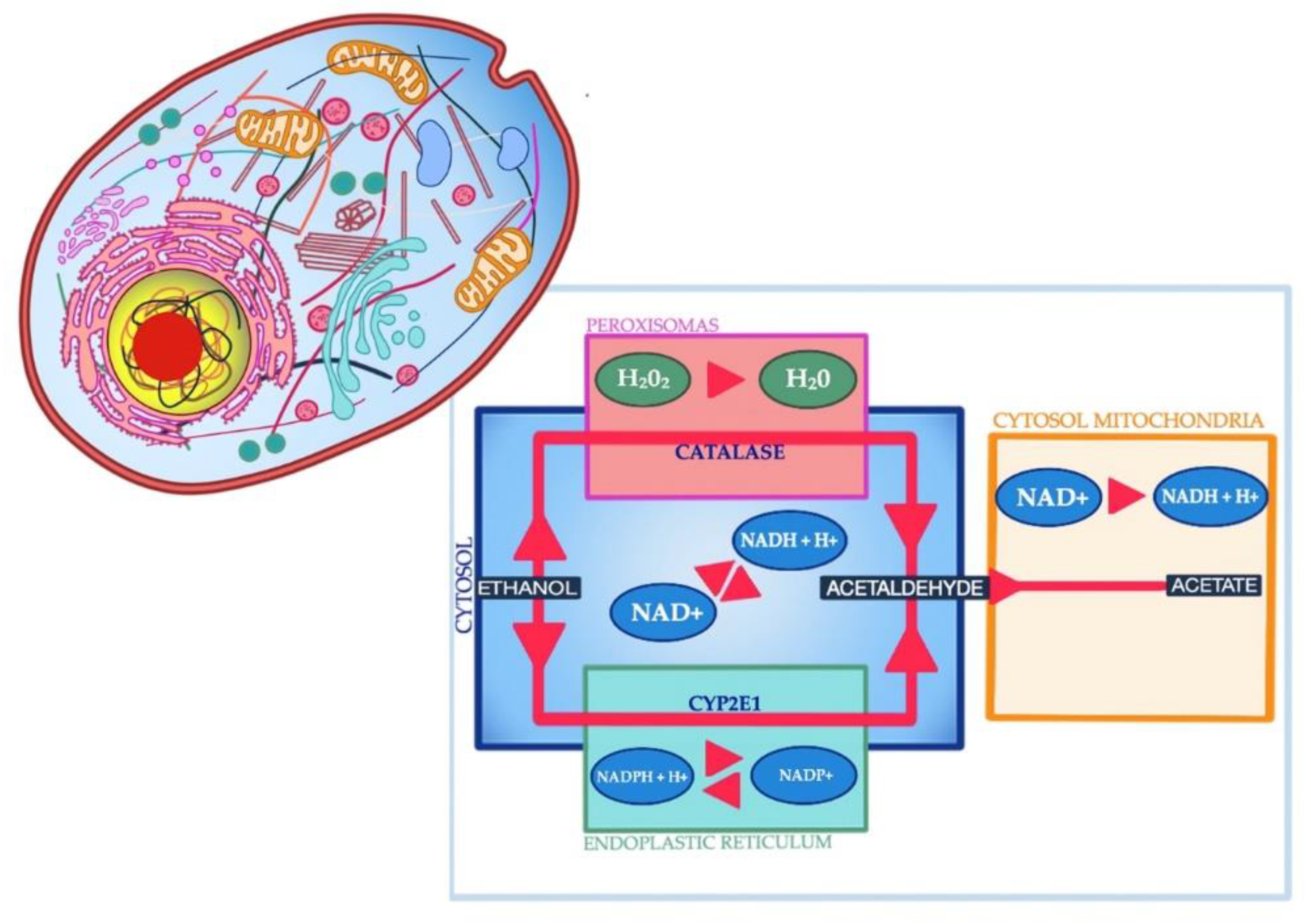
3. Acid-Base Disturbances
4. Phosphorus Deficit
5. Imbalances in Magnesium and Calcium Levels in Plasma
6. Plasma Potassium Deficit
7. Sodium Deficit
8. Selenium Deficit
9. Zinc Concentration Disturbance
10. Chromium Deficit
11. Conclusions
Author Contributions
Funding
Conflicts of Interest
References
- Muller, A. Alcohol consumption and community hospital admissions in the United States: A dynamic regression analysis, 1950–1992. Addiction 1996, 91, 231–242. [Google Scholar] [CrossRef] [PubMed]
- Kang, Y.J.; Zhou, Z. Zinc prevention and treatment of alcoholic liver disease. Mol. Asp. Med. 2005, 26, 391–404. [Google Scholar] [CrossRef]
- Grochowski, C.; Blicharska, E.; Baj, J.; Mierzwińska, A.; Brzozowska, K.; Forma, A.; Maciejewski, R. Serum iron, Magnesium, Copper, and Manganese Levels in Alcoholism: A Systematic Review. Molecules 2019, 24, 1361. [Google Scholar] [CrossRef]
- Grochowski, C.; Blicharska, E.; Bogucki, J.; Proch, J.; Mierzwińska, A.; Baj, J.; Litak, J.; Podkowinkski, A.; Flieger, J.; Teresinski, G.; et al. Increased Aluminum Content in Certain Brain Structures is Correlated with Higher Silicon Concentration in Alcoholic Use Disorder. Molecules 2019, 24, 1721. [Google Scholar] [CrossRef]
- Barrio, P.; Wurst, F.M.; Gual, A. New Alcohol Biomarkers. New challenges. Alcohol Alcohol. 2018, 53, 762–763. [Google Scholar] [CrossRef]
- Moreno Otero, R.; Cortés, J.R. Nutrition and chronic alcohol abuse. Nutr. Hosp. 2008, 23, 3–7. [Google Scholar]
- Boye, A. Metabolic derivatives of alcohol and the molecular culprits of fibro-hepatocarcinogenesis: Allies or enemies? World J. Gastroenterol. 2016, 22, 50. [Google Scholar] [CrossRef]
- Biff, F.; Palmer, M.D.; Clegg, D.J. Electrolyte Disturbances in Patients with Chronic Alcohol-Use Disorder. N. Engl. J. Med. 2017, 377, 1368–1377. [Google Scholar]
- Barrio, P.; Gual, A. Patient-centered care interventions for the management of alcohol use disorders: A systematic review of randomized controlled trials. Patient Prefer. Adher. 2016, 10, 1823–1845. [Google Scholar] [CrossRef]
- Addolorato, G.; Mirijello, A.; Barrio, P.; Gual, A. Treatment of alcohol use disorders in patients with alcoholic liver disease. J. Hepatol. 2016, 65, 618–630. [Google Scholar] [CrossRef]
- Adamson, D.J.; Laing, R.B.; Nathwani, D. Alcoholism, hyponatremia and central neurological damage. Scott. Med. J. 1992, 37, 1006–1012. [Google Scholar] [CrossRef]
- Burin, M.R.; Cook, C.C. Alcohol withdrawal and hypokalemia: A case report. Alcohol Alcohol. 2000, 35, 188–189. [Google Scholar] [CrossRef][Green Version]
- Carl, G.; Holzbach, E. Reversible hypokalemia and hypomagnesemia during alcohol withdrawal syndrome. Nervenarzt 1994, 65, 206–211. [Google Scholar]
- Cunha, D.F.; Monteiro, J.P.; Ortega, L.S.; Alves, L.G.; Cunha, S.F. Serum electrolytes in hospitalized pellagra alcoholics. Eur. J. Clin. Nutr. 2000, 54, 440–442. [Google Scholar] [CrossRef][Green Version]
- Stasiukyniene, V. Blood plasma potassium, sodiumand magnesiumlevelsin chronic alcoholic patientsduring alcohol withdrawal. Medicina 2002, 38, 892–895. [Google Scholar]
- Vetter, W.R. Hypokalemia and Electrocardiographic Abnormalities During Acute Alcohol Withdrawal. Arch. Intern. Med. 1967, 120, 536. [Google Scholar] [CrossRef]
- Grochowski, C.; Szukała, M.; Litak, J.; Budny, A.; Proch, J.; Majerek, D.; Blicharska, E.; Niedzielski, P. Correlations between Trace Elements in Selected Locations of the Human Brain in Individuals with Alcohol Use Disorder. Molecules 2020, 25, 359. [Google Scholar] [CrossRef]
- Grochowski, C.; Blicharska, E.; Krukow, P.; Jonak, K.; Maciejewski, M.; Szczepanek, D.; Jonak, K.; Flieger, J.; Maciejewski, R. Analysis of Trace Elements in Human Brain: Its Aim, Methods, and Concentration Levels. Front. Chem. 2019, 7, 115. [Google Scholar] [CrossRef]
- Skórzyńska-Dziduszko, K.E.; Kimber-Trojnar, Ż.; Patro-Małysza, J.; Olszewska, A.; Zaborowski, T.; Małecka-Massalska, T. An Interplay between Obesity and Inflammation in Gestational Diabetes Mellitus. Curr. Pharm. Biotechnol. 2016, 17, 603–613. [Google Scholar]
- Dolar-Szczasny, J.; Święch, A.; Flieger, J.; Tatarczak-Michalewska, M.; Niedzielski, P.; Proch, J.; Majerek, D.; Kawka, J.; Mackiewicz, J. Levels of Trace Elements in the Aqueous Humor of Cataract Patients Measured by the Inductively Coupled Plasma Optical Emission Spectrometry. Molecules 2019, 24, 4127. [Google Scholar] [CrossRef]
- Gonzalez-Quintela, A.; Campos, J.; Loidi, L.; Quinteiro, C.; Perez, L.-F.; Gude, F. Serum TNF-α levels in relation to alcohol consumption and common TNF gene polymorphisms. Alcohol 2008, 42, 513–518. [Google Scholar] [CrossRef]
- Hong, F.; Kim, W.-H.; Tian, Z.; Jaruga, B.; Ishac, E.; Shen, X.; Gao, B. Elevated interleukin-6 during ethanol consumption acts as a potential endogenous protective cytokine against ethanol-induced apoptosis in the liver: Involvement of induction of Bcl-2 and Bcl-xL proteins. Oncogene 2002, 21, 32–43. [Google Scholar] [CrossRef]
- Seth, D.; D’Souza El-Guindy, N.B.; Apte, M.; Mari, M.; Dooley, S.; Neuman, M.; Haber, P.S.; Kundu, G.C.; Darwanto, A.; de Villiers, W.J.; et al. Alcohol, signaling, and ECM turnover. Alcohol Clin. Exp. Res. 2010, 34, 4–18. [Google Scholar] [CrossRef]
- Zima, T.; Fialová, L.; Mestek, O.; Janebová, M.; Crkovská, J.; Malbohan, I.; Stipek, S.; Mikulikova, L.; Popov, P. Oxidative stress, metabolism of ethanol and alcohol-related diseases. J. Biomed. Sci. 2001, 8, 59–70. [Google Scholar] [CrossRef]
- Chen, C.H.; Pan, C.H.; Chen, C.C.; Huang, M.C. Increased oxidative DNA damage in patients with alcohol dependence and its correlation with alcohol withdrawal severity. Alcohol Clin. Exp. Res. 2011, 35, 338–344. [Google Scholar] [CrossRef]
- Peng, F.C.; Tang, S.H.; Huang, M.C.; Chen, C.C.; Kuo, T.L.; Yin, S.J. Oxidative status in patients with alcohol dependence: A clinical study in Taiwan. J. Toxicol. Env. Health A 2005, 68, 1497–1509. [Google Scholar] [CrossRef]
- Vatsalya, V.; Kong, M.; Cave, M.C.; Liu, N.; Schwandt, M.L.; McClain, C.J. Association of serum zinc with markers of liver injury in very heavy drinking alcohol-dependent patients. J. Nutr. Biochem. 2018, 59, 49–55. [Google Scholar] [CrossRef]
- Bates, J.; McClain, C.J. The effect of severe zinc deficiency on serum levels of albumin, transferrin, and prealbumin in man. Am. J. Clin. Nutr. 1981, 34, 1655–1660. [Google Scholar] [CrossRef]
- Vatsalya, V.; Cave, M.C.; Kumar, R.; Srivastava, S.; Khanal, S.; Jenson, A.B.; Schwandt, M.L.; Barve, S.S.; Ramchandani, V.A.; McClain, C.J. Alterations in Serum Zinc and Polyunsaturated Fatty Acid Concentrations in Treatment-Naive HIV-Diagnosed Alcohol-Dependent Subjects with Liver Injury. AIDS Res. Hum. Retrovir. 2019, 35, 92–99. [Google Scholar] [CrossRef] [PubMed]
- Ordak, M.; Bulska, E.; Jablonka-Salach, K.; Luciuk, A.; Maj-_urawska, M.; Matsumoto, H.; Nasierowski, T.; Wojnar, M.; Matras, J.; Muszynska, E. Effect of Disturbances of Zinc and Copper on the Physical and Mental Health Status of Patients with Alcohol Dependence. Biol. Trace Elem. Res. 2018, 183, 9–15. [Google Scholar] [CrossRef] [PubMed]
- Shahsavari, D.; Ahmed, Z.; Karikkineth, A.; Williams, R.; Zigel, C. Zinc-deficiency acrodermatitis in a patient with chronic alcoholism and gastric bypass: A case report. J. Community Hosp. Intern. Med. Perspect. 2014, 31, 4. [Google Scholar] [CrossRef] [PubMed][Green Version]
- Mehta, A.J.; Yeligar, S.M.; Elon, L.; Brown, L.A.; Guidot, D.M. Alcoholism Causes Alveolar Macrophage Zinc Deficiency and Immune Dysfunction. Am. J. Respir. Crit. Care Med. 2013, 188, 716–723. [Google Scholar] [CrossRef] [PubMed]
- Lorentzen, H.F.; Fugleholm, A.M.; Weismann, K. Zinc deficiency and pellagra in alcohol abuse. Ugeskr Laeger 2000, 162, 6854–6856. [Google Scholar] [PubMed]
- Van Gossum, A.; Closset, P.; Noel, E.; Cremer, M.; Neve, J. Deficiency in antioxidant factors in patients with alcohol-related chronic pancreatitis. Dig. Dis. Sci. 1996, 41, 1225–1231. [Google Scholar] [CrossRef]
- Rui, M.; Rua, M.; Ojeda, L.; Nogales, F.; Rubio, J.M.; Romero-Gomez, M.; Funuyet, J.; Murillo, M.L.; Carreras, O. Serum selenium levels and oxidative balance as differential markers in hepatic damage caused by alcohol. Life Sci. 2014, 94, 158–163. [Google Scholar] [CrossRef]
- Tanner, A.R.; Bantock, I.; Hinks, L.; Lloyd, B.; Turner, N.R.; Wright, R. Depressed selenium and vitamin E levels in an alcoholic population. Possible relationship to hepatic injury through increased lipid peroxidation. Dig. Dis. Sci. 1986, 31, 1307–1312. [Google Scholar] [CrossRef]
- Dworkin, B.M.; Rosenthal, W.S.; Gordon, G.G.; Jankowski, R.H. Diminished blood selenium levels in alcoholics. Alcohol Clin. Exp. Res. 1984, 8, 535–538. [Google Scholar] [CrossRef]
- Skalny, A.V.; Berezkina, E.S.; Kiyaeva, E.V.; Alidzhanova, I.E.; Grabeklis, A.R.; Tinkov, A.A. The effect of alcohol consumption on maternal and cord blood electrolyte and trace element levels. Acta Sci. Pol. Technol. Aliment. 2016, 15, 439–445. [Google Scholar] [CrossRef]
- Kishore, B.; Thurlow, V.; Kessel, B. Hypokalaemic rhabdomyolysis. Ann. Clin. Biochem. 2007, 44 Pt 3, 308–311. [Google Scholar] [CrossRef]
- Smets, Y.F.; Bokani, N.; de Meijer, P.H.; Meinders, A.E. Tetany due to excessive use of alcohol: A possible magnesium deficiency. Ned. Tijdschr. Geneeskd. 2004, 148, 641–644. [Google Scholar]
- Mancinelli, R.; Barlocci, E.; Ciprotti, M.; Senofonte, O.; Fidente, R.M.; Draisci, R.; Attilia, M.L.; Vitali, M.; Fiore, M.; Ceccanti, M. Blood thiamine, zinc, selenium, lead and oxidative stress in a population of male and female alcoholics: Clinical evidence and gender differences. Ann. Ist. Super. Sanita 2013, 49, 65–72. [Google Scholar] [PubMed]
- Sobral-Oliveira, M.B.; Faintuch, J.; Guarita, D.R.; Oliveira, C.P.; Carrilho, F.J. Nutritional profile of asymptomatic alcoholic patients. Arq. Gastroenterol. 2011, 48, 112–118. [Google Scholar] [CrossRef] [PubMed]
- Gonzalez-Reimers, E.; Galindo-Martin, L.; Santolaria-Fernandez, F.; Sanchez-Perez, M.J.; Alvisa-Negrin, J.; Garcia-Valdecasas-Campelo, E.; Gonzalez-Perez, J.M.; Martin-Gonzalez, M.C. Prognostic Value of Serum Selenium Levels in Alcoholics. Biol. Trace Elem. Res. 2008, 125, 22–29. [Google Scholar] [CrossRef] [PubMed]
- Wrenn, K.D.; Slovis, C.M.; Minion, G.E.; Rutkowski, R. The syndrome of alcoholic ketoacidosis. Am. J. Med. 1991, 91, 119–128. [Google Scholar] [CrossRef]
- Halperin, M.L.; Hammeke, M.; Josse, R.G.; Jungas, R.L. Metabolic acidosis in the alcoholic: A pathophysiologic approach. Metabolism 1983, 32, 308–315. [Google Scholar] [CrossRef]
- Palmer, B.F.; Clegg, D.J. Electrolyte and acid-base disturbances in patients with diabetes mellitus. N. Engl. J. Med. 2015, 373, 548–559. [Google Scholar] [CrossRef]
- Fulop, M.; Hoberman, H.D. Alcoholic detosis. Diabetes 1975, 24, 785–790. [Google Scholar] [CrossRef]
- Palmer, B.F.; Clegg, D.J.; Taylor, S.I.; Weir, M.R. Diabetic ketoacidosis, sodium glucose transporter-2 inhibitors and the kidney. J. Diabetes Complicat. 2016, 30, 1162–1166. [Google Scholar] [CrossRef]
- Madison, L.L.; Lochner, A.; Wulff, J. Ethanol-induced hypoglycemia. II. Mechanism of suppression of hepatic gluconeogenesis. Diabetes 1967, 16, 252–258. [Google Scholar] [CrossRef]
- Elisaf, M.S.; Siamopoulos, K.C. Mechanisms of hypophosphataemia in alcoholic patients. Int. J. Clin. Pract. 1997, 51, 501–503. [Google Scholar]
- Stein, J.H.; Smith, W.O.; Ginn, H.E. Hypophosphatemia in acute alcoholism. Am. J. Med. Sci. 1966, 252, 78–83. [Google Scholar] [CrossRef] [PubMed]
- De Marchi, S.; Cecchin, E.; Basile, A.; Bertotti, A.; Nardini, R.; Bartoli, E. Renal tubular dysfunction in chronic alcohol abuse—Effects of abstinence. N. Engl. J. Med. 1993, 329, 1927–1934. [Google Scholar] [CrossRef] [PubMed]
- Parenti, P.; Giordana, B.; Hanozet, G.M. In vitro effect of ethanol on sodium and glucose transport in rabbit renal brush border membrane vesicles. Biochim. Biophys. Acta 1991, 1070, 92–98. [Google Scholar] [CrossRef]
- Rothman, A.; Proverbio, T.; Proverbio, F. Inhibitory effect of ethanol on the Na(+)- ATPase activity of rat kidney proximal tubular cell plasma membranes. Physiol. Res. 1996, 45, 205–211. [Google Scholar] [PubMed]
- Curthoys, N.P.; Moe, O.W. Proximal tubule function and response to acidosis. Clin. J. Am. Soc. Nephrol. 2014, 9, 1627–1638. [Google Scholar] [CrossRef]
- Laitinen, K.; Tähtelä, R.; Välimäki, M. The dose-dependency of alcohol-induced hypoparathyroidism, hypercalciuria, and hypermagnesuria. Bone Miner. 1992, 19, 75–83. [Google Scholar] [CrossRef]
- Knochel, J.P. Hypophosphatemia in the Alcoholic. Arch. Intern. Med. 1980, 140, 613. [Google Scholar] [CrossRef]
- Knochel, J.P. Hypophosphatemia and rhabdomyolysis. Am. J. Med. 1992, 92, 455–457. [Google Scholar] [CrossRef]
- Anderson, R.; Cohen, M.; Haller, R.; Elms, J.; Carter, N.W.; Knochel, J.P. Skeletal muscle phosphorus and magnesium deficiency in alcoholic myopathy. Miner. Electrolyte Metab. 1980, 4, 106–112. [Google Scholar]
- Haller, R.G.; Knochel, J.P. Skeletal muscle disease in alcoholism. Med. Clin. N. Am. 1984, 68, 91–103. [Google Scholar] [CrossRef]
- Ferguson, E.R.; Blachley, J.D.; Carter, N.W.; Knochel, J.P. Derangements of muscle composition, ion transport, and oxygen consumption in chronically alcoholic dogs. Am. J. Physiol. 1984, 246, F700–F709. [Google Scholar] [CrossRef] [PubMed]
- Goff, J.P. Calcium and Magnesium Disorders. Vet. Clin. N. Am. Food Anim. Pract. 2014, 30, 359–381. [Google Scholar] [CrossRef] [PubMed]
- Elisaf, M.; Bairaktari, E.; Kalaitzidis, R.; Siamopoulos, K.C. Hypomagnesemia in alcoholic patients. Alcohol Clin. Exp. Res. 1998, 22, 134–139. [Google Scholar] [CrossRef]
- Langley, W.F.; Mann, D. Central Nervous System Magnesium Deficiency. Arch. Intern. Med. 1991, 151, 593–596. [Google Scholar] [CrossRef]
- Diringer, M. Neurologic manifestations of major electrolyte abnormalities. In Handbook of Clinical Neurology. Critical Care Neurology Part II; Elsevier: Amsterdam, The Netherlands, 2017; pp. 705–713. [Google Scholar]
- Hall, R.C.; Joffe, J.R. Hypomagnesemia. Physical and Psychiatric Symptoms. JAMA 1973, 224, 1749–1751. [Google Scholar] [CrossRef]
- Baaij, J.H.F.D.; Hoenderop, J.G.J.; Bindels, R.J.M. Magnesium in Man: Implications for Health and Disease. Physiol. Rev. 2015, 95, 1–46. [Google Scholar] [CrossRef]
- Abrantes, C.; Brigas, D.; Casimiro, H.J.; Madeira, M. Hypocalcaemia in an Adult: The Importance of Not Overlooking the Cause. BMJ Case Rep. 2018, 2018. [Google Scholar] [CrossRef]
- Newman, D.B.; Fidahussein, S.S.; Kashiwagi, D.T.; Kennel, K.A.; Kashani, K.B.; Wang, Z.; Altayar, O.; Murad, M.H. Reversible cardiac dysfunction associated with hypocalcemia: A systematic review and meta-analysis of individual patient data. Heart Fail. Rev. 2013, 19, 199–205. [Google Scholar] [CrossRef]
- Cooper, M.S.; Gittoes, N.J.L. Diagnosis and management of hypocalcaemia. BMJ 2008, 336, 1298–1302. [Google Scholar] [CrossRef]
- Peacock, M. Calcium Metabolism in Health and Disease. Clin. J. Am. Soc. Nephrol. 2010, 5 (Suppl. 1), S23–S30. [Google Scholar] [CrossRef]
- Sterns, R.H.; Silver, S.M. Complications and Management of Hyponatremia. Curr. Opin. Nephrol. Hypertens. 2016, 25, 114–119. [Google Scholar] [CrossRef] [PubMed]
- Portales-Castillo, I.; Sterns, R.H. Allostasis and the Clinical Manifestations of Mild to Moderate Chronic Hyponatremia: No Good Adaptation Goes Unpunished. Am. J. Kidney Dis. 2019, 73, 391–399. [Google Scholar] [CrossRef] [PubMed]
- Kogika, M.M.; Morais, H.A.D. A Quick Reference on Hypokalemia. Vet. Clin. N. Am. Small Anim. Pract. 2017, 47, 229–234. [Google Scholar] [CrossRef] [PubMed]
- Tachi, T.; Yokoi, T.; Goto, C.; Umeda, M.; Noguchi, Y.; Yasuda, M.; Minamitani, M.; Mizui, T.; Tsuchiya, T.; Teramachi, H. Hyponatremia and hypokalemia as risk factors for falls. Eur. J. Clin. Nutr. 2014, 69, 205–210. [Google Scholar] [CrossRef] [PubMed]
- Viera, A.J.; Wouk, N. Potassium Disorders: Hypokalemia and Hyperkalemia. Am. Fam. Physician 2015, 92, 487–495. [Google Scholar]
- Marti, G.; Schwarz, C.; Leichtle, A.B.; Fiedler, G.-M.; Arampatzis, S.; Exadaktylos, A.K.; Lindner, G. Etiology and symptoms of severe hypokalemia in emergency department patients. Eur. J. Emerg. Med. 2014, 21, 46–51. [Google Scholar] [CrossRef] [PubMed]
- Ariyoshi, N.; Nogi, M.; Ando, A.; Watanabe, H.; Umekawa, S. Cardiovascular Consequences of Hypophosphatemia. Panminerva Med. 2017, 59, 230–240. [Google Scholar]
- Ariyoshi, N.; Nogi, M.; Ando, A.; Watanabe, H.; Umekawa, S. Hypophosphatemia-induced Cardiomyopathy. Am. J. Med. Sci. 2016, 352, 317–323. [Google Scholar] [CrossRef]
- Pesta, D.H.; Tsirigotis, D.N.; Befroy, D.E.; Caballero, D.; Jurczak, M.J.; Rahimi, Y.; Cline, G.W.; Dufour, S.; Birkenfeld, A.L.; Rothman, D.L.; et al. Hypophosphatemia promotes lower rates of muscle ATP synthesis. FASEB J. 2016, 30, 3378–3387. [Google Scholar] [CrossRef]
- Leung, J.; Crook, M. Disorders of phosphate metabolism. J. Clin. Pathol. 2019, 72, 741–747. [Google Scholar] [CrossRef]
- Christov, M.; Jüppner, H. Phosphate homeostasis disorders. Best Pract. Res. Clin. Endocrinol. Metab. 2018, 32, 685–706. [Google Scholar] [CrossRef] [PubMed]
- Schomburg, L.; Orho-Melander, M.; Struck, J.; Bergmann, A.; Melander, O. Selenoprotein-P Deficiency Predicts Cardiovascular Disease and Death. Nutrients 2019, 11, 1852. [Google Scholar] [CrossRef] [PubMed]
- Vinceti, M.; Filippini, T.; Wise, L.A. Environmental Selenium and Human Health: An Update. Curr. Environ. Health Rep. 2018, 5, 464–485. [Google Scholar] [CrossRef] [PubMed]
- Huang, Z.; Rose, A.H.; Hoffmann, P.R. The Role of Selenium in Inflammation and Immunity: From Molecular Mechanisms to Therapeutic Opportunities. Redox Signal. 2012, 16, 705–743. [Google Scholar] [CrossRef] [PubMed]
- Mehdi, Y.; Hornick, J.-L.; Istasse, L.; Dufrasne, I. Selenium in the Environment, Metabolism and Involvement in Body Functions. Molecules 2013, 18, 3292–3311. [Google Scholar] [CrossRef] [PubMed]
- Mangiapane, E.; Pessione, A.; Pessione, E. Selenium and Selenoproteins: An Overview on Different Biological Systems. Curr. Protein Pept. Sci. 2014, 15, 598–607. [Google Scholar] [CrossRef]
- Babür, E.; Tan, B.; Yousef, M.; Cinbaş, S.; Süer, C.; Dursun, N. Deficiency but Not Supplementation of Selenium Impairs the Hippocampal Long-Term Potentiation and Hippocampus-Dependent Learning. Biol. Trace Elem. Res. 2019, 192, 252–262. [Google Scholar] [CrossRef]
- Skalny, A.V.; Skalnaya, M.G.; Grabeklis, A.R.; Skalnaya, A.A.; Tinkov, A.A. Zinc deficiency as a mediator of toxic effects of alcohol abuse. Eur. J. Nutr. 2017, 57, 2313–2322. [Google Scholar] [CrossRef]
- Elitt, C.M.; Fahrni, C.J.; Rosenberg, P.A. Zinc homeostasis and zinc signaling in white matter development and injury. Neurosci. Lett. 2019, 707, 134247. [Google Scholar] [CrossRef]
- Sandstead, H.H.; Freeland-Graves, J.H. Dietary phytate, zinc and hidden zinc deficiency. J. Trace Elem. Med. Biol. 2014, 28, 414–417. [Google Scholar] [CrossRef]
- Ogawa, Y.; Kinoshita, M.; Shimada, S.; Kawamura, T. Zinc and Skin Disorders. Nutrients 2018, 10, 199. [Google Scholar] [CrossRef] [PubMed]
- Vincent, J.B.; Lukaski, H.C. Chromium. Adv. Nutr. 2018, 9, 505–506. [Google Scholar] [CrossRef] [PubMed]
- Martin, K.J.; González, E.A.; Slatopolsky, E. Clinical Consequences and Management of Hypomagnesemia. J. Am. Soc. Nephrol. 2008, 20, 2291–2995. [Google Scholar] [CrossRef] [PubMed]
- Areco, V.; Rivoira, M.A.; Rodriguez, V.; Marchionatti, A.M.; Carpentieri, A.; Tolosa de Talamoni, N. Dietary and pharmacological compounds altering intestinal calcium absorption in humans and animals. Nutr. Res. Rev. 2015, 28, 83–99. [Google Scholar] [CrossRef]
- Emkey, R.D.; Emkey, G.R. Calcium metabolism and correcting calcium deficiencies. Endocrinol. Metab. Clin. N. Am. 2012, 41, 527–556. [Google Scholar] [CrossRef]
- Siesjö, B.K. Calcium in the Brain under Physiological and Pathological Conditions. Eur. Neurol. 1990, 30, 3–9. [Google Scholar] [CrossRef]
- Frazier, H.N.; Maimaiti, S.; Anderson, K.L.; Brewer, L.D.; Gant, J.C.; Porter, N.M.; Thibault, O. Calciums role as nuanced modulator of cellular physiology in the brain. Biochem. Biophys. Res. Commun. 2017, 483, 981–987. [Google Scholar] [CrossRef]
- Fleet, J.C.; Schoch, R.D. Molecular mechanisms for regulation of intestinal calcium absorption by vitamin D and other factors. Crit. Rev. Clin. Lab. Sci. 2010, 47, 181–195. [Google Scholar] [CrossRef] [PubMed]
- Schuster, R.; Koopmann, A.; Grosshans, M.; Reinhard, I.; Spanagel, R.; Kiefer, F. Association of plasma calcium concentrations with alcohol craving: New data on potential pathways. Eur. Neuropsychopharmacol. 2017, 27, 42–47. [Google Scholar] [CrossRef]
- Ilias, I.; Paparrigopoulos, T.; Tzavellas, E.; Karaiskos, D.; Meristoudis, G.; Liappas, A.; Liappas, I. Inpatient alcohol detoxification and plasma calcitonin (with original findings). Hell. J. Nucl. Med. 2011, 14, 177–178. [Google Scholar]
- Vantyghem, M.C.; Danel, T.; Marcelli-Tourvieille, S.; Moriau, J.; Leclerc, L.; Cardot-Bauters, C.; Docao, C.; Carnaille, B.; Wemeau, J.L.; D’Herbomez, M. Calcitonin levels do not decrease with weaning in chronic alcoholism. Thyroid 2007, 17, 213–217. [Google Scholar] [CrossRef]
- Beto, J.A. The role of calcium in human aging. Clin. Nutr. Res. 2015, 4, 1–8. [Google Scholar] [CrossRef] [PubMed]
- Zhu, K.; Prince, R.L. Lifestyle and osteoporosis. Curr. Osteoporos. Rep. 2015, 13, 52–59. [Google Scholar] [CrossRef] [PubMed]
- Vodoz, J.F.; Luisier, M.; Donath, A.; Courvoisier, B.; Garcia, B. Decrease of intestinal absorption of 47-calcium in chronic alcoholism. Med. Wochenschr. 1977, 107, 1525–1529. [Google Scholar]
- Laitinen, K.; Tahtela, R.; Luomanmaki, K.; Valimaki, M.J. Mechanisms of hypocalcemia and markers of bone turnover in alcohol-intoxicated drinkers. Bone Min. 1994, 24, 171–179. [Google Scholar] [CrossRef]
- O’Brien, C.C. Experimental evidence in the treatment of alcoholism by intensive calcium therapy. J. Am. Osteopath. Assoc. 1952, 51, 393–394. [Google Scholar]
- Spanagel, R.; Vengeliene, V.; Jandeleit, B.; Fischer, W.N.; Grindstaff, K.; Zhang, X.; Gallop, M.A.; Krstew, E.V.; Lawrence, A.J.; Kiefer, F. Acamprosate produces its anti-relapse effects via calcium. Neuropsychopharmacology 2014, 39, 783–791. [Google Scholar] [CrossRef]
- Kalk, N.J.; Lingford-Hughes, A.R. The clinical pharmacology of acamprosate. Br. J. Clin. Pharmacol. 2014, 77, 315–323. [Google Scholar] [CrossRef]
- Mann, K.; Hoffmann, S.; Pawlak, C.R. Does acamprosate really produce its anti-relapse effects via calcium? No support from the PREDICT study in human alcoholics. Neuropsychopharmacology 2016, 41, 659–660. [Google Scholar] [CrossRef][Green Version]
- N’Gouemo, P. Voltage-Sensitive Calcium Channels in the Brain: Relevance to Alcohol Intoxication and Withdrawal. In Handbook of Experimental Pharmacology; Springer: Cham, Switzerland, 2018; Volume 248, pp. 263–280. [Google Scholar]
- Elisaf, M.; Liberopoulos, E.; Bairaktari, E.; Siamopoulos, K. Hypokalaemia in alcoholic patients. Drug Alcohol Rev. 2002, 21, 73–76. [Google Scholar] [CrossRef]
- Bahr, M.; Sommer, N.; Petersen, D.; Wietholtre, H.; Dichgans, J. Central Pontine Myelinosis Associated with Low Potassium Levels in Alcoholism. J. Neurol. 1990, 237, 275–276. [Google Scholar] [CrossRef] [PubMed]
- Mascarenhas, J.V.; Jude, E.B. Central pontine myelinolysis: Electrolytes and beyond. Case Rep. 2014, 2014. [Google Scholar] [CrossRef] [PubMed]
- Palmer, B.F.; Clegg, D.J. Physiology and pathophysiology of potassium homeostasis. Adv. Physiol. Educ. 2016, 40, 480–490. [Google Scholar] [CrossRef] [PubMed]
- Huang, C.L.; Kuo, E. Mechanism of hypokalemia in magnesium deficiency. J. Am. Soc. Nephrol. 2007, 18, 2649–2652. [Google Scholar] [CrossRef] [PubMed]
- Tõnisson, M.; Tillmann, V.; Kuudeberg, A.; Väli, M. Plasma glucose, lactate, sodium, and potassium levels in children hospitalized with acute alcohol intoxication. Alcohol 2010, 44, 565–571. [Google Scholar] [CrossRef]
- Benedict, N.J.; Wong, A.; Cassidy, E.; Lohr, B.R.; Pizon, A.F.; Smithburger, P.L.; Falcione, B.A.; Kirisci, L.; Kane-Gill, S.L. Predictors of resistant alcohol withdrawal (RAW): A retrospective case-control study. Drug Alcohol Depend. 2018, 192, 303–308. [Google Scholar] [CrossRef]
- Goodson, C.M.; Clark, B.J.; Douglas, I.S. Predictors of Severe Alcohol Withdrawal Syndrome: A Systematic Review and Meta-Analysis. Alcohol. Clin. Exp. Res. 2014, 38, 2664–2677. [Google Scholar] [CrossRef]
- Sterns, R.H. Disorders of plasma sodium—Causes, consequences, and correction. N. Engl. J. Med. 2015, 372, 55–65. [Google Scholar] [CrossRef]
- Geoffrey, C.; Aiken, A. History of medical understanding and misunderstanding of acid base balance. J. Clin. Diagn. Res. 2013, 7, 2038–2041. [Google Scholar]
- Carrillo, J.M.Y.; Dobrynin, A.V. Salt effect on osmotic pressure of polyelectrolyte solutions: Simulation study. Polymers 2014, 6, 1897–1913. [Google Scholar] [CrossRef]
- Wright, E.M.; Loo, D.D.F.; Hirayama, B.A. Biology of human sodium glucose transporters. Physiol. Rev. 2011, 91, 733–794. [Google Scholar] [CrossRef] [PubMed]
- Hilber, B.; Scholze, P.; Dorostkar, M.M.; Sandtner, W.; Holy, M.; Boehm, S.; Singer, E.A.; Sitte, H.H. Serotonin-transporter mediated efflux: A pharmacological analysis of amphetamines and non-amphetamines. Neuropharmacology 2005, 49, 811–819. [Google Scholar] [CrossRef] [PubMed]
- Kanner, B.I.; Zomot, E. Sodium-coupled neurotransmitter transporters. Chem. Rev. 2008, 108, 1654–1668. [Google Scholar] [CrossRef] [PubMed]
- Yu, F.H.; Catterall, W.A. Overview of the voltage-gated sodium channel family. Genome Biol. 2003, 4, 207. [Google Scholar] [CrossRef] [PubMed]
- Bosmans, F.; Martin-Eauclaire, M.F.; Swartz, K.J. Deconstructing voltage sensor function and pharmacology in sodium channels. Nature 2008, 456, 202–208. [Google Scholar] [CrossRef]
- Kratz, A.; Ferraro, M.; Sluss, P.M.; Lewandrowski, K.B. Laboratory reference values. N. Engl. J. Med. 2004, 351, 1548–1563. [Google Scholar] [CrossRef]
- Tietz, N.W. Clinical Guide to Laboratory Tests, 3rd ed.; W. B. Saunders: Philadelphia, PA, USA, 1995. [Google Scholar]
- Sunde, R.A. Selenium. In Modern Nutrition in Health and Disease, 11th ed.; Ross, A.C., Caballero, B., Cousins, R.J., Eds.; Lippincott Williams & Wilkins: Philadelphia, PA, USA, 2012; p. 225. [Google Scholar]
- Versieck, J.; De Rudder, J.; Barbier, F. Serum Chromium Levels. JAMA 1979, 242, 1613. [Google Scholar] [CrossRef]
- Wiernsperger, N.; Rapin, J. Trace elements in glucometabolic disorders: An update. Diabetol. Metab. Syndr. 2010, 2, 70. [Google Scholar] [CrossRef]
- Liamis, G.L.; Milionis, H.J.; Rizos, E.C.; Siamopoulos, K.C.; Elisaf, M.S. Mechanisms of hynatraemia in alcohol patients. Alcohol Alcohol. 2000, 35, 612–616. [Google Scholar] [CrossRef]
- Papazachariou, I.M.; Martinez-Isla, A.; Efthimiou, E.; Williamson, R.C.N.; Girgis, S.I. Magnesium deficiency in patients with chronic pancreatitis identified by an intravenous loading test. Clin. Chim. Acta 2000, 302, 145–154. [Google Scholar] [CrossRef]
- Aagaard, N.K.; Andersen, H.; Vilstrup, H.; Clausen, T.; Jakobsen, J.; Dżrup, I. Muscle strength, Na, K-pumps magnesium and potassium in patients with alcoholic liver cirrhosis—Relation to spironolactone. J. Intern. Med. 2002, 252, 56–63. [Google Scholar] [CrossRef]
- Lindsay, A. Profound hyponatremia in cirrhosis: A case report. Cases J. 2010, 3, 77. [Google Scholar] [CrossRef]
- Majdanik, S.; Borowiak, K.; Orowicz, W. Stezenia wybranych biopierwiastkow w moczu osob znajdujacych sie w chwili smierci w stanie upojenia alkoholowego. Biul. Magnezol. 2001, 6, 594–598. [Google Scholar]
- Burst, V. Etiology and Epidemiology of Hyponatremia. Disorders of Fluid and Electrolyte Metabolism. Front. Horm. Res. 2019, 24–35. [Google Scholar] [PubMed]
- Rondon-Berrios, H.; Agaba, E.I.; Tzamaloukas, A.H. Hyponatremia: Pathophysiology, classification, manifestations and management. Int. Urol. Nephrol. 2014, 46, 2153–2165. [Google Scholar] [CrossRef] [PubMed]
- Huang, M.-C.; Chen, C.-H.; Peng, F.-C.; Tang, S.-H.; Chen, C.-C. Alterations in Oxidative Stress Status during Early Alcohol Withdrawal in Alcoholic Patients. J. Formos. Med Assoc. 2009, 108, 560–569. [Google Scholar] [CrossRef]
- Sahay, M.; Sahay, R. Hyponatremia: A practical approach. Indian J. Endocrinol. Metab. 2014, 18, 760. [Google Scholar] [CrossRef]
- Ordak, M.; Maj-Zurawska, M.; Matsumoto, H.; Bujalska Zadrozny, M.; Nasierowski, T.; Muszynska, E.; Wojnar, M. Hyponatremia effect in patients with alcohol dependence on their physical and mental health status. Alcohol 2016, 57, 49–53. [Google Scholar]
- Eisenhofer, G.; Johnson, R.H. Effect of ethanol ingestion on plasma vasopressin and water balance in humans. Am. J. Physiol. 1982, 242, R522–R527. [Google Scholar] [CrossRef] [PubMed]
- Helderman, J.H.; Vestal, R.E.; Rowe, J.W.; Tobin, J.D.; Andres, R.; Robertson, G.L. The response of arginine vasopressin to intravenous ethanol and hypertonic saline in man: The impact of aging. J. Gerontol. 1978, 33, 39–47. [Google Scholar] [CrossRef]
- Elisaf, M.; Kalaitzidis, R. Metabolic abnormalities in alcoholic patients: Focus on acid base and electrolyte disorders. J. Alcohol Drug Depend. 2015, 3, 185–190. [Google Scholar]
- Gwinup, G.; Chelvam, R.; Jabola, R.; Meister, L. Beer drinker’s hyponatremia. Inappropriate concentration of the urine during ingestion of beer. Calif. Med. 1972, 116, 78–81. [Google Scholar] [PubMed]
- Ouellette, L.; Michel, K.; Riley, B.; Jones, J. Beer potomania: Atypical cause of severe hyponatremia in older alcoholics. Am. J. Emerg. Med. 2018, 36, 1303. [Google Scholar] [CrossRef]
- Bhttarai, N.; Kafle, P.; Panda, M. Beer Potomania: A Case Report. BMJ Case Rep. 2010, 2010. [Google Scholar] [CrossRef] [PubMed]
- Sanghvi, S.R.; Kellerman, P.S.; Nanovic, L. Beer Potomania: An Unusual Cause of Hyponatremia at High Risk of Complications from Rapid Correction. Am. J. Kidney Dis. 2007, 50, 673–680. [Google Scholar] [CrossRef]
- Lodhi, M.; Saleem, T.; Kuzel, A.R.; Khan, D.; Syed, I.A.; Rahim, U.; Iqbal, H.I.; Rahim, M. “Beer Potomania”—A Syndrome of Severe Hyponatremia with Unique Pathophysiology: Case Studies and Literature Review. Cureus 2017, 9, e2000. [Google Scholar] [CrossRef]
- Ahluwalia, V.; Wade, J.B.; Thacker, L.; Kraft, K.A.; Sterling, R.K.; Stravitz, R.T.; Fuchs, M.; Bouneva, I.; Puri, P.; Luketic, V.; et al. Differential Impact of Hyponatremia and Hepatic Encephalopathy on Health-Related Quality of Life and Brain Metabolite Abnormalities in Cirrhosis. J. Hepatol. 2013, 59, 467–473. [Google Scholar] [CrossRef]
- Fujisawa, H.; Sugimura, Y.; Takagi, H.; Mizoguchi, H.; Takeuchi, H.; Izumida, H.; Nakashima, K.; Ochiai, H.; Takeuchi, S.; Kiyota, A.; et al. Chronic Hyponatremia Causes Neurologic and Psychologic Impairments. J. Am. Soc. Nephrol. 2015, 27, 766–780. [Google Scholar] [CrossRef]
- Costa, K.N.; Nakamura, H.M.; Cruz, L.R.D.; De Miranda, L.S.V.F.; Santos-Neto, R.C.D.; Cosme, S.D.L.; Casulari, L.A. Hyponatremia and brain injury: Absence of alterations of serum brain natriuretic peptide and vasopressin. Arq. Neuro-Psiquiatr. 2009, 67, 1037–1044. [Google Scholar] [CrossRef][Green Version]
- Podestà, M.A.; Faravelli, I.; Cucchiari, D.; Reggiani, F.; Oldani, S.; Fedeli, C.; Graziani, G. Neurological Counterparts of Hyponatremia: Pathological Mechanisms and Clinical Manifestations. Curr. Neurol. Neurosci. Rep. 2015, 15, 18. [Google Scholar] [CrossRef]
- Adrogué, H.J. Consequences of Inadequate Management of Hyponatremia. Am. J. Nephrol. 2005, 25, 240–249. [Google Scholar] [CrossRef] [PubMed]
- Hellem, T.; Shi, X.; Latendresse, G.; Renshaw, P.F. The Utility of Magnetic Resonance Spectroscopy for Understanding Substance Use Disorders. J. Am. Psychiatr. Nurses Assoc. 2015, 21, 244–275. [Google Scholar] [CrossRef] [PubMed]
- Demnitz, N.; Topiwala, A.; Zsoldos, E.; Stagg, C.J.; Emir, U.E.; Johansen-Berg, H.; Ebmeier, K.P.; Sexton, C.E. Alcohol consumption is associated with reduced creatine levels in the hippocampus of older adults. Psychiat. Res.-Neuroim. 2020, 295, 111019. [Google Scholar] [CrossRef] [PubMed]
- McNamara, P.H.; Williams, J.; McCabe, D.J.H.; Walsh, R.A. Striking Central Pontine Myelinolysis in a Patient with Alcohol Dependence Syndrome without Hyponatremia. JAMA Neurol. 2015, 73, 234–235. [Google Scholar] [CrossRef] [PubMed]
- Rayman, M.P. The importance of selenium to human health. Lancet 2000, 356, 233–241. [Google Scholar] [CrossRef]
- Johansson, U.; Johnsson, F.; Joelsson, B.; Berglund, M.; Akesson, B. Selenium status in patients with liver cirrhosis and alcoholism. Br. J. Nutr. 1986, 55, 227–233. [Google Scholar] [CrossRef] [PubMed]
- Papp, L.V.; Lu, J.; Holmgren, A.; Khanna, K.K. From selenium to selenoproteins: Synthesis, identity, and their role in human health. Antioxid. Redox Signal. 2007, 9, 775–806. [Google Scholar] [CrossRef]
- Tinggi, U. Selenium: Its role as antioxidant in human health. Environ. Health Prev. Med. 2008, 13, 102–108. [Google Scholar] [CrossRef]
- Martinez-Peinado, M.; Nogueras-Lopez, F.; Arcos-Cebrian, A.; Agil, A.; Navarro-Alarcon, M. Serum selenium levels in cirrhotic patients are not influenced by the disease severity index. Nutr. Res. 2010, 30, 574–578. [Google Scholar] [CrossRef]
- Rossi, R.E.; Conte, D.; Massironi, S. Diagnosis and treatment of nutritional deficiencies in alcoholic liver disease: Overview of available evidence and open issues. Dig. Liver Dis. 2015, 47, 819–825. [Google Scholar] [CrossRef]
- Bergheim, I.; Parlesak, A.; Dierks, C.; Bode, J.C.; Bode, C. Nutritional deficiencies in German middle-class male alcohol consumers: Relation to dietary intake and severity of liver disease. Eur. J. Clin. Nutr. 2003, 57, 431–438. [Google Scholar] [CrossRef] [PubMed]
- Ojeda, M.L.; Vazquez, B.; Nogales, F.; Murillo, M.L.; Carreras, O. Ethanol consumption by Wistar rat dams affects selenium bioavailability and antioxidant balance in their progeny. Int. J. Environ. Res. Public Health 2009, 6, 2139–2149. [Google Scholar] [CrossRef] [PubMed]
- Simonoff, M.; Simonoff, G. Le sélénium et la vie; Masson: Paris, France, 1991; p. 242. [Google Scholar]
- Burk, R.F.; Hill, K.E. Selenoprotein P: An extracellular protein with unique physical characteristics and a role in selenium homeostasis. Annu. Rev. Nutr. 2005, 25, 215–235. [Google Scholar] [CrossRef]
- Zhu, R.; Wang, Y.; Zhang, L.; Guo, Q. Oxidative stress and liver disease. Hepatol. Res. 2012, 42, 741–749. [Google Scholar] [CrossRef] [PubMed]
- Zhu, H.; Jia, Z.; Misra, H.; Li, Y.R. Oxidative stress and redox signaling mechanisms of alcoholic liver disease: Updated experimental and clinical evidence. J. Dig. Dis. 2012, 13, 133–142. [Google Scholar] [CrossRef]
- Tuma, D.J. Role of malondialdehyde-acetaldehyde adducts in liver injury. Free Radic. Biol. Med. 2002, 32, 303–308. [Google Scholar] [CrossRef]
- Singh, M.; Gupta, S.; Singhal, U.; Pandey, R.; Aggarwal, S.L. Evaluation of the Oxidative Stress in Chronic Alcoholics. J. Clin. Diag. Res. 2013, 7, 1568–1571. [Google Scholar]
- Perez-Hernandez, O.; Gonzalez-Reimers, E.; Quintero-Platt, G.; Abreu-Gonzalez, P.; de la Vega-Prieto, M.J.; Sanchez-Perez, M.J.; Martin-Gonzalez, C.; Martinez-Riera, A.; Santolaria-Fernandez, F. Malondialdehyde as a Prognostic Factor in Alcoholic Hepatitis. Alcohol Alcohol. 2017, 52, 305–310. [Google Scholar] [CrossRef][Green Version]
- Gonzalez-Reimers, E.; Monedero-Prieto, M.J.; Gonzalez-Perez, J.M.; Duran-Castellon, M.C.; Galindo-Martin, L.; Abreu-Gonzalez, P.; Sanchez-Perez, M.J.; Santolaria-Fernandez, F. Relative and Combined Effects of Selenium, Protein Deficiency and Ethanol on Hepatocyte Ballooning and Liver Steatosis. Biol. Trace Elem. Res. 2013, 154, 281–287. [Google Scholar] [CrossRef]
- Zhang, Z.; Guo, Y.; Qiu, C.; Deng, G.; Guo, M. Protective Action of Se-Supplement Against Acute Alcoholism Is Regulated by Selenoprotein P (SelP) in the Liver. Biol. Trace Elem. Res. 2016, 175, 375–387. [Google Scholar] [CrossRef]
- Livingstone, C. Zinc: Physiology, Deficiency, and Parenteral Nutrition. Nutr. Clin. Pract. 2015, 30, 371–382. [Google Scholar] [CrossRef] [PubMed]
- Marger, L.; Schubert, C.; Bertrand, D. Zinc: An underappreciated modulatory factor of brain function. Biochem. Pharmacol. 2014, 91, 426–435. [Google Scholar] [CrossRef]
- Najafabadi, H.S.; Mnaimneh, S.; Schmitges, F.W.; Garton, M.; Lam, K.N.; Yang, A.; Albu, M.; Weirauch, M.T.; Radovani, E.; Kim, P.M.; et al. C2H2 zinc finger proteins greatly expand the human regulatory lexicon. Nat. Biotechnol. 2015, 33, 555–562. [Google Scholar] [CrossRef] [PubMed]
- Altemose, N.; Noor, N.; Bitoun, E.; Tumian, A.; Imbeault, M.; Chapman, J.R.; Aricescu, A.R.; Myers, S.R. A map of human PRDM9 binding provides evidence for novel behaviors of PRDM9 and other zinc-finger proteins in meiosis. eLife 2017, 6, e28383. [Google Scholar] [CrossRef] [PubMed]
- Cousins, R.J. Zinc. In Present Knowledge in Nutrition, 7th ed.; Ziegler, E.E., Filer, I.J., Jr., Eds.; ILSI Press: Washington, DC, USA, 1996; pp. 293–306. [Google Scholar]
- Fraker, P.J.; Jardieu, P.; Cook, J. Zinc deficiency and immune function. Arch. Dermatol. 1987, 123, 1699–1701. [Google Scholar] [CrossRef]
- Zhou, Z. Zinc and Alcoholic Liver Disease. Dig. Dis. 2010, 28, 745–750. [Google Scholar] [CrossRef]
- Sun, Q.; Li, Q.; Zhong, W.; Zhang, J.; Sun, X.; Tan, X.; Yin, X.; Sun, X.; Zhang, X.; Zhou, Z. Dysregulation of hepatic zinc transporters in a mouse model of alcoholic liver disease. Am. J. Physiol. Gastrointest. Liver Physiol. 2014, 307, G313–G322. [Google Scholar] [CrossRef]
- Sun, Q.; Zhong, W.; Zhang, W.; Li, Q.; Sun, X.; Tan, X.; Sun, X.; Dong, D.; Zhou, Z. Zinc deficiency mediates alcohol-induced apoptotic cell death in the liver of rats through activating ER and mitochondrial cell death pathways. Am. J. Physiol. Gastrointest. Liver Physiol. 2015, 308, G757–G766. [Google Scholar] [CrossRef]
- Moghe, A. Histone modifications and alcohol-induced liver disease: Are altered nutrients the missing link? World J. Gastroenterol. 2011, 17, 2465. [Google Scholar] [CrossRef]
- Gonzalez-Reimers, E.; Martinez-Riera, A.; Santolaria-Fernandez, F.; Mas-Pascual, A.; Rodriguez-Moreno, F.; Galindo-Martin, L.; Molina-Perez, M.; Barros-Lopez, N. Relative and Combined Effects of Ethanol and Protein Deficiency on Zinc, Iron, Copper, and Manganese Contents in Different Organs and Urinary and Fecal Excretion. Alcohol 1998, 16, 7–12. [Google Scholar] [CrossRef]
- Zhou, Z.; Wang, L.; Song, Z.; Saari, J.T.; Mcclain, C.J.; Kang, Y.J. Zinc Supplementation Prevents Alcoholic Liver Injury in Mice through Attenuation of Oxidative Stress. Am. J. Pathol. 2005, 166, 1681–1690. [Google Scholar] [CrossRef]
- Menzano, E.; Carlen, P.L. Zinc deficiency and corticosteroids in the pathogenesis of alcoholic brain dysfunction—A review. Alcohol. Clin. Exp. Res. 1994, 18, 895–901. [Google Scholar] [CrossRef] [PubMed]
- Ham, B.J.; Choi, I.G. Psychiatric Implications of Nutritional Deficiencies in Alcoholism. Psychiatry Investig. 2005, 2, 44–59. [Google Scholar]
- Gueguen, S.; Pirollet, P.; Leroy, P.; Guilland, J.C.; Arnaud, J.; Paille, F.; Siest, G.; Visvikis, S.; Hercberg, S.; Herbeth, B. Changes in Serum Retinol, α-Tocopherol, Vitamin C.; Carotenoids, Zinc and Selenium after Micronutrient Supplementation during Alcohol Rehabilitation. J. Am. Coll. Nutr. 2003, 22, 303–310. [Google Scholar] [CrossRef] [PubMed]
- Zhong, W.; Mcclain, C.J.; Cave, M.; Kang, Y.J.; Zhou, Z. The role of zinc deficiency in alcohol-induced intestinal barrier dysfunction. Am. J. Physiol. Gastrointest. Liver Physiol. 2010, 298, G625–G633. [Google Scholar] [CrossRef]
- Anderson, R.A. Nutritional role of chromium. Sci. Total Environ. 1981, 17, 13–29. [Google Scholar] [CrossRef]
- European Food Safety Authority. Scientific opinion on dietary reference values for chromium. EFSA J. 2014, 12, 3845. [Google Scholar] [CrossRef]
- Vincent, J.B. The Bioinorganic Chemistry of Chromium; John Wiley & Sons: Chichester, UK, 2013. [Google Scholar]
- Walter, L.R.; Marel, E.; Harbury, R.; Wearne, J. Distribution of Chromium and Cobalt ions in various blood fractions after resurfacing hip arthroplasty. J. Arthroplast. 2008, 23, 814–821. [Google Scholar] [CrossRef]
- De Smet, K.; De Haan, R.; Calistri, A.; Campbell, P.A.; Embramzadeh, E.; Pattyn, C.; Gill, H.S. Metal ion measurement as a diagnostic tool to identify problems with metal-on-metal hip resurfacing. J. Bone Jt. Surg. A 2008, 90, 202–208. [Google Scholar] [CrossRef]
- Costello, R.B.; Dwyer, J.T.; Bailey, R.L. Chromium supplements for glycemic control in type 2 diabetes: Limited evidence of effectiveness. Nutr. Rev. 2016, 74, 455–468. [Google Scholar] [CrossRef]
- Cefalu, W.T.; Rood, J.; Pinsonat, P.; Qin, J.; Sereda, O.; Levitan, L.; Anderson, R.; Zhang, X.H.; Martin, J.M.; Martin, C.; et al. Characterization of the metabolic and physiologic response to chromium supplementation in subjects with type 2 diabetes mellitus. Metabolism 2010, 59, 755–762. [Google Scholar] [CrossRef]
- Sharma, S.; Agrawal, R.P.; Choudhary, M.; Jain, S.; Goyal, S.; Agarwal, V. Beneficial effect of chromium supplementation on glucose, HbA1C and lipid variables in individuals with newly onset type-2 diabetes. J. Trace Elem. Med. Biol. 2011, 25, 149–153. [Google Scholar] [CrossRef] [PubMed]
- Drake, T.C.; Rudser, K.D.; Seaquist, E.R.; Saeed, A. Chromium infusion in hospitalized patients with severe insulin resistance: A retrospective analysis. Endocr. Pract. 2012, 18, 394–398. [Google Scholar] [CrossRef] [PubMed]
- Surani, S.R.; Ratnani, I.; Guntupalli, B.; Bopparaju, S. Severe insulin resistance treatment with intravenous chromium in septic shock patient. World J. Diabetes 2012, 3, 170–173. [Google Scholar] [CrossRef] [PubMed]
- Jain, S.K.; Kahlon, G.; Morehead, L.; Dhawan, R.; Lieblong, B.; Stapleton, T.; Caldito, G.; Hoeldtke, R.; Levine, S.N.; Bass, P.F., III. Effect of chromium dinicocysteinate supplementation on circulating levels of insulin, TNF-α, oxidative stress, and insulin resistance in type 2 diabetic subjects: Randomized, double-blind, placebo-controlled study. Mol. Nutr. Food Res. 2012, 56, 1333–1341. [Google Scholar] [CrossRef] [PubMed]
- Lewicki, S.; Zdanowski, R.; Krzy_owska, M.; Lewicka, A.; D_bski, B.; Niemcewicz, M.; Goniewicz, M. The role of Chromium III in the organism and its possible use in diabetes and obesity treatment. Ann. Agric. Environ. Med. 2014, 21, 331–335. [Google Scholar] [CrossRef]
- Balk, E.M.; Tatsioni, A.; Lichtenstein, A.H.; Lau, J.; Pittas, A.G. Effect of chromium supplementation on glucose metabolism and lipids: A systematic review of randomized controlled trials. Diabetes Care 2007, 30, 2154–2163. [Google Scholar] [CrossRef]
- Wang, Z.Q.; Cefalu, W.T. Current concepts about chromium supplementation in type 2 diabetes and insulin resistance. Curr. Diab. Rep. 2010, 10, 145–151. [Google Scholar] [CrossRef]
- Masharani, U.; Gjerde, C.; McCoy, S.; Maddux, B.A.; Hessler, D.; Goldfine, I.D.; Youngren, J.F. Chromium supplementation in non-obese non-diabetic subjects is associated with decline in insulin sensitivity. BMC Endocr. Disord. 2012, 12, 31. [Google Scholar] [CrossRef] [PubMed]
- Kuryl, T.; Debski, B.; Martinik, K. The effect of microelements supplementation on beta-oxidation activity in healthy and type 1 diabetic rats. Cent. Eur. J. Public Health 2008, 16, 205–208. [Google Scholar] [CrossRef] [PubMed]
- Lau, F.C.; Bagchi, M.; Sen, C.K.; Bagchi, D. Nutrigenomic basis of beneficial effects of chromium(III) on obesity and diabetes. Mol. Cell Biochem. 2008, 317, 1–10. [Google Scholar] [CrossRef] [PubMed]
- Tuzcu, M.; Sahin, N.; Orhan, C.; Agca, C.A.; Akdemir, F.; Tuzcu, Z.; Komorowski, J.; Sahin, K. Impact of chromium histidinate on high fat diet induced obesity in rats. Nutr. Metab. 2011, 8, 28. [Google Scholar] [CrossRef] [PubMed]


| Sample Source | Concomitant Diseases | Patients | Determined Element | Results of Other Measured Parameters/Treatment | Ref. |
|---|---|---|---|---|---|
| serum | without | 108 (male and female) | 40% of subjects had low Zn levels | -the low serum zinc group had the higher AST: ALT ratio, the lower albumin levels, and the higher CRP, lower bilirubin levels. | [27] |
| serum | without | 7 patients | low Zn levels | -albumin, transferrin, and prealbumin were depressed in zinc-deficient patients. | [28] |
| serum | HIV infection | 7 (AD + HIV), 7 (AD) | lower Zn in AD + HIV patients. | -AD + HIV: elevation in LA, ALA, ALT. | [29] |
| erythrocytes in blood plasma | without | 100 AD patients, 50 the control group | AD had reduced level of Zn | -zinc in erythrocytes/copper in blood plasma ratio differs from alcohol-dependent patients in comparison to the control group. | [30] |
| serum | gastric bypass surgery | a case report (n = 1) | Zn deficiency | -hypomagnesemia (0.9 mg/dL [1.6–2.3 mg/dL]) and hypophosphatemia (2.2 mg/dL [2.5–4.5 mg/dL]) were observed, -Hb 8.7 g/dL (normal 13.5–17.5 g/dL), and liver functions consistent with alcoholism (AST 139 U/L [8–20 U/L], ALT 52 U/L [8–20 U/L], alkaline phosphate 196 U/L [20–70 U/L], and total bilirubin 2.9 mg/dL [0.1–1.0 mg/dL]). INR was 1.3. | [31] |
| serum, alveolar macrophages | without | alcoholics (n = 17) and control (n = 17) | AD had normal serum Zn | -AD patients had decreased alveolar macrophage intracellular zinc levels; bacterial phagocytosis; and expression of granulocyte-macrophage colony–stimulating factor receptor β subunit. | [32] |
| serum | pellagra | 2 cases | Zn deficiency | -supplementation with niacin and zinc. | [33] |
| serum | chronic pancreatitis | 23 males and 12 females, with a disease, 14 controls (6 males and 8 females) | Zn level was not different | -hemoglobin (130 ± 16 vs. control 143 ± 15 g/L), vitamin E (8 ± 5 vs. 16 ± 9 mg/L), vitamin A (30 ± 11 vs. 49 ± 12 μg/dL), selenium (54 ± 20 vs. 87 ± 11 μg/L), and plasma glutathione peroxidase (903 ± 313 vs. 1326 ± 168 units/L) were significantly lower in AD patients than in controls (p < 0.05), -white blood cell count, C-reactive protein, and plasma copper levels were significantly higher in patients than in controls, -cholesterol, triglycerides, iron, ferritin, total proteins, zinc, and malondialdehyde were not different. Vitamin E was lower in patients with steatorrhea, while vitamin A was lower in patients with concomitant diabetes mellitus. | [34] |
| serum | liver disease | alcoholics without liver disease, alcoholics with liver disease, and non-alcoholics with liver disease; and healthy volunteers | Se levels were lower in AD | -lower the activity of GPx, the antioxidant activities of GR, SOD in the non-healthy groups, -higher concentrations of PC, MDA. | [35] |
| serum | alcoholic liver disease | AD patients and controls | Se was more depressed in subjects with liver disease | -lower vitamin E levels in AD patients, -no changes in serum GPx activity, -higher activity of lipid peroxides, and transaminase (AST). | [36] |
| plasma, whole blood, and red blood cell | without | 30 AD patients (group I) and 20 controls (group II). | Lower Se levels -plasma 0.065 μg/mL (I)vs. 0.100 μg/mL (II) | -percentage of ideal body weight, midarm muscle circumference, serum albumin, and total lymphocyte count revealed no differences, -elevated AST and/or alkaline phosphatase | [37] |
| maternal (1 and 3 trimesters) and umbilical cord blood | without | A total of 30 pregnant women (15 women consuming alcohol and 15 controls) | AD patients had increased Ca and Na in the first trimester of pregnancy. In the third trimester, Co levels increased. | -maternal alcohol consumption results in fetal Co and Mn deficiency, -in the third trimester in women consuming alcohol, we detected a close association between maternal whole blood and cord blood levels for Ca, Cd, and Pb. | [38] |
| serum | non-traumatic rhabdomyolysis | a case report (n = 1) | hypokalemic, Mg deficiency. | -abrupt withdrawal may exacerbate hypokalemia and hypomagnesemia increasing the risk of rhabdomyolysis, -other measured parameters: potassium of 1.4 mmol/L; magnesium 0.40 mmol/L; phosphate 1.40 mmol/L; adjusted calcium 1.87 mmol/L and creatine kinase 6421 U/L. | [39] |
| serum | tetany | 2 alcoholic patients, (a woman aged 64 and a man aged 69 years) | low plasma levels of Ca, Mg, and K. | -hypomagnesemia leads to suppression of parathyroid-hormone secretion, parathyroid-hormone resistance, and vitamin-D suppression, resulting in hypocalcemia, -hypomagnesemia causes kaliuresis leading to hypokalemia, -supplementation with magnesium is crucial in the treatment of this combined electrolyte disorder. | [40] |
| blood samples | without | 58 AD patients (17 females and 41 males) | Zn and Se did not differ from reference values | -alcoholics show a significant increase in blood oxidative stress and Pb and decrease in thiamine, -T/TDP was lower than controls (p < 0.005), -ROMs were higher than the healthy population only in female abusers. | [41] |
| blood samples | chronic alcoholic pancreatitis | chronic alcoholic pancreatitis n = 12, alcoholics without visceral disease n = 16, and controls n = 20 | Mg was depleted, | -alcoholic populations suffered from reduced lean body mass (p = 0.001), with well-maintained body fat, -depletion of vitamin D and B12, -LDL and total cholesterol were increased in alcoholics without pancreatitis (p = 0.04), but not in those with visceral damage, -C-reactive protein and serum amyloid A correlated with the duration of excessive drinking (p = 0.01). | [42] |
| serum | pancreatitis alcoholic liver disease, liver cirrhosis | 76 AD patients (ten women), 16 the control group (three women) | Se was lower in AD patients, with the highest mortality. | -patients with liver cirrhosis showed significantly lower Se values than those without cirrhosis -no relation was observed between Se and alcohol intake, -prothrombin activity- as a marker of liver function-shows independent prognostic value. | [43] |
| Element | Clinical Manifestations Associated with Deficiency |
|---|---|
| Magnesium (Mg) | Electrolyte disturbance: hypokalemia (renal potassium wasting, decreased intracellular potassium), hypocalcemia [62] |
| Neuromuscular and central nervous system: positive Chvostek and Trousseau signs, spontaneous carpal-pedal spasm, seizures, vertigo, ataxia, nystagmus, athetoid, and choreiform movements, muscular: weakness, tremor, fasciculation, and wasting [64,65] | |
| Psychiatric: depression, psychosis [65,66] | |
| Cardiovascular: cardiac arrhythmia, (electrocardiographic: prolonged PR and QT intervals, U waves, atrial tachycardia, premature contractions, fibrillation, hypertension, junctional arrhythmias, ventricular premature contractions, tachycardia, fibrillation, sensitivity to digitalis intoxication), myocardial ischemia/infarction, hypertension, atherosclerotic vascular disease, preeclampsia, atrial tachycardias, fibrillation, supraventricular arrhythmias, ventricular arrhythmias, torsade de pointes, digoxin sensitivity [67] | |
| Miscellaneous: migraine, asthma, chronic fatigue syndrome, athletic performance [68] | |
| Bone and mineral metabolism: hypocalcemia (impaired parathyroid hormone secretion, renal and skeletal resistance to parathyroid hormone, resistance to vitamin D), osteoporosis [67] | |
| Complications of magnesium deficiency: altered glucose homeostasis, atherosclerotic vascular disease, hypertension, myocardial infarction [65] | |
| Calcium (Ca) | Cardiovascular: the characteristic electrocardiographic finding in hypocalcemia is a prolonged QTc. Cardiomyopathy or congestive heart failure may rarely result from prolonged hypocalcemia, cardiac arrhythmias [69] |
| Increased neuromuscular excitability: tetany (when caused by respiratory alkalosis: hyperventilation-induced tetany), paresthesia, muscle spasms, and cramps, Chvostek’s sign, Trousseau’s sign, seizures [70,71] | |
| Central nervous system: ranging from generalized fatigue and depression to confusion, delirium or coma, calcification of the basal ganglia and other intracerebral calcifications [68] | |
| Sodium (Na) | Mild symptoms: anorexia, nausea, vomiting, headache, muscle cramps [72] |
| Moderate symptoms: muscle weakness, lethargy, confusion [73] | |
| Severe symptoms: seizures, altered consciousness, coma [73] | |
| Potassium (K) | Muscular: muscle weakness, paralysis, muscle cramps and spasms, deep tendon reflexes [74,75] |
| Cardiovascular: cardiac arrhythmias (e.g., premature atrial and ventricular complexes, ventricular fibrillation [76] | |
| Other symptoms: nausea, vomiting, constipation, fatigue, polyuria [74,77] | |
| Phosphorous (P) | Cardiovascular system: hypophosphatemic cardiomyopathy, and arrhythmias [78,79] |
| Muscle composition: total phosphorous and magnesium are abnormally low, whereas sodium, chloride, and calcium are abnormally elevated. Potassium content of skeletal muscle is low. The muscle weakness and osteomalacia can occur in severe chronic depletion of profound hypophosphatemia may be accompanied by rhabdomyolysis, especially in acute alcoholism [80] | |
| Respiratory insufficiency: hypoxia, respiratory acidosis, peripheral neuropathy, weakness, or muscle paralysis [81,82] | |
| Erythrocyte/leukocyte dysfunction/platelet disorders: hematologic disturbances of profound hypophosphatemia include hemolytic anemia, decreased release of oxygen from hemoglobin, as well as impaired leukocyte and platelet function. 50% reduction in chemotactic, phagocytic, bactericidal activity of granulocytes, 2,3-DPG in erythrocytes [81] | |
| Nervous system: neuromuscular disturbances, progressive encephalopathy, seizures, coma, and death, brain stem dysfunction, peripheral neuropathy, encephalitis [65] | |
| Metabolic acidosis: mobilization of bone mineral and hypercalciuria, an increase in intracellular pH [82] | |
| Selenium (Se) | Cardiovascular: Keshan disease is congestive cardiomyopathy associated with heart failure, cardiac enlargement, electrocardiogram (ECG) abnormalities, gallop rhythm, and even cardiogenic shock [83] |
| Endocrine System: of the 35 selenoproteins that have been identified so far, 3 of them are called Iodothyronine deiodinases, and they play a role in thyroid hormone metabolism. The thyroid contains the maximum concentration of selenium of all the organs in our bodies. One of the iodothyronine deiodinases converts inactive thyroxine to its active form. The second one is abundant in the central nervous system (CNS), brown fat and skeletal muscles and also plays a role in the activation of thyroid hormones. The third has a role in deactivating thyroid hormones [84] | |
| Immune System: glutathione peroxidase (G-Px) is a selenium-dependent enzyme that protects cell membranes and lipid-containing organelles from peroxidative damage by inhibition and destruction. It acts in combination with vitamin E to maintain the integrity of the cell membranes, participating in redox reactions with hydrogen peroxide-producing glutathione. Selenium deficiency exacerbates the redox by-product toxicity and oxidative damage to cell membranes [85] | |
| Musculoskeletal: Kashin–Beck disease is a disabling deformity of bones, cartilage, and joints leading to enlarged joints and restricted movements. Both Keshan disease and muscular syndrome have been described in patients on total parenteral nutrition (TPN) who did not have selenium added to their supplement. Symptoms described included intermittent myalgias and tenderness as well as eventual white fingernail beds [86] | |
| Neurological and psychiatric: studies have shown that selenium deprivation can lead to depressed mood and more hostile behavior. The turnover rate of some neurotransmitters has also been found to be affected by selenium deficiency. Selenium concentration in the brain of patients with Alzheimer’s disease was found in one study to be about 60% of that of controls [87,88] | |
| Reproductive System: selenium is essential for testosterone biosynthesis and the formation and development of normal spermatozoa. Testicular tissue contains large concentrations of selenium and is responsible for sperm quality and male fertility health [86] | |
| Zinc (Zn) | Brain: neuropsychiatric disorders, neurosensory disorders, decreased nerve conduction, mental lethargy [89,90] |
| Thymus: thymic atrophy [91] | |
| Skin: skin lesions, acrodermatitis, decreased wound healing [92] | |
| Reproductive system: hypogonadism [91] | |
| Chromium (Cr) | Chromium insufficiency has been hypothesized to be a contributing factor to the development of type 2 diabetes and atherosclerosis [93] |
| Psychiatric: an abrupt rise in anxiety levels, a sudden decrease in energy level, chronic fatigue, signs of muscle weakness, slow growth rate, mood swings [93] |
| Trace/Macro Elements | The Reference Range | Minerals Deficiency Conditions | Trace/Macro Elements | The Reference Range | Minerals Deficiency Conditions |
|---|---|---|---|---|---|
| sodium | 135–145 mEq/L (135–145 mmol/L) | Hyponatremia: serum concentration < 135 mEql/L Hypotonic hyponatremia: serum osmolality < 280 mOsmol/kg Hypertonic hyponatremia: serum osmolality > 295 mOsmol/kg Isotonic hyponatremia: serum osmolality 280–295 mOsmol/kg | calcium | 9 to 10.5 mg/dL (2.2–2.6 mmol/L) | Total serum calcium concentration < 8.5 mg/dL (<2.12 mmol/L), or ionized (or free) calcium concentration < 4.65 mg/dL (<1.16 mmol/L) |
| potassium | 3.5 to 5 mmol/L | serum level < 3.5 mEq/L | magnesium | 1.8–3.0 mg/dL (0.8–1.2 mmol/L) | serum Mg < 1.6 mg/dl |
| chromium | −0.05–0.5 μg/L (1–10 μmole/L) | No data | selenium | less than 8 μg/dL | No data |
| zinc | 84–159 μg/dL | No data | phosphorous | 3.0–4.5 mg/dL (1.0–1.5 mmol/L) | serum phosphate < 2.5 mg/dL (0.81 mmol/L). |
© 2020 by the authors. Licensee MDPI, Basel, Switzerland. This article is an open access article distributed under the terms and conditions of the Creative Commons Attribution (CC BY) license (http://creativecommons.org/licenses/by/4.0/).
Share and Cite
Baj, J.; Flieger, W.; Teresiński, G.; Buszewicz, G.; Sitarz, R.; Forma, A.; Karakuła, K.; Maciejewski, R. Magnesium, Calcium, Potassium, Sodium, Phosphorus, Selenium, Zinc, and Chromium Levels in Alcohol Use Disorder: A Review. J. Clin. Med. 2020, 9, 1901. https://doi.org/10.3390/jcm9061901
Baj J, Flieger W, Teresiński G, Buszewicz G, Sitarz R, Forma A, Karakuła K, Maciejewski R. Magnesium, Calcium, Potassium, Sodium, Phosphorus, Selenium, Zinc, and Chromium Levels in Alcohol Use Disorder: A Review. Journal of Clinical Medicine. 2020; 9(6):1901. https://doi.org/10.3390/jcm9061901
Chicago/Turabian StyleBaj, Jacek, Wojciech Flieger, Grzegorz Teresiński, Grzegorz Buszewicz, Ryszard Sitarz, Alicja Forma, Kaja Karakuła, and Ryszard Maciejewski. 2020. "Magnesium, Calcium, Potassium, Sodium, Phosphorus, Selenium, Zinc, and Chromium Levels in Alcohol Use Disorder: A Review" Journal of Clinical Medicine 9, no. 6: 1901. https://doi.org/10.3390/jcm9061901
APA StyleBaj, J., Flieger, W., Teresiński, G., Buszewicz, G., Sitarz, R., Forma, A., Karakuła, K., & Maciejewski, R. (2020). Magnesium, Calcium, Potassium, Sodium, Phosphorus, Selenium, Zinc, and Chromium Levels in Alcohol Use Disorder: A Review. Journal of Clinical Medicine, 9(6), 1901. https://doi.org/10.3390/jcm9061901