Article

Feasibility and Potential Clinical Ramifications of Using Bacteriophage Therapy for S. aureus Necrotizing Fasciitis

1
The Doub Laboratory of Translational Bacterial Research, University of Maryland School of Medicine, Baltimore, MD 21201, USA
2
Division of Clinical Care and Research, Institute of Human Virology, University of Maryland School of Medicine, Baltimore, MD 21201, USA
3
Department of Orthopedic Surgery, University of Pittsburgh School of Medicine, Pittsburgh, PA 15260, USA
*
Author to whom correspondence should be addressed.
J. Clin. Med. 2025, 14(16), 5609; https://doi.org/10.3390/jcm14165609
Submission received: 27 June 2025 / Revised: 31 July 2025 / Accepted: 6 August 2025 / Published: 8 August 2025
(This article belongs to the Special Issue Clinical Advances in Orthopedic Infections)

Abstract

Background: Staphylococcus aureus necrotizing fasciitis is a life-threatening infection requiring aggressive surgical and medical management. Despite these interventions, tremendous morbidity and mortality occur. Thus, novel agents are needed to reduce these negative outcomes. Consequently, the aims of this translational study were to evaluate the feasibility of using bacteriophages and the potential clinical ramifications of using bacteriophages in treatment of S. aureus necrotizing fasciitis. Methods: Necrotizing fasciitis clinical isolates (n = 6) were tested against different Staphylococcal bacteriophages (n = 4) to assess for activity. After exposure to bacteriophages that had growth inhibition for more than 16 h, the ability of S. aureus to change phenotypic expression of numerous enzymes was evaluated, and the ability to reduce bacterial virulence was measured with the Caenorhabditis elegans assay. Results: Staphylococcal myoviridae bacteriophages were able to lyse most clinical isolates (83%). Interestingly, after exposure to myoviridae bacteriophages, S. aureus isolates had no expression of hemolysin, secreted coagulase, or lecthinase, or the ability to ferment mannitol. These same bacteriophages also caused statistically significant decreases in bacterial virulence (p < 0.05). Neither findings were observed for bacteriophages of the podoviridae family. Conclusions: To use bacteriophages for S. aureus necrotizing fasciitis, cocktails of Staphylococcal myoviridae are likely needed to allow for broad host ranges, mitigating the need for in vitro sensitivity testing. Moreover, Staphylococcal myoviridae have the potential to reduce specific enzyme expression and global virulence of residual S. aureus. Thus, bacteriophages may aid in reducing necrotizing fasciitis morbidity by not only lysing S. aureus but also by reducing S. aureus virulence.

1. Introduction

Necrotizing fasciitis (NF) is a devastating infection where virulent pathogens instigate profound inflammatory responses leading to tissue necrosis, exposing patients to significant morbidity and mortality [1,2]. Categorization of NF is based on microbial causes, with Type I being polymicrobial and Type II being associated with either Staphylococcus aureus or Streptococcus pyogenes [2]. Treatment requires prompt surgical debridement in combination with intravenous antibiotics [1,2,3]. Surgical debridement is paramount to remove necrotic tissues, which harbor virulent pathogens. Yet even with these aggressive treatments, many patients need numerous surgical interventions, and amputations are common, especially with S. aureus NF [1,2,3]. This occurs because NF S. aureus strains secrete numerous enzymes, triggering robust inflammatory responses leading to rapid tissue necrosis [1,2,3]. Consequently, novel approaches are drastically needed to reduce the morbidity and mortality associated with S. aureus NF by decreasing microbial burdens and diminishing bacterial virulence.
Some novel approaches, such as hyperbaric oxygen therapy and intravenous immunoglobulin therapy, have been theorized to be advantageous by enhancing the host’s innate immune response [4,5,6]. However, these interventions have limited data to support their use as adjuvant agents in S. aureus NF [4,5,6]. Another novel therapeutic that has yet to be evaluated for S. aureus NF is bacteriophage therapy. This therapy has the potential to not only aid in eradicating S. aureus through bacterial lysis but also reduce bacterial virulence [7]. Yet the use of bacteriophage therapy is complicated in part by the narrow spectrum of activity, and a paucity of translational research hinders the ability of clinicians to determine how bacteriophages will enhance NF treatments [8]. Consequently, the aim of this pilot translational study was to evaluate the ability of Staphylococcal bacteriophages to lyse S. aureus NF clinical isolates and reduce S. aureus virulence and enzyme expression to thereby devise ways to use bacteriophages for S. aureus NF.

2. Materials and Methods

2.1. S. aureus NF Clinical Isolates

This study was approved by the University of Maryland internal review board (HP-00109342) and the Institutional Biosafety Committee (IBC-00007724). Cases of necrotizing fasciitis from 1 January 2022–1 January 2024 were identified by use of the International Statistical Classification of Diseases and Related Health Problems-10 code M72.6. Patients who had type I necrotizing fasciitis were excluded. Patients who had type II NF (either S. pyogenes or S. aureus) were included. Manual review of medical records was performed to ensure the NF diagnosis. Cases that did not undergo surgical intervention to confirm NF were excluded. Preserved S. aureus NF clinical isolates from the January 2022–January 2024 cases that had been frozen at −80 °C were unfrozen and swabbed onto tryptic soy agar plates and incubated at 37 °C. The isolates that grew were utilized for translational studies.

2.2. Bacteriophage Activity Againts S. aureus NF Clinical Isolates

Evaluation of bacteriophage activity against the S. aureus NF clinical isolates was conducted by growing the bacterial isolates overnight in tryptic soy broth. Concentrations of this bacterial growth were then diluted to 1 × 10⁶ colony-forming units per mL (CFU/mL). Plaque-forming assays and growth inhibition assays were performed with 4 different bacteriophages (K, Remus, 44A, and 68). 44A and 68 are podoviridae, and Remus and K are myoviridae Staphylococcal bacteriophages. The ability to form plaques was recorded, and then the ability to inhibit bacterial growth for more than 16 h was also evaluated. This was conducted by adding 1 × 10⁸ PFU/mL of each individual bacteriophage to 1 × 10⁶ CFU/mL of each individual clinical isolate and monitoring changes in optical density (600 nm) over time with SpectraMax iD5 (Molecular devices, Sunnyvale, CA, USA). Combinations of bacteriophages were also evaluated for enhanced activity.

2.3. Phenotypic Enzyme Variation Before and After Bacteriophage Therapy

To evaluate the phenotypic enzyme variation after exposure to different bacteriophages, individual S. aureus strains were first grown overnight and then swabbed onto DNase agar, mannitol fermentation agar, Baird-Parker agar, and blood agar plates. Furthermore, slide coagulase test and test tube coagulation test were conducted. The same strains were then exposed to bacteriophages that could form plaques and inhibit growth for more than 16 h. Bacteria that overgrew the bacteriophages after periods of growth inhibition (˃16 h) were then swabbed onto the same agar plates as indicated above, and the slide and test tube coagulase tests were conducted. When there were changes to phenotypic expression after exposure to bacteriophages, these isolates were then grown in the absence of bacteriophages on tryptic soy agar and retested using the same agar plates and coagulase tests. The experiments were conducted in triplicate and repeated.

2.4. Changes in Global Virulence Seen with Caenorhabditis Elegans Assay

Changes in bacterial virulence were assessed by use of the C. elegans assay, as has been discussed by others [9,10]. Again, bacterial isolates were grown overnight and then diluted to concentrations of 1 × 10⁶ CFU/mL. These bacteria were then placed on plates where ten L4 C. elegans were present and observed for survival over 7 days. This was repeated for the bacteria that overgrew the bacteriophages after periods of growth inhibition (˃16 h). Pairwise comparisons of individual S. aureus strain survival after exposure to podoviridae or myoviridae were compared to the same strains before exposure to bacteriophages. Statistical evaluation was conducted using GraphPad Prism 10 (Graphpad software Inc., Boston, MA, USA), in which Chi-squared testing and log-rank test were utilized for comparison. A p-value < 0.05 was considered significant. The experiments were conducted in triplicate and repeated.

3. Results

From 1 January 2022–1 January 2024, there were 72 patients who had type II NF cases at the University of Maryland Medical Center who underwent surgical debridement and antibiotic therapy. From these cases, 40 were caused by S. aureus cases and 32 were caused by S. pyogenes. From this period, six S. aureus NF clinical isolates had been preserved, allowing for evaluation with the translational studies described above.
All six strains had at least one bacteriophage (K, Remus, 44A, or 68) that formed plaques and could inhibit growth for more than 16 h (Figure 1). However, only the bacteriophages in the myoviridae family (K, Remus) had activity against 83% of the clinical isolates individually (Table 1). When the myoviridae bacteriophages were combined, they had activity against all the clinical isolates, which did not occur when combining the podoviridae bacteriophages (Table 1).
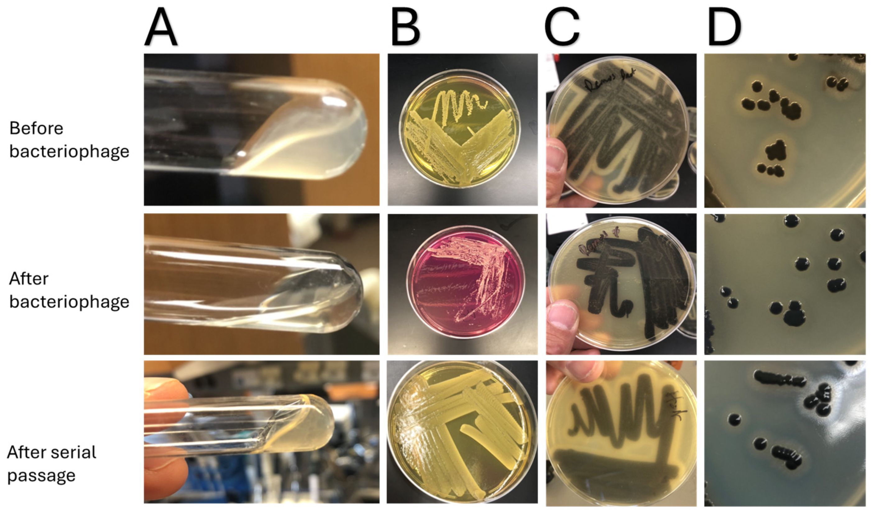
Before exposure to bacteriophages, all the S. aureus strains could hemolyze red blood cells (beta-hemolysis on blood agar plates), had DNase activity (seen on DNase plates), could ferment mannitol, and had lecithinase activity (seen on Baird-Parker agar) (Figure 2). Moreover, all had positive test tube and slide coagulase tests (Figure 1). After exposure to the podoviridae bacteriophages (44a and/or 68) that had activity against the bacterial isolates, both clinical NF isolates still retained all the enzymatic phenotypic activities assessed. However, when the S. aureus strains were exposed to myoviridae bacteriophages (K or Remus) that had activity against the strains, all lost the ability to hemolyze red blood cells, ferment mannitol, lost lecithinase activity, and lost the ability to form coagulase in the test tube test (Figure 1). Yet all the strains retained DNase activity and slide coagulase ability. When these same bacteria were grown on tryptic soy agar devoid of myoviridae bacteriophages, these same strains regained baseline enzymatic abilities after two passages.
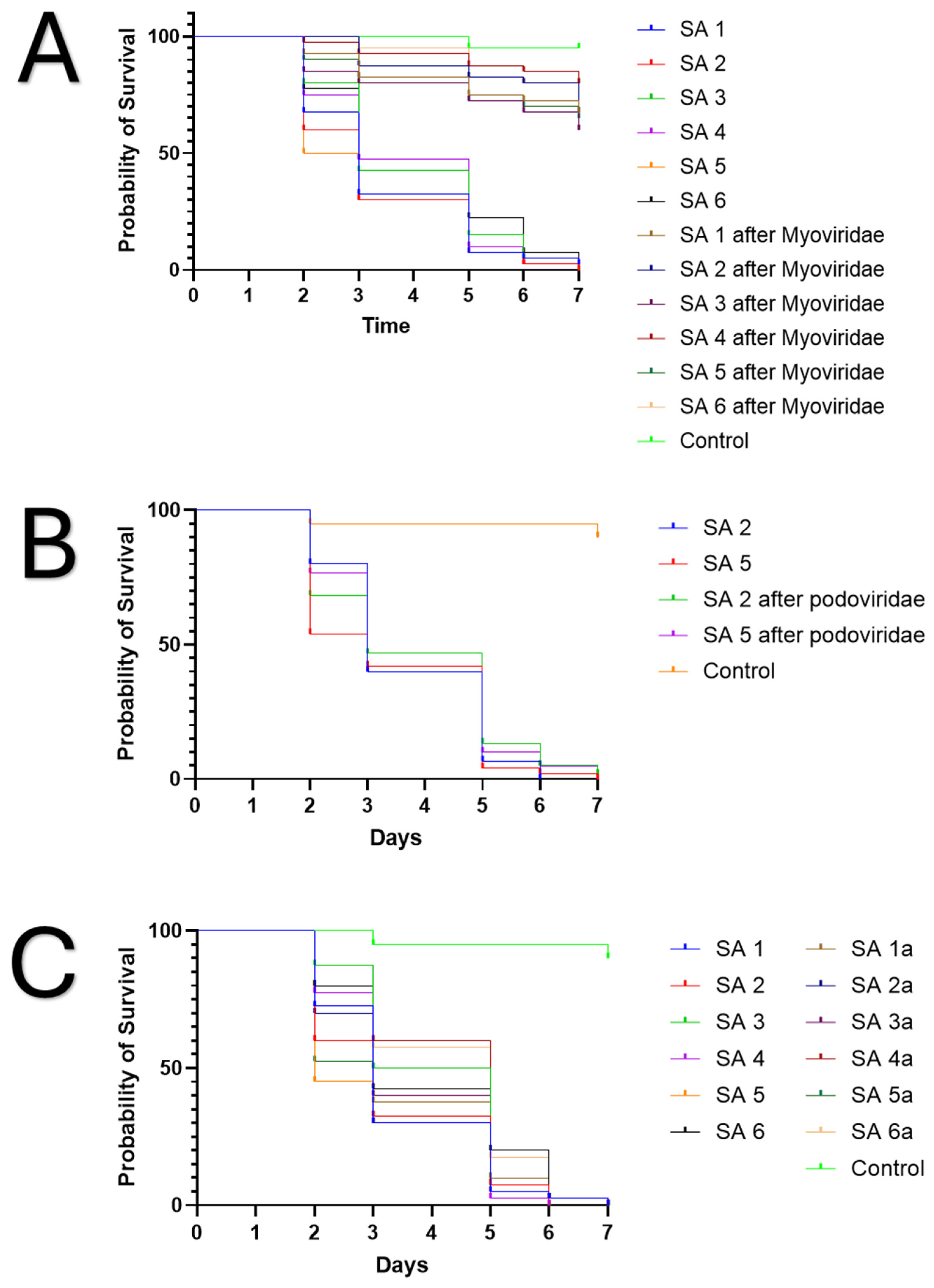
Furthermore, before exposure to bacteriophages, all the strains killed C. elegans within six days. Yet after exposure to myoviridae bacteriophages, which had activity against the individual bacteria, all the strains had a statistically significant decrease in the ability to kill the worms over 7 days (Figure 3A). This was not seen after exposure to podoviridae bacteriophages, in which there was no statistical difference in viability of worms after 7 days (Figure 3B). Moreover, after passage of the strains on tryptic soy agar devoid of bacteriophages, all of them regained their ability to kill C. elegans, similar to that seen before exposure to bacteriophages after two passages (Figure 3C). Pairwise statistical calculations can be seen in Supplementary Table S1.

4. Discussion

At this nascent stage, bacteriophage therapy is a novel therapeutic that has typically been relegated to aid in treating chronic recalcitrant infections, given that the current treatment paradigm requires a clinical isolate to ensure a bacteriophage therapeutic’s activity in vitro before clinical use [8,11]. This stems from the theoretical narrow spectrum of activity of some bacteriophages, which hinders the ability to use this therapeutic in acute infections, as there is not ample time to conduct this analysis. Yet some bacteriophages are promiscuous with broad host ranges. The ability to have broader host ranges is the result of bacteriophages using binding sites on attachment receptors that are highly conserved amongst a wide variety of bacterial strains [12,13]. As a result, creating therapeutics that utilize these highly conserved binding sites allows the potential to use bacteriophages empirically in acute infections. Thus, understanding bacteriophage attachment receptors is vital to utilizing these agents in acute infections.
Staphylococcal bacteriophages utilize wall teichoic acid (WTA) for attachment, but the two main clinically relevant families of Staphylococcal bacteriophages, podoviridae and myoviridae, bind at different locations on WTA [13]. Staphylococcal podoviridae bacteriophages bind to the distal end of WTA that can have widely different affinities based on the WTA glycosylation patterns [12,13]. On the other hand, Staphylococcal myoviridae bacteriophages bind to the proximal end of WTA that are less impacted by WTA glycosylation [12,13]. Consequently, Staphylococcal myoviridae can have broad host ranges amongst S. aureus strains (Table 1) and as seen by others [14,15]. This is important for creating Staphylococcal bacteriophage NF therapeutics, given that there is not ample time to determine in vitro activity. Rather, in order to use bacteriophages for S. aureus NF, use of myoviridae bacteriophage cocktails will offer a broad host range and robust lytic activity to numerous clinical strains of S. aureus. This will mitigate the need to ensure in vitro activity initially and allow for empiric use of bacteriophage therapy for a large percentage of S. aureus NF clinical isolates.
While it is assumed that bacteriophages will lyse all bacteria present, in nature, bacteriophages have evolved to live in predator-prey dynamics with bacteria, and thus the rise and fall of phage populations follow the rise and fall of bacterial populations. Bacteriophages will eradicate a large portion of bacteria, but do not fully eradicate entire bacterial populations. This is commonly seen in vitro after bacteriophage bacterial growth inhibition assays, where bacteria overgrow bacteriophages after a temporal delay [11]. Bacteria evade bacteriophage predation via several mechanisms, but the most common way is by modifying bacteriophage attachment receptors, thereby not allowing for phage attachment [16,17]. For S. aureus this is associated with modifications of WTA, which is a major virulence factor [18,19]. Therefore, there might be an evolutionary trade-off in that with WTA modification, there are associated phenotypic changes that can be utilized clinically. In S. aureus NF, a reduction in enzyme expression with respect to degradation of lipids and red blood cells, and formation of clots may translate into reduced inflammation and tissue necrosis, thereby reducing morbidity and mortality.
In this study we utilized this principle and evaluated the bacterial overgrowth that occurred after more than 16 h of bacteriophage-induced bacterial growth inhibition. Here we show that all the S. aureus NF clinical isolates that evaded Staphylococcal myoviridae bacteriophage predation had less enzymatic activity with respect to hemolysis, lecithinase, secreted coagulase activity, and ability to ferment mannitol (Figure 2). The reduction in enzymatic expression was not universal with respect to the different families of bacteriophages. For podoviridae, no changes in enzyme expression were seen. This likely occurred because podoviridae Staphylococcal bacteriophages bind to distal ends of WTA, and thus post-translational modifications to these regions with respect to glycosylation can easily alter bacteriophage affinity and activity but have little impact on enzyme expression [12,13]. For bacteria to evade Staphylococcal myoviridae bacteriophages, WTA alterations are likely much more pronounced, given that the phage attachment regions are on more proximal portions of WTA and thus, more radical modifications are needed, which drastically change bacterial phenotypic expression of enzymes. Further reinforcing this are other studies implicating WTA in translocation of S. aureus enzymes across the cell wall [18]. These findings are important to realize, given that bacteriophages can not only lyse bacterial cells but also reduce specific S. aureus enzyme activity, allowing for an NF therapeutic with dual mechanisms of activity.
Interestingly all bacteria, regardless of exposure to myoviridae or podoviridae, still retained DNase activity and slide coagulase activity. This likely occurred because DNAse activity is used by S. aureus to degrade extracellular DNA, thereby allowing for escape and spread to other environments, allowing means to escape from phage predation. As for the retention of slide coagulase activity, this likely demonstrates that despite modifications in WTA, S. aureus still retains the presence of membrane-bound coagulase, also known as clumping factor. This suggests that global membrane alterations likely do not occur when S. aureus evades myoviridae bacteriophage predation, but rather only through subtle changes to WTA. However, this will need to be clarified by use of nuclear magnetic resonance or mass spectrometry to correlate WTA changes with phenotypic changes seen here.
Reducing enzyme expression is important for NF, but the ability to reduce inflammatory processes that are associated with tissue necrosis is also paramount. Here we assessed this by evaluating changes in global bacterial virulence as seen with viability of C. elegans to the S. aureus strains before and after exposure to bacteriophages. After evasion of myoviridae bacteriophages, S. aureus became statistically less virulent compared to the same strain before bacteriophage exposure (Figure 3 and Supplementary Table S1). This did not occur with podoviridae bacteriophages. We hypothesize this likely occurred given the WTA modifications that are needed to evade bacteriophage predation. Yet this was a simplistic approach to assess global virulence, and follow-up studies are needed to assess specific alterations in immune responses that are associated with these findings in other models, such as wax moths or zebra fish [20,21]. Nonetheless, this further demonstrates that Staphylococcal myoviridae have immense promise as S. aureus NF therapeutics.
Overall, this was a pilot study that had important clinical findings, but it had several limitations. For one, this study only evaluated gross phenotypic changes. Follow-up studies with nuclear magnetic resonance or mass spectrometry are needed to clarify the mechanism associated with the changes seen here. However, the ability of the bacteria to revert back to virulent states after removing bacteriophage pressures suggests an inducible modification that supports our WTA hypothesis stated above. Secondly, this study only evaluated six NF S. aureus strains, and larger studies are needed to confirm the findings seen here and devise cocktails with ample activity against regionally diverse NF clinical isolates. However, this may require prospectively collecting NF S. aureus strains, given the potential lack of preserved S. aureus NF clinical isolates. Lastly, the clinical strains used did not undergo genomic evaluation to ensure that the same strains were present before and after bacteriophage predation. Yet the similar growth inhibition times after serial passage strongly suggest that these were the same bacterial strains (Supplementary Figure S1).

5. Conclusions

In conclusion, S. aureus NF is a life-threatening infection that drastically needs novel therapeutics to reduce morbidity and mortality. To use bacteriophage therapy for S. aureus NF cocktails of Staphylococcal myoviridae are likely needed to allow for broad host ranges, mitigating the need for in vitro sensitivity testing. Moreover, Staphylococcal myoviridae have the ability to not only lyse S. aureus but also reduce specific enzyme expression and global virulence of residual S. aureus. Therefore, as outlined here, the use of Staphylococcal myoviridae cocktails could potentially be a powerful adjuvant treatment for S. aureus NF. Yet follow-up studies are needed to clarify the mechanism associated with the phenotypic changes seen here and to conduct early clinical studies evaluating safety of myoviridae bacteriophages in S. aureus NF.

Supplementary Materials

The following supporting information can be downloaded at: https://www.mdpi.com/article/10.3390/jcm14165609/s1, Figure S1: Bacteriophage induced growth inhibtion curves; Table S1: Pairwise statistical comparisons of S. aureus clinical isolates virulence as seen with C. elegans survival before and after exposure to various bacteriophages.

Author Contributions

J.B.D. devised the experimentation and conducted the experimentation. D.D., S.J., M.M., L.B., J.M., B.A., R.M. and M.J. all helped review records and procure samples. K.L.U. helped oversee the experimentation. All authors have read and agreed to the published version of the manuscript.

Funding

We acknowledge the support of the University of Maryland, Baltimore; Institute for Clinical & Translational Research (ICTR); and the National Center for Advancing Translational Sciences (NCATS) Clinical Translational Science Award (CTSA) grant number 1UL1TR003098.

Institutional Review Board Statement

This study was approved by the University of Maryland internal review board (HP-00109342) and the Institutional Biosafety Committee (IBC-00007724). This was approved on 20 March 2024.

Informed Consent Statement

Patient consent was waived given this study was deemed not to require patient consent by the above mentioned Institutional Review Board.

Data Availability Statement

The data generated and analyzed during the current study is available upon reasonable request to the corresponding author.

Acknowledgments

We appreciate the help of the microbiology lab staff at the University of Maryland, Baltimore, in preserving these samples.

Conflicts of Interest

The authors declare no conflicts of interest.

References

  1. Bonne, S.L.; Kadri, S.S. Evaluation and Management of Necrotizing Soft Tissue Infections. Infect. Dis. Clin. N. Am. 2017, 31, 497–511. [Google Scholar] [CrossRef] [PubMed] [PubMed Central]
  2. Allaw, F.; Wehbe, S.; Kanj, S.S. Necrotizing fasciitis: An update on epidemiology, diagnostic methods, and treatment. Curr. Opin. Infect. Dis. 2024, 37, 105–111. [Google Scholar] [CrossRef] [PubMed]
  3. Puvanendran, R.; Huey, J.C.; Pasupathy, S. Necrotizing fasciitis. Can. Fam. Physician 2009, 55, 981–987. [Google Scholar] [PubMed]
  4. Koch, C.; Hecker, A.; Grau, V.; Padberg, W.; Wolff, M.; Henrich, M. Intravenous immunoglobulin in necrotizing fasciitis—A case report and review of recent literature. Ann. Med. Surg. 2015, 4, 260–263. [Google Scholar] [CrossRef] [PubMed]
  5. McDermott, J.; Kao, L.S.; Keeley, J.A.; Grigorian, A.; Neville, A.; de Virgilio, C. Necrotizing Soft Tissue Infections: A Review. JAMA Surg. 2024, 159, 1308–1315. [Google Scholar] [CrossRef] [PubMed]
  6. Chiang, K.J.; Wang, Y.T.; Kang, E.; Wu, Y.C.; Huang, C.U.; Lin, X.Y.; Tsai, F.C.; Tsai, C.S.; Chen, Y.H.; Wang, F.Y.; et al. Is Prompt Hyperbaric Oxygen Adjunctive Therapy Able to Reduce Mortality and Amputation in Management of Necrotizing Soft-Tissue Infection? Surg. Infect. 2024, 25, 659–667. [Google Scholar] [CrossRef] [PubMed]
  7. Doub, J.B.; Shishido, A.; Srikumaran, U.; Haskoor, J.; Tran-Nguyen, P.; Lee, M.; Würstle, S.; Lee, A.; Kortright, K.; Chan, B.K. Salphage: Salvage bacteriophage therapy for a recalcitrant Klebsiella pneumoniae prosthetic shoulder infection—A case report. Acta Orthop. 2022, 93, 756–759. [Google Scholar] [CrossRef] [PubMed] [PubMed Central]
  8. Doub, J.B. Bacteriophage therapy: Are we running before we have learned to walk? Eur. J. Clin. Microbiol. Infect. Dis. 2023, 42, 1281–1283. [Google Scholar] [CrossRef] [PubMed]
  9. Cohen, L.B.; Troemel, E.R. Microbial pathogenesis and host defense in the nematode C. elegans. Curr. Opin. Microbiol. 2015, 23, 94–101. [Google Scholar] [CrossRef]
  10. Kamaladevi, A.; Balamurugan, K. Response of Caenorhabditis elegans during Klebsiella pneumoniae pathogenesis. BMC Infect. Dis. 2014, 14, 8. [Google Scholar] [CrossRef]
  11. Suh, G.A.; Lodise, T.P.; Tamma, P.D.; Knisely, J.M.; Alexander, J.; Aslam, S.; Barton, K.D.; Bizzell, E.; Totten, K.M.C.; Campbell, J.L.; et al. Antibacterial Resistance Leadership Group. Considerations for the Use of Phage Therapy in Clinical Practice. Antimicrob. Agents Chemother. 2022, 66, e0207121. [Google Scholar] [CrossRef] [PubMed] [PubMed Central]
  12. Doub, J.B.; Urish, K.; Lee, M.; Fackler, J. Impact of Bacterial Phenotypic Variation with Bacteriophage therapy: A Pilot Study with Prosthetic Joint Infection Isolates. Int. J. Infect. Dis. 2022, 119, 44–46. [Google Scholar] [CrossRef] [PubMed]
  13. Moller, A.G.; Lindsay, J.A.; Read, T.D. Determinants of Phage Host Range in Staphylococcus Species. Appl. Environ. Microbiol. 2019, 85, e00209-19. [Google Scholar] [CrossRef] [PubMed] [PubMed Central]
  14. Azam, A.H.; Tanji, Y. Peculiarities of Staphylococcus aureus phages and their possible application in phage therapy. Appl. Microbiol. Biotechnol. 2019, 103, 4279–4289. [Google Scholar] [CrossRef]
  15. Totten, K.M.C.; Patel, R. Phage Activity against Planktonic and Biofilm Staphylococcus aureus Periprosthetic Joint Infection Isolates. Antimicrob. Agents Chemother. 2022, 66, e0187921. [Google Scholar] [CrossRef]
  16. Egido, J.E.; Costa, A.R.; Aparicio-Maldonado, C.; Haas, P.J.; Brouns, S.J.J. Mechanisms and clinical importance of bacteriophage resistance. FEMS Microbiol. Rev. 2022, 46, fuab048. [Google Scholar] [CrossRef]
  17. Bleriot, I.; Pacios, O.; Blasco, L.; Fernández-García, L.; López, M.; Ortiz-Cartagena, C.; Barrio-Pujante, A.; García-Contreras, R.; Pirnay, J.P.; Wood, T.K. Improving phage therapy by evasion of phage resistance mechanisms. JAC Antimicrob Resist. 2024, 6, dlae017. [Google Scholar] [CrossRef] [PubMed]
  18. Brignoli, T.; Douglas, E.; Duggan, S.; Fagunloye, O.G.; Adhikari, R.; Aman, M.J.; Massey, R.C. Wall Teichoic Acids Facilitate the Release of Toxins from the Surface of Staphylococcus aureus. Microbiol. Spectr. 2022, 10, e0101122. [Google Scholar] [CrossRef] [PubMed] [PubMed Central]
  19. Wanner, S.; Schade, J.; Keinhörster, D.; Weller, N.; George, S.E.; Kull, L.; Bauer, J.; Grau, T.; Winstel, V.; Stoy, H.; et al. Wall teichoic acids mediate increased virulence in Staphylococcus aureus. Nat. Microbiol. 2017, 2, 16257. [Google Scholar] [CrossRef] [PubMed]
  20. Sharma, S.; Singh, K.; Chaurasiya, A.; Banerjee, T.; Singh, R.; Yadav, G.; Kumar, A. Comparative study of phenotypic and genotypic expression of virulence factors in colonizing and pathogenic carbapenem resistant Acinetobacter baumannii (CRAB). BMC Microbiol. 2025, 25, 13. [Google Scholar] [CrossRef] [PubMed] [PubMed Central]
  21. Gálvez-Silva, M.; Varas, M.A.; Allende, M.L.; Chávez, F.P.; Marcoleta, A.E. Zebrafish Larvae Microinjection and Automated Fluorescence Microscopy for Studying Klebsiella pneumoniae Infection and the Host Immune Response. Methods Mol. Biol. 2025, 2852, 171–179. [Google Scholar] [CrossRef] [PubMed]
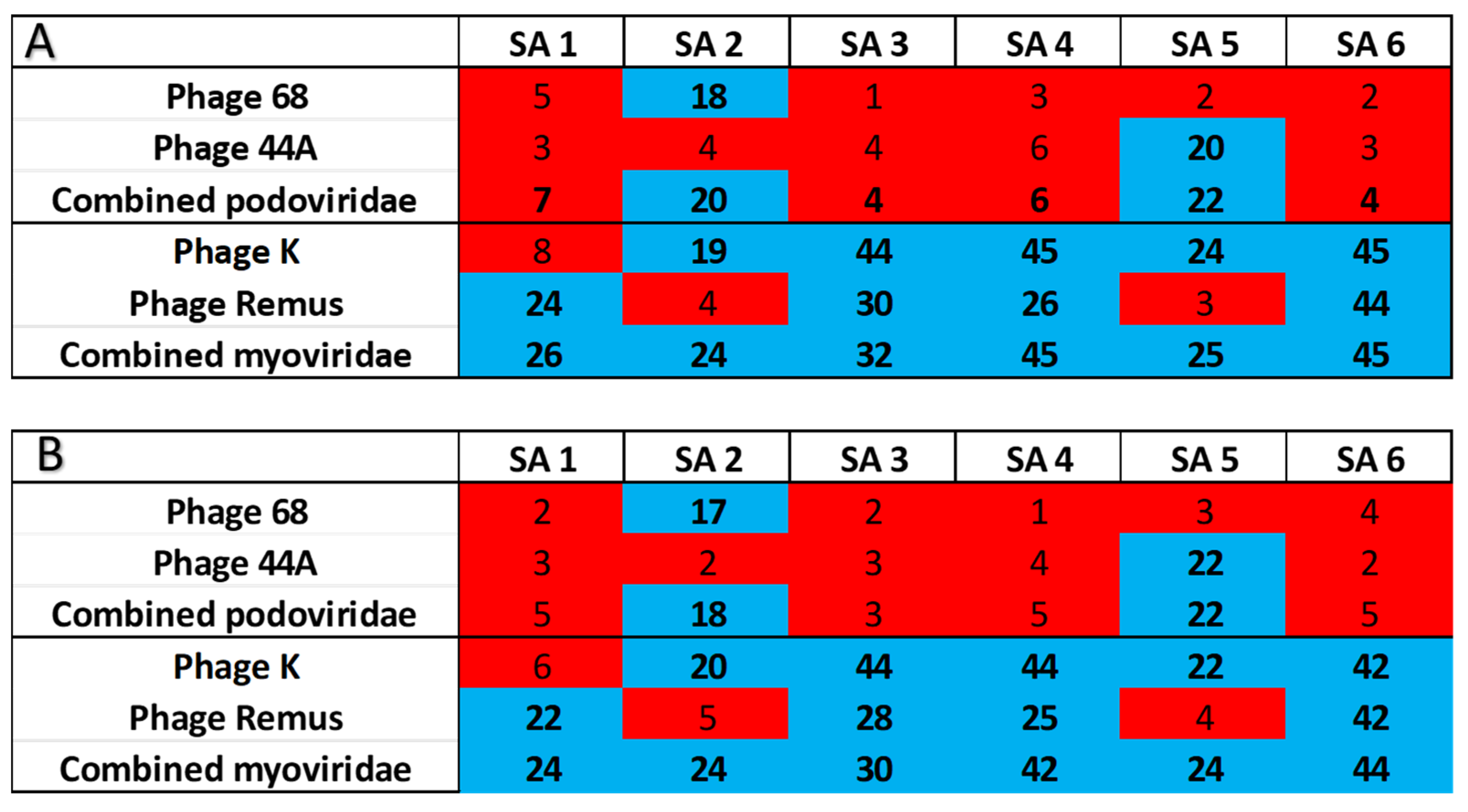
Figure 1. Results of bacteriophage-induced growth inhibition assays for each S. aureus clinical isolate (SA1-6) with individual bacteriophages and combined bacteriophages of the same family. Growth inhibition times are listed, where growth inhibition above 16 h is represented by blue rectangles and red rectangles represent times less than 10 h: (A) Growth inhibition times with initial exposure of S. aureus isolates to the different bacteriophages. (B) After S. aureus strains were exposed to bacteriophages and then serially passed on tryptic soy agar, similar growth inhibition patterns were observed.
Jcm 14 05609 g001
Figure 2. Changes to the phenotypic expression of enzymes before and after exposure to various bacteriophages, as well as after serial passage on tryptic soy agar plates: (A) Changes in phenotypic expression of secreted coagulase as seen with the test tube coagulase test. The top picture shows a positive coagulase test before exposure to myoviridae Staphylococcal bacteriophages, and the middle picture shows the same S. aureus strain after exposure to Staphylococcal myoviridae in which no coagulase activity is observed. The bottom picture shows phenotypic results after serial passage on agar plates in which reversion back to the wild-type phenotype, which expressed secreted coagulase, was seen. (B) Changes in the ability to ferment mannitol. The top picture shows the strains before exposure to the myoviridae Staphylococcal phage in which the bacteria can ferment mannitol. The middle picture shows the same strains after exposure to Staphylococcal myoviridae in which the strains could not ferment mannitol. The bottom picture refers to after serial passage on agar plates in which reversion back to the wild-type phenotype with the ability to ferment mannitol was seen. (C,D) Changes in lecithinase activity as seen on Baird-Parker agar with addition of egg yolk. All of the S. aureus strains before and after exposure to the bacteriophages were capable of reducing tellurite, as seen with black colonies on agar. Before exposure to Staphylococcal myoviridae bacteriophages, lecithinase activity was seen with an opaque zone around colonies (top (C,D)). Yet after exposure to myoviridae, the S. aureus strains did not express this enzyme (middle pictures). However, after serial passage on agar plates, reversion back to the wild-type phenotype in which lectithinase activity was observed (bottom pictures).
Jcm 14 05609 g002
Figure 3. Changes in NF S. aureus virulence after exposure to the Podoviridae and Myoviridae Staphylococcal bacteriophages: (A) C. elegans Kaplan–Meier survival curve, which shows the log-rank test with statistically significant (p < 0.0001) decreases in virulence of the S. aureus strains before and after exposure to myoviridae as quantified by C. elegans mortality after phage predation with myoviridae. (B) C. elegans survival assay, which shows no statistical difference (p = 0.4047) in virulence as quantified by C. elegans mortality after phage predation with Podoviridae. (C) C. elegans survival assay, which shows no statistical difference (p = 0.1106) in virulence after isolates were grown on agar devoid of myoviridae bacteriophages.
Jcm 14 05609 g003
Table 1. Isolate coverage as seen with plaque assays and growth inhibition greater than 16 h.
Podoviridae (P68, 44A) Myoviridae (K, Remus)
Combined coverage 2/6 (33.3%) 6/6 (100%)
Single phage 1/6 (16.7%) 15/6 (83.3%) 2
1 P68 and 44A bacteriophages were only able to have adequate activity against one isolate. 2 K and Remus bacteriophages had adequate activity against five of the six isolates.
Disclaimer/Publisher’s Note: The statements, opinions and data contained in all publications are solely those of the individual author(s) and contributor(s) and not of MDPI and/or the editor(s). MDPI and/or the editor(s) disclaim responsibility for any injury to people or property resulting from any ideas, methods, instructions or products referred to in the content.