Article

Impact of Corneal Crosslinking on Endothelial and Biomechanical Parameters in Keratoconus

by
Maria-Silvia Dina
1,*,
Maria-Cristina Marinescu
1,
Cătălina-Gabriela Corbu
2,
Mihaela-Monica Constantin
2,
Cătălina-Ioana Tătaru
3 and
Călin-Petru Tătaru
3
1
Doctoral School, Carol Davila University of Medicine and Pharmacy, 020021 Bucharest, Romania
2
Oftaclinic Ophthalmology Clinic, 040254 Bucharest, Romania
3
Ophthalmology Department, “Carol Davila” University of Medicine and Pharmacy, 020021 Bucharest, Romania
*
Author to whom correspondence should be addressed.
J. Clin. Med. 2025, 14(13), 4489; https://doi.org/10.3390/jcm14134489
Submission received: 2 May 2025 / Revised: 26 May 2025 / Accepted: 21 June 2025 / Published: 25 June 2025
(This article belongs to the Section Ophthalmology)

Abstract

Background/Objectives: Keratoconus (KC) is a corneal ectatic disease, characterized by the progressive thinning of the cornea, myopia, and astigmatism, which lead to a decrease in visual acuity. Corneal collagen crosslinking (CXL) is an efficient method of stopping the progression of the disease. The objective of this study is to investigate the endothelial and biomechanical properties of the cornea in keratoconus patients, before and after undergoing corneal collagen crosslinking. Methods: A total of 66 eyes were diagnosed with progressive keratoconus and were recommended epi-off corneal crosslinking. Before the procedure, they were investigated with corneal topography (for minimum, maximum, average keratometry, and corneal astigmatism), specular microscopy (for the following endothelial cell parameters: number, density, surface, variability, and hexagonality), and an ocular response analyzer (for the following biomechanical parameters: corneal hysteresis and resistance factor). All measurements were repeated 1 month and 6 months after the intervention. Results: Several parameters differ according to the Amsler–Krumeich stage of keratoconus: in more advanced stages, patients present higher endothelial cell variability, a lower number of endothelial cells in the paracentral region of the cornea, lower CCT and CRF, and higher keratometry and astigmatism. Endothelial cell variability and number correlate with average keratometry, and there are also strong correlations between topography and CH and CRF. After CXL, the paracentral number of endothelial cells decreased; cell variability and average cell surface increased. Conclusions: More advanced keratoconus cases present with altered corneal biomechanics and topographical parameters, the endothelial layer also being affected proportional to the stage of the disease and also slightly affected after corneal collagen crosslinking.

1. Introduction

Keratoconus (KC) is a corneal ectatic disease, characterized by a progressive thinning of the cornea, myopia, and astigmatism, which lead to a decrease in visual acuity [1]. This disease is often found in younger people, appearing during puberty, progressing throughout the second and third decades, and stabilizing around the age of 40 years old [2]. Incidence is estimated to be in the range of 1.5–25 cases per 100,000 persons/year, with a higher rate in Middle Eastern and Asian patients [3].
One significant step in the treatment of keratoconus was represented by corneal collagen crosslinking (CXL), introduced in clinical practice in 2003, which halts ectasia progression [4]. CXL aims to stabilize corneal ectasia, being a surgical procedure that is used to increase the biomechanical strength of the cornea by forming chemical bonds between collagen fibrils, which leads to the inhibition of keratoconus progression, therefore maintaining visual acuity over time and preventing complications [5,6,7].
The epithelium-off CXL technique was first applied to human keratoconus corneas in 1998 and consists of epithelial removal, after which the corneal stroma is saturated with the photosensitizer riboflavin (vitamin B2) for 30 min. Then, ultraviolet A radiation (UVA, 370 nm) is applied, which produces reactive oxygen species, which in turn induce interfibrillar collagen crosslinks [8]. The reason for the removal of the epithelium has been described as allowing the adequate penetration of riboflavin into the stromal tissue, where it absorbs UVA light and produces actual crosslinking between collagen fibrils in the corneal stroma. The disadvantage of epithelial removal is that it causes significant pain and discomfort in the first few days postoperatively, in addition to a 3–8% chance of epithelial healing problems [9,10].
Specular microscopy is a noninvasive technique that allows us to visualize and analyze the corneal endothelium. Corneal endotheliopathy is a broad term used to classify several diseases and clinical conditions that affect the structure and function of the corneal endothelium. One of the physiological functions of the endothelium is to secrete a collagen matrix that forms the Descemet membrane, but the primary physiological function of the corneal endothelium is to maintain the health and transparency of the corneal stroma [11].
Corneal biomechanical properties present more and more significance in ocular pathology, such as glaucoma [12] and refractive errors (myopia [13] and hyperopia [14]). An Ocular Response Analyzer (ORA) is a device that measures two essential biomechanical parameters: corneal hysteresis (CH) and the corneal resistance factor (CRF). In keratoconus patients, studies have shown that CH and CRF values are statistically significantly lower compared to healthy individuals, and the values are even lower the more advanced the keratoconus is. Also, the CRF value is lower compared to the CH value in keratoconus patients, so the difference between CH and CRF is positive and increases as the condition progresses [15].
The objective of this study is to investigate the endothelial and biomechanical properties of the cornea in keratoconus patients, before and after undergoing corneal collagen crosslinking.

2. Materials and Methods

This study is designed as a retrospective, non-randomized, interventional study. All patients who presented at the Oftaclinic Clinic in Bucharest between 2019 and 2022 were screened for inclusion in this study. This study’s cohort was formed by applying the inclusion and exclusion criteria. All patients, and, in the case of minors, their legal representatives, offered informed consent. This study was approved by the Oftaclinic Ethics Committee (Approval 2/4 February 2019).
The inclusion criterion was the diagnosis of progressive keratoconus. Keratoconus was diagnosed and staged according to Amsler–Krumeich staging, and progression was defined as an increase of at least 1 diopter (D) within a year of follow-up of the steepest corneal meridian (Kmax), decreased visual acuity, or thinning of the cornea by more than 5% of the corneal thickness [16].
Patients were excluded from this study if they presented other ocular pathologies (vitreoretinal pathology, glaucoma, ocular hypertension, cataract, and history of corneal surgery or corneal disease—particularly Fuch’s endothelial dystrophy), if they were pregnant or breastfeeding, or if they were not compliant with the investigations needed (for example, low waveform score in Ocular Response Analyzer testing). All patients had normal intraocular pressure (under 21 mmHg) both at the first consultation and at the consultations following the intervention.
Furthermore, patients were excluded if they presented any of the following known CXL contraindications: known sensitivity against ingredients used during the procedure, corneal thickness under 400 microns, and history of corneal pathology, such as herpes simplex keratitis [17].
All patients underwent a complete ocular consultation, after being instructed to stop wearing soft contact lenses for at least 2 weeks or rigid contact lenses for at least 4 weeks before the consultation [18]. During the consultation, the following assessments were also included:
-
Aladdin corneal topography (Topcon, Tokyo, Japan)—to determine maximum, minimum, average keratometry, and corneal astigmatism.
-
Specular microscopy (Nidek, Gamagori, Japan) for the determination of the following corneal endothelial parameters: number of cells, cell density, average cell surface, cell variability, and hexagonality—these parameters being calculated both centrally and paracentrally, and central corneal thickness.
-
Ocular Response Analyzer (ORA) (Reichert Ophthalmic Instruments Inc., Depew, NY, USA)—in order to determine the following corneal biomechanical properties: corneal hysteresis (CH) and corneal resistance factor (CRF). The Goldmann-correlated intraocular pressure calculated with the ORA over 21 represented an exclusion criterion.
Topographic evaluation and specular microscopy were performed after hydrating the ocular surface, considering that patients with keratoconus have a shorter tear film break-up time compared to the general population [19].
Patients were classified according to the Amsler–Krumeich classification of keratoconus (one criterion being needed to assign a certain stage to the eye), which accounted for myopia and astigmatism degree, mean keratometry, and minimal central corneal thickness [20].
The crosslinking procedure was performed in cases of progressive keratoconus, following the inclusion and exclusion criteria [21].
The crosslinking intervention followed the standard protocol. Stopping the progression of keratoconus was achieved by photo-oxidative crosslinking “epi-off” technique, a method that involves epithelial removal. In order to perform the procedure, local topical anesthesia is required, which does not influence the surgical procedure. After administering anesthesia and cleaning the regional tissue with Betadine (Povidone Iodate), a sterile field was applied. A speculum was applied to keep the eyelids open. After epithelial removal mechanically, the corneal stroma was saturated with riboflavin (vitamin B2) 0.1% in 20% dextran by photosensitization for a certain period of time (1 drop every 2 min for 30 min). Subsequently, ultraviolet A radiation (UVA, 370 nm) was applied for 30 min, continuing the infusion of riboflavin at 2 min (the “Dresden” technique) [21,22].
After the procedure, the patient was fitted with a therapeutic contact lens for 7 days, which plays a role in pain relief and the mechanical protection of the cornea [23].
Postoperative treatment consisted of the application of anti-inflammatory drops, topical antibiotics, and artificial tears for 14 days, which play a role in pain relief and the prevention of inflammation [23,24].
The data analyzed within this study originate from the pre-intervention consultation and from the consultations performed 1 month and 6 months after the CXL intervention.
The data analyzed in this article were tested with Levene’s Test, followed by the dependent t test, in order to identify significant differences between examinations (same patient—before the intervention, and 1 month and 6 months after the intervention). The 4 keratoconus stages were compared using the One-way ANOVA test, followed by the post hoc analysis Tukey test.
Pearson’s correlation coefficient (“Pearson’s r”) was calculated to determine the degree of correlation between variables (weak correlation: Pearson’s r between 0.3 and −0.3, moderate correlation: 0.3–0.5 or −0.3–−0.5, and strong correlation: over 0.5, under −0.5). The p value of 0.05 was considered the threshold for statistical significance. Statistical analysis was performed using the Statistical Package IBM SPSS Statistics for Windows, version 26 (IBM Corp., Armonk, NY, USA).

3. Results

3.1. Descriptive Results of the Entire Cohort

This study includes 47 patients, which provided 66 eyes for analysis (diagnosed with progressive keratoconus and recommended corneal crosslinking). A total of 17.02% of patients were female and 82.98% were male. The average age was 25.40 years old (SD 6.68). According to the Amsler–Krumeich classification, 48.48% of eyes were stage I keratoconus (32 eyes), 33.33% stage II (22 eyes), 12.12% stage III (8 eyes), and 6.06% stage IV (4 eyes). The average values of the parameters followed are in Table 1, before the procedure and after 1 and 6 months.
When analyzing the entire cohort, the endothelial layer was only minimally modified after CXL: cell variability increased significantly at 1 month after CXL (p 0.003), and more changes emerged at 6 months: cell variability was still significantly higher than before the procedure (p 0.001), average cell size was significantly higher (p 0.014), and the paracentral number of cells was significantly lower (p 0.038) (Figure 1) (Table 1).
The endothelial parameters did not differ significantly between the different KC stages: as the ectasia was more advanced, the cell variability increased (p value 0.033) (mean value of 24.81% in stage I, 24.77% in stage II, 26.00% in stage III, and 28.75% in stage IV); however, hexagonality was not significantly modified (67.19% in stage I, 69.96% in stage II, 65.50% in stage III, and 71.25% in stage IV). However, other parameters differed significantly: CRF was smaller (p value 0.006) and the central cornea was thinner (p < 0.001). Also, topographical parameters were more advanced, with higher minimum, maximum, and average keratometry and astigmatism (<0.001) in more advanced keratoconus cases (see Table 2).

3.2. Stage I Keratoconus Group—Evolution After Corneal Crosslinking

The stage I keratoconus group was composed of 32 eyes—all mean values are presented in Table 3. The paired T-test, comparing pre-CXL variables with post-CXL values at one and six months, reveals that very few parameters were modified after the procedure: the number of cells (central and paracentral), the hexagonality and paracentral cell density are significantly decreased at 6 months compared to the baseline, and cell variability is significantly increased both at 1 and at 6 months compared to the baseline—see Table 4.

3.3. Stage II Keratoconus Group—Evolution After Corneal Crosslinking

The stage II keratoconus group was composed of 22 eyes—all mean values are presented in Table 5. The paired T-test, comparing pre-CXL variables with post-CXL values at one and six months, reveals that few parameters were modified after the procedure, and the differences become significant only after 6 months (See Table 6). Cell density is significantly decreased at 6 months compared to the baseline; cell average surface area and variability are significantly increased at 6 months compared to the baseline. Furthermore, both Kmax and Kaverage are significantly lower at 6 months post-procedure.

3.4. Stage III Keratoconus—Evolution After Corneal Crosslinking

The stage III keratoconus group was composed of 8 eyes—all mean values are presented in Table 7. The paired T-test, comparing pre-CXL variables with post-CXL values at one and six months, reveals that CRF significantly improves after 1 month, and paracentral hexagonality is affected only after 6 months (See Table 8).

3.5. Stage IV Keratoconus—Evolution After Corneal Crosslinking

The stage IV keratoconus group was composed of 4 eyes—all mean values are presented in Table 9. The paired T-test, comparing pre-CXL variables with post-CXL values at one and six months, reveals only a modified CRF average at one month after the procedure (see Table 10).

3.6. Correlations in the Entire Cohort

Correlations were calculated between endothelial, biomechanical, and topographical parameters, in the initial pre-crosslinking cohort. However, no significant correlations were found between either of the endothelial parameters or the biomechanical parameters. On the other hand, there were several statistically significant correlations between paracentral endothelial parameters and topographic variables in the initial cohort. There are significant, weak, positive correlations between paracentral cell variability and either average keratometry and minimum keratometry. There are also significant, moderate, negative correlations between the paracentral number of endothelial cells and either maximum, minimum, or average keratometry—see Table 11.
Also, as expected, there are significant correlations between the biomechanical parameters and topographic parameters—CH has negative, moderate correlations with minimum, maximum, and average keratometry; CRF has negative, moderate correlations with minimum, maximum, and average keratometry and astigmatism; and CCT has negative, moderate–strong correlations with minimum, maximum, and average keratometry—see Table 12, Figure 2.

3.7. Correlations Between Baseline Values and Post-CXL Changes

By correlating the degree of change in several variables to the baseline values, we can understand how crosslinking influences both biomechanical and endothelial data. The degree of change in several endothelial parameters depended on the baseline biomechanical and topographical characteristics of the cornea—see Table 13. Firstly, there was a weak–moderate positive correlation between age and cell variability increase after CXL—the older the patient, the greater the variability increase in the endothelial layer—see Figure 3.
There was a weak–moderate negative correlation between CH and Avg cell surface increase at 6 months, and a negative moderate correlation between CRF and CV increase at 1 month, revealing that the higher the biomechanical parameters, the lower the damage to the endothelial layer in terms of cell surface and variability. There were also weak–moderate positive correlations between Kavg and the increase in Avg cell surface and variability at 6 months—the steeper the cornea (e.g., the more advanced the corneal ectasia), the greater the endothelial damage in terms of cell surface and variability.
There were also significant correlations between the baseline endothelial layer characteristics and the degree of change in the endothelial layer—see Table 14. There was a weak negative correlation between CV and the number of paracentral cells lost at 6 months, and a weak positive correlation between CV and the increase in cell surface at 6 months—the lower the variability in the endothelial layer, the lower the endothelial damage in terms of cells lost and surface increase—see Figure 4.

4. Discussion

In this study, we identified several differences between keratoconus eyes, depending on how advanced the corneal ectasia was, although this study was conducted on a small number of patients. According to the Amsler–Krumeich classification, the higher the stage, the greater the corneal astigmatism and average keratometry, and the thinner the cornea, these significant differences being found in our cohort as well.
Sturbaum and Peiffer (1993) found that in the early stages of keratoconus, the endothelium appears normal [25]. As the condition progresses and the corneal stroma thins, the endothelial cells flatten. This likely reflects the stretching of the endothelial cells as they attempt to maintain continuity across the progressively ectatic posterior surface [26]. Furthermore, the endothelium may be damaged in keratoconus through Descemet membrane breaks, contact lens wear and excessive eye rubbing, and thin corneas, which allow a greater deal of UV damage [27]. Later in the course of the disease, endothelial changes include pleomorphism, polymegathism, endothelial cell degeneration, and exposure of fibrin, inflammatory cells, or both on the endothelial surface. Endothelial and Descemet membrane ruptures occur in 11% to 35% of keratoconic corneas, and the healing of these defects can also lead to pleomorphism and polymegathism [26]. While advancing endothelial cell loss may decrease visual acuity through the loss of stromal transparency, unfortunately there is no definitive treatment to halt the loss of cells, except corneal transplantation in advanced cases [28].
There is research in the literature hinting at an altered endothelial cell layer in keratoconus. A large study of over 700 eyes revealed that more advanced cases present lower cell density and a higher variability [29]. This is probably due to the small number of patients with advanced stages included in our study. These results are similar to ours, however only the variability was significant in our cohort. On the other hand, there are studies in the literature that found no differences in density, variability, or hexagonality between Amsler–Krumeich KC stages [30].
A study applying deep learning technologies has found that an average number of cells in keratoconus is 175.00, comparable but slightly higher than our value of approximately 137 found in the present study [31]. More importantly, this average differed significantly to that of myopic eyes, serving as a control group [31]. Another recent study included Egyptian keratoconus patients, with Amsler–Krumeich stages 1–3, and found a significant decrease in cell density and an increase in variability [27]. Compared with our cohort, these parameters revealed a more significant alteration in the endothelial layer, suggesting that more research is needed to understand the endothelial behavior of keratoconus in different populations and which factors may influence endothelial health. Importantly, in our cohort, the average central endothelial cell density was sufficient to maintain corneal transparency (over 2900 cells/mm2), and the decrease in cell density was non-significant following CXL.
Keratoconus is a disease that involves the degradation of collagen fibers. Histologically, KC is characterized by a lower number of collagen fibers, in conjunction with a thinner stroma, abnormal keratocytes, affected Bowman’s membrane, and basement membrane with iron deposits [32]. Increased expression of the matrix metalloproteinase (MMP-1) leads to extracellular matrix degradation, affecting collagen, and a decreased expression of an enzyme responsible for natural collagen crosslinking contributes to a weaker corneal structure in keratoconus [33], with the addition of a higher lacrimal concentration of inflammatory markers in keratoconus patients [34]. As collagen is responsible for the elasticity and resistance of the cornea, it is known that CH and CRF are lowered in keratoconus. A large comparative study found an average of 7.5 mmHg and 6.2 mmHg for CH and CRF, respectively, significantly lower than the 10.8 and 11.0 averages in normal eyes [35]. These values are comparable with the averages obtained in our study. Furthermore, as expected, keratoconus variables (average keratometry and corneal thickness) correlate well with CH and CRF, similar to other results in the literature [36].
A biomechanical evaluation of the cornea holds great value as focal alterations may appear before the topographical alterations become evident, therefore holding great potential for early ectasia diagnosis [37]. CH, and even moreso, CRF, present good accuracy in differentiating normal and ectatic corneas [37]. Similarly, in our study, only CRF, not CH, differed significantly between stages of keratoconus, supporting the role of diagnostic and staging in the variations in CRF.
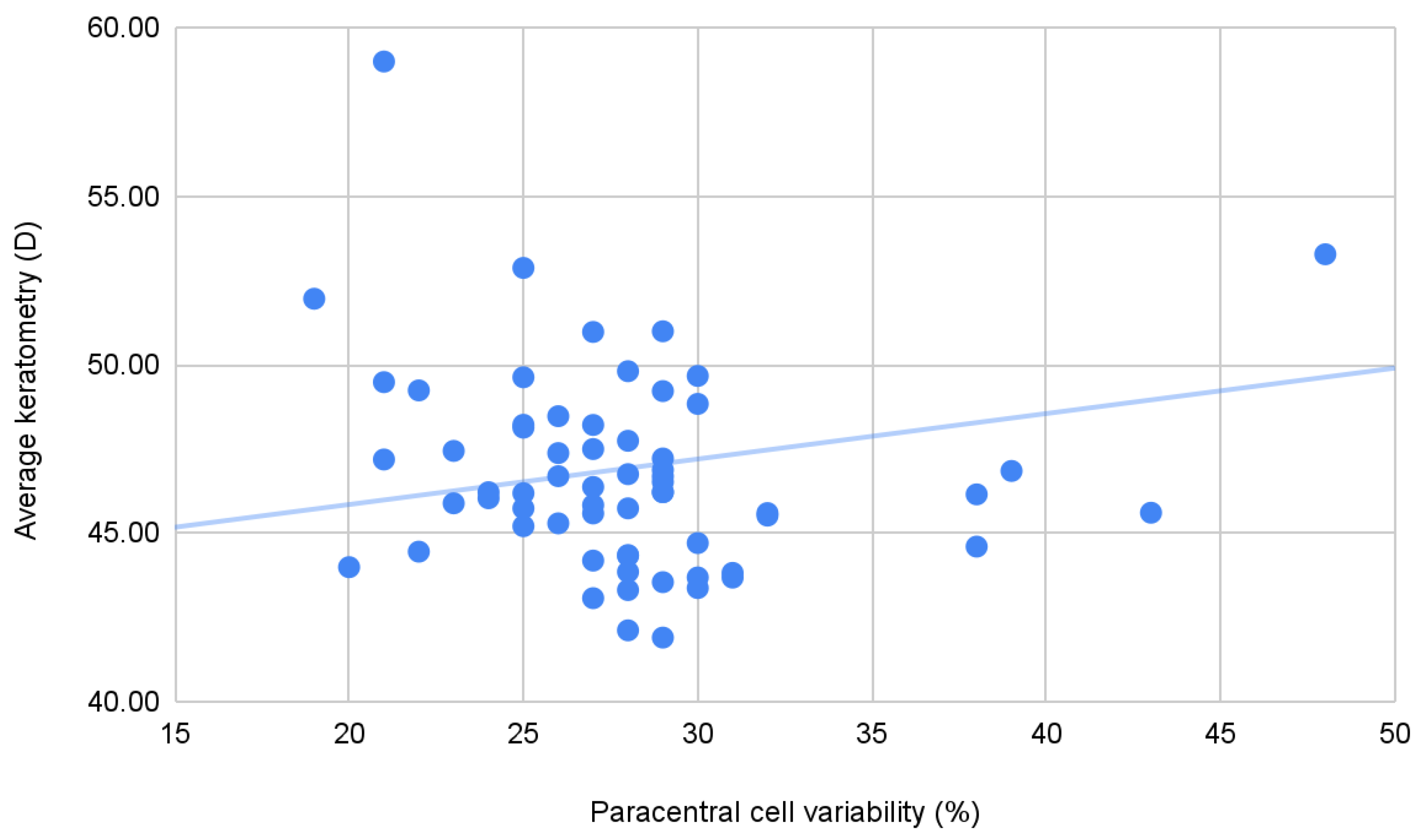
Besides the variable relationship with the disease stage, in our cohort, endothelial properties correlated with other parameters specific to KC—namely corneal astigmatism and keratometry readings (minimum, maximum, and average). These findings have been previously suggested in the literature, with endothelial cell density being negatively correlated with the steepest and flattest corneal meridian and positively correlated with minimum corneal thickness [38].
Corneal crosslinking is a well-established treatment in keratoconus that is used to stop KC evolution by increasing the biomechanical strength of the cornea. Using confocal microscopy, the corneal changes that occur after CXL could be examined. This procedure leads to microscopic changes, such as the appearance of a demarcation line, the apoptosis of keratocytes, and stroma edema—however, these changes generally resolve within six months and are accompanied by visual acuity increase [39].
In our study, there were no statistically significant changes in terms of corneal topography or biomechanics, as the follow-up reported here was only up to 6 months. Studies reporting from 1 year up to 7 years of follow-up describe significant improvements in keratometry and refractive errors, however less so in biomechanical parameters [40,
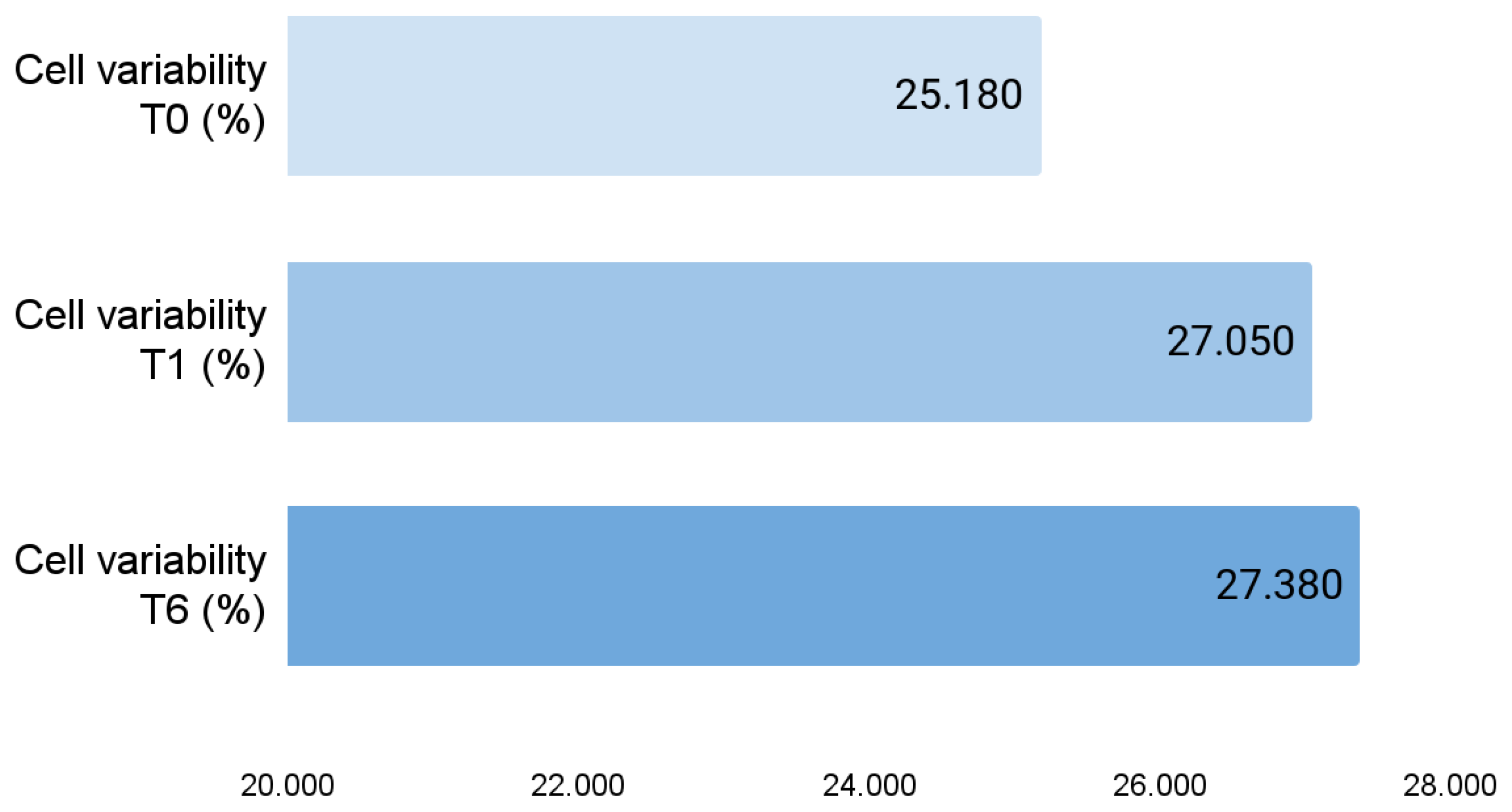
41]. On the other hand, certain endothelial alterations have been observed in our cohort, such as increased cell variability and average cell area, and decreased paracentral number of cells.
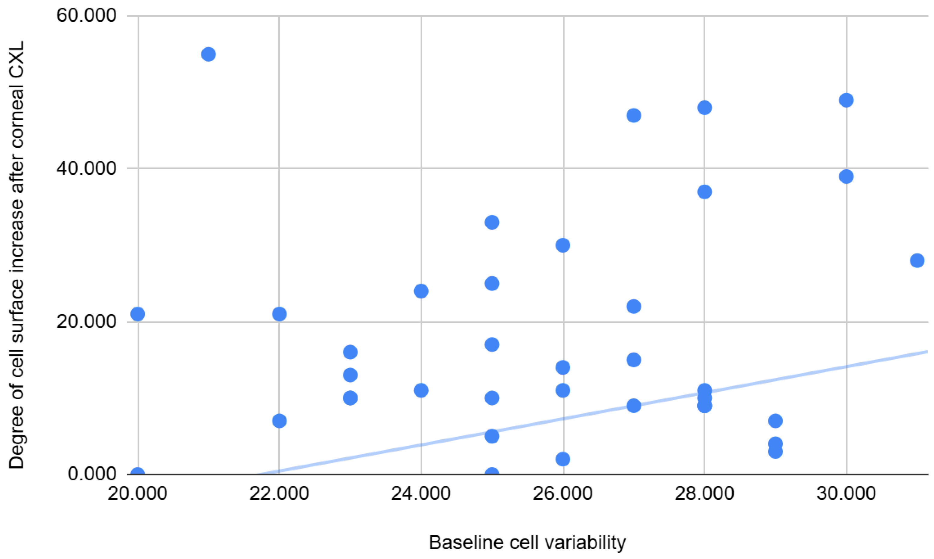
The literature states that increased cell variability is the first sign of cell damage, and the cell surface area increases with increasing CV [42]. Although in our cohort no patient had a coefficient higher than the normal limit, our study showed that a higher value of cellular variability before CXL was correlated with a decrease in the number of endothelial cells, but most importantly with an increase in cell surface after the procedure. A recent study that measured the effect of CXL on corneal endothelial cell morphology in patients with KC observed that the coefficient of variation was also statistically significantly higher at 6 months postoperatively than the initial value and there was a slight change in the percentage of hexagonal cells [43]. These observations can also be found in our cohort in stages I-III. Thus, although the corneal endothelium is not affected per se, the possibility of minor damage before CXL, suggested by the higher value of the coefficient of variation, which probably also increases after the CXL procedure, leads to an increase in the surface area of endothelial cells.
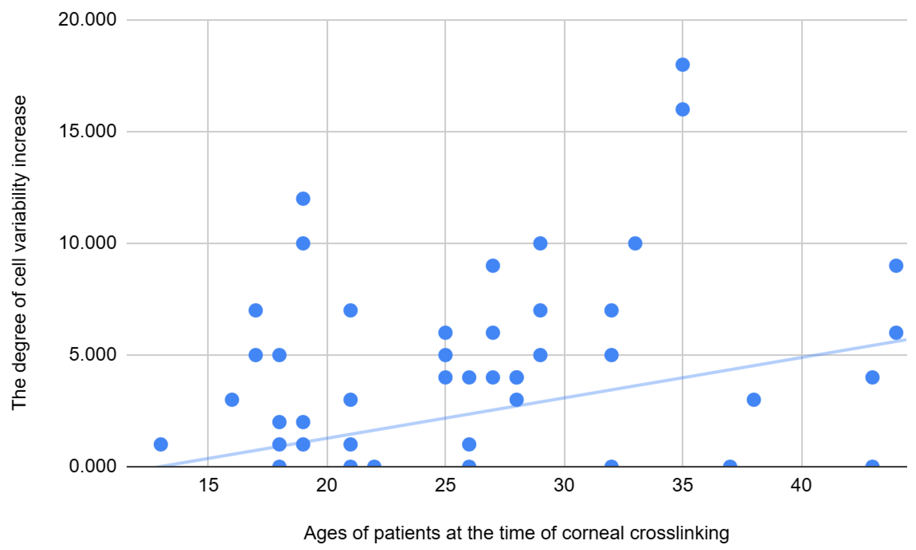
A study of 400 eyes of patients with keratoconus who underwent CXL and were followed for 4 years showed that the procedure stopped the progression of KC in all age groups, but the functional and morphological results were better in patients aged 18 to 39 years [44]. Similarly, our study showed a halt in the evolution of keratoconus after the crosslinking procedure in all patients, but the older the age, the greater the CV difference. On the other hand, it is known that in the healthy population, the corneal endothelial layer has a very high cell density at birth, which decreases with age at a rate of approximately 0.3–0.6% per year [45]. A study conducted on healthy volunteers aged 11 to 88 years in Thailand, which aimed to determine the influence of age on corneal endothelial thickness and morphology, showed a significant decrease in corneal thickness, cell density, and hexagonality, while endothelial cell surface area and cellular variation increased with age [46]. Although the age of the patients in our cohort does not exceed 50 years, the increase in the coefficient of endothelial variation after the crosslinking procedure at older ages could be explained by the overlap of some physiological processes.
Given the limitations of our study regarding the proportion of patients in stages III and IV compared to stages I and II, the correlation of corneal endothelial changes post-CXL at each stage is also limited. It is important, however, that the stages that included the largest number of patients, respectively I and II, showed morphological changes in endothelial cells that included a significant increase in the coefficient of variation and changes in the surface and hexagonal shape of endothelial cells at 6 months post-intervention. This is also found in a study comparing the effects of corneal crosslinking on the corneal endothelium in patients with KC compared to those with post-Lasik ectasia, which found that CV was statistically significantly higher 3 months after surgery compared to preoperative data in KC, and the percentage of hexagonal cells showed the smallest changes. In addition, similar to our study, a decrease in cell density occurred 6 months after CXL, which in their study recovered after one year [47].
A study similar to ours, with a follow-up of one year, has not found any differences in terms of cell density; however, it has not investigated other endothelial parameters that we found to be different before and after CXL [48]. Another recent study revealed a statistically significant decrease in cell density after 6 months; however, the final density did not impede endothelial function and corneal clarity [49]. The significant variation in endothelial parameters in the paracentral area after crosslinking obtained in our study could also be explained by the fact that the paracentral area in the patients in this study is the most deformed area and has the lowest corneal resistance (it coincides with the area of corneal ectasia). While these modifications are statistically significant, it is unlikely that they are clinically significant. The usage of riboflavin prevents UVA damage to endothelial cells, and more studies have reported no density decrease and far fewer have reported a decrease in endothelial cell density after CXL [50]. Most likely, significant changes occur in very thin keratoconic corneas, where the endothelium experiences a higher dose of UVA [51]. The UVA reaction from the crosslinking process damages subbasal nerves [47,52], which are essential in the endothelial pump. In this way, changes occur in the endothelial cells. Over time, endothelial cells that have not been affected tend to replace damaged cells [50].
In our study, a significant increase in CRF was also found in patients with stage III AK at 1 month post-CXL. However, this is inconsistent with stage IV. In any case, the small number of subjects in stages III and IV AK is a limitation of our study; therefore, more research is needed to determine if the biomechanical changes occurring post-CXL are more evident in more advanced ectatic corneas. Current studies on this topic are still under debate, with some studies finding no changes in either CH or CRF [53], and others showing increases in these parameters [54].
The methodology chosen for this study was a non-randomized one, because the patients followed treatment according to the clinical indication and could not be randomized, the only method to stop the evolution of keratoconus nowadays being the corneal crosslinking method. Otherwise, without the application of this treatment, with the progression of the disease, severe damage to the structure of the cornea leads to the need for a corneal transplant through different techniques (Bowman layer transplantation, deep anterior lamellar keratoplasty, or penetrating keratoplasty), techniques that bring with them the risk of damage to the endothelial layer, unrelated to keratoconic damage as much as the procedure itself [55,56,57].
Another significant limitation of this study is the minimal clinical data we have on our patients: no further data are known regarding atopic background or behavior, such as eye rubbing, which are known KC risk factors [58]. Furthermore, no data regarding visual acuity or method of visual correction (glasses, or rigid or soft contact lenses) are analyzed in this study, while it is known that, more often, visual correction in early KC stages is performed using spectacles or soft contact lenses, and in more advanced cases rigid lenses are more successfully used [59]. More research is needed through expanding the cohort of advanced KC (stage IV) and analyzing the influence of more factors on the progression of the disease and the response to corneal crosslinking.

5. Conclusions

In more advanced keratoconus, there is a decrease in the number of paracentral endothelial cells and an increase in variability, and a correlation between the characteristics of the endothelial layer and KC specific parameters—minimum, maximum, and average keratometry. This cellular layer is also affected after corneal crosslinking, the differences not being, however, clinically impactful.
More research is needed to determine if different corneal crosslinking protocols have a different effect on the endothelial layer, and also if the long-term follow-up of these patients would reveal a clinical significance of these endothelial alterations.

Author Contributions

Conceptualization, M.-S.D. and M.-C.M.; methodology, C.-G.C.; software, M.-S.D.; validation, M.-S.D., M.-C.M. and C.-I.T.; formal analysis, M.-M.C.; investigation, C.-I.T.; resources, C.-G.C.; data curation, M.-M.C.; writing—original draft preparation, M.-S.D.; writing—review and editing, M.-S.D.; visualization, M.-M.C.; supervision, C.-P.T.; project administration, C.-G.C. All authors have read and agreed to the published version of the manuscript.

Funding

This research received no external funding.

Institutional Review Board Statement

This study was conducted in accordance with the Declaration of Helsinki, and approved by the Oftaclinic Ethics Committee (Approval 2/4 February 2019).

Informed Consent Statement

Written informed consent was obtained from all subjects involved in this study.

Data Availability Statement

The datasets generated during and/or analyzed during the current study are available from the corresponding authors on reasonable request.

Acknowledgments

We would like to express our gratitude to Alexandra Vlagali for her valuable input and support with data management and statistical analysis.

Conflicts of Interest

The authors declare no conflicts of interest.

Abbreviations

The following abbreviations are used in this manuscript:
CXLCorneal collagen crosslinking
CHCorneal hysteresis
CRFCorneal resistance factor
IOPgGoldmann-correlated intraocular pressure
IOPccCornea-correlated IOP
KCKeratoconus

References

  1. Burcel, M.G.; Corbu, C.; Coviltir, V.; Potop, V.; Constantin, M.; Dascalescu, D.; Ionescu, C.; Ciuluvica, R.; Voinea, L.M. Corneal Crosslinking in Progressive Keratoconus: Comparison of Dextran-Based and Hydroxypropyl Methylcellulose—Based Riboflavin Solutions—Differences in Demarcation Line Depth and 1 Year Outcomes. Rev. Chim. 2019, 70, 1152–1156. [Google Scholar] [CrossRef]
  2. Tharini, B.; Sahebjada, S.; Borrone, M.A.; Vaddavalli, P.; Ali, H.; Reddy, J.C. Keratoconus in Pre-Teen Children: Demographics and Clinical Profile. Indian J. Ophthalmol. 2022, 70, 3508. [Google Scholar] [CrossRef] [PubMed]
  3. Santodomingo-Rubido, J.; Carracedo, G.; Suzaki, A.; Villa-Collar, C.; Vincent, S.J.; Wolffsohn, J.S. Keratoconus: An Updated Review. Cont. Lens Anterior Eye 2022, 45, 101559. [Google Scholar] [CrossRef] [PubMed]
  4. Wollensak, G.; Spoerl, E.; Seiler, T. Riboflavin/ultraviolet-a-Induced Collagen Crosslinking for the Treatment of Keratoconus. Am. J. Ophthalmol. 2003, 135, 620–627. [Google Scholar] [CrossRef]
  5. Kymionis, G.D.; Grentzelos, M.A.; Plaka, A.D.; Stojanovic, N.; Tsoulnaras, K.I.; Mikropoulos, D.G.; Rallis, K.I.; Kankariya, V.P. Evaluation of the Corneal Collagen Cross-Linking Demarcation Line Profile Using Anterior Segment Optical Coherence Tomography. Cornea 2013, 32, 907–910. [Google Scholar] [CrossRef]
  6. Yam, J.C.; Chan, C.W.; Cheng, A.C. Corneal Collagen Cross-Linking Demarcation Line Depth Assessed by Visante OCT After CXL for Keratoconus and Corneal Ectasia. J. Refract. Surg. 2012, 28, 475–481. [Google Scholar] [CrossRef]
  7. Doors, M.; Tahzib, N.G.; Eggink, F.A.; Berendschot, T.T.J.M.; Webers, C.A.B.; Nuijts, R.M.M.A. Use of Anterior Segment Optical Coherence Tomography to Study Corneal Changes After Collagen Cross-Linking. Am. J. Ophthalmol. 2009, 148, 844–851.e2. [Google Scholar] [CrossRef]
  8. Constantin, M.M.; Corbu, C.; Ionita, G. EPR Study on the Role of Riboflavin Used in Photo-Oxidative Collagen Cross-Linking. Rev. Chim. 2010, 61, 495–497. [Google Scholar]
  9. Salgado, J.P.; Khoramnia, R.; Lohmann, C.P.; von Mohrenfels, C.W. Corneal Collagen Crosslinking in Post-LASIK Keratectasia. Br. J. Ophthalmol. 2011, 95, 493–497. [Google Scholar] [CrossRef]
  10. Spadea, L. Corneal Collagen Cross-Linking with Riboflavin and UVA Irradiation in Pellucid Marginal Degeneration. J. Refract. Surg. 2010, 26, 375–377. [Google Scholar] [CrossRef]
  11. Philllips, C.; Laing, R. Yee Specular Microscopy. In Cornea E-Book; Krachmer, J.H., Mannis, M.J., Holland, E.J., Eds.; Elsevier Health Sciences: Philadelphia, PA, USA, 2005; pp. 261–277. ISBN 9780323081177. [Google Scholar]
  12. Potop, V.; Coviltir, V.; Corbu, C.; Burcel, M.G.; Ionescu, C.I.; Dascalescu, D.M.C. Corneal Hysteresis, a Glaucoma Risk Factor Independent of the Intraocular Pressure. Rev. Roum. Sci. Tech.–Électrotech. Énergétique 2019, 64, 297–300. [Google Scholar]
  13. Sun, Y.; Guo, Y.; Pang, R.; Peng, J.; Cao, K.; Wang, N. Corneal Biomechanical Properties and Potential Influencing Factors in Varying Degrees of Myopia. Sci. Rep. 2024, 14, 20626. [Google Scholar] [CrossRef] [PubMed]
  14. Marinescu, M.-C.; Dascalescu, D.-M.-C.; Constantin, M.-M.; Coviltir, V.; Potop, V.; Stanila, D.; Constantin, F.; Alexandrescu, C.; Ciuluvica, R.-C.; Voinea, L.-M. Particular Anatomy of the Hyperopic Eye and Potential Clinical Implications. Medicina 2023, 59, 1660. [Google Scholar] [CrossRef]
  15. Piñero, D.P.; Alcón, N. Corneal Biomechanics: A Review. Clin. Exp. Optom. 2015, 98, 107–116. [Google Scholar] [CrossRef] [PubMed]
  16. Kuechler, S.J.; Tappeiner, C.; Epstein, D.; Frueh, B.E. Keratoconus Progression After Corneal Cross-Linking in Eyes with Preoperative Maximum Keratometry Values of 58 Diopters and Steeper. Cornea 2018, 37, 1444–1448. [Google Scholar] [CrossRef] [PubMed]
  17. Mohammadpour, M.; Masoumi, A.; Mirghorbani, M.; Shahraki, K.; Hashemi, H. Updates on Corneal Collagen Cross-Linking: Indications, Techniques and Clinical Outcomes. J. Curr. Ophthalmol. 2017, 29, 235. [Google Scholar] [CrossRef]
  18. Sedaghat, M.-R.; Momeni-Moghaddam, H.; Roberts, C.J.; Maddah, N.; Ambrósio, R.; Hosseini, S.R. Corneal Biomechanical Parameters in Keratoconus Eyes with Abnormal Elevation on the Back Corneal Surface Only versus Both Back and Front Surfaces. Sci. Rep. 2021, 11, 11971. [Google Scholar] [CrossRef]
  19. Constantin, M.M.; Corbu, C.; Potop, V.; Burcel, M.; Mocanu, S.; Ionita, G. Evaluation of Dry Eye Symptomatology at Patients with Keratoconus. Rev. Chim. 2019, 70, 92–95. [Google Scholar] [CrossRef]
  20. Giannaccare, G.; Murano, G.; Carnevali, A.; Yu, A.C.; Vaccaro, S.; Scuteri, G.; Maltese, L.; Scorcia, V. Comparison of Amsler–Krumeich and Sandali Classifications for Staging Eyes with Keratoconus. Appl. Sci. 2021, 11, 4007. [Google Scholar] [CrossRef]
  21. Papachristoforou, N.; Ueno, A.; Ledwos, K.; Bartuś, J.; Nowińska, A.; Karska-Basta, I. A Review of Keratoconus Cross-Linking Treatment Methods. J. Clin. Med. 2025, 14, 1702. [Google Scholar] [CrossRef]
  22. Qureshi, M.; Watson, S.L.; Kandel, H. Pulsed Corneal Crosslinking in the Treatment of Keratoconus: A Systematic Review and Meta-Analysis. Graefes Arch. Clin. Exp. Ophthalmol. 2025, 263, 589–601. [Google Scholar] [CrossRef] [PubMed]
  23. Knutsson, K.A.; Genovese, P.N.; Paganoni, G.; Ambrosio, O.; Ferrari, G.; Zennato, A.; Cataldo, M.; Caccia, M.; Rama, P. Evaluation of a Post-Operative Therapy Protocol after Epithelium-Off Corneal Cross-Linking in Patients Affected by Keratoconus. J. Clin. Med. 2022, 11, 7093. [Google Scholar] [CrossRef] [PubMed]
  24. van der Valk Bouman, E.S.; Pump, H.; Borsook, D.; Severinsky, B.; Wisse, R.P.; Saeed, H.N.; Moulton, E.A. Pain Mechanisms and Management in Corneal Cross-Linking: A Review. BMJ Open Ophthalmol. 2021, 6, e000878. [Google Scholar] [CrossRef] [PubMed]
  25. Sturbaum, C.W.; Peiffer, R.L., Jr. Pathology of Corneal Endothelium in Keratoconus. Ophthalmologica 1993, 206, 192–208. [Google Scholar] [CrossRef]
  26. Masoud, M. Corneal Endothelial Changes in Keratoconus Patients; Benha University: Benha, Egypt, 2016. [Google Scholar]
  27. Elmassry, A.; Osman, A.; Sabry, M.; Elmassry, M.; Katkat, M.; Hatata, M.Y.; El-Kateb, M. Corneal Endothelial Cells Changes in Different Stages of Keratoconus: A Multi-Centre Clinical Study. BMC Ophthalmol. 2021, 21, 143. [Google Scholar] [CrossRef]
  28. Feizi, S. Corneal Endothelial Cell Dysfunction: Etiologies and Management. Ther. Adv. Ophthalmol. 2018, 10, 2515841418815802. [Google Scholar] [CrossRef]
  29. Goebels, S.; Eppig, T.; Seitz, B.; Szentmàry, N.; Cayless, A.; Langenbucher, A. Endothelial Alterations in 712 Keratoconus Patients. Acta Ophthalmol. 2018, 96, e134–e139. [Google Scholar] [CrossRef]
  30. El-Agha, M.S.; El Sayed, Y.M.; Harhara, R.M.; Essam, H.M. Correlation of Corneal Endothelial Changes with Different Stages of Keratoconus. Cornea 2014, 33, 707–711. [Google Scholar] [CrossRef]
  31. Wan, Q.; Wei, R.; Ma, K.; Yin, H.; Deng, Y.-P.; Tang, J. Deep Learning-Based Automatic Diagnosis of Keratoconus with Corneal Endothelium Image. Ophthalmol. Ther. 2023, 12, 3047–3065. [Google Scholar] [CrossRef]
  32. Labiris, G.; Gatzioufas, Z.; Sideroudi, H.; Giarmoukakis, A.; Kozobolis, V.; Seitz, B. Biomechanical Diagnosis of Keratoconus: Evaluation of the Keratoconus Match Index and the Keratoconus Match Probability. Acta Ophthalmol. 2013, 91, e258–e262. [Google Scholar] [CrossRef]
  33. Volatier, T.L.A.; Figueiredo, F.C.; Connon, C.J. Keratoconus at a Molecular Level: A Review. Anat. Rec. 2020, 303, 1680–1688. [Google Scholar] [CrossRef] [PubMed]
  34. Burcel, M.G.; Constantin, M.; Ionita, G.; Dascalescu, D.; Ionescu, C.; Stanila, D.; Potop, V.; Coviltir, V. Levels of Lactoferrin, Lysozyme and Albumin in the Tear Film of Keratoconus Patients and Their Correlations with Important Parameters of the Disease. Rev. Romana Med. Lab. 2020, 28, 153–161. [Google Scholar] [CrossRef]
  35. Ortiz, D.; Piñero, D.; Shabayek, M.H.; Arnalich-Montiel, F.; Alió, J.L. Corneal Biomechanical Properties in Normal, Post-Laser in Situ Keratomileusis, and Keratoconic Eyes. J. Cataract Refract. Surg. 2007, 33, 1371–1375. [Google Scholar] [CrossRef] [PubMed]
  36. Viswanathan, D.; Kumar, N.L.; Males, J.J.; Graham, S.L. Relationship of Structural Characteristics to Biomechanical Profile in Normal, Keratoconic, and Crosslinked Eyes. Cornea 2015, 34, 791–796. [Google Scholar] [CrossRef]
  37. Herber, R.; Hasanli, A.; Lenk, J.; Vinciguerra, R.; Terai, N.; Pillunat, L.E.; Raiskup, F. Evaluation of Corneal Biomechanical Indices in Distinguishing Between Normal, Very Asymmetric, and Bilateral Keratoconic Eyes. J. Refract. Surg. 2022, 38, 364–372. [Google Scholar] [CrossRef]
  38. Bozkurt, B.; Yılmaz, M.; Meşen, A.; Kamış, Ü.; Köktekir, B.E.; Okudan, S. Correlation of Corneal Endothelial Cell Density with Corneal Tomographic Parameters in Eyes with Keratoconus. Turk. J. Ophthalmol. 2017, 47, 255–260. [Google Scholar] [CrossRef]
  39. Khaled, M.L.; Helwa, I.; Drewry, M.; Seremwe, M.; Estes, A.; Liu, Y. Molecular and Histopathological Changes Associated with Keratoconus. BioMed Res. Int. 2017, 2017, 7803029. [Google Scholar] [CrossRef]
  40. Eslami, M.; Ghaseminejad, F.; Dubord, P.J.; Yeung, S.N.; Iovieno, A. Delayed Topographical and Refractive Changes Following Corneal Cross-Linking for Keratoconus. J. Clin. Med. 2022, 11, 1950. [Google Scholar] [CrossRef]
  41. Sedaghat, M.; Bagheri, M.; Ghavami, S.; Bamdad, S. Changes in Corneal Topography and Biomechanical Properties after Collagen Cross Linking for Keratoconus: 1-Year Results. Middle East Afr. J. Ophthalmol. 2015, 22, 212–219. [Google Scholar]
  42. Epshtein, D. What Specular Microscopy Reveals About Your Patients. Available online: https://www.reviewofcontactlenses.com/article/what-specular-microscopy-reveals-about-your-patients (accessed on 22 June 2025).
  43. Ellakwa, A.F.; Ibrahim, A.M.; El-Ghoubashy, E.S.; ELSaka, O.M.S. Effect of Collagen Cross-Linking on Corneal Endothelial Cell Count in Keratoconus Patients Using Specular Microscopy. Menoufia Med. J. 2022, 35, 85. [Google Scholar]
  44. Vinciguerra, R.; Romano, M.R.; Camesasca, F.I.; Azzolini, C.; Trazza, S.; Morenghi, E.; Vinciguerra, P. Corneal Cross-Linking as a Treatment for Keratoconus: Four-Year Morphologic and Clinical Outcomes with Respect to Patient Age. Ophthalmology 2013, 120, 908–916. [Google Scholar] [CrossRef]
  45. Balyen, L. The Physiology of Cornea. Available online: http://juniperpublishers.com/jojo/JOJO.MS.ID.555764.php (accessed on 10 June 2025).
  46. Tananuvat, N.; Khumchoo, N. Corneal Thickness and Endothelial Morphology in Normal Thai Eyes. BMC Ophthalmol. 2020, 20, 167. [Google Scholar] [CrossRef] [PubMed]
  47. Badawi, A.E. Corneal Endothelial Changes after Accelerated Corneal Collagen Cross-Linking in Keratoconus and postLASIK Ectasia. Clin. Ophthalmol. 2016, 10, 1891–1898. [Google Scholar] [CrossRef]
  48. Sun, L.; Zhao, J.; Zhang, X.; Shen, Y.; Tian, M.; Zhou, X. Transepithelial Accelerated Corneal Crosslinking for Keratoconus Eyes with Maximum Keratometry Values Larger than 58 Diopters. J. Cataract Refract. Surg. 2021, 48, 208–214. [Google Scholar] [CrossRef]
  49. Bahar, T.S.; Şahin, V.; Ayaz, Y.; Ünal, M. Long-Term Outcomes in Crosslinking Therapy for Patients with Progressive Keratoconus. Diagnostics 2025, 15, 626. [Google Scholar] [CrossRef] [PubMed]
  50. Subasinghe, S.K.; Ogbuehi, K.C.; Dias, G.J. Current Perspectives on Corneal Collagen Crosslinking (CXL). Arbeitsphysiologie 2018, 256, 1363–1384. [Google Scholar] [CrossRef] [PubMed]
  51. Yurttaser Ocak, S.; Mangan, M.S. Endothelial Cell Loss after Accelerated Corneal Crosslinking Using Pachymetry-Guided Hypo-Osmolar Riboflavin Dosing in Thin Keratoconic Corneas. J. Cataract Refract. Surg. 2021, 47, 1530–1534. [Google Scholar] [CrossRef]
  52. Mazzotta, C.; Hafezi, F.; Kymionis, G.; Caragiuli, S.; Jacob, S.; Traversi, C.; Barabino, S.; Randleman, J.B. In Vivo Confocal Microscopy after Corneal Collagen Crosslinking. Ocul. Surf. 2015, 13, 298–314. [Google Scholar] [CrossRef]
  53. Sedaghat, M.-R.; Momeni-Moghaddam, H.; Ambrósio, R., Jr.; Roberts, C.J.; Yekta, A.-A.; Danesh, Z.; Reisdorf, S.; Khabazkhoob, M.; Heidari, H.-R.; Sadeghi, J. Long-Term Evaluation of Corneal Biomechanical Properties After Corneal Cross-Linking for Keratoconus: A 4-Year Longitudinal Study. J. Refract. Surg. 2018, 34, 849–856. [Google Scholar] [CrossRef]
  54. Küçümen, R.B.; Şahan, B.; Yıldırım, C.A.; Çiftçi, F. Evaluation of Corneal Biomechanical Changes After Collagen Crosslinking in Patients with Progressive Keratoconus by Ocular Response Analyzer. Turk. J. Ophthalmol. 2018, 48, 160–165. [Google Scholar] [CrossRef]
  55. Fogla, R. Deep Anterior Lamellar Keratoplasty in the Management of Keratoconus. Indian J. Ophthalmol. 2013, 61, 465–468. [Google Scholar] [CrossRef] [PubMed]
  56. Vabres, B.; Bosnjakowski, M.; Bekri, L.; Weber, M.; Pechereau, A. Deep lamellar keratoplasty versus penetrating keratoplasty for keratoconus. J. Fr. Ophtalmol. 2006, 29, 361–371. [Google Scholar] [CrossRef] [PubMed]
  57. De Clerck, E.E.B.; Bravetti, G.E.; Kropp, M.; Massa, H.; Pajic, B.; Thumann, G.; Guber, I. Bowman Layer Transplantation for Treating Keratoconus-Preliminary Findings. J. Clin. Med. 2023, 12, 2402. [Google Scholar] [CrossRef] [PubMed]
  58. Najmi, H.; Mobarki, Y.; Mania, K.; Altowairqi, B.; Basehi, M.; Mahfouz, M.S.; Elmahdy, M. The Correlation between Keratoconus and Eye Rubbing: A Review. Int. J. Ophthalmol. 2019, 12, 1775–1781. [Google Scholar] [CrossRef]
  59. Deshmukh, R.; Ong, Z.Z.; Rampat, R.; del Barrio, J.L.A.; Barua, A.; Ang, M.; Mehta, J.S.; Said, D.G.; Dua, H.S.; Ambrósio, R., Jr.; et al. Management of Keratoconus: An Updated Review. Front. Med. 2023, 10, 1212314. [Google Scholar] [CrossRef]
Figure 1. Bar charts representing average endothelial cell variability before CXL, after 1 month, and after 6 months.
Jcm 14 04489 g001
Figure 2. Correlations between average keratometry and paracentral cell variability in the entire study cohort before crosslinking.
Jcm 14 04489 g002
Figure 3. Correlations between the age of the patients and cell variability increase at 6 months after the procedure.
Jcm 14 04489 g003
Figure 4. Correlations between the baseline cell variability and the degree of cell surface increase after CXL.
Jcm 14 04489 g004
Table 1. Mean values and standard deviation (SD) in the entire cohort before (T0), 1 month after CXL (T1), and 6 months after CXL (T6). Bold items signify statistically significant differences compared to the baseline value.
Mean ± SD Mean ± SD
Number of cellsT0136.86 ± 48.38Number of cells (paracentral)T01110.45 ± 406.01
T1140.97 ± 52.48T11060.82 ± 487.53
T6127.30 ± 46.74T61003.98 ± 429.42
Cell density (cell/mm2)T02941.02 ± 281.53Cell density (paracentral) (cell/mm2)T02927.65 ± 253.68
T12950.27 ± 264.00T12932.82 ± 258.00
T62914.53 ± 276.43T62913.44 ± 247.38
Cell variability (%)T025.18 ± 2.76Cell variability (paracentral) (%)T028.11 ± 5.36
T127.05 ± 4.54T129.70 ± 9.57
T627.38 ± 5.20T629.38 ± 7.41
Cell hexagonality (%)T068.15 ± 6.18Cell hexagonality (paracentral) (%)T066.50 ± 4.31
T167.23 ± 5.47T166.94 ± 4.49
T667.11 ± 6.19T666.97 ± 4.33
Average cell surface (micrometers)T0342.73 ± 35.11Central corneal thickness (μm)T0496.27 ± 36.06
T1341.82 ± 32.43T1495.26 ± 40.99
T6348.61 ± 33.50T6495.86 ± 40.29
Corneal hysteresis (mmHg)T08.75 ± 1.37Corneal resistance factor (mmHg)T07.82 ± 1.74
T18.75 ± 1.73T17.83 ± 1.83
T68.76 ± 1.95T67.89 ± 2.34
Flattest meridian—Kmin (D)T045.05 ± 3.39Steepest meridian—Kmax (D)T049.51 ± 4.88
T145.10 ± 3.52T149.56 ± 4.92
T645.21 ± 3.48T649.50 ± 4.82
Average keratometry—Kavg (D)T047.50 ± 4.05Corneal astigmatism (D)T04.57 ± 2.99
T147.32 ± 4.03T14.50 ± 2.90
T647.34 ± 3.92T64.29 ± 3.04
Table 2. Mean values of significantly different parameters between keratoconus Amsler–Krumeich stages (before corneal collagen crosslinking).
KC Stages CVCRFCCTKavgCorneal Astigmatism
Stage IMean ± SD24.81 ± 2.668.55 ± 1.62507.31 ± 35.2544.94 ± 1.452.52 ± 1.31
Stage IIMean ± SD24.77 ± 3.0237.44 ± 1.65494.18 ± 27.5948.48 ± 2.264.97 ± 2.02
Stage IIIMean ± SD26.00 ± 1.3096.76 ± 1.57494.37 ± 10.0149.39 ± 3.258.98 ±0.96
Stage IVMean ± SD28.75 ± 1.506.42 ± 1.13423.25 ± 36.3558.82 ± 3.919.57 ± 3.40
Table 3. Mean values and standard deviation (SD) in stage I KC, before CXL, and after 1 and 6 months. NrCells = number of cells, CD = cell density, Avg = average cell surface, CV = cell variability, Hex = cell hexagonality, CCT = central corneal thickness, NrCellsPC = number of paracentral cells, CDPC = paracentral cell density, CVPC = paracentral cell variability, HexPC = paracentral hexagonality, CH = corneal hysteresis, CRF = corneal resistance factor, Kmin = minimum keratometry, Kmax = maximum keratometry, Kavg = average keratometry, and Astigm = astigmatism. Bold items signify statistically significant differences compared to the baseline value.
Mean ± SD Mean ± SD
Endothelial variables (both central and paracentral)
NrCells137.00 ± 48.7NrCellsPC1130.62 ± 452.2
NrCells I136.53 ± 48.42NrCellsPC I1053.50± 502.7
NrCells6120.06 ± 43.4NrCellsPC6943.00 ± 449.6
CD2909.87 ± 312.8CDPC2939.25 ± 267.8
CD I2956.50 ± 289.4CDPC I2950.53 ± 290.8
CD62915.53 ± 313.6CDPC62904.81 ± 272.4
CV24.81 ± 2.7CVPC28.75 ± 4.4
CV I28.69 ± 5.1CVPC I29.59 ± 6.2
CV628.19 ± 5.4CVPC629.69 ± 4.6
Hex67.19 ± 6.9HexPC65.28 ± 4.6
Hex I65.56 ± 6.3HexPC I65.34 ± 5.1
Hex665.00 ± 6.8HexPC665.63 ± 5
Avg346.56 ± 39.9CCT507.31 ± 35.2
Avg I341.69 ± 36CCT I509.03 ± 38
Avg6347.62 ± 39.5CCT6507.75 ± 38.2
Biomechanical corneal properties
CH9.12 ± 1.3CRF8.54 ± 1.6
CH I9.08 ± 1.4CRF I8.33 ± 1.6
CH69.03 ± 2.3CRF68.51 ± 2.6
Topographical properties
Kmin43.68 ± 1.5Kmax46.2 ± 1.7
Kmin I43.79 ± 1.7Kmax I46.45 ± 2
Kmin644.0 ± 2Kmax646.46 ± 2.3
Kavg44.94 ± 1.45Astigm2.52 ± 1.3
KavgI45.12 ± 1.7AstigmI2.66 ± 1.5
Kavg645.23 ± 1.9Astigm62.47 ± 2.06
Table 4. Significant differences in parameters before CXL, after CXL at 1 and 6 months, and p value of difference in stage I KC. NrCells = number of cells, CV = cell variability, Hex = cell hexagonality, NrCellsPC = number of paracentral cells, and CDPC = paracentral cell density.
Mean Difference (After CXL—Baseline) (p Value)
NrCells at 6 months vs. Baseline NrCells−16.94 (0.009)
CV at 1 month vs. Baseline CV3.87 (<0.001)
CV at 6 months bs. Baseline CV3.37 (0.002)
Hex at 6 months vs. Baseline Hex−2.19 (0.017)
NrCellsPC at 6 months vs. Baseline NrCellsPC−187.62 (0.013)
CDPC at 6 months vs. Baseline CDPC−34.437 (0.011)
Table 5. Mean values and standard deviation (SD) in stage II KC before CXL, and after 1 and 6 months. NrCells = number of cells, CD = cell density, Avg = average cell surface, CV = cell variability, Hex = cell hexagonality, CCT = central corneal thickness, NrCellsPC = number of paracentral cells, CDPC = paracentral cell density, CVPC = paracentral cell variability, HexPC = paracentral hexagonality, CH = corneal hysteresis, CRF = corneal resistance factor, Kmin = minimum keratometry, Kmax = maximum keratometry, Kavg = average keratometry, and Astigm = astigmatism. Bold items signify statistically significant differences compared to the baseline value.
Mean ± SD Mean ± SD
Endothelial variables (both central and paracentral)
NrCells135.91 ± 54.6NrCellsPC1125.36 ± 422.1
NrCells I148.27 ± 62.3NrCellsPC I1035.09 ± 532.1
NrCells6143.0 ± 50.5NrCellsPC61086.18 ± 484.5
CD2941.9 ± 296.2CDPC2908.32 ± 266.7
CD I2894.86 ± 272CDPC I2866.45 ± 255.6
CD62873.54 ± 267.1CDPC62876.23 ± 246.6
CV24.77 ± 3.0CVPC26.77 ± 2.8
CV I25.73 ± 3.1CVPC I29 ± 8.5
CV626.5 ± 4.5CVPC628.09 ± 4.8
Hex69.96 ± 5.2HexPC68 ± 3.9
Hex I69.04 ± 3.9HexPC I68.5 ± 3.4
Hex669.86 ± 4.2HexPC668.09 ± 3.3
CCT494.18 ± 27.6Avg343.36 ± 35.6
CCT I484.91 ± 39Avg I348.4 ± 32.9
CCT6485.05 ± 38Avg6352.73 ± 31.1
Biomechanical corneal properties
CH8.432 ± 1.446CRF7.441 ± 1.646
CH I8.718 ± 2.059CRF I7.586 ± 2.151
CH68.755 ± 1.122CRF67.696 ± 1.532
Topographical properties
Kmin45.995 ± 2.739Kmax50.962 ± 2.183
Kmin I45.812 ± 2.575Kmax I50.860 ± 2.415
Kmin645.725 ± 2.452Kmax650.614 ± 2.086
Kavg48.481 ± 2.262Astigm4.967 ± 2.017
KavgI48.301 ± 2.318AstigmI5.116 ± 1.970
Kavg648.169 ± 2.110Astigm64.890 ± 1.708
Table 6. Significant differences in parameters before CXL, after CXL at 1 and 6 months, and p value of difference in stage II KC. CD = cell density, Avg = average cell surface, CV = cell variability, Kmax = maximum keratometry, and Kavg = average keratometry.
Mean Difference (After CXL—Baseline) (p Value)
CD at 6 months vs. Baseline CD −68.36 (0.026)
Avg at 6 months vs. Baseline Avg9.36 (0.034)
CV at 6 months vs. Baseline CV1.73 (0.027)
Kmax at 6 months vs. Baseline Kmax−0.35 (0.042)
Kavg at 6 months vs. Baseline Kavg−0.31 (0.048)
Table 7. Mean values and standard deviation (SD) in stage III KC before CXL, and after 1 and 6 months. NrCells = number of cells, CD = cell density, Avg = average cell surface, CV = cell variability, Hex = cell hexagonality, CCT = central corneal thickness, NrCellsPC = number of paracentral cells, CDPC = paracentral cell density, CVPC = paracentral cell variability, HexPC = paracentral hexagonality, CH = corneal hysteresis, CRF = corneal resistance factor, Kmin = minimum keratometry, Kmax = maximum keratometry, Kavg = average keratometry, and Astigm = astigmatism. Bold items signify statistically significant differences compared to the baseline value.
Mean ± SD Mean ± SD
Endothelial variables (both central and paracentral)
NrCells151.87 ± 36.4NrCellsPC1066.87 ± 267.2
NrCells I156.12 ± 42.7NrCellsPC I1248.62 ± 420.3
NrCells6137.5 ± 29.8NrCellsPC61084.75 ± 238.5
CD3017.62 ± 114.5CDPC2993.75 ± 53.6
CD I3002.75 ± 1178CDPC I3009.75 ± 93.9
CD62971.5 ± 143.5CDPC62984.87 ± 87.1
CV26 ± 1.3CVPC27.75 ± 5.3
CV I25.37 ± 3.7CVPC I26.62 ± 4.1
CV626.87 ± 3.6CVPC627.37 ± 3.4
Hex65.5 ± 3.6HexPC66.12 ± 2.6
Hex I67 ± 3.5HexPC I67.75 ± 2.9
Hex665.5 ± 4.9HexPC668.5 ± 1.9
CCT494.37 ± 10Avg331.75 ± 13
CCT I501.62 ± 14Avg I333.37 ± 13.4
CCT6502.37 ± 23.7Avg6337.12 ± 16.1
Biomechanical corneal properties
CH8.62 ± 1.2CRF6.76 ± 1.6
CH I8.04 ± 1.8CRF I7.51 ± 1.3
CH68.47 ± 2.4CRF66.96 ± 2.9
Topographical properties
Kmin43.2 ± 1.9Kmax52.18 ± 2.7
Kmin I43.35 ± 1.6Kmax I51.85 ± 3
Kmin643.45 ± 1.5Kmax651.72 ± 3.5
Kavg49.39 ± 3.2Astigm8.98 ±0.9
KavgI47.60 ± 2.2AstigmI8.5 ± 1.8
Kavg649.39 ± 3.2Astigm68.28 ± 2.3
Table 8. Significant differences in parameters before CXL, after CXL at 1 and 6 months, and p value of difference in stage III KC. HexPC = paracentral hexagonality; CRF = corneal resistance factor.
Mean Difference (After CXL—Baseline)
(p Value)
HexPC at 6 months vs. Baseline Hex2.37 (0.004)
CRF at 1 month vs. Baseline CRF0.75 (0.004)
Table 9. Mean values and standard deviation (SD) in stage IV KC before CXL, and after 1 and 6 months. NrCells = number of cells, CD = cell density, Avg = average cell surface, CV = cell variability, Hex = cell hexagonality, CCT = central corneal thickness, NrCellsPC = number of paracentral cells, CDPC = paracentral cell density, CVPC = paracentral cell variability, HexPC = paracentral hexagonality, CH = corneal hysteresis, CRF = corneal resistance factor, Kmin = minimum keratometry, Kmax = maximum keratometry, Kavg = average keratometry, and Astigm = astigmatism. Bold items signify statistically significant differences compared to the baseline value.
Mean ± SD Mean ± SD
Endothelial variables (both central and paracentral)
NrCells111 ± 24.08NrCellsPC954.25 ± 30.8
NrCells I106 ± 36.6NrCellsPC I885.25 ± 59.3
NrCells678.5 ± 45.9NrCellsPC6878.25 ± 55.9
CD3032 ± 156.6CDPC2809 ± 337.7
CD I3100.250± 184.8CDPC I3002.25 ± 180.9
CD63018 ± 238.3CDPC63044.25 ± 261.5
CV28.75 ± 1.5CVPC34 ± 14.5
CV I24.5 ± 4.6CVPC I40.5 ± 29.3
CV626.75 ± 9.8CVPC638 ± 25.5
Hex71.25 ± 7.4HexPC68.75 ± 4.5
Hex I71 ± 5.2HexPC I69.5 ± 3.4
Hex672 ± 5.3HexPC668.5 ± 4.7
Avg330.5 ± 17.5CCT423.25 ± 36.4
Avg I323.5 ± 20.5CCT I429.25 ± 40.5
Avg6356.75 ± 14.8CCT6431 ± 46.8
Biomechanical corneal properties
CH7.9 ± 1.2CRF6.425 ± 1.1
CH I7.77 ± 1.3CRF I5.950 ± 0.9
CH67.25 ± 1.3CRF65.875 ± 1.4
Topographical properties
Kmin54.59 ± 2.9Kmax63.045 ± 5.7
Kmin I55.19 ± 4.5Kmax I63.078 ± 6.4
Kmin655.37 ± 2.6Kmax662.483 ± 6.3
Kavg58.82 ± 3.9Astigm9.568 ± 3.4
KavgI59.14 ± 4.9AstigmI7.883 ± 5.1
Kavg658.92 ± 3.9Astigm67.115 ± 5.6
Table 10. Significant differences in parameters before CXL, after CXL at 1 and 6 months, and p value of difference in stage IV KC. CRF = corneal resistance factor.
Mean Difference (After CXL—Baseline) (p Value)
CRF at 1 month vs. Baseline CRF0.475 (0.036)
Table 11. Significant correlations between topographical and endothelial corneal properties (before corneal collagen crosslinking).
Pair of VariablesPearson Coefficient of Correlation r (p Value)
Paracentral cell variability—Kmin0.329 (0.007)
Paracentral cell variability—Kmax0.272 (0.027)
Paracentral cell variability—Kavg0.294 (0.016)
Paracentral number of cells—Kmax−0.305 (0.014)
Paracentral number of cells—Kmin−0.323 (0.009)
Paracentral number of cells—Kavg−0.329 (0.007)
Table 12. Significant correlations between topographical and biomechanical corneal properties (before corneal collagen crosslinking).
Pair of VariablesPearson Coefficient of Correlation r (p Value)Pair of VariablesPearson Coefficient of Correlation r (p Value)
Corneal hysteresis—Kmax−0.302 (0.014)CCT—Kmax−0.492 (<0.001)
Corneal hysteresis—Kmin−0.329 (0.007)CCT—Kmin−0.535 (<0.001)
Corneal hysteresis—Kavg−0.330 (0.007)CCT—Kavg−0.536 (<0.001)
Corneal resistance factor—Kmax−0.504 (<0.001)Corneal resistance factor—Kmin−0.387 (0.001)
Corneal resistance factor—Kavg−0.479 (<0.001)Corneal resistance factor—Astigmatism−0.385 (0.002)
Table 13. Significant correlations between age, topographical and biomechanical baseline corneal properties, and the degree of difference in endothelial parameters.
Pair of VariablesPearson Correlation (p Value)
Age—CV 1-00.377 (0.002)
Age—CV 6-00.298 (0.015)
CH—Avg 6-0−0.285 (0.02)
CRF—CV 1-0−0.308 (0.012)
Kavg—Avg 6-00.349 (0.004)
Kavg—CV 6-00.250 (0.043)
Table 14. Significant correlations between baseline endothelial properties and the degree of difference in endothelial parameters.
Pair of VariablesPearson Correlation (p Value)
CV—NrCells 6-0−0.288 (0.019)
CV—Avg 6-00.248 (0.045)
Disclaimer/Publisher’s Note: The statements, opinions and data contained in all publications are solely those of the individual author(s) and contributor(s) and not of MDPI and/or the editor(s). MDPI and/or the editor(s) disclaim responsibility for any injury to people or property resulting from any ideas, methods, instructions or products referred to in the content.

