

Determination of Correlated Color Temperature in Ex Vivo Porcine Eyes during Intraocular Illumination

Abstract
1. Introduction
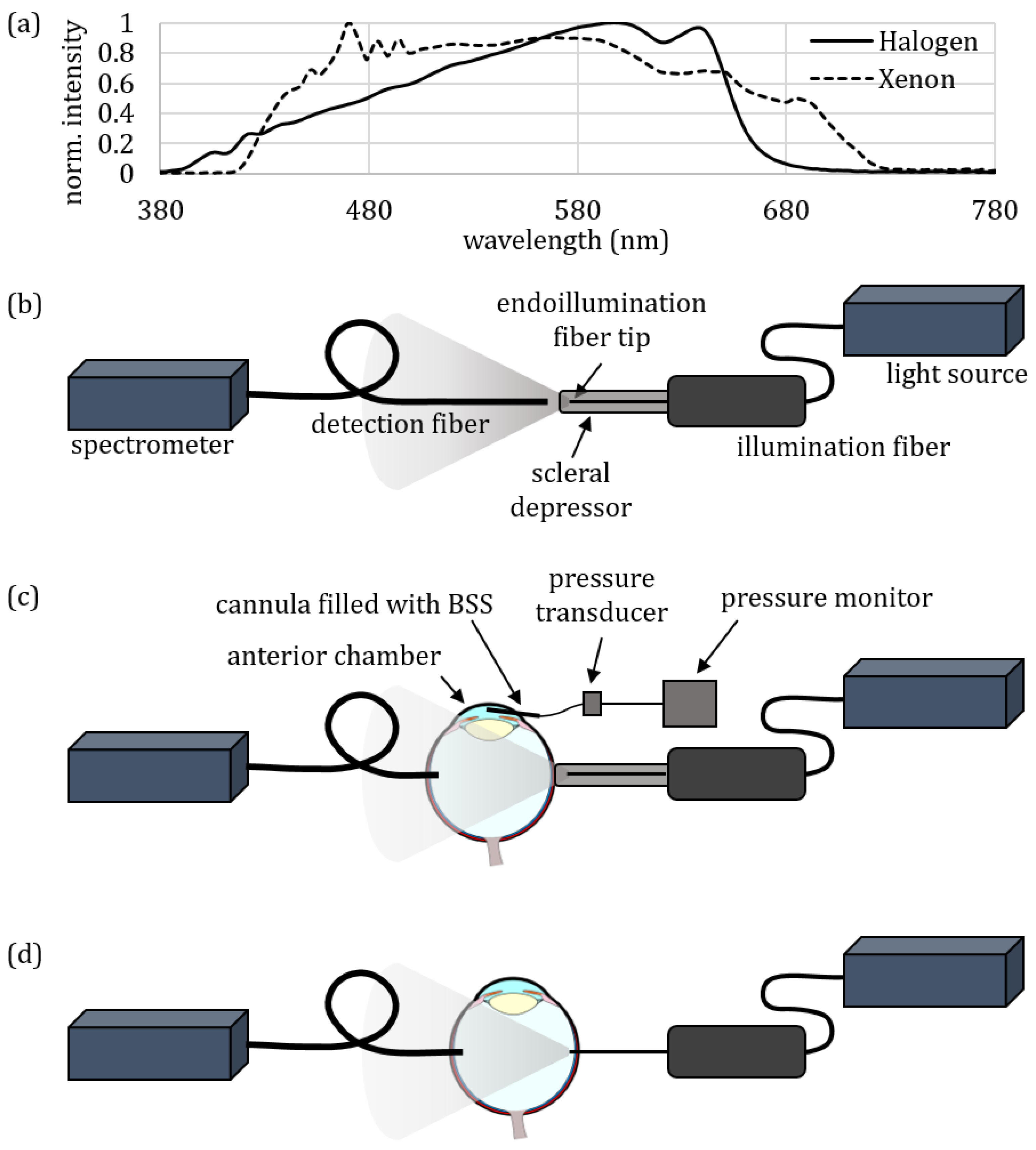
2. Materials and Methods
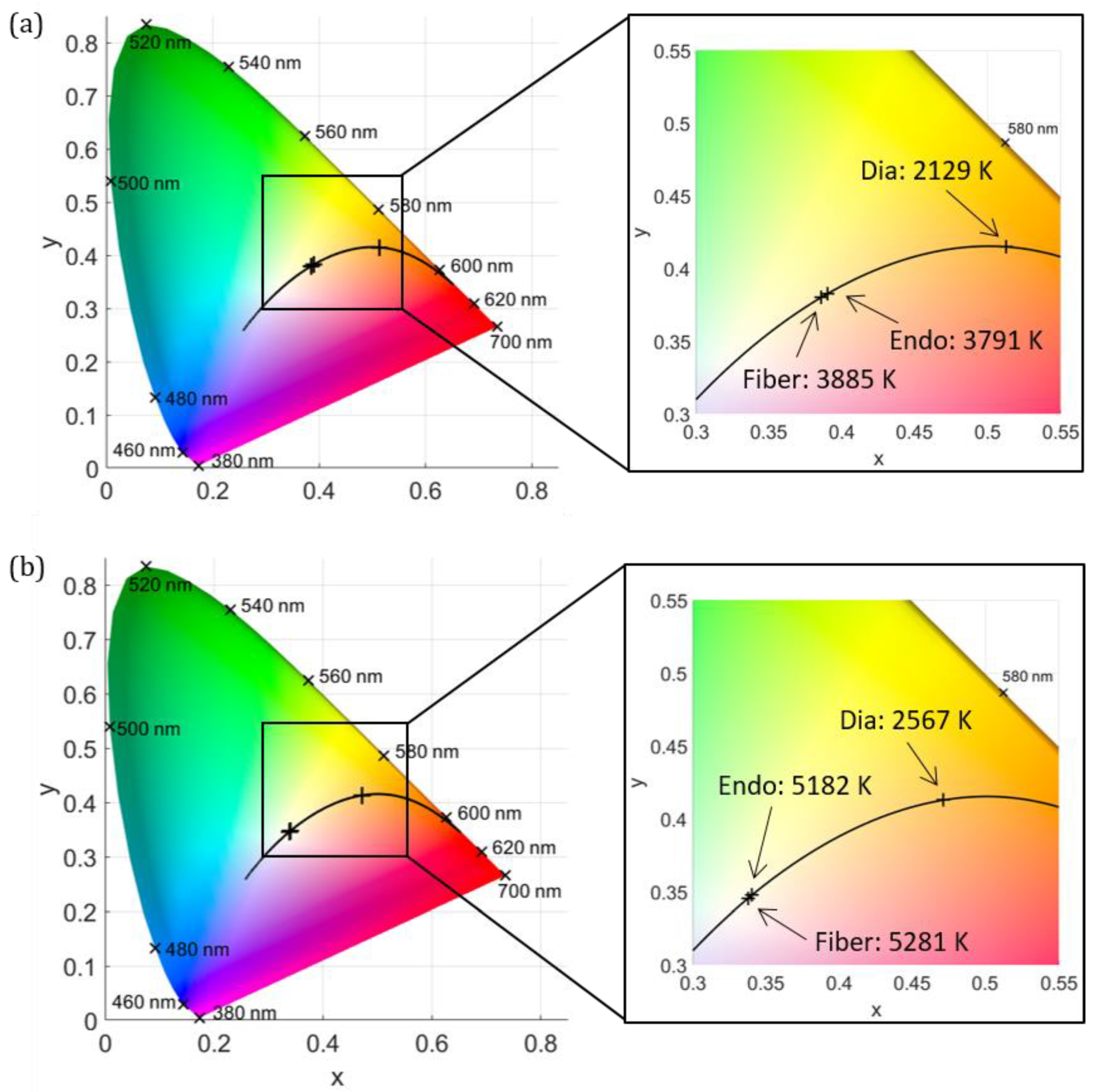
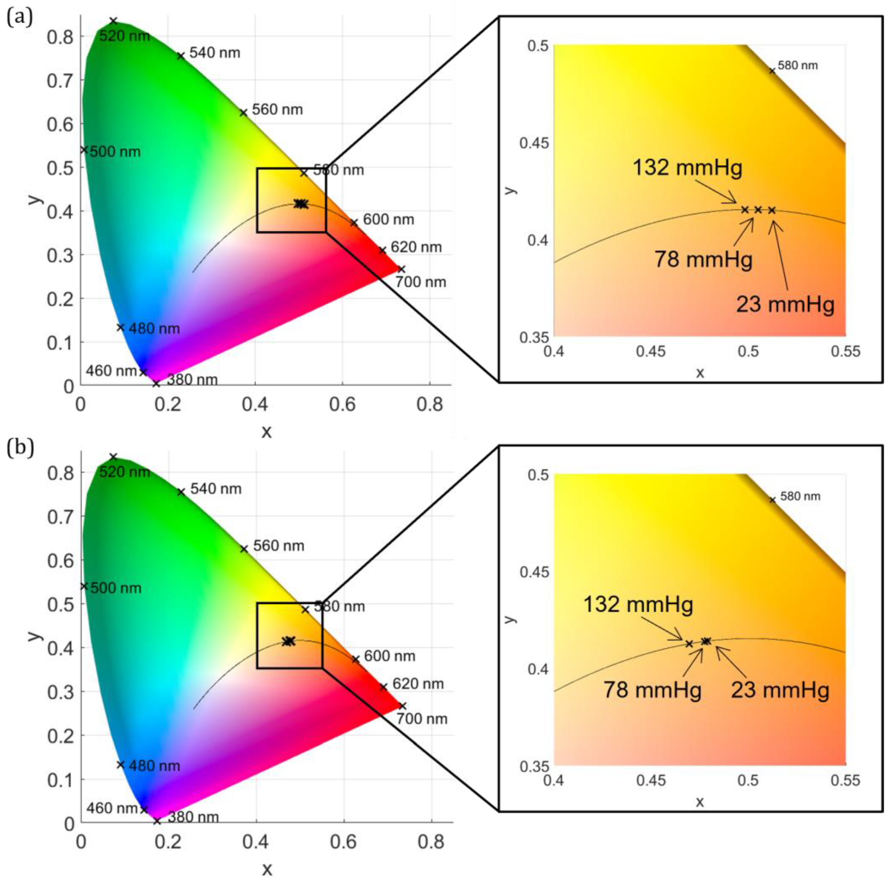
3. Results
4. Discussion
5. Conclusions
Author Contributions
Funding
Institutional Review Board Statement
Informed Consent Statement
Data Availability Statement
Conflicts of Interest
References
- Gea, M.; Schilirò, T.; Iacomussi, P.; Degan, R.; Bonetta, S.; Gilli, G. Cytotoxicity and genotoxicity of light emitted by incandescent, halogen, and LED bulbs on ARPE-19 and BEAS-2B cell lines. J. Toxicol. Environ. Health A 2018, 81, 998–1014. [Google Scholar] [CrossRef] [PubMed]
- Saxena, S.; Meyer, C.H. A trichrome RGB endoillumination prototype system: A novel application for chromovitrectomy. Ophthalmologica 2013, 230 (Suppl. S2), 73–76. [Google Scholar] [CrossRef]
- Brambilla, A.; Hu, W.; Samangouei, R.; Cadorin, R.; Davis, W. How correlated colour temperature manipulates human thermal perception and comfort. Build. Environ. 2020, 177, 106929. [Google Scholar] [CrossRef]
- Chaopu, Y.; Wenqing, F.; Jiancheng, T.; Fan, Y.; Yanfeng, L.; Chun, L. Change of blue light hazard and circadian effect of LED backlight displayer with color temperature and age. Opt. Express 2018, 26, 27021–27032. [Google Scholar] [CrossRef] [PubMed]
- Mohammadian, F.; Mehri, A.; Abedi, K.; Ebrahimi, H.; Laal, F.; Falahi, H. A Simplified Approach to Evaluate Retinal Blue Light Hazard Using the Correlated Color Temperature of LED Light Sources. J. Environ. Health Sci. Eng. 2021, 9, 299–310. [Google Scholar] [CrossRef]
- Bullough, J.D.; Bierman, A.; Rea, M.S. Evaluating the blue-light hazard from solid state lighting. Int. J. Occup. Saf. Ergon. 2019, 25, 311–320. [Google Scholar] [CrossRef] [PubMed]
- Jin, M.; Li, X.; Yan, F.; Chen, W.; Jiang, L.; Zhang, X. The effects of low-color-temperature dual-primary-color light-emitting diodes on three kinds of retinal cells. J. Photochem. Photobiol. B Biol. 2021, 214, 112099. [Google Scholar] [CrossRef]
- Xie, C.; Zhu, H.; Chen, S.; Wen, Y.; Jin, L.; Zhang, L.; Tong, J.; Shen, Y. Chronic retinal injury induced by white LED light with different correlated color temperatures as determined by microarray analyses of genome-wide expression patterns in mice. J. Photochem. Photobiol. B Biol. 2020, 210, 111977. [Google Scholar] [CrossRef] [PubMed]
- Litorja, M.; Brown, S.; Ohno, Y.; Lin, C. Illumination with Enhanced Contrast as a Visualization Tool for Clinical Diagnostics and Surgery; Litorja, M., Brown, S., Ohno, Y., Lin, C., Eds.; Biomedical Optics: San Jose, CA, USA, 2009. [Google Scholar]
- Argyraki, A.; Clemmensen, L.K.H.; Petersen, P.M. Does correlated color temperature affect the ability of humans to identify veins? J. Opt. Soc. Am. A 2016, 33, 141–148. [Google Scholar] [CrossRef]
- Hürzeler, D. Blue light endoscopy. Laryngoscope 1975, 85, 1374–1378. [Google Scholar] [CrossRef]
- Gono, K.; Obi, T.; Yamaguchi, M.; Ohyama, N.; Machida, H.; Sano, Y.; Yoshida, S.; Hamamoto, Y.; Endo, T. Appearance of enhanced tissue features in narrow-band endoscopic imaging. J. Biomed. Opt. 2004, 9, 568–577. [Google Scholar] [CrossRef] [PubMed]
- Litorja, M.; Brown, S.W.; Nadal, M.E.; Allen, D.; Gorbach, A. Development of surgical lighting for enhanced color contrast. In Proceedings of the SPIE—The International Society for Optical Engineering, San Diego, CA, USA, 17–22 February2007; Volume 6515, pp. 65150K-1–65150K-11. [Google Scholar] [CrossRef]
- Charles, S. Illumination and phototoxicity issues in vitreoretinal surgery. Retina 2008, 28, 1–4. [Google Scholar] [CrossRef] [PubMed]
- Sarao, V.; Veritti, D.; Borrelli, E.; Sadda, S.V.R.; Poletti, E.; Lanzetta, P. A comparison between a white LED confocal imaging system and a conventional flash fundus camera using chromaticity analysis. BMC Ophthalmol. 2019, 19, 231. [Google Scholar] [CrossRef] [PubMed]
- Mundinger, J.J.; Houser, K.W. Adjustable correlated colour temperature for surgical lighting. Light. Res. Technol. 2019, 51, 280–290. [Google Scholar] [CrossRef]
- Koelbl, P.S.; Sieber, N.; Lingenfelder, C.; Koch, F.H.J.; Deuchler, S.; Hessling, M. Pressure dependent direct transtissue transmission of eyewall, sclera and vitreous body in the range of 350–1050 nm. Z. Med. Phys. 2020, 30, 201–210. [Google Scholar] [CrossRef]
- Fehler, N.; Lingenfelder, C.; Kupferschmid, S.; Hessling, M. Intraocular reflectance of the ocular fundus and its impact on increased retinal hazard. Zeitschrift für Medizinische Physik 2022, 32, 453–465. [Google Scholar] [CrossRef] [PubMed]
- Vogel, A.; Dlugos, C.; Nuffer, R.; Birngruber, R. Optical properties of human sclera, and their consequences for transscleral laser applications. Lasers Surg. Med. 1991, 11, 331–340. [Google Scholar] [CrossRef] [PubMed]
- Sieber, N.; Kölbl, P.; Lingenfelder, C.; Stucke-Straub, K.; Kupferschmid, S.; Hessling, M. Measurement of the retinal irradiation exposure during diaphanoscopic illumination. Curr. Dir. Biomed. Eng. 2020, 6, 276–279. [Google Scholar] [CrossRef]
- Chan, E.K.; Sorg, B.; Protsenko, D.; O’Neil, M.; Motamedi, M.; Welch, A.J. Effects of compression on soft tissue optical properties. IEEE J. Sel. Top. Quantum Electron. 1996, 2, 943–950. [Google Scholar] [CrossRef]
- Koelbl, P.S.; Klante, P.; Koch, F.; Lingenfelder, C.; Werner, J.U.; Enders, C.; Hessling, M. Location and pressure dependent transmission of human and porcine sclera: An anterior to posterior examination. Graefes Arch. Clin. Exp. Ophthalmol. 2017, 255, 2185–2198. [Google Scholar] [CrossRef] [PubMed]
- Hessling, M.; Koelbl, P.S.; Lingenfelder, C.; Koch, F. Miniature LED Endoilluminators For Vitreoretinal Surgery. In Medical Laser Applications and Laser-Tissue Interactions VII: Proceedings of the European Conference on Biomedical Optics 2015, Munich, Germany, 21–25 June 2015; SPIE Proceedings Paper 95421A; Optica Publishing Group: Washington, DC, USA, 2015; Volume 9542. [Google Scholar]
- Nicoli, S.; Ferrari, G.; Quarta, M.; Macakuso, C.; Govoni, P.; Dallatana, D.; Santi, P. Porcine sclera as a model of human sclera for in vitro transport experiments histology, SEM, and comparative permeability. Mol. Vis. 2009, 15, 259–266. [Google Scholar] [PubMed]
- Sanchez, I.; Martin, R.; Ussa, F.; Fernandez-Bueno, I. The parameters of the porcine eyeball. Graefes Arch. Clin. Exp. Ophthalmol. 2011, 249, 475–482. [Google Scholar] [CrossRef]
- Beauchemin, M.L. The fine structure of the pig′s retina. Graefes Arch. Clin. Exp. Ophthalmol. 1974, 190, 27–45. [Google Scholar] [CrossRef]
- Olsen, T.W.; Sanderson, S.; Feng, X.; Hubbard, W.C. Porcine sclera: Thickness and surface area. Investig. Ophthalmol. Vis. Sci. 2002, 43, 2529–2532. [Google Scholar]
- De Schaepdrijver, L.; Simoens, P.; Pollet, L.; Lauwers, H.; De Laey, J.-J. Morphologic and clinical study of the retinal circulation in the miniature pig. B: Fluorescein angiography of the retina. Exp. Eye Res. 1992, 54, 975–985. [Google Scholar] [CrossRef] [PubMed]
- Chandler, M.J.; Smith, P.J.; Samuelson, D.A.; MacKay, E.O. Photoreceptor density of the domestic pig retina. Vet. Ophthalmol. 1999, 2, 179–184. [Google Scholar] [CrossRef] [PubMed]
- Koelbl, P.S.; Werner, J.U.; Enders, C.; Lingenfelder, C.; Koch, F.H.J.; Hessling, M. Short-Term Intraocular Pressure Rise during Locally Induced Force by Ophthalmologic Surgery Applications. Ophthalmic Res. 2019, 61, 159–167. [Google Scholar] [CrossRef]
- Fehler, N.; Lingenfelder, C.; Kupferschmid, S.; Hessling, M. Determination of the intraocular irradiance and potential retinal hazards at various positions in the eye during transscleral equatorial illumination for different applied pressures. Z. Med. Phys. 2022. [Google Scholar] [CrossRef] [PubMed]
- Bashkatov, A.N.; Tuchin, V.V.; Genina, E.A.; Sinichkin, Y.P.; Lakodina, N.A.; Kochubey, V.I. Human sclera dynamic spectra: In-vitro and in-vivo measurements. In Proceedings of the SPIE 3591, Ophthalmic Technologies IX, San Jose, CA, USA, 18 June 1999; Volume 3591, p. 311. [Google Scholar] [CrossRef]
- Smith, R.S.; Stein, M.N. Ocular Hazards of Transscleral Laser Radiation. Am. J. Ophtalmol. 1968, 66, 21–31. [Google Scholar] [CrossRef] [PubMed]
- Menon, I.A.; Wakeham, D.C.; Persad, S.D.; Avaria, M.; Trope, G.E.; Basu, P.K. Quantitative determination of the melanin contents in ocular tissues from human blue and brown eyes. J. Ocul. Pharmacol. Ther. 1992, 8, 35–42. [Google Scholar] [CrossRef]
- Durairaj, C.; Chastain, J.E.; Kompella, U.B. Intraocular distribution of melanin in human, monkey, rabbit, minipig and dog eyes. Exp. Eye Res. 2012, 98, 23–27. [Google Scholar] [CrossRef]
- Bashkatov, A.N.; Genina, E.A.; Kochubey, V.I.; Tuchin, V.V. Optical properties of human sclera in spectral range 370–2500 nm. Opt. Spectrosc. 2010, 109, 197–204. [Google Scholar] [CrossRef]
- Rönnerstam, R. Fundus Examination Using Transscleral Illumination. Graefes Arch. Clin. Exp. Ophthalmol. 1977, 201, 295–296. [Google Scholar] [CrossRef] [PubMed]
- Wood, E.H. Study of transillumination of the eye. Arch. Ophthalmol. 1939, 22, 653–666. [Google Scholar] [CrossRef]
- Terasaki, H.; Miyake, Y.; Awaya, S.; Arai, M.; Ando, F.; Kyogane, I. Identification of Anterior Uveal Tumor Border by Transscleral Transillumination and an Ophthalmic Endoscope. Am. J. Ophthalmol. 1997, 123, 138–140. [Google Scholar] [CrossRef]
- Laqua, H.; Völcker, H.E. Pars plana vitrectomy in eyes with malignant melanoma. Graefes Arch. Clin. Exp. Ophthalmol. 1983, 220, 279–284. [Google Scholar] [CrossRef] [PubMed]
- Finger, P.T.; Iezzi, R.; Romero, J.M.; Rosen, R.B.; Szechter, A.; Hegde, H. Plaque-mounted diode-light transillumination for localization around intraocular tumors. Arch. Ophthalmol. 1999, 117, 179–183. [Google Scholar] [CrossRef]
- Krohn, J.; Seland, J.H.; Monge, O.R.; Rekstad, B.L. Transillumination for accurate placement of radioactive plaques in brachytherapy of choroidal melanoma. Am. J. Ophthalmol. 2001, 132, 418–419. [Google Scholar] [CrossRef]
- Wecker, T.; Jordan, J.F.; van Oterendorp, C. Diaphanoskopie bei der Zyklophotokoagulation. Ophthalmologe 2016, 113, 171–174. [Google Scholar] [CrossRef] [PubMed]
- Schmidt, J.C.; Nietgen, G.W.; Hesse, L.; Kroll, P. External diaphanoscopic illuminator: A new device for visualization in pars plana vitrectomy. Retina 2000, 20, 103–106. [Google Scholar] [CrossRef] [PubMed]
- Veckeneer, M.; Wong, D. Visualising vitreous through modified trans-scleral illumination by maximising the Tyndall effect. Br. J. Ophthalmol. 2009, 93, 268–270. [Google Scholar] [CrossRef] [PubMed]
- Koch, F.H.; Scholtz, S.; Koss, M.; Krueger, H.; Lambert, M.; Al Saraireh, F.; Al-Ahmar, Y.; Pfister, M.; Deuchler, S.; Singh, P. External Illumination Options for Vitreoretinal Service Provided in the Operating Room, an Ambulatory Surgery Center or in the Office. Investig. Ophthalmol. Vis. Sci. 2011, 52, 544. [Google Scholar]
- Shanmugam, P.M.; Ramanjulu, R.; Mishra, K.C.D.; Sagar, P. Novel techniques in scleral buckling. Indian J. Ophthalmol. 2018, 66, 909–915. [Google Scholar] [CrossRef]
- Mariacher, S.; Khalil, H.; Bolz, M. Enhancing scleral buckling surgery using an illuminated scleral depressor. Retina 2021. [Google Scholar] [CrossRef]
- Lytvynchuk, L.M.; Grzybowski, A.; Lorenz, B.; Ansari-Shahrezaei, S.; Binder, S. New Scleral Depressor Marker for Retinal Detachment Surgery. Ophthalmol. Retin. 2019, 3, 73–76. [Google Scholar] [CrossRef] [PubMed]
- Ham, W.T.; Mueller, H.A.; Sliney, D.H. Retinal sensitivity to damage from short wavelength light. Nature 1976, 260, 153–155. [Google Scholar] [CrossRef]
- Ham, W.T.; Ruffolo, J.J.; Mueller, H.A.; Clarke, A.M.; Moon, M.E. Histologic analysis of photochemical lesions produced in rhesus retina by short-wave-length light. Investig. Ophthalmol. Vis. Sci. 1978, 17, 1029–1035. [Google Scholar]
- Ueda, T.; Nakanishi-Ueda, T.; Yasuhara, H.; Koide, R.; Dawson, W.W. Eye damage control by reduced blue illumination. Exp. Eye Res. 2009, 89, 863–868. [Google Scholar] [CrossRef]
- Shang, Y.-M.; Wang, G.-S.; Sliney, D.H.; Yang, C.-H.; Lee, L.-L. Light-emitting-diode induced retinal damage and its wavelength dependency in vivo. Int. J. Ophthalmol. 2017, 10, 191–202. [Google Scholar] [CrossRef]
- ISO 15004-2:2007; DIN Deutsches Institut für Normung e. V. Ophthalmische Instrumente—Grundlegende Anforderungen und Prüfverfahren-Teil 2: Schutz Gegen Gefährdung Durch Licht. Deutsche Fassung EN ISO 15004-2:2007. Beuth Verlag GmbH: Berlin, Germany, 2007.


| CCT (K) | Fiber Emission | Endoillumination | Diaphanoscopy |
|---|---|---|---|
| Halogen lamp | 3885 ± 62 | 3791 ± 213 | 2129 ± 134 |
| Xenon lamp | 5281 ± 100 | 5182 ± 406 | 2567 ± 197 |
Disclaimer/Publisher’s Note: The statements, opinions and data contained in all publications are solely those of the individual author(s) and contributor(s) and not of MDPI and/or the editor(s). MDPI and/or the editor(s) disclaim responsibility for any injury to people or property resulting from any ideas, methods, instructions or products referred to in the content. |
© 2023 by the authors. Licensee MDPI, Basel, Switzerland. This article is an open access article distributed under the terms and conditions of the Creative Commons Attribution (CC BY) license (https://creativecommons.org/licenses/by/4.0/).
Share and Cite
Fehler, N.; Hessling, M. Determination of Correlated Color Temperature in Ex Vivo Porcine Eyes during Intraocular Illumination. J. Clin. Med. 2023, 12, 3034. https://doi.org/10.3390/jcm12083034
Fehler N, Hessling M. Determination of Correlated Color Temperature in Ex Vivo Porcine Eyes during Intraocular Illumination. Journal of Clinical Medicine. 2023; 12(8):3034. https://doi.org/10.3390/jcm12083034
Chicago/Turabian StyleFehler, Nicole, and Martin Hessling. 2023. "Determination of Correlated Color Temperature in Ex Vivo Porcine Eyes during Intraocular Illumination" Journal of Clinical Medicine 12, no. 8: 3034. https://doi.org/10.3390/jcm12083034
APA StyleFehler, N., & Hessling, M. (2023). Determination of Correlated Color Temperature in Ex Vivo Porcine Eyes during Intraocular Illumination. Journal of Clinical Medicine, 12(8), 3034. https://doi.org/10.3390/jcm12083034