
Evaluation of Ocular Surface after Cataract Surgery—A Prospective Study


Abstract
:1. Introduction
2. Materials and Methods
- n—sample size;
- ∆—the size of the sample error (for this study, the size of the sample error of 5% was chosen);
- N—general whole (it was estimated that per year an average of 50 patients who would fit the inclusion criteria and would not have exclusion criteria are undergoing cataract surgery with this one specific surgeon at the Lithuanian University of Health Sciences Kaunas Clinics).
- To evaluate the secretion of tears, the Schirmer’s I test was performed without topical anesthesia. Schirmer paper strips were placed over the lower lid margin, midway between the middle and outer third of the lid. After 5 min strips were taken off and the wet area was measured: the normal secretion in this study was more than 10 mm.
- Tear break-up time (TBUT) was measured using fluorescein dye. The result was the interval between the last complete blink and the first appearance of a disruption of the tear film. The normal TBUT was ≤10 s.
- Corneal sensitivity was measured using a Cochet–Bonnet esthesiometer (Luneau; Pruneay-Le-Gillon, France). A nylon filament was placed in the center of the cornea perpendicular to the surface. Patients were asked to look straight ahead and indicate when they felt the stimulus. The result was checked twice before it was noted in the protocol.
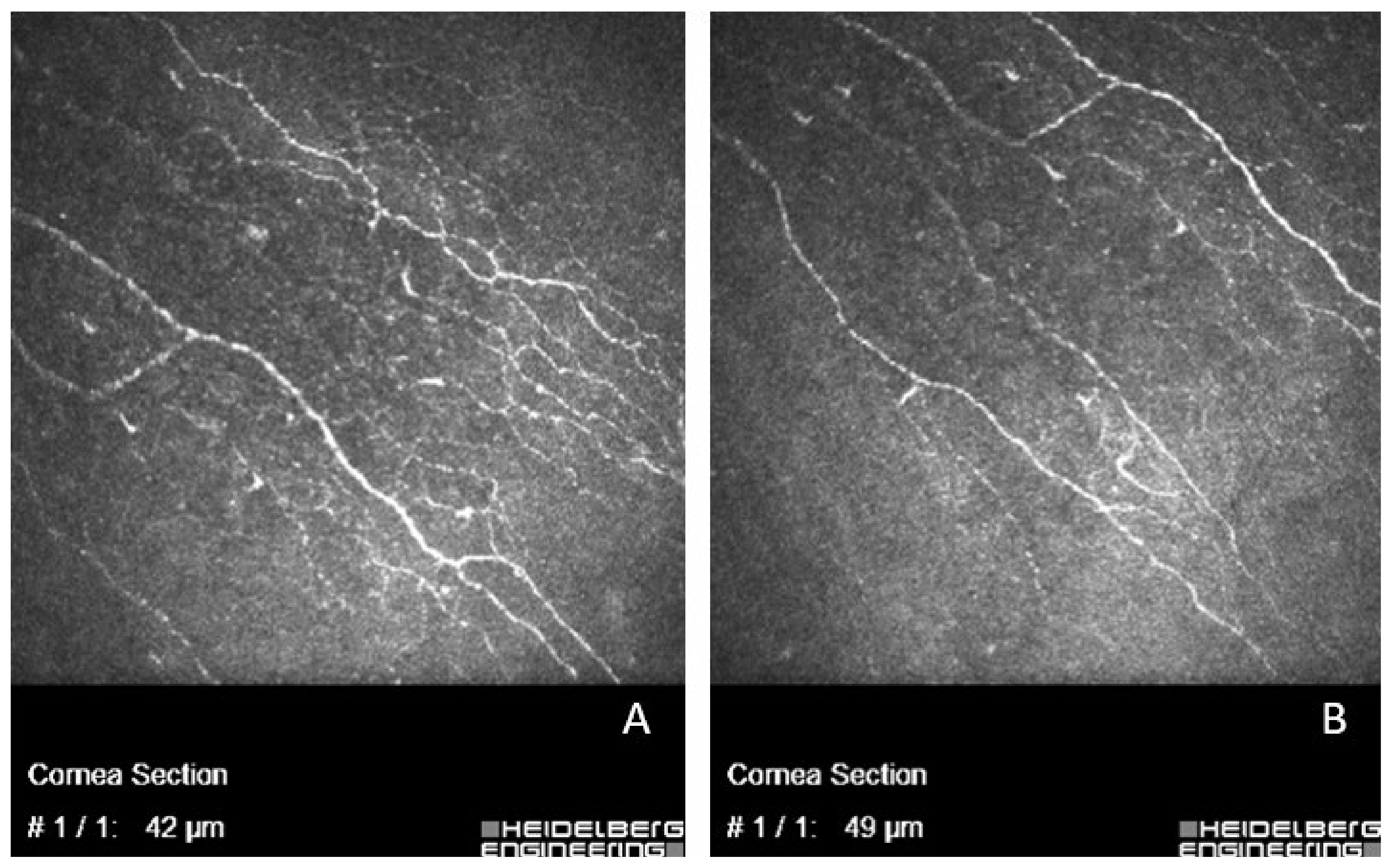
- In vivo confocal microscopy was carried out by the same investigator at the central cornea to evaluate it with laser scanning confocal microscope Heidelberg Retinal Tomograph (HRT II RCM Heidelberg Engineering Inc, Heidelberg, Germany; Rostock Cornea Module). The laser source is a diode laser at a wavelength of 670 nm. The acquired 2D images have a definition of 384 × 384 pixels over an area of 400 × 400 μm. Before the test, one drop of local anesthetic was instilled in the conjunctival sac (Proxymetacaine hydrochloride 0.5%). The single use contact element in sterile packaging (TomoCap) was used during the test which was lubricated with sterile gel (Carbomer 2.5 mg/g) for better image quality. In each case, another independent investigator picked from eight to 10 images which were analyzed with the Image J program (National Institutes of Health, Bethesda, Maryland, USA), Neuron J extension (Biomedical Imaging Group, Lausanne, Switzerland). The evaluation consisted of two parameters: the density of corneal nerves (it showed the length of nerve fibers in one square millimeter, mm/mm2) and the number of fibers (measured in one image). Confocal microscopy was conducted for not all the study participants because some patients refused to repeat this test after the cataract surgery, or images were not representable, so such cases (n = 26) were not included in the final analysis.
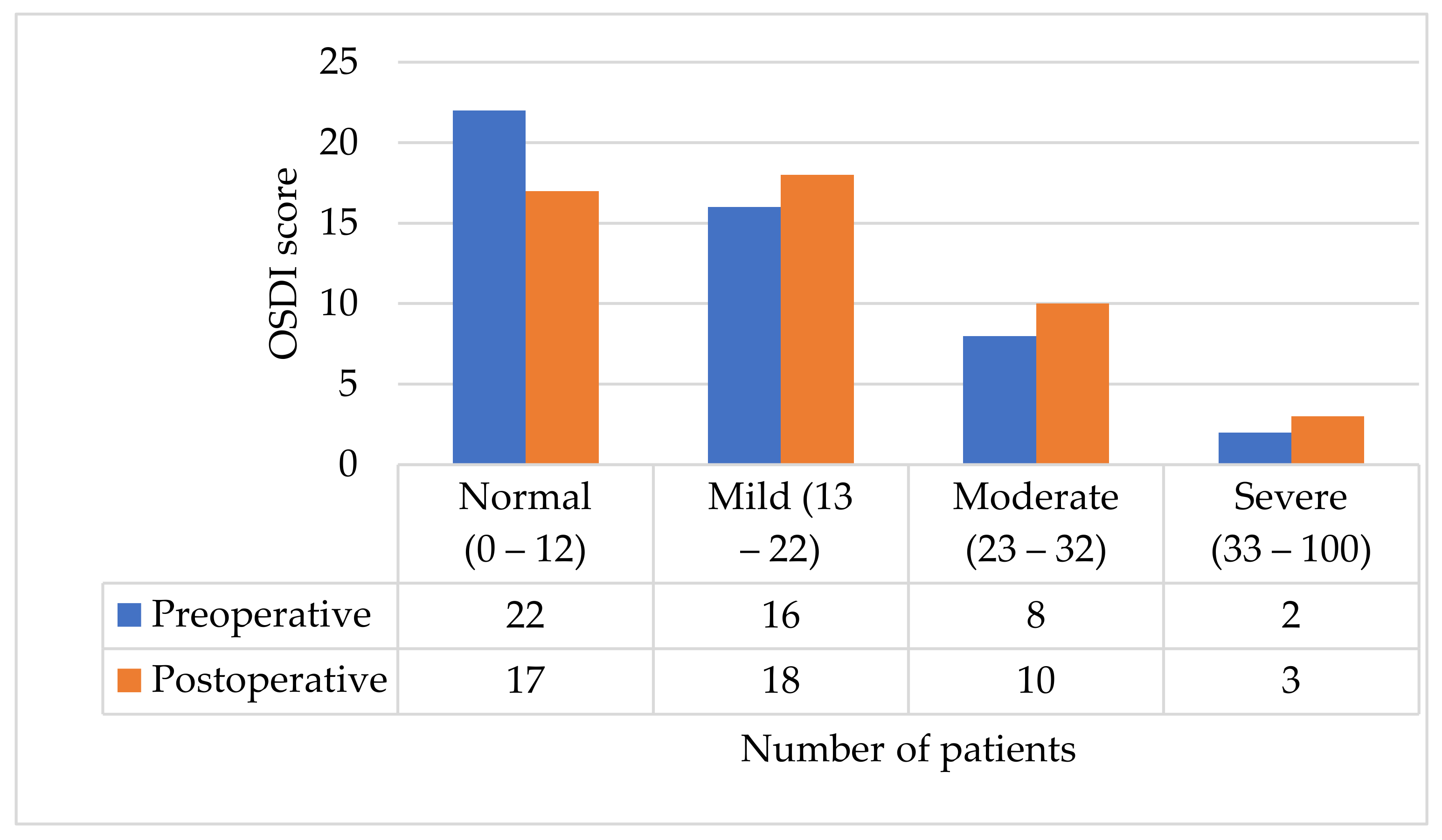
- Dry eye symptoms were assessed with a validated OSDI questionnaire [24]. It consists of 12 questions about symptoms and the patient chooses from ‘never’ (0 points) to ‘all the time’ (4 points). The result was calculated using the following formula: OSDI score = (sum of all answered questions) × 100/total number of answered questions) × 4. The OSDI scores range from 0 to 100. Scores from 0 to 12 were considered normal; from 13 to 22—mild dry eye disease; from 23 to 32—moderate dry eye disease; from 33 to 100—severe dry eye disease.
3. Results
3.1. OSDI Questionnaire: Results
3.2. Corneal Sensitivity
3.3. Schirmer’s I Test
3.4. TBUT
3.5. The Density of Corneal Nerves and the Fiber Number
4. Discussion
5. Conclusions
Author Contributions
Funding
Institutional Review Board Statement
Informed Consent Statement
Data Availability Statement
Conflicts of Interest
References
- Zhang, X.; Vadoothker, S.; Munir, W.M.; Saeedi, O. Ocular Surface Disease and Glaucoma Medications: A Clinical Approach. Eye Contact Lens 2019, 45, 11–18. [Google Scholar] [CrossRef] [PubMed]
- Pflugfelder, S.C.; de Paiva, C.S. The Pathophysiology of Dry Eye Disease: What We Know and Future Directions for Research. Ophthalmology 2017, 124, S4–S13. [Google Scholar] [CrossRef] [PubMed]
- Mikalauskiene, L.; Grzybowski, A.; Zemaitiene, R. Ocular Surface Changes Associated with Ophthalmic Surgery. J. Clin. Med. 2021, 10, 1642. [Google Scholar] [CrossRef] [PubMed]
- Matossian, C.; McDonald, M.; Donaldson, K.E.; Nichols, K.K.; MacIver, S.; Gupta, P.K. Dry Eye Disease: Consideration for Women’s Health. J. Women’s Health 2019, 28, 502–514. [Google Scholar] [CrossRef] [PubMed]
- Farrand, K.F.; Fridman, M.; Stillman, I.Ö.; Schaumberg, D.A. Prevalence of Diagnosed Dry Eye Disease in the United States among Adults Aged 18 Years and Older. Am. J. Ophthalmol. 2017, 182, 90–98. [Google Scholar] [CrossRef] [PubMed] [Green Version]
- Messmer, E.M. The pathophysiology, diagnosis, and treatment of dry eye disease. Dtsch. Arztebl. Int. 2015, 112, 71–82. [Google Scholar] [CrossRef] [Green Version]
- Stern, M.E.; Schaumburg, C.S.; Pflugfelder, S.C. Dry eye as a mucosal autoimmune disease. Int. Rev. Immunol. 2013, 32, 19–41. [Google Scholar] [CrossRef]
- Stevenson, W.; Chauhan, S.K.; Dana, R. Dry eye disease: An immune-mediated ocular surface disorder. Arch. Ophthalmol. 2012, 130, 90–100. [Google Scholar] [CrossRef] [Green Version]
- Alhatem, A.; Cavalcanti, B.; Hamrah, P. In vivo confocal microscopy in dry eye disease and related conditions. Semin. Ophthalmol. 2012, 27, 138–148. [Google Scholar] [CrossRef] [Green Version]
- Matsumoto, Y.; Ibrahim, O.M.A. Application of In Vivo Confocal Microscopy in Dry Eye Disease. Investig. Ophthalmol. Vis. Sci. 2018, 59, DES41–DES47. [Google Scholar] [CrossRef]
- Belmonte, C.; Nichols, J.J.; Cox, S.M.; Brock, J.A.; Begley, C.G.; Bereiter, D.A.; Dartt, D.A.; Galor, A.; Hamrah, P.; Ivanusic, J.J.; et al. TFOS DEWS II pain and sensation report. Ocul. Surf. 2017, 15, 404–437. [Google Scholar] [CrossRef] [Green Version]
- Machetta, F.; Fea, A.M.; Actis, A.G.; de Sanctis, U.; Dalmasso, P.; Grignolo, F.M. In vivo confocal microscopic evaluation of corneal langerhans cells in dry eye patients. Open Ophthalmol. J. 2014, 8, 51–59. [Google Scholar] [CrossRef] [Green Version]
- Kheirkhah, A.; Rahimi Darabad, R.; Cruzat, A.; Hajrasouliha, A.R.; Witkin, D.; Wong, N.; Dana, R.; Hamrah, P. Corneal Epithelial Immune Dendritic Cell Alterations in Subtypes of Dry Eye Disease: A Pilot In Vivo Confocal Microscopic Study. Investig. Ophthalmol. Vis. Sci. 2015, 56, 7179–7185. [Google Scholar] [CrossRef]
- Benítez-Del-Castillo, J.M.; Acosta, M.C.; Wassfi, M.A.; Díaz-Valle, D.; Gegúndez, J.A.; Fernandez, C.; García-Sánchez, J. Relation between corneal innervation with confocal microscopy and corneal sensitivity with noncontact esthesiometry in patients with dry eye. Investig. Ophthalmol. Vis. Sci. 2007, 48, 173–181. [Google Scholar] [CrossRef] [Green Version]
- Benítez del Castillo, J.M.; Wasfy, M.A.; Fernandez, C.; Garcia-Sanchez, J. An in vivo confocal masked study on corneal epithelium and subbasal nerves in patients with dry eye. Investig. Ophthalmol. Vis. Sci. 2004, 45, 3030–3035. [Google Scholar] [CrossRef] [Green Version]
- Villani, E.; Galimberti, D.; Viola, F.; Mapelli, C.; Ratiglia, R. The cornea in Sjogren’s syndrome: An in vivo confocal study. Investig. Ophthalmol. Vis. Sci. 2007, 48, 2017–2022. [Google Scholar] [CrossRef]
- Prokofyeva, E.; Wegener, A.; Zrenner, E. Cataract prevalence and prevention in Europe: A literature review. Acta Ophthalmol. 2013, 91, 395–405. [Google Scholar] [CrossRef]
- Han, K.E.; Yoon, S.C.; Ahn, J.M.; Nam, S.M.; Stulting, R.D.; Kim, E.K.; Seo, K.Y. Evaluation of dry eye and meibomian gland dysfunction after cataract surgery. Am. J. Ophthalmol. 2014, 157, 1144–1150.e1. [Google Scholar] [CrossRef]
- Cho, Y.K.; Kim, M.S. Dry eye after cataract surgery and associated intraoperative risk factors. Korean J. Ophthalmol. 2009, 23, 65–73. [Google Scholar] [CrossRef] [Green Version]
- Kasetsuwan, N.; Satitpitakul, V.; Changul, T.; Jariyakosol, S. Incidence and pattern of dry eye after cataract surgery. PLoS ONE 2013, 8, e78657. [Google Scholar] [CrossRef]
- Trattler, W.B.; Majmudar, P.A.; Donnenfeld, E.D.; McDonald, M.B.; Stonecipher, K.G.; Goldberg, D.F. The Prospective Health Assessment of Cataract Patients’ Ocular Surface (PHACO) study: The effect of dry eye. Clin. Ophthalmol. 2017, 11, 1423–1430. [Google Scholar] [CrossRef] [PubMed] [Green Version]
- Iglesias, E.; Sajnani, R.; Levitt, R.C.; Sarantopoulos, C.D.; Galor, A. Epidemiology of persistent dry eye-like symptoms after cataract surgery: Persistent post-surgical pain after cataract surgery. Cornea 2018, 37, 893. [Google Scholar] [CrossRef] [PubMed]
- Miller, K.M.; Oetting, T.A.; Tweeten, J.P.; Carter, K.; Lee, B.S.; Lin, S.; Nanji, A.A.; Shorstein, N.H.; Musch, D.C.; American Academy of Ophthalmology Preferred Practice Pattern Cataract/Anterior Segment Panel. Cataract in the Adult Eye Preferred Practice Pattern. Ophthalmology 2022, 129, P1–P126. [Google Scholar] [CrossRef] [PubMed]
- Kubiliūtė, A.; Pakulienė, G. Validation of Lithuanian Version of Ocular Surface Disease Index Questionnaire. In Proceedings of the 17th International and 59th Polish Conference “Juvenes Pro Medicina”, Lodz, Poland, 14–16 May 2021; The Book of Abstracts/Medical University of Lodz; Kwas, K., Gromek, W., Kwaśniewska, O., Eds.; Students’ Scientific Association of the Medical University of Lodz: Łódź, Poland, 2021. [Google Scholar]
- Gupta, P.K.; Drinkwater, O.J.; VanDusen, K.W.; Brissette, A.R.; Starr, C.E. Prevalence of ocular surface dysfunction in patients presenting for cataract surgery evaluation. J. Cataract Refract. Surg. 2018, 44, 1090–1096. [Google Scholar] [CrossRef]
- Xue, W.; Xu, X.; Zou, H. A rating scale is a proper method to evaluate changes in quality of life due to dry eye symptoms. Int. Ophthalmol. 2019, 39, 563–569. [Google Scholar] [CrossRef] [PubMed]
- Xue, W.; Zhu, M.M.; Zhu, B.J.; Huang, J.N.; Sun, Q.; Miao, Y.Y.; Zou, H.D. Long-term impact of dry eye symptoms on vision-related quality of life after phacoemulsification surgery. Int. Ophthalmol. 2019, 39, 419–429. [Google Scholar] [CrossRef] [Green Version]
- Kohli, P.; Arya, S.K.; Raj, A.; Handa, U. Changes in ocular surface status after phacoemulsification in patients with senile cataract. Int. Ophthalmol. 2019, 39, 1345–1353. [Google Scholar] [CrossRef]
- Garg, P.; Gupta, A.; Tandon, N.; Raj, P. Dry Eye Disease after Cataract Surgery: Study of Its Determinants and Risk Factors. Turk. J. Ophthalmol. 2020, 50, 133–142. [Google Scholar] [CrossRef]
- Cung, L.X.; Nga, N.T.T.; Nga, D.M.; Hiep, N.X.; Pham, D.T. Cataract Surgery Destabilises Temporary the Tear Film of the Ocular Surface. Klin. Monbl. Augenheilkd. 2021, 238, 282–287. [Google Scholar] [CrossRef]
- Sullivan, D.A.; Rocha, E.M.; Aragona, P.; Clayton, J.A.; Ding, J.; Golebiowski, B.; Hampel, U.; McDermott, A.M.; Schaumberg, D.A.; Srinivasan, S.; et al. TFOS DEWS II Sex, Gender, and Hormones Report. Ocul. Surf. 2017, 15, 284–333. [Google Scholar] [CrossRef]
- Zamora, M.G.; Caballero, E.F.; Maldonado, M.J. Short-term changes in ocular surface signs and symptoms after phacoemulsification. Eur. J. Ophthalmol. 2020, 30, 1301–1307. [Google Scholar] [CrossRef]
- Sahu, P.K.; Das, G.K.; Malik, A.; Biakthangi, L. Dry Eye Following Phacoemulsification Surgery and Its Relation to Associated Intraoperative Risk Factors. Middle East Afr. J. Ophthalmol. 2015, 22, 472–477. [Google Scholar] [CrossRef]
- Cetinkaya, S.; Mestan, E.; Acir, N.O.; Cetinkaya, Y.F.; Dadaci, Z.; Yener, H.I. The course of dry eye after phacoemulsification surgery. BMC Ophthalmol. 2015, 15, 68. [Google Scholar] [CrossRef] [Green Version]
- Vehof, J.; Sillevis Smitt-Kamminga, N.; Nibourg, S.A.; Hammond, C.J. Sex differences in clinical characteristics of dry eye disease. Ocul. Surf. 2018, 16, 242–248. [Google Scholar] [CrossRef] [Green Version]
- Li, W.; Lin, M.C. Sex Disparity in How Pain Sensitivity Influences Dry Eye Symptoms. Cornea 2019, 38, 1291–1298. [Google Scholar] [CrossRef] [Green Version]
- Labbé, A.; Liang, Q.; Wang, Z.; Zhang, Y.; Xu, L.; Baudouin, C.; Sun, X. Corneal nerve structure and function in patients with non-Sjogren dry eye: Clinical correlations. Investig. Ophthalmol. Vis. Sci. 2013, 54, 5144–5150. [Google Scholar] [CrossRef] [Green Version]
- Labbé, A.; Alalwani, H.; Van Went, C.; Brasnu, E.; Georgescu, D.; Baudouin, C. The relationship between subbasal nerve morphology and corneal sensation in ocular surface disease. Investig. Ophthalmol. Vis. Sci. 2012, 53, 4926–4931. [Google Scholar] [CrossRef]
- Kheirkhah, A.; Saboo, U.S.; Abud, T.B.; Dohlman, T.H.; Arnoldner, M.A.; Hamrah, P.; Dana, R. Reduced Corneal Endothelial Cell Density in Patients with Dry Eye Disease. Am. J. Ophthalmol. 2015, 159, 1022–1026.e2. [Google Scholar] [CrossRef] [Green Version]
- Xu, J.; Chen, P.; Yu, C.; Liu, Y.; Hu, S.; Di, G. In vivo Confocal Microscopic Evaluation of Corneal Dendritic Cell Density and Subbasal Nerve Parameters in Dry Eye Patients: A Systematic Review and Meta-analysis. Front. Med. 2021, 8, 578233. [Google Scholar] [CrossRef]
- Giannaccare, G.; Bernabei, F.; Pellegrini, M.; Guaraldi, F.; Turchi, F.; Torrazza, C.; Vagge, A. Bilateral morphometric analysis of corneal sub-basal nerve plexus in patients undergoing unilateral cataract surgery: A preliminary in vivo confocal microscopy study. Br. J. Ophthalmol. 2021, 105, 174–179. [Google Scholar] [CrossRef]
- Lum, E.; Corbett, M.C.; Murphy, P.J. Corneal sensitivity after ocular surgery. Eye Contact Lens 2019, 45, 226–237. [Google Scholar] [CrossRef]
- Kobashi, H.; Kamiya, K.; Sambe, T.; Nakagawa, R. Factors influencing subjective symptoms in dry eye disease. Int. J. Ophthalmol. 2018, 11, 1926–1931. [Google Scholar] [CrossRef]
- De Cillà, S.; Fogagnolo, P.; Sacchi, M.; Orzalesi, N.; Carini, E.; Ceresara, G.; Rossetti, L. Corneal involvement in uneventful cataract surgery: An in vivo confocal microscopy study. Ophthalmologica 2014, 231, 103–110. [Google Scholar] [CrossRef]
- Misra, S.L.; Goh, Y.W.; Patel, D.V.; Riley, A.F.; McGhee, C.N. Corneal microstructural changes in nerve fiber, endothelial and epithelial density after cataract surgery in patients with diabetes mellitus. Cornea 2015, 34, 177–181. [Google Scholar] [CrossRef]
- Cagini, C.; Torroni, G.; Mariniello, M.; Di Lascio, G.; Martone, G.; Balestrazzi, A. Trehalose/sodium hyaluronate eye drops in post-cataract ocular surface disorders. Int. Ophthalmol. 2021, 41, 3065–3071. [Google Scholar] [CrossRef]
- Cagini, C.; Di Lascio, G.; Torroni, G.; Mariniello, M.; Meschini, G.; Lupidi, M.; Messina, M. Dry eye and inflammation of the ocular surface after cataract surgery: Effectiveness of a tear film substitute based on trehalose/hyaluronic acid vs. hyaluronic acid to resolve signs and symptoms. J. Cataract Refract. Surg. 2021, 47, 1430–1435. [Google Scholar] [CrossRef]
- Kim, S.; Shin, J.; Lee, J.E. A randomised, prospective study of the effects of 3% diquafosol on ocular surface following cataract surgery. Sci. Rep. 2021, 11, 9124. [Google Scholar] [CrossRef]
- Kang, M.S.; Shin, J.; Kwon, J.M.; Huh, J.; Lee, J.E. Efficacy of 0.05% cyclosporine A on the lipid layer and meibomian glands after cataract surgery: A randomized, double-masked study. PLoS ONE 2021, 16, e0245329. [Google Scholar] [CrossRef]
- Favuzza, E.; Cennamo, M.; Vicchio, L.; Giansanti, F.; Mencucci, R. Protecting the Ocular Surface in Cataract Surgery: The Efficacy of the Perioperative Use of a Hydroxypropyl Guar and Hyaluronic Acid Ophthalmic Solution. Clin. Ophthalmol. 2020, 14, 1769–1775. [Google Scholar] [CrossRef]
- Hovanesian, J.A.; Berdy, G.J.; Epitropoulos, A.; Holladay, J.T. Effect of Cyclosporine 0.09% Treatment on Accuracy of Preoperative Biometry and Higher Order Aberrations in Dry Eye Patients Undergoing Cataract Surgery. Clin. Ophthalmol. 2021, 15, 3679–3686. [Google Scholar] [CrossRef]
Publisher’s Note: MDPI stays neutral with regard to jurisdictional claims in published maps and institutional affiliations. |
© 2022 by the authors. Licensee MDPI, Basel, Switzerland. This article is an open access article distributed under the terms and conditions of the Creative Commons Attribution (CC BY) license (https://creativecommons.org/licenses/by/4.0/).
Share and Cite
Sidaraite, A.; Mikalauskiene, L.; Grzybowski, A.; Zemaitiene, R. Evaluation of Ocular Surface after Cataract Surgery—A Prospective Study. J. Clin. Med. 2022, 11, 4562. https://doi.org/10.3390/jcm11154562
Sidaraite A, Mikalauskiene L, Grzybowski A, Zemaitiene R. Evaluation of Ocular Surface after Cataract Surgery—A Prospective Study. Journal of Clinical Medicine. 2022; 11(15):4562. https://doi.org/10.3390/jcm11154562
Chicago/Turabian StyleSidaraite, Agne, Lina Mikalauskiene, Andrzej Grzybowski, and Reda Zemaitiene. 2022. "Evaluation of Ocular Surface after Cataract Surgery—A Prospective Study" Journal of Clinical Medicine 11, no. 15: 4562. https://doi.org/10.3390/jcm11154562
APA StyleSidaraite, A., Mikalauskiene, L., Grzybowski, A., & Zemaitiene, R. (2022). Evaluation of Ocular Surface after Cataract Surgery—A Prospective Study. Journal of Clinical Medicine, 11(15), 4562. https://doi.org/10.3390/jcm11154562