Cognitive Fatigue in Multiple Sclerosis: An Objective Approach to Diagnosis and Treatment by Transcranial Electrical Stimulation

Abstract
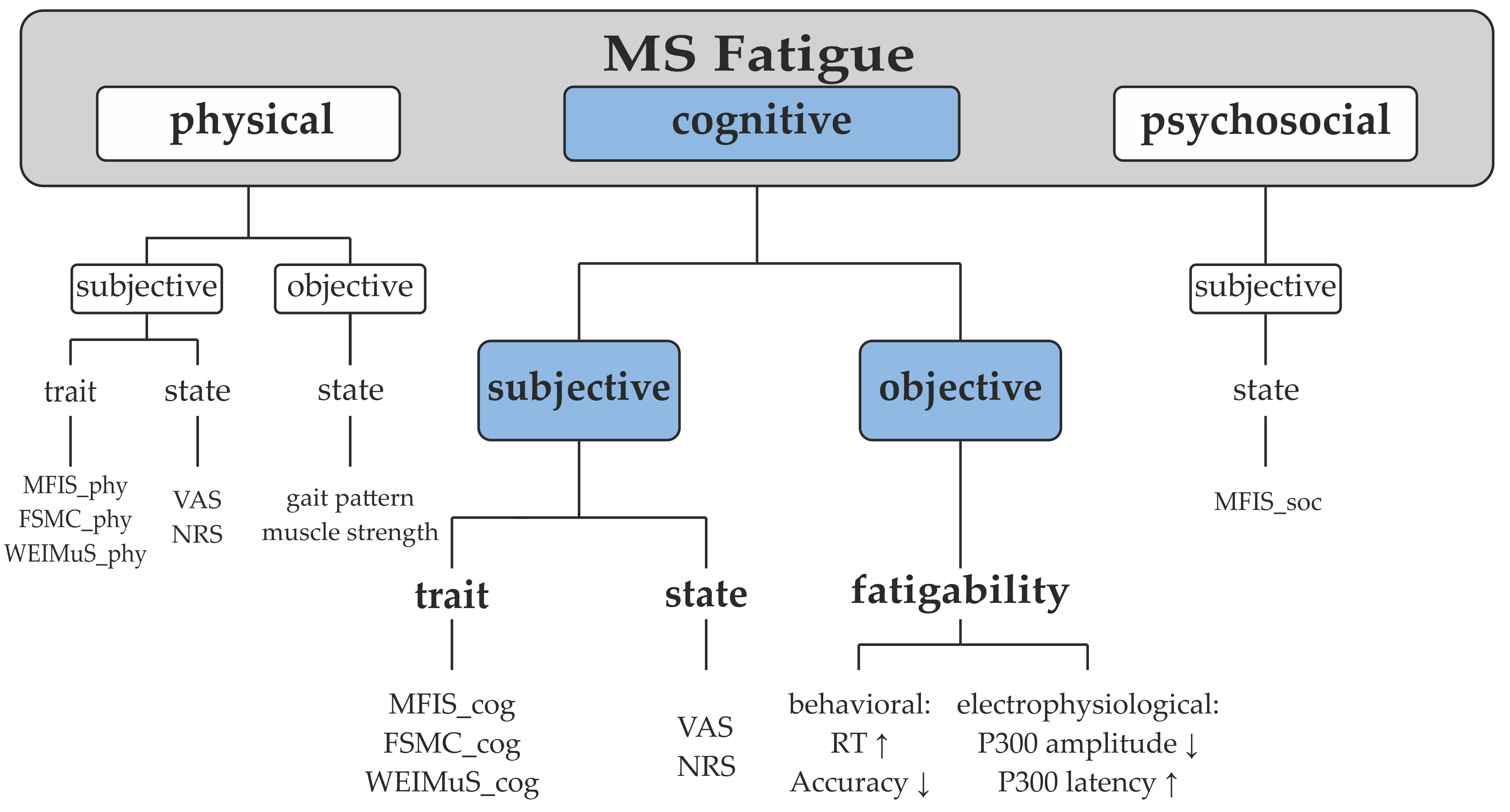
:1. Introduction
2. Search Strategies
3. Objective Measurement of Cognitive Fatigue
3.1. Behavioral Measures
3.2. Electrophysiological Measures
3.3. Sensory Gating Parameter
4. Relationship between Objective and Subjective Fatigue
5. Therapeutic Potential of tES for Cognitive Fatigue
5.1. Neuromodulation of the Fatigue Circuit by tDCS
5.2. Role of Neural Oscillations in Cognitive Fatigue and its Modulation by tACS
6. Conclusions and Outlook
Author Contributions
Funding
Conflicts of Interest
References
- Oh, J.; Vidal-Jordana, A.; Montalban, X. Multiple sclerosis: Clinical aspects. Curr. Opin. Neurol. 2018, 31, 752–759. [Google Scholar] [CrossRef] [PubMed]
- Fisk, J.D.; Pontefract, A.; Ritvo, P.G.; Archibald, C.J.; Murray, T. The Impact of Fatigue on Patients with Multiple Sclerosis. Can. J. Neurol. Sci./J. Can. des Sci. Neurol. 1994, 21, 9–14. [Google Scholar] [CrossRef]
- Kos, D.; Kerckhofs, E.; Nagels, G.; D’hooghe, M.B.; Ilsbroukx, S. Origin of fatigue in multiple sclerosis: Review of the literature. Neurorehabil Neural Repair 2008, 1, 91–100. [Google Scholar] [CrossRef] [PubMed]
- Simmons, R.D.; Tribe, K.L.; McDonald, E.A. Living with multiple sclerosis: Longitudinal changes in employment and the importance of symptom management. J. Neurol. 2010, 257, 926–936. [Google Scholar] [CrossRef]
- Ayache, S.S.; Chalah, M.A. Fatigue in multiple sclerosis - Insights into evaluation and management. Neurophysiol. Clin. Neurophysiol. 2017, 47, 139–171. [Google Scholar] [CrossRef]
- Chaudhuri, A.; Behan, P.O. Fatigue and basal ganglia. J. Neurol. Sci. 2000, 179, 34–42. [Google Scholar] [CrossRef]
- Chalah, M.A.; Riachi, N.; Ahdab, R.; Créange, A.; Lefaucheur, J.-P.; Ayache, S.S. Fatigue in Multiple Sclerosis: Neural Correlates and the Role of Non-Invasive Brain Stimulation. Front. Cell. Neurosci. 2015, 9, 155. [Google Scholar] [CrossRef] [PubMed]
- Sepulcre, J.; Masdeu, J.; Goñi, J.; Arrondo, G.; De Mendizábal, N.V.; Bejarano, B.; Villoslada, P. Fatigue in multiple sclerosis is associated with the disruption of frontal and parietal pathways. Mult. Scler. J. 2009, 15, 337–344. [Google Scholar] [CrossRef]
- Pardini, M.; Bonzano, L.; Mancardi, G.L.; Roccatagliata, L. Frontal networks play a role in fatigue perception in multiple sclerosis. Behav. Neurosci. 2010, 124, 329–336. [Google Scholar] [CrossRef] [PubMed]
- Roelcke, U.; Kappos, L.; Lechner-Scott, J.; Brunnschweiler, H.; Huber, S.; Ammann, W.; Plohmann, A.; Dellas, S.; Maguire, R.P.; Missimer, J.; et al. Reduced glucose metabolism in the frontal cortex and basal ganglia of multiple sclerosis patients with fatigue: A 18F-fluorodeoxyglucose positron emission tomography study. Neurology 1997, 48, 1566–1571. [Google Scholar] [CrossRef]
- Riccitelli, G.; Rocca, M.A.; Forn, C.; Colombo, B.; Comi, G.; Filippi, M. Voxelwise Assessment of the Regional Distribution of Damage in the Brains of Patients with Multiple Sclerosis and Fatigue. Am. J. Neuroradiol. 2011, 32, 874–879. [Google Scholar] [CrossRef]
- Rocca, M.A.; Parisi, L.; Pagani, E.; Copetti, M.; Rodegher, M.; Colombo, B.; Comi, G.; Falini, A.; Filippi, M. Regional but Not Global Brain Damage Contributes to Fatigue in Multiple Sclerosis. Radiology 2014, 273, 511–520. [Google Scholar] [CrossRef] [PubMed]
- Colombo, B.; Boneschi, F.M.; Rossi, P.; Rovaris, M.; Maderna, L.; Filippi, M.; Comi, G. MRI and motor evoked potential findings in nondisabled multiple sclerosis patients with and without symptoms of fatigue. J. Neurol. 2000, 247, 506–509. [Google Scholar] [CrossRef] [PubMed]
- Pellicano, C.; Gallo, A.; Li, X.; Ikonomidou, V.N.; Evangelou, I.E.; Ohayon, J.M.; Stern, S.K.; Ehrmantraut, M.; Cantor, F.; McFarland, H.F.; et al. Relationship of Cortical Atrophy to Fatigue in Patients With Multiple Sclerosis. Arch. Neurol. 2010, 67, 447–453. [Google Scholar] [CrossRef] [PubMed]
- Yaldizli, Ö.; Glassl, S.; Sturm, D.; Papadopoulou, A.; Gass, A.; Tettenborn, B.; Putzki, N. Fatigue and progression of corpus callosum atrophy in multiple sclerosis. J. Neurol. 2011, 258, 2199–2205. [Google Scholar]
- Yaldizli, Ö.; Penner, I.K.; Frontzek, K.; Naegelin, Y.; Amann, M.; Papadopoulou, A.; Gass, A. The relationship between total and regional corpus callosum atrophy, cognitive impairment and fatigue in multiple sclerosis patients. Mult. Scler. 2014, 3, 356–364. [Google Scholar] [CrossRef]
- Gobbi, C.; Rocca, M.; Pagani, E.; Riccitelli, G.; Pravatà, E.; Radaelli, M.; Martinelli-Boneschi, F.; Falini, A.; Copetti, M.; Comi, G.; et al. Forceps minor damage and co-occurrence of depression and fatigue in multiple sclerosis. Mult. Scler. J. 2014, 20, 1633–1640. [Google Scholar] [CrossRef]
- Niepel, G.; Tench, C.R.; Morgan, P.S.; Evangelou, N.; Auer, D.P.; Constantinescu, C.S. Deep gray matter and fatigue in MS: A T1 relaxation time study. J. Neurol. 2006, 7, 896–902. [Google Scholar] [CrossRef]
- Inglese, M.; Park, S.-J.; Johnson, G.; Babb, J.S.; Miles, L.; Jaggi, H.; Grossman, R.I. Deep gray matter perfusion in multiple sclerosis: Dynamic susceptibility contrast perfusion magnetic resonance imaging at 3 T. Arch. Neurol. 2007, 2, 196–202. [Google Scholar] [CrossRef]
- Téllez, N.; Alonso, J.; Río, J.; Tintoré, M.; Nos, C.; Montalban, X.; Rovira, A. The basal ganglia: A substrate for fatigue in multiple sclerosis. Neuroradiology 2008, 1, 17–23. [Google Scholar] [CrossRef]
- Multiple Sclerosis Council for Clinical Practice Guidelines. Fatigue and Multiple Sclerosis: Evidence-Based Management Strategies for Fatigue in Multiple Sclerosis; Paralyzed Veterans of America: Washington, DC, USA, 1998. [Google Scholar]
- Krupp, L.B.; LaRocca, N.G.; Muir-Nash, J.; Steinberg, A.D. The fatigue severity scale. Application to patients with multiple sclerosis and systemic lupus erythematosus. Arch. Neurol. 1989, 46, 1121–1123. [Google Scholar] [CrossRef]
- Penner, I.; Raselli, C.; Stöcklin, M.; Opwis, K.; Kappos, L.; Calabrese, P. The Fatigue Scale for Motor and Cognitive Functions (FSMC): Validation of a new instrument to assess multiple sclerosis-related fatigue. Mult. Scler. J. 2009, 15, 1509–1517. [Google Scholar] [CrossRef]
- Flachenecker, P.; Müller, G.; König, H.; Meissner, H.; Toyka, K.V.; Rieckmann, P. “Fatigue” in multiple sclerosis. Development and and validation of the “Würzburger Fatigue Inventory for MS”. Nervenarzt 2006, 77, 165–174. [Google Scholar] [CrossRef]
- Fiene, M.; Rufener, K.S.; Kuehne, M.; Matzke, M.; Heinze, H.-J.; Zaehle, T. Electrophysiological and behavioral effects of frontal transcranial direct current stimulation on cognitive fatigue in multiple sclerosis. J. Neurol. 2018, 265, 607–617. [Google Scholar] [CrossRef]
- Flachenecker, P.; Kümpfel, T.; Kallmann, B.; Gottschalk, M.; Grauer, O.; Rieckmann, P.; Trenkwalder, C.; Toyka, K.V. Fatigue in multiple sclerosis: A comparison of different rating scales and correlation to clinical parameters. Mult. Scler. J. 2002, 8, 523–526. [Google Scholar] [CrossRef]
- Barak, Y.; Achiron, A. Cognitive fatigue in multiple sclerosis: Findings from a two-wave screening project. J. Neurol. Sci. 2006, 245, 73–76. [Google Scholar] [CrossRef]
- Holtzer, R.; Shuman, M.; Mahoney, J.R.; Lipton, R.; Verghese, J. Cognitive fatigue defined in the context of attention networks. Neuropsychol Dev. Cogn B Aging Neuropsychol. Cogn. 2011, 1, 108–128. [Google Scholar] [CrossRef]
- Kluger, B.M.; Krupp, L.B.; Enoka, R.M. Fatigue and fatigability in neurologic illnesses: Proposal for a unified taxonomy. Neurology 2013, 4, 409–416. [Google Scholar] [CrossRef]
- Genova, H.M.; Rajagopalan, V.; DeLuca, J.; Das, A.; Binder, A.; Arjunan, A.; Chiaravalloti, N.; Wylie, G. Examination of Cognitive Fatigue in Multiple Sclerosis using Functional Magnetic Resonance Imaging and Diffusion Tensor Imaging. PLOS ONE 2013, 8, 11. [Google Scholar] [CrossRef]
- DeLuca, J. Fatigue As a Window to the Brain. Issues in Clinical and Cognitive Neuropsychology; MIT Press: Cambridge, MA, USA, 2005. [Google Scholar]
- Bailey, A.; Channon, S.; Beaumont, J.G. The relationship between subjective fatigue and cognitive fatigue in advanced multiple sclerosis. Mult. Scler. J. 2007, 13, 73–80. [Google Scholar] [CrossRef]
- Claros-Salinas, D.; Bratzke, D.; Greitemann, G.; Nickisch, N.; Ochs, L.; Schröter, H. Fatigue-related diurnal variations of cognitive performance in multiple sclerosis and stroke patients. J. Neurol. Sci. 2010, 295, 75–81. [Google Scholar] [CrossRef] [PubMed]
- Andreasen, A.K.; Spliid, P.E.; Andersen, H.; Jakobsen, J. Fatigue and processing speed are related in multiple sclerosis. Eur. J. Neurol. 2010, 2, 212–218. [Google Scholar] [CrossRef] [PubMed]
- Beatty, W.W.; Goretti, B.; Siracusa, G.; Zipoli, V.; Portaccio, E.; Amato, M.P. Changes in Neuropsychological Test Performance Over the Workday in Multiple Sclerosis. Clin. Neuropsychol. 2003, 17, 551–560. [Google Scholar] [CrossRef]
- Bruce, J.M.; Bruce, A.S.; Arnett, P.A. Response variability is associated with self-reported cognitive fatigue in multiple sclerosis. Neuropsychology 2010, 24, 77–83. [Google Scholar] [CrossRef]
- Johnson, S.K.; Lange, G.; DeLuca, J.; Korn, L.R.; Natelson, B. The Effects of Fatigue on Neuropsychological Performance in Patients With Chronic Fatigue Syndrome, Multiple Sclerosis, and Depression. Appl. Neuropsychol. 1997, 4, 145–153. [Google Scholar] [CrossRef]
- Krupp, L.B.; Elkins, L.E. Fatigue and declines in cognitive functioning in multiple sclerosis. Neurology 2000, 55, 934–939. [Google Scholar] [CrossRef]
- Neumann, M.; Sterr, A.; Claros-Salinas, D.; Gütler, R.; Ulrich, R.; Dettmers, C. Modulation of alertness by sustained cognitive demand in MS as surrogate measure of fatigue and fatigability. J. Neurol. Sci. 2014, 340, 178–182. [Google Scholar] [CrossRef] [PubMed]
- Aldughmi, M.; Bruce, J.; Siengsukon, C.F. Relationship Between Fatigability and Perceived Fatigue Measured Using the Neurological Fatigue Index in People with Multiple Sclerosis. Int. J. MS Care 2017, 19, 232–239. [Google Scholar] [CrossRef]
- Bryant, D.; Chiaravalloti, N.D.; DeLuca, J. Objective Measurement of Cognitive Fatigue in Multiple Sclerosis. Rehabilitation Psychol. 2004, 49, 114–122. [Google Scholar] [CrossRef]
- Chinnadurai, S.A.; Venkatesan, S.A.; Shankar, G.; Samivel, B.; Ranganathan, L.N. A study of cognitive fatigue in Multiple Sclerosis with novel clinical and electrophysiological parameters utilizing the event related potential P300. Mult. Scler. Relat. Disord. 2016, 10, 1–6. [Google Scholar] [CrossRef]
- Gossmann, A.; Eling, P.; Kastrup, A.; Hildebrandt, H. No effect of cooling on cognitive fatigue, vigilance and autonomic functioning in multiple sclerosis. J. Mult. Scler. 2014, 1, 112. [Google Scholar] [CrossRef]
- Kos, D.; Kerckhofs, E.; Nagels, G.; Geentjens, L. Cognitive fatigue in multiple sclerosis: Comment on Schwid SR, Tyler CM, Scheid EA, Weinstein A, Goodman AD and McDermott MR. Mult. Scler. 2004, 3, 337. [Google Scholar] [CrossRef]
- Kujala, P.; Portin, R.; Revonsuo, A.; Ruutiainen, J. Attention related performance in two cognitively different subgroups of patients with multiple sclerosis. J. Neurol. Neurosurg. N.a. 1995, 59, 77–82. [Google Scholar] [CrossRef]
- Moore, T.M.; Key, A.P.; Thelen, A.; Hornsby, B.W.Y. Neural Mechanisms of Mental Fatigue Elicited by Sustained Auditory Processing. Neuropsychologia 2017, 106, 371–382. [Google Scholar] [CrossRef]
- Claros-Salinas, D.; Dittmer, N.; Neumann, M.; Sehle, A.; Spiteri, S.; Willmes, K.; Schoenfeld, M.A.; Dettmers, C. Induction of cognitive fatigue in MS patients through cognitive and physical load. Neuropsychol. Rehabilitation 2013, 23, 182–201. [Google Scholar] [CrossRef]
- Hanken, K.; Bosse, M.; Möhrke, K.; Eling, P.; Kastrup, A.; Antal, A.; Hildebrandt, H. Counteracting Fatigue in Multiple Sclerosis with Right Parietal Anodal Transcranial Direct Current Stimulation. Front. Neurol. 2016, 7, 112. [Google Scholar]
- Heesen, C.; Schulz, K.; Fiehler, J.; Von Der Mark, U.; Otte, C.; Jung, R.; Poettgen, J.; Krieger, T.; Gold, S. Correlates of cognitive dysfunction in multiple sclerosis. Brain Behav. Immun. 2010, 24, 1148–1155. [Google Scholar] [CrossRef]
- Schwid, S.R.; Tyler, C.M.; A Scheid, E.; Weinstein, A.; Goodman, A.D.; McDermott, M.P. Cognitive fatigue during a test requiring sustained attention: A pilot study. Mult. Scler. J. 2003, 9, 503–508. [Google Scholar] [CrossRef]
- Kluckow, S.W.; Rehbein, J.-G.; Schwab, M.; Witte, O.W.; Bublak, P. What you get from what you see: Parametric assessment of visual processing capacity in multiple sclerosis and its relation to cognitive fatigue. Cortex 2016, 83, 167–180. [Google Scholar] [CrossRef]
- Lehmann, P.; Eling, P.; Kastrup, A.; Grothues, O.; Hildebrandt, H. Self-reported sleep problems, but not fatigue, lead to decline in sustained attention in MS patients. Mult. Scler. J. 2012, 19, 490–497. [Google Scholar] [CrossRef]
- Walker, L.; Berard, J.; Berrigan, L.; Rees, L.; Freedman, M. Detecting cognitive fatigue in multiple sclerosis: Method matters. J. Neurol. Sci. 2012, 316, 86–92. [Google Scholar] [CrossRef]
- Hanken, K.; Eling, P.; Hildebrandt, H. Is there a cognitive signature for MS-related fatigue? Mult. Scler. 2015, 4, 376–381. [Google Scholar] [CrossRef]
- Derache, N.; Grassiot, B.; Mézenge, F.; Emmanuelle Dugué, A.; Desgranges, B.; Constans, J.-M.; Defer, G.-L. Fatigue is associated with metabolic and density alterations of cortical and deep gray matter in Relapsing-Remitting-Multiple Sclerosis patients at the earlier stage of the disease: A PET/MR study. Mult. Scler. Relat. Disord. 2013, 4, 362–369. [Google Scholar] [CrossRef]
- Calabrese, M.; Rinaldi, F.; Grossi, P.; Mattisi, I.; Bernardi, V.; Favaretto, A.; Perini, P.; Gallo, P. Basal ganglia and frontal/parietal cortical atrophy is associated with fatigue in relapsing—remitting multiple sclerosis. Mult. Scler. J. 2010, 16, 1220–1228. [Google Scholar] [CrossRef]
- Engström, M.; Flensner, G.; Landtblom, A.-M.; Ek, A.-C.; Karlsson, T. Thalamo-striato-cortical determinants to fatigue in multiple sclerosis. Brain Behav. 2013, 3, 715–728. [Google Scholar] [CrossRef]
- Hidalgo de Cruz, M.; d’Ambrosio, A.; Valsasina, P.; Pagani, E.; Colombo, B.; Rodegher, M.; Rocca, M.A. Abnormal functional connectivity of thalamic sub-regions contributes to fatigue in multiple sclerosis. Mult. Scler. 2018, 9, 1183–1195. [Google Scholar] [CrossRef]
- Huolman, S.; Hämäläinen, P.; Vorobyev, V.; Ruutiainen, J.; Parkkola, R.; Laine, T.; Hämäläinen, H. The effects of rivastigmine on processing speed and brain activation in patients with multiple sclerosis and subjective cognitive fatigue. Mult. Scler. 2011, 11, 1351–1361. [Google Scholar] [CrossRef]
- Cehelyk, E.K.; Harvey, D.Y.; Grubb, M.L.; Jalel, R.; El-Sibai, M.S.; Markowitz, C.E.; Chahin, S. Uncovering the association between fatigue and fatigability in multiple sclerosis using cognitive control. Mult. Scler. Relat. Disord. 2019, 27, 269–275. [Google Scholar] [CrossRef]
- Psychological Test Systems. Available online: https://www.psytest.net/ (accessed on 30 April 2019).
- Jennekens-Schinkel, A.; Sanders, E.; Lanser, J.; Van Der Velde, E. Reaction time in ambulant multiple sclerosis patients. J. Neurol. Sci. 1988, 85, 173–186. [Google Scholar] [CrossRef]
- DeLuca, J.; Genova, H.M.; Hillary, F.G.; Wylie, G. Neural correlates of cognitive fatigue in multiple sclerosis using functional MRI. J. Neurol. Sci. 2008, 270, 28–39. [Google Scholar] [CrossRef]
- Paul, R.H.; Beatty, W.W.; Schneider, R.; Blanco, C.R.; Hames, K.A. Cognitive and Physical Fatigue in Multiple Sclerosis: Relations Between Self-Report and Objective Performance. Appl. Neuropsychol. 1998, 5, 143–148. [Google Scholar] [CrossRef]
- Bodling, A.M.; Denney, D.R.; Lynch, S.G. Individual variability in speed of information processing: An index of cognitive impairment in multiple sclerosis. Neuropsychology 2012, 26, 357–367. [Google Scholar] [CrossRef]
- Gronwall, D.M.A. Paced Auditory Serial-Addition Task: A Measure of Recovery from Concussion. Percept. Ski. 1977, 44, 367–373. [Google Scholar] [CrossRef]
- Snyder, P.J.; Cappelleri, J.C.; Archibald, C.J.; Fisk, J.D. Improved detection of differential information-processing speed deficits between two disease-course types of multiple sclerosis. Neuropsychology 2001, 15, 617–625. [Google Scholar] [CrossRef]
- Sandry, J.; Genova, H.M.; Dobryakova, E.; DeLuca, J.; Wylie, G. Subjective Cognitive Fatigue in Multiple Sclerosis Depends on Task Length. Front. Neurol. 2014, 5, 214. [Google Scholar] [CrossRef]
- Spiteri, S.; Hassa, T.; Claros-Salinas, D.; Dettmers, C.; Schoenfeld, M.A. Neural correlates of effort-dependent and effort-independent cognitive fatigue components in patients with multiple sclerosis. Mult. Scler. J. 2017, 25, 256–266. [Google Scholar] [CrossRef]
- Berard, J.A.; Smith, A.M.; Walker, L.A. A Longitudinal Evaluation of Cognitive Fatigue on a Task of Sustained Attention in Early Relapsing-Remitting Multiple Sclerosis. Int. J. MS Care 2018, 20, 55–61. [Google Scholar] [CrossRef]
- Crivelli, L.; Farez, M.F.; González, C.D.; Fiol, M.; Amengual, A.; Leiguarda, R.; Correale, J. Alerting Network Dysfunction in Early Multiple Sclerosis. J. Int. Neuropsychol. Soc. 2012, 18, 757–763. [Google Scholar] [CrossRef]
- Beatty, W.W.; Goodkin, D.E.; Hertsgaard, D.; Monson, N. Clinical and Demographic Predictors of Cognitive Performance in Multiple Sclerosis. Arch. Neurol. 1990, 47, 305. [Google Scholar] [CrossRef]
- Sutton, S.; Braren, M.; Zubin, J.; John, E.R. Evoked-Potential Correlates of Stimulus Uncertainty. Science 1965, 150, 1187–1188. [Google Scholar] [CrossRef]
- Picton, T.W. The P300 Wave of the Human Event-Related Potential. J. Clin. Neurophysiol. 1992, 9, 456–479. [Google Scholar] [CrossRef]
- Polich, J. Updating P300: An integrative theory of P3a and P3b. Clin. Neurophysiol. 2007, 118, 2128–2148. [Google Scholar] [CrossRef]
- Piras, M.R.; Magnano, I.; Canu, E.; Paulus, K.; Satta, W.; Soddu, A.; Conti, M.; Achene, A.; Solinas, G.; Aiello, I. Longitudinal study of cognitive dysfunction in multiple sclerosis: Neuropsychological, neuroradiological, and neurophysiological findings. J. Neurol. Neurosurg. N.a. 2003, 74, 878–885. [Google Scholar] [CrossRef]
- Polich, J.; Romine, J.S.; Sipe, J.C.; Aung, M.; Dalessio, D.J. P300 in multiple sclerosis: A preliminary report. Int. J. Psychophysiol. 1992, 12, 155–163. [Google Scholar] [CrossRef]
- Pokryszko-Dragan, A.; Zagrajek, M.; Slotwinski, K.; Bilinska, M.; Gruszka, E.; Podemski, R. Event-related potentials and cognitive performance in multiple sclerosis patients with fatigue. Neurol. Sci. 2016, 37, 1545–1556. [Google Scholar] [CrossRef] [PubMed]
- Van Der Linden, D.; Massar, S.A.; Schellekens, A.F.; Ellenbroek, B.A.; Verkes, R.-J. Disrupted sensorimotor gating due to mental fatigue: Preliminary evidence. Int. J. Psychophysiol. 2006, 62, 168–174. [Google Scholar] [CrossRef]
- Aleksandrov, A.A.; Dmitrieva, E.S.; Stankevich, L.N.; Knyazeva, V.M.; Shestakova, A.N. The Development of Muscle Fatigue Suppresses Auditory Sensory Gating (P50) during Sustained Contraction. Front. Syst. Neurosci. 2016, 10, 181. [Google Scholar] [CrossRef] [PubMed]
- Deloire, M.S.; Bonnet, M.C.; Salort, E.; Arimone, Y.; Boudineau, M.; Petry, K.G.; Brochet, B. How to detect cognitive dysfunction at early stages of multiple sclerosis? Mult. Scler. J. 2006, 12, 445–452. [Google Scholar] [CrossRef] [PubMed]
- Hulst, H.E.; Steenwijk, M.D.; Versteeg, A.; Pouwels, P.J.; Vrenken, H.; Uitdehaag, B.M.; Polman, C.H.; Geurts, J.J.; Barkhof, F. Cognitive impairment in MS: Impact of white matter integrity, gray matter volume, and lesions. Neurology 2013, 80, 1025–1032. [Google Scholar] [CrossRef] [PubMed]
- Karadayi, H.; Arisoy, O.; Altunrende, B.; Boztaş, M.H.; Sercan, M. The relationship of cognitive impairment with neurological and psychiatric variables in multiple sclerosis patients. Int. J. N.a. Clin. Pr. 2014, 18, 45–51. [Google Scholar] [CrossRef]
- Morrow, S.; Weinstock-Guttman, B.; Munschauer, F.; Hojnacki, D.; Benedict, R. Subjective fatigue is not associated with cognitive impairment in multiple sclerosis: cross-sectional and longitudinal analysis. Mult. Scler. J. 2009, 15, 998–1005. [Google Scholar] [CrossRef] [PubMed]
- Niino, M.; Mifune, N.; Kohriyama, T.; Mori, M.; Ohashi, T.; Kawachi, I.; Shimizu, Y.; Fukaura, H.; Nakashima, I.; Kusunoki, S.; et al. Apathy/depression, but not subjective fatigue, is related with cognitive dysfunction in patients with multiple sclerosis. BMC Neurol. 2014, 14, 3. [Google Scholar] [CrossRef]
- A Parmenter, B.; Denney, D.R.; Lynch, S.G. The cognitive performance of patients with multiple sclerosis during periods of high and low fatigue. Mult. Scler. J. 2003, 9, 111–118. [Google Scholar] [CrossRef]
- Sundgren, M.; Maurex, L.; Wahlin, Å.; Piehl, F.; Brismar, T. Cognitive Impairment Has a Strong Relation to Nonsomatic Symptoms of Depression in Relapsing-Remitting Multiple Sclerosis. Arch. Clin. Neuropsychol. 2013, 28, 144–155. [Google Scholar] [CrossRef] [PubMed]
- Weinges-Evers, N.; Brandt, A.U.; Bock, M.; Pfueller, C.F.; Dörr, J.; Bellmann-Strobl, J.; Scherer, P.; Urbanek, C.; Boers, C.; Ohlraun, S.; et al. Correlation of self-assessed fatigue and alertness in multiple sclerosis. Mult. Scler. J. 2010, 16, 1134–1140. [Google Scholar] [CrossRef] [PubMed]
- Greim, B.; Benecke, R.; Zettl, U.K. Qualitative and quantitative assessment of fatigue in multiple sclerosis (MS). J. Neurol. 2007, 254, II58–II64. [Google Scholar] [CrossRef] [PubMed]
- Hanken, K.; Eling, P.; Hildebrandt, H. The Representation of Inflammatory Signals in the Brain – A Model for Subjective Fatigue in Multiple Sclerosis. Front. Neurol. 2014, 5, 264. [Google Scholar] [CrossRef]
- Yavari, F.; Jamil, A.; Vidor, L.P.; Nitsche, M.A.; Samani, M.M. Basic and functional effects of transcranial Electrical Stimulation (tES)—An introduction. Neurosci. Biobehav. Rev. 2018, 85, 81–92. [Google Scholar] [CrossRef] [PubMed]
- Sale, M.V.; Mattingley, J.B.; Zalesky, A.; Cocchi, L. Imaging human brain networks to improve the clinical efficacy of non-invasive brain stimulation. Neurosci. Biobehav. Rev. 2015, 57, 187–198. [Google Scholar] [CrossRef] [PubMed]
- Borragán, G.; Gilson, M.; Guerrero-Mosquera, C.; Di Ricci, E.; Slama, H.; Peigneux, P. Transcranial Direct Current Stimulation Does Not Counteract Cognitive Fatigue, but Induces Sleepiness and an Inter-Hemispheric Shift in Brain Oxygenation. Front. Psychol. 2018, 9, 2351. [Google Scholar] [CrossRef] [PubMed]
- McIntire, L.K.; McKinley, R.A.; Goodyear, C.; Nelson, J. A Comparison of the Effects of Transcranial Direct Current Stimulation and Caffeine on Vigilance and Cognitive Performance During Extended Wakefulness. Brain Stimul. 2014, 7, 499–507. [Google Scholar] [CrossRef]
- McIntire, L.K.; McKinley, R.A.; Nelson, J.M.; Goodyear, C. Transcranial direct current stimulation versus caffeine as a fatigue countermeasure. Brain Stimul. 2017, 10, 1070–1078. [Google Scholar] [CrossRef]
- Nelson, J.T.; McKinley, R.A.; Golob, E.J.; Warm, J.S.; Parasuraman, R. Enhancing vigilance in operators with prefrontal cortex transcranial direct current stimulation (tDCS). NeuroImage 2014, 85, 909–917. [Google Scholar] [CrossRef]
- Sarasso, P.; Ninghetto, M.; Salatino, A.; Ronga, I.; Bongiardina, A.; Iarrobino, I.; Neppi-Modona, M.; Ricci, R. Everything is (still) illuminated: Dual right cathodal-left anodal tDCS of PPC prevents fatigue on a visual detection task. Brain Stimul. 2019, 12, 187–189. [Google Scholar] [CrossRef]
- Loffler, B.S.; Stecher, H.I.; Fudickar, S.; De Sordi, D.; Otto-Sobotka, F.; Hein, A.; Herrmann, C.S. Counteracting the Slowdown of Reaction Times in a Vigilance Experiment With 40-Hz Transcranial Alternating Current Stimulation. IEEE Trans. N.a. Syst. Rehabilitation Eng. 2018, 26, 2053–2061. [Google Scholar] [CrossRef]
- Clayton, M.S.; Yeung, N.; Kadosh, R.C. Electrical stimulation of alpha oscillations stabilizes performance on visual attention tasks. J. Exp. Psychol. 2019, 148, 203–220. [Google Scholar] [CrossRef]
- Nitsche, M.A.; Liebetanz, D.; Antal, A.; Lang, N.; Tergau, F.; Paulus, W. Chapter 27 Modulation of cortical excitability by weak direct current stimulation—Technical, safety and functional aspects. Suppl. Clin. Neurophys. 2003, 56, 255–276. [Google Scholar] [CrossRef]
- Nitsche, M.A.; Paulus, W. Excitability changes induced in the human motor cortex by weak transcranial direct current stimulation. J. Physiol. 2000, 527, 633–639. [Google Scholar] [CrossRef]
- Nitsche, M.A.; Tergau, F.; Liebetanz, D.; Paulus, W. Pharmacological approach to the mechanisms of transcranial DC-stimulation-induced after-effects of human motor cortex excitability. Brain 2002, 125, 2238–2247. [Google Scholar]
- Monte-Silva, K.; Kuo, M.-F.; Hessenthaler, S.; Fresnoza, S.; Liebetanz, D.; Paulus, W.; Nitsche, M.A. Induction of Late LTP-Like Plasticity in the Human Motor Cortex by Repeated Non-Invasive Brain Stimulation. Brain Stimul. 2013, 6, 424–432. [Google Scholar] [CrossRef]
- Ayache, S.S.; Palm, U.; Chalah, M.A.; Al-Ani, T.; Brignol, A.; Abdellaoui, M.; Dimitri, D.; Sorel, M.; Créange, A.; Lefaucheur, J.-P. Prefrontal tDCS Decreases Pain in Patients with Multiple Sclerosis. Front. Neurosci. 2016, 10, 57. [Google Scholar] [CrossRef]
- Chalah, M.A.; Riachi, N.; Ahdab, R.; Mhalla, A.; Abdellaoui, M.; Créange, A.; Lefaucheur, J.-P.; Ayache, S.S. Effects of left DLPFC versus right PPC tDCS on multiple sclerosis fatigue. J. Neurol. Sci. 2017, 372, 131–137. [Google Scholar] [CrossRef]
- Charvet, L.E.; Dobbs, B.; Shaw, M.T.; Bikson, M.; Datta, A.; Krupp, L.B. Remotely supervised transcranial direct current stimulation for the treatment of fatigue in multiple sclerosis: Results from a randomized, sham-controlled trial. Mult. Scler. 2018, 13, 1760–1769. [Google Scholar] [CrossRef]
- Saiote, C.; Goldschmidt, T.; Timäus, C.; Steenwijk, M.D.; Opitz, A.; Antal, A.; Paulus, W.; A Nitsche, M. Impact of transcranial direct current stimulation on fatigue in multiple sclerosis. Restor. Neurol. Neurosci. 2014, 32, 423–436. [Google Scholar]
- Cancelli, A.; Cottone, C.; Giordani, A.; Migliore, S.; Lupoi, D.; Porcaro, C.; Tecchio, F. Personalized, bilateral whole-body somatosensory cortex stimulation to relieve fatigue in multiple sclerosis. Mult. Scler. 2018, 10, 1366–1374. [Google Scholar] [CrossRef]
- Tecchio, F.; Cancelli, A.; Cottone, C.; Ferrucci, R.; Vergari, M.; Zito, G.; Pasqualetti, P.; Filippi, M.M.; Ghazaryan, A.; Lupoi, D.; et al. Brain Plasticity Effects of Neuromodulation Against Multiple Sclerosis Fatigue. Front. Neurol. 2015, 6, 141. [Google Scholar] [CrossRef]
- Tecchio, F.; Cancelli, A.; Cottone, C.; Zito, G.; Pasqualetti, P.; Ghazaryan, A.; Rossini, P.M.; Filippi, M.M. Multiple sclerosis fatigue relief by bilateral somatosensory cortex neuromodulation. J. Neurol. 2014, 261, 1552–1558. [Google Scholar] [CrossRef]
- Ferrucci, R.; Vergari, M.; Cogiamanian, F.; Bocci, T.; Ciocca, M.; Tomasini, E.; De Riz, M.; Scarpini, E.; Priori, A. Transcranial direct current stimulation (tDCS) for fatigue in multiple sclerosis. NRE 2014, 34, 121–127. [Google Scholar]
- Ayache, S.S.; Lefaucheur, J.-P.; Chalah, M.A. Long term effects of prefrontal tDCS on multiple sclerosis fatigue: A case study. Brain Stimul. 2017, 10, 1001–1002. [Google Scholar] [CrossRef]
- Chalah, M.A.; Lefaucheur, J.-P.; Ayache, S.S. Long-term effects of tDCS on fatigue, mood and cognition in multiple sclerosis. Clin. Neurophysiol. 2017, 128, 2179–2180. [Google Scholar] [CrossRef]
- Buyukturkoglu, K.; Porcaro, C.; Cottone, C.; Cancelli, A.; Inglese, M.; Tecchio, F. Simple index of functional connectivity at rest in Multiple Sclerosis fatigue. Clin. Neurophysiol. 2017, 128, 807–813. [Google Scholar] [CrossRef]
- Vecchio, F.; Miraglia, F.; Porcaro, C.; Cottone, C.; Cancelli, A.; Rossini, P.M.; Tecchio, F. Electroencephalography-derived sensory and motor network topology in multiple sclerosis fatigue. Neurorehabil. Neural. Repair 2017, 1, 56–64. [Google Scholar] [CrossRef]
- Klimesch, W. EEG alpha and theta oscillations reflect cognitive and memory performance: A review and analysis. Brain Rev. 1999, 29, 169–195. [Google Scholar] [CrossRef]
- Boksem, M.A.; Meijman, T.F.; Lorist, M.M. Effects of mental fatigue on attention: An ERP study. Cogn. Brain 2005, 25, 107–116. [Google Scholar] [CrossRef]
- Tran, Y.; Wijesuriya, N.; Craig, A.; Nguyen, H. Regional brain wave activity changes associated with fatigue. Psychophysiology 2012, 49, 574–582. [Google Scholar]
- Wascher, E.; Rasch, B.; Sänger, J.; Hoffmann, S.; Schneider, D.; Rinkenauer, G.; Heuer, H.; Gutberlet, I. Frontal theta activity reflects distinct aspects of mental fatigue. Boil. Psychol. 2014, 96, 57–65. [Google Scholar] [CrossRef]
- Shigihara, Y.; Tanaka, M.; Ishii, A.; Kanai, E.; Funakura, M.; Watanabe, Y. Two types of mental fatigue affect spontaneous oscillatory brain activities in different ways. Behav. Brain Funct. 2013, 9, 2. [Google Scholar] [CrossRef]
- Dimitrakopoulos, G.N.; Kakkos, I.; Dai, Z.; Wang, H.; Sgarbas, K.; Thakor, N.; Bezerianos, A.; Sun, Y. Functional Connectivity Analysis of Mental Fatigue Reveals Different Network Topological Alterations Between Driving and Vigilance Tasks. IEEE Trans. N.a. Syst. Rehabilitation Eng. 2018, 26, 740–749. [Google Scholar] [CrossRef]
- Liu, J.-P.; Zhang, C.; Zheng, C.-X. Estimation of the cortical functional connectivity by directed transfer function during mental fatigue. Appl. Ergon. 2010, 42, 114–121. [Google Scholar] [CrossRef]
- Sun, Y.; Lim, J.; Kwok, K.; Bezerianos, A. Functional cortical connectivity analysis of mental fatigue unmasks hemispheric asymmetry and changes in small-world networks. Brain Cogn. 2014, 85, 220–230. [Google Scholar] [CrossRef]
- Cavanagh, J.F.; Cohen, M.X.; Allen, J.J.B. Prelude to and resolution of an error: EEG phase synchrony reveals cognitive control dynamics during action monitoring. J. Neurosci. 2009, 29, 98–105. [Google Scholar] [CrossRef]
- Van De Vijver, I.; Ridderinkhof, K.R.; Cohen, M.X. Frontal Oscillatory Dynamics Predict Feedback Learning and Action Adjustment. J. Cogn. Neurosci. 2011, 23, 4106–4121. [Google Scholar] [CrossRef]
- Clayton, M.S.; Yeung, N.; Kadosh, R.C. The roles of cortical oscillations in sustained attention. N.a. Cogn. Sci. 2015, 19, 188–195. [Google Scholar] [CrossRef]
- Mathewson, K.E.; Beck, D.M.; Ro, T.; Maclin, E.L.; Low, K.A.; Fabiani, M.; Gratton, G. Dynamics of alpha control: Preparatory suppression of posterior alpha oscillations by frontal modulators revealed with combined EEG and event-related optical signal (EROS). J. Cogn. Neurosci. 2014, 26, 2400–2415. [Google Scholar] [CrossRef]
- Reato, D.; Rahman, A.; Bikson, M.; Parra, L.C. Effects of weak transcranial alternating current stimulation on brain activity—A review of known mechanisms from animal studies. Front. Hum. Neurosci. 2013, 7, 687. [Google Scholar] [CrossRef]
- Tavakoli, A.V.; Yun, K. Transcranial Alternating Current Stimulation (tACS) Mechanisms and Protocols. Front. Cell. Neurosci. 2017, 11, 214. [Google Scholar] [CrossRef]
- Fiene, M.; Schwab, B.C.; Misselhorn, J.; Herrmann, C.S.; Schneider, T.R.; Engel, A.K. Manipulation of Oscillatory Phase by Transcranial Alternating Current Stimulation. bioRxiv 2019, 579631. [Google Scholar]
- Schwab, B.C.; Misselhorn, J.; Engel, A.K. Modulation of large-scale cortical coupling by transcranial alternating current stimulation. bioRxiv 2018, 484014. [Google Scholar]
- Vosskuhl, J.; Strüber, D.; Herrmann, C.S. Non-invasive Brain Stimulation: A Paradigm Shift in Understanding Brain Oscillations. Front. Hum. Neurosci. 2018, 12, 211. [Google Scholar]
- Zaehle, T.; Rach, S.; Herrmann, C.S. Transcranial Alternating Current Stimulation Enhances Individual Alpha Activity in Human EEG. PLOS ONE 2010, 5, e13766. [Google Scholar] [CrossRef]
- Thibaut, A.; Di Perri, C.; Chatelle, C.; Bruno, M.-A.; Bahri, M.A.; Wannez, S.; Piarulli, A.; Bernard, C.; Martial, C.; Heine, L.; et al. Clinical Response to tDCS Depends on Residual Brain Metabolism and Grey Matter Integrity in Patients With Minimally Conscious State. Brain Stimul. 2015, 8, 1116–1123. [Google Scholar] [CrossRef]


| Reference | Parameter | Sample Size | EDSS Score | Duration of MS (in Years) | Conceptualization | Fatigability | Correlation with Subjective Fatigue |
|---|---|---|---|---|---|---|---|
| Cognitive fatigue over an extended time | |||||||
| Andreasen et al., 2010 [34] | Processing speed | 60 MS (all RR), 18 HC | PF: 3.0 (1–3.5) b SF: 2.5 (2–3.5) b NF: 2 (1.5–−3.5) b | PF: 5.0 (1–14) b SF: 3.5 (0–16) b NF: 3.0 (0–9) b | Processing speed across two testing blocks | No: processing speed improved in second testing block | Yes: negative correlation between subjective trait-fatigue and cognitive performance |
| Bailey et al., 2007 [32] | RT, Accuracy | 14 MS (all PP + SP), 17 HC | 7.7 (0.4) a | 27.2 (8–59) c | Performance in 0-back (attention) and 1-back task (working memory) | Yes: accuracy decreased over time | No: no correlation between subjective state-fatigue and fatigability |
| Beatty et al., 2003 [35] | Processing speed | 17 MS (13 RR, 4 SP), 12 HC | 2.9 (2.3) a | 14.2 (7.4) a | Performance in cognitive tests (list recall, letter-number sequence, SDMT, PASAT) before and after workday | No: no performance decline from first to second testing block | No: no correlation between subjective state-fatigue and cognitive performance after workday |
| Bruce et al., 2010 [36] | RT, RT variability | 87 MS (70 RR, 17 SP), 24 HC | 4.5 (1.6) a | 10.9 (7.9) a | Performance across three blocks of CARB | No: shorter RT and smaller variability over time | Yes: positive correlation between subjective trait-fatigue and cognitive performance |
| Claros-Salinas et al., 2010 [33] | RT | 20 MS, 76 HC, 22 stroke | - | 8.2 (7.2) a | Performance in three TAP subtests at three different time points of the day | Yes: cognitive performance decreased over time only in MS patients | Not mentioned |
| DeLuca et al., 2008 [63] | RT | 15 MS (12 RR, 3 PP), 15 HC | - | 6.4 (4.9) a | Performance across four blocks of modified SDMT | No: faster RT over time | Not mentioned |
| Fiene et al., 2018 [25] | RT, P300 amplitude and latency | 15 MS (14 RR, 1 SP) | 3.5 (1.9) a | 9.6 (8.6) a | Performance across three blocks of SRT and auditory oddball paradigm | Yes: increasing RT, shorter amplitude and longer latencies of P300 over time | Yes: correlation between subjective state-fatigue and fatigability (negative with P300 amplitude and positive with latency) |
| Huolman et al., 2011 [59] | Processing speed, RT | 15 MS (all RR), 13 HC | 1.5 (0.9) a | 4.2 (3.6) a | Performance of the last 20 items across four blocks of a modified version of the PVSAT | Yes: group differences increased over time | Not mentioned |
| Johnson et al., 1997 [37] | Processing speed | 15 MS, 15 CFS, 15 MD, 15 HC | 1.8 (1.2) a | - | PASAT performance across four testing blocks | No: performance unchanged across blocks | Not mentioned |
| Sandry et al., 2014 [68] | RT, Accuracy | 32 MS (24 RR, 1 PP, 3 SP, 1 PR),24 HC | AI *: 2.4 (2.5) a | 11.9 (7.1) a | Task performance (processing speed and working memory task) across four testing blocks | No: RT improved across blocks, no changes in accuracy | No: no correlation between subjective state-fatigue and cognitive performance across blocks |
| Cognitive fatigue after challenging mental or physical exertion | |||||||
| Claros-Salinas et al., 2013 [47] | RT | 32 MS (20 RR, 2 PP, 10 SP), 20 HC | 3.6 (1.6) a | 7.7 (5.4) a | Performance in TAP subtests before and after physical and cognitive load for 2.5 hours | Yes: people with MS showed a significant increase in RT after cognitive load | Yes: positive correlation between subjective trait as well as state-fatigue and fatigability |
| Jennekens- Schinkel et al., 1988 [62] | RT | 39 MS (20 RR, 19 PP + SP), 25 HC | 3.5 (0–7) c | 12.0 (1–48) c | Performance in SRT before and after neuropsychological assessment for four hours | No: no group differences in task-related performance decline | Not mentioned |
| Krupp & Elkins, 2000 [38] | Neuropsychological test battery | 45 MS (24 RR, 8 PP, 13 SP), 14 HC | 3.8 (1.7) a | - | Performance in neuropsychological test battery before and after cognitive demanding task | Yes: performance of people with MS worsened after cognitive task | Not mentioned |
| Neumann et al., 2014 [39] | RT | 30 MS (23 RR, 1 PP, 6 SP), 15 HC | F: 3.8 (1.2) a NF: 3.7 (0.6) a | F: 9.9 (6.7) a NF: 13.6 (6.8) a | Performance in TAP alertness test before and after cognitive load and after a one hour resting time | Yes: increased RT in MS group after cognitive load; after rest RT returned to baseline in most patients | Yes: positive correlation between subjective trait-fatigue and cognitive performance |
| Paul et al., 1998 [64] | Accuracy, memory performance | 39 MS, 19 HC | AI: 4.1 (2.5) a | 12.2 (4.8) a | Performance in Word List Learning Task and vigilance test before and after a cognitive work battery that lasted 30 min | No: neither patients nor controls showed changes in cognitive performance after 30 min task | Not mentioned |
| Spiteri et al., 2017 [69] | RT | 40 MS (25 RR, 2 PP, 13 SP), 22 HC | 3.5 (1.5) a | 14.1 (8.8) a | Performance in alertness test before and after a cognitive demanding task (n-back) | Yes: patients responded slower and with greater variability after n-back task | No: no correlation between subjective trait as well as state-fatigue and cognitive performance |
| Cognitive fatigue during sustained mental effort | |||||||
| Berard et al., 2018 [70] | Processing speed | 32 MS (all RR), 32 HC | 1.8 (1.2) a | 4.4 (3.1) a | Performance in first third versus last third of PASAT | Yes: poorer performance in last third of PASAT | No: no correlation between subjective trait-fatigue and fatigability |
| Bryant et al., 2004 [41] | Processing speed | 56 MS, 39 HC | - | SG1: 5.8 (1.6) a SG2: 10.6 (1.8) a | Performance in first versus second half of each of four PASAT testing blocks | Yes: percent dyads declined earlier in time in MS subgroup | No: no correlation between subjective trait-fatigue and cognitive performance |
| Cehelyk et al., 2019 [60] | RT | 21 MS (19 RR, 2 SP) | 3.5 (1.6) a | 13.3 (8.7) a | Performance in first versus fourth quarter of Blocked Cyclic Naming Task | Yes: RT increased from first to fourth quarter | Yes: positive correlation between subjective state-fatigue and fatigability |
| Chinnadurai et al., 2016 [42] | Processing speed, P300 | 50 MS (36 RR, 2 PP, 12 SP), 50 HC | 4.6 (1.9) a | 6.0 (7.4) a | Performance in 60 and 180 sec version of Stroop Task, SDMT, serial addition task and ratio between first and last 50 items in P300 oddball paradigm | Yes: performance decline and increasing P300 latencies in last 50 items only in people with MS | Not mentioned |
| Crivelli et al., 2012 [71] | RT | 27 MS (all RR), 27 HC | 1.03 (0.8) a | 0.7 (0.7) a | Performance in third compared to first block of three attentional network tests (alertness, orienting, executive control) | No: performance improved over time | Not mentioned |
| DeLuca et al., 2008 [63] | RT, Accuracy | 15 MS (12 RR, 3 PP), 15 HC | - | 6.4 (4.9) a | Performance in second compared to first half in each of four blocks of modified SDMT | No: both groups responded faster in second half of each block | Not mentioned |
| Gossmann et al., 2014 [43] | Accuracy | 31 MS (all RR), 10 HC | 3.6 (2.1) a | 10.4 (9.2) a | Omissions in second half compared to first half of a 30 min auditory vigilance test | Yes: only in MS group performance declined significantly during the task | Yes: positive correlation between subjective state-fatigue and fatigability |
| Hanken et al., 2016 [48] | RT | 46 MS (18 RR, 28 PP + SP) | LF: 3.7 (1.8) a HF: 4.7 (1.1) a | LF: 13.5 (8.8) a HF: 10.9 (7.8) a | Performance in first 5 min compared to last 5 min of a 20 min visual vigilance task | Yes: RT increased with time-on-task | Not mentioned |
| Kluckow et al., 2016 [51] | Processing speed | 36 MS (all RR), 36 HC | 1.9 (1.2) a | 2.8 (6.6) a | Performance in PASAT during the last 20 items compared to first 20 items and performance change in TVA from first to fourth block | Yes: processing speed of MS group declined in second half of TVA (especially in high-fatigue patients) | Not mentioned |
| Kos et al., 2004 [44] | Processing speed | 50 MS, 21 HC | 6.4 (1.2) a | - | Performance in the first ten items compared to the last ten items in PASAT | Yes: 21.1% performance decline in MS group | No: no correlation between subjective trait-fatigue and fatigability |
| Kujala et al., 1995 [45] | RT, Accuracy | 45 MS (22 RR, 17 PP, 6 SP), 35 HC | CP: 5.0 (1.8) a CD: 5.5 (1.3) a | CP: 8.7 (5.9) a CD: 8.7 (6.0) a | Performance in visual vigilance test over 15 min | Yes: slower RT with time-on-task; the cognitively preserved MS group also showed decline in accuracy | Not mentioned |
| Lehmann et al., 2012 [52] | RT, Accuracy | 42 MS (all RR), 11 HC | F: 2.8 (1.4) a NF: 4.3 (2.7) a | - | Performance decline from first to second half of a 10 min 2-back task | No: no task-related performance changes with time-on-task | Not mentioned |
| Schwid et al., 2003 [50] | Processing speed | 20 MS (10 RR, 2 PP, 8 SP), 21 HC | 3.8 (1.5) a | - | Performance in first 20 items compared to last 20 items in PASAT | Yes: performance decline over time only in people with MS | Yes: correlation between subjective trait-fatigue and fatigability |
| Walker et al., 2012 [53] | Processing speed | 70 MS (all RR), 70 HC | 1.8 (1.2) a | 4.4 (3.1) a | Performance during first compared to second half in PASAT and CTIP | Yes: ability of MS group to meet task demands declined over time | Yes: negative correlation between subjective trait-fatigue and fatigability |
| Reference | Parameter | Sample Size | Stimulation Design | Study Design | Results |
|---|---|---|---|---|---|
| tDCS Studies | |||||
| Borragan et al., 2018 [93] | RT, Accuracy | 20 HC | Position: DLPFC Parameters: 1.5 mA for 25 min Average current density: 0.06 mA/cm2 | Three blocks of PVT; Between first and second block of PVT participants performed cognitive demanding working memory task; From the beginning to the second block participants received anodal or sham tDCS (within-subject design) | Anodal tDCS had no impact on behavioral performance decrements over time; tDCS-related interhemispheric shift in cortical oxygenation after stimulation offset |
| Fiene et al., 2018 [25] | RT, P300 amplitude and latency | 15 MS (14 RR, 1 SP) | Position: DLPFC Parameters: 1.5 mA for circa 30 min Average current density: 0.06 mA/cm2 | Three blocks of SRT task and auditory oddball paradigm; During second block, participants received anodal or sham tDCS (within-subject design) | Anodal tDCS caused a decrease in RT and an increase in P300 amplitude which persisted after the end of stimulation |
| Hanken et al., 2016 [48] | RT, Accuracy | Study I: 52 HC Study II: 46 MS (18 RR, 28 PP + SP) | Position: right parietal (Study I + II) or right frontal (Study I) Parameters: 1.5 mA for 20 min Average current density: 0.04 mA/cm2 | Visual vigilance task for 40 min (Study I) or 20 min (Study II); Anodal or sham tDCS for 20 min (between-subject design) | Anodal tDCS counteracted the time-on-task RT decrements (in people with MS and healthy controls) |
| McIntire et al., 2014 [94] | RT, Accuracy | 30 HC | Position: DLPFC Parameters: 2 mA for 30 min Average current density: 0.199 mA/cm2 | Five blocks of PVT every two hours after initial baseline assessment; Anodal tDCS with placebo gum or sham tDCS with placebo or caffeine gum after 22 h of wakefulness (between-subject design) | Anodal tDCS prevented vigilance decrements over time and led to better subjective ratings of fatigue, drowsiness and energy; Positive effects lasted at least six hours |
| McIntire et al., 2017 [95] | RT, Accuracy | 50 HC | Position: DLPFC Parameters: 2 mA for 30 min Average current density: 0.199 mA/cm2 | Five blocks of PVT every two hours after initial baseline assessment; Anodal tDCS with placebo gum or sham tDCS with placebo or caffeine gum early or late in the experiment (after 18 or 22 h of wakefulness) (between-subject design) | Anodal tDCS applied early in the experiment led to improved attentional accuracy and RT lasting for six hours |
| Nelson et al., 2014 [96] | RT, Accuracy | 19 HC | Position: DLPFC Parameters: 1 mA for 10 min Average current density: 0.028 mA/cm2 | Anodal, cathodal, or sham tDCS early (first 10 min) or late (last 10 min) during a 40 min vigilance task (within-subject design) | Especially early anodal and cathodal tDCS significantly improved task performance |
| Sarasso et al., 2019 [97] | Accuracy | 45 HC | Position: PPC Parameters: 1.5 mA for 15 min Average current density: 0.06 mA/cm2 | Two blocks of a visual vigilance task; Between blocks participants received right-anodal-left-cathodal, right-cathodal- eft-anodal, or sham tDCS (between-subject design) | Right-cathodal-left-anodal tDCS counteracted the time-on-task decrease in performance accuracy |
| tACS Studies | |||||
| Loeffler et al., 2018 [98] | RT, Accuracy | 24 HC | 40 Hz gamma tACS over visual cortex Parameters: 1 mA for 30 min | tACS was applied during the second block of a vigilance task (the first block taken as a baseline) | tACS significantly decreased the time-on-task related slowdown of RT |
| Clayton et al., 2019 [99] | RT, Accuracy | 178 HC in four studies | 10 Hz alpha tACS over posterior cortex Parameters: 2 mA for 11 min | Visual and auditory sustained attention task performance across four blocks; 10 Hz, 50 Hz or sham tACS were applied during second and third block | Alpha tACS exerted a stabilizing effect on accuracy and RT and generally limited the slope of performance deteriorations or improvements over time (specific to visual domain) |
© 2019 by the authors. Licensee MDPI, Basel, Switzerland. This article is an open access article distributed under the terms and conditions of the Creative Commons Attribution (CC BY) license (http://creativecommons.org/licenses/by/4.0/).
Share and Cite
Linnhoff, S.; Fiene, M.; Heinze, H.-J.; Zaehle, T. Cognitive Fatigue in Multiple Sclerosis: An Objective Approach to Diagnosis and Treatment by Transcranial Electrical Stimulation. Brain Sci. 2019, 9, 100. https://doi.org/10.3390/brainsci9050100
Linnhoff S, Fiene M, Heinze H-J, Zaehle T. Cognitive Fatigue in Multiple Sclerosis: An Objective Approach to Diagnosis and Treatment by Transcranial Electrical Stimulation. Brain Sciences. 2019; 9(5):100. https://doi.org/10.3390/brainsci9050100
Chicago/Turabian StyleLinnhoff, Stefanie, Marina Fiene, Hans-Jochen Heinze, and Tino Zaehle. 2019. "Cognitive Fatigue in Multiple Sclerosis: An Objective Approach to Diagnosis and Treatment by Transcranial Electrical Stimulation" Brain Sciences 9, no. 5: 100. https://doi.org/10.3390/brainsci9050100
APA StyleLinnhoff, S., Fiene, M., Heinze, H.-J., & Zaehle, T. (2019). Cognitive Fatigue in Multiple Sclerosis: An Objective Approach to Diagnosis and Treatment by Transcranial Electrical Stimulation. Brain Sciences, 9(5), 100. https://doi.org/10.3390/brainsci9050100