1. Introduction
Dental plaque is a structurally complex biofilm composed of multilayered bacterial communities embedded within an extracellular matrix that adheres to the tooth surface [
1]. Although plaque is typically translucent and difficult to detect visually, its biochemical composition renders it susceptible to extrinsic pigmentation [
2]. In clinical practice, the identification and effective removal of dental biofilm are central objectives of professional oral hygiene procedures, which aim not only to eliminate supragingival and subgingival deposits but also to promote long-term oral health through patient education and motivation [
3].
Plaque-disclosing agents have therefore been widely adopted as adjunctive tools in both professional and home-care settings [
4]. By selectively staining plaque deposits, these agents facilitate the assessment of plaque distribution and maturity, support targeted mechanical debridement, and provide immediate visual feedback that may improve patient motivation and adherence to oral hygiene instructions [
5]. Plaque disclosants are available in multiple formulations, including tablets, liquid solutions, gels, and mouthrinses, all of which function through dye adsorption onto biofilm-covered surfaces. Variations in pigment molecular size and chemical affinity influence penetration within the biofilm, staining intensity, and persistence over time [
2].
In routine chairside practice, plaque disclosure is frequently followed by immediate removal of stained deposits using manual brushing or professional prophylactic procedures such as rubber-cup polishing with prophylaxis paste or air-polishing systems. Contemporary preventive strategies, including Guided Biofilm Therapy, formalize this disclosure-based approach by integrating visualization with site-specific biofilm removal, often employing low-abrasive air-polishing powders [
6,
7]. Beyond facilitating mechanical plaque removal, visual disclosure has been shown to improve patient engagement and self-care compliance by providing immediate and tangible feedback [
8].
Despite their clinical advantages, plaque-disclosing agents are not entirely selective for bacterial biofilm. In addition to staining plaque, these dyes may adsorb onto dental hard tissues, restorative materials, and adjacent soft tissues [
9,
10]. Surface characteristics play a critical role in this interaction, as increased surface roughness promotes plaque accumulation and enhances the retention of chromogenic substances [
11]. From an esthetic standpoint, extrinsic tooth discoloration primarily reflects the deposition of pigments within the acquired pellicle and biofilm rather than intrinsic alterations in enamel mineral content. Importantly, perceived tooth color is influenced not only by intrinsic optical properties but also by surface-dependent parameters, including gloss, texture, and light scattering [
12]. Consequently, even superficial deposits or microtopographical changes may result in clinically perceptible alterations in appearance.
Previous investigations have demonstrated that plaque-disclosing agents can induce measurable color changes in tooth-colored restorative materials and surface sealants, with some studies reporting incomplete color recovery following professional cleaning or repolishing [
2,
10,
13]. These findings suggest a material-dependent interaction between disclosing dyes and dental substrates. Although the majority of available evidence has focused on restorative materials, similar mechanisms of dye adsorption and retention may be relevant for natural enamel surfaces, where pellicle formation, residual biofilm, and surface texture may favor extrinsic staining following disclosure procedures. Moreover, observations of staining on both hard and soft oral tissues indicate that disclosing dyes can extend beyond their intended target during clinical use [
9,
10].
To date, evidence remains limited on how commonly used prophylactic cleaning modalities simultaneously affect the removal of plaque disclosing agent-associated extrinsic discoloration and enamel surface properties. Therefore, this in vitro study compared manual brushing, rubber-cup prophylaxis polishing, and erythritol-based air-polishing in terms of discloser-associated enamel color change and concomitant changes in surface roughness and gloss within a combined esthetic and surface-property framework. The null hypothesis was that no significant differences would be observed among the tested modalities with respect to color change, surface roughness, or gloss.
2. Materials and Methods
2.1. Ethical Approval and Specimen Selection
This study was approved by the Trakya University Non-Interventional Scientific Research Ethics Committee (Approval No: TUTF-GOBAEK 2025/538) and conducted in accordance with the Declaration of Helsinki. Written informed consent was obtained from all patients prior to extraction, allowing the use of the extracted teeth for research purposes.
Human permanent maxillary anterior teeth extracted for orthodontic or periodontal indications were collected. After extraction, residual soft tissues and visible calculus deposits were carefully removed, and the specimens were disinfected in a 0.1% chloramine solution. The buccal enamel surfaces were examined under ×20 magnification using a stereomicroscope, and teeth presenting cracks, fractures, surface abrasion, or carious defects were excluded to ensure sample homogeneity.
2.2. Application of the Plaque Disclosing Agent
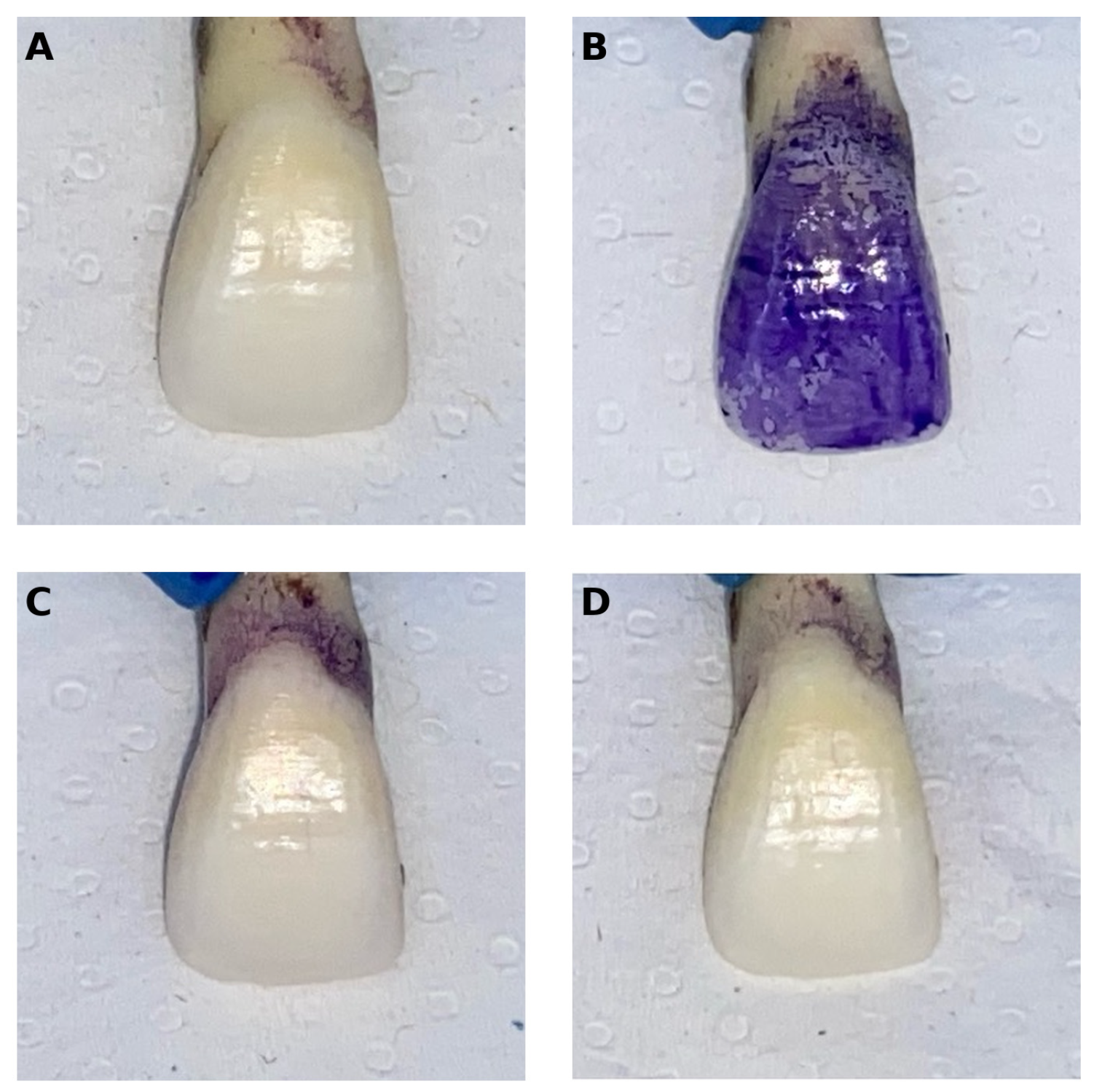
To generate standardized extrinsic discoloration, a plaque disclosing agent aligned with contemporary guided biofilm removal protocols was applied to the vestibular enamel surfaces. The disclosing solution (Biofilm Discloser; EMS, Nyon, Switzerland) contains erythrosine, a two-tone dye commonly used for biofilm visualization. The agent was applied using preloaded sponge pellets to ensure uniform delivery and minimal handling variability. The solution was left in contact with the enamel for 2 min, followed by gentle rinsing with water spray for 30 s and air-drying. This procedure produced consistent extrinsic staining prior to cleaning interventions.
2.3. Experimental Design and Removal Protocols
After discoloration induction, the specimens were randomly allocated in a 1:1:1 ratio to three cleaning protocols using a computer-generated randomization sequence prepared by an independent investigator. All procedures were performed by a single trained operator (E.A.A.) under standardized conditions.
Group 1—Manual brushing: Specimens were brushed using a flat-trim, medium-bristle manual toothbrush and a conventional toothpaste (Colgate Total; Colgate-Palmolive Company, New York, NY, USA; RDA ≈ 70) [
14]. Each specimen was brushed for approximately 10 s (4–5 circular strokes). This brushing duration was selected to represent the estimated per-tooth contact time during routine oral hygiene. The estimation was based on a typical full-mouth brushing time of approximately 2 min, corresponding to about 30 s per quadrant. Considering that each quadrant contains approximately 7–8 teeth and that toothbrush bristles are in simultaneous contact with 2–3 teeth, the quadrant-level brushing time was proportionally distributed across multiple teeth. This calculation yields an estimated per-tooth exposure time of approximately 10 s under active brushing conditions. Brushing was performed using the modified Bass technique by a single trained operator. A standardized and controlled pressure was applied throughout all procedures. Specimens were stabilized in a vertical position to ensure consistency of brushing dynamics across specimens.
Group 2—Rubber-cup prophylaxis polishing: Polishing was performed using a rubber cup and a perlite-containing prophylaxis paste (Cleanic; Kerr GmbH, Biberach, Germany; particle size ≈ 45 µm; RDA 27) [
15]. Each specimen was polished at 1000 rpm for 5 s using approximately 0.5 g of paste [
16]. The polishing duration was selected based on previously reported standardized clinical protocols, in which a single consecutive 5 s rubber-cup application at moderate speed (~1000 rpm) is used per tooth or tooth surface during prophylactic procedures. This short, standardized exposure was chosen to represent a single controlled polishing cycle, allowing isolation of the mechanical effect while minimizing cumulative abrasion that may occur in extended full-mouth clinical applications. Although polishing duration in clinical practice may vary according to patient-specific factors and operator technique, the selected protocol provides a reproducible and standardized unit of application suitable for experimental comparison. Polishing was carried out by a single trained operator using light, controlled, and consistent pressure to ensure uniform surface treatment across all specimens.
Group 3—Air-polishing: Professional mechanical biofilm removal was performed using an air-polishing device (AIR-FLOW Master Piezon
®, EMS Electro Medical Systems, Nyon, Switzerland) with an erythritol-based powder (PLUS powder
®, EMS) containing 99.7% erythritol and 0.3% chlorhexidine, with an average particle size of 14 µm. The nozzle was positioned approximately 3 mm from the enamel surface at an angulation of 30–60°. Each specimen was treated for 5 s at 50% power and 100% water settings, following manufacturer recommendations [
7]. During application, specimens were manually rotated while the nozzle was moved in gentle circular and sweeping motions from the cervical region toward the incisal edge to ensure homogeneous surface exposure.
2.4. Outcome Measures and Evaluation Time Points
Outcome measures were recorded at predefined time points relative to disclosing-agent application and subsequent removal procedures. Surface roughness (Ra) and surface gloss (GU) were evaluated at baseline and immediately after the removal interventions. Enamel color was assessed at three time points: baseline, immediately after stain removal, and after 1 week of storage in artificial saliva prepared in accordance with ASTM E2720 and ISO 10271 standards (Testonic Laboratories, Colin Kimya San. Tic. A.Ş., İstanbul, Turkey). Importantly, color change (ΔE00) was calculated relative to baseline enamel values rather than relative to the post-disclosure condition. For other parameters, pre-post differences were calculated and statistically compared among the three removal methods. All measurements were performed by an examiner blinded to group allocation.
2.5. Color Measurement
Enamel color was measured using a clinical spectrophotometer (VITA Easyshade V; VITA Zahnfabrik, Bad Säckingen, Germany). To enhance reproducibility, the probe tip was positioned at the geometric center of the labial surface, and triplicate measurements were recorded at each time point. To minimize potential diurnal variation, measurements were performed at consistent times of day. Color change was calculated using the CIEDE2000 formula (ΔE
00) [
17] under standardized background and lighting conditions, with all measurements performed by the same operator.
In this model, ΔL′, ΔC′, and ΔH′ denote the differences in lightness, chroma, and hue between two measurements. The weighting functions (, , and ) adjust the contribution of each component to better reflect perceptual non-uniformity in the CIELAB color space, while the rotation term (RT) accounts for the interaction of chroma and hue differences, particularly in the blue region. The parametric factors (, , and ) were included to reflect experimental conditions.
2.6. Surface Gloss Measurement
Surface gloss (GU) was assessed using a glossmeter (Novo-Curve, Rhopoint Instrumentation, East Sussex, UK) at a 60° geometry. The device was calibrated using a manufacturer-provided black glass standard with a reference value of 93.3 GU. Three readings were obtained per specimen, rotating the specimen by 90° between measurements, and the mean value was recorded.
2.7. Surface Roughness Measurement
Surface roughness (Ra) was measured using a calibrated contact profilometer (Surtronic S128, Taylor Hobson Ltd., Leicester, UK) with a 0.8 mm cut-off length and a stylus speed of 0.6 mm/s. The device was calibrated before each measurement session using a certified reference block (Ra = 5.81 µm). Three measurements were obtained from different regions of each specimen, and the mean value was recorded.
2.8. Statistical Analysis
An a priori power analysis was performed using G*Power (version 3.1.9.7) to determine the minimum sample size, based on the study by Mazzitelli et al. [
2] Assuming an effect size of f = 0.525, a statistical power of 80%, and a significance level of α = 0.05, the required total sample size for a three-group design was calculated as 39 specimens. To compensate for potential specimen loss, 15 specimens were included per group.
All statistical analyses were performed using SPSS software (IBM SPSS Statistics, version 31.0.1.0; IBM Corp., Armonk, NY, USA). Data normality was assessed using the Shapiro–Wilk test, and distributional characteristics were further evaluated using boxplots and Q–Q plots to assess symmetry, dispersion, and potential outliers. No data were excluded; all observations were retained to preserve the natural variability of the dataset.
Based on the normality assessment, surface roughness (ΔRa) and surface gloss (ΔGU) demonstrated a normal distribution with homogeneous variance and therefore were analyzed using one-way analysis of variance (ANOVA). In contrast, color change data (ΔE00) did not meet parametric assumptions and were analyzed using the Kruskal–Wallis test.
Intragroup comparisons between baseline and post-treatment values were performed using the Wilcoxon signed-rank test. Associations between variables, including ΔE00, ΔRa, and ΔGU, were evaluated using Spearman’s rank correlation coefficient.
Descriptive statistics are presented as mean ± standard deviation. The level of statistical significance was set at α = 0.05 for all analyses.
4. Discussion
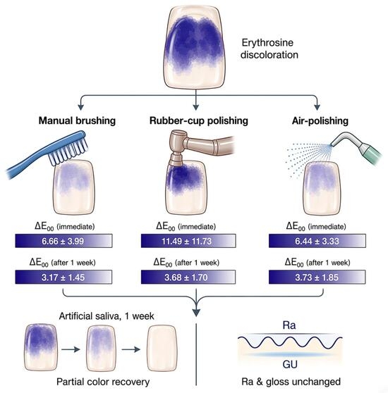
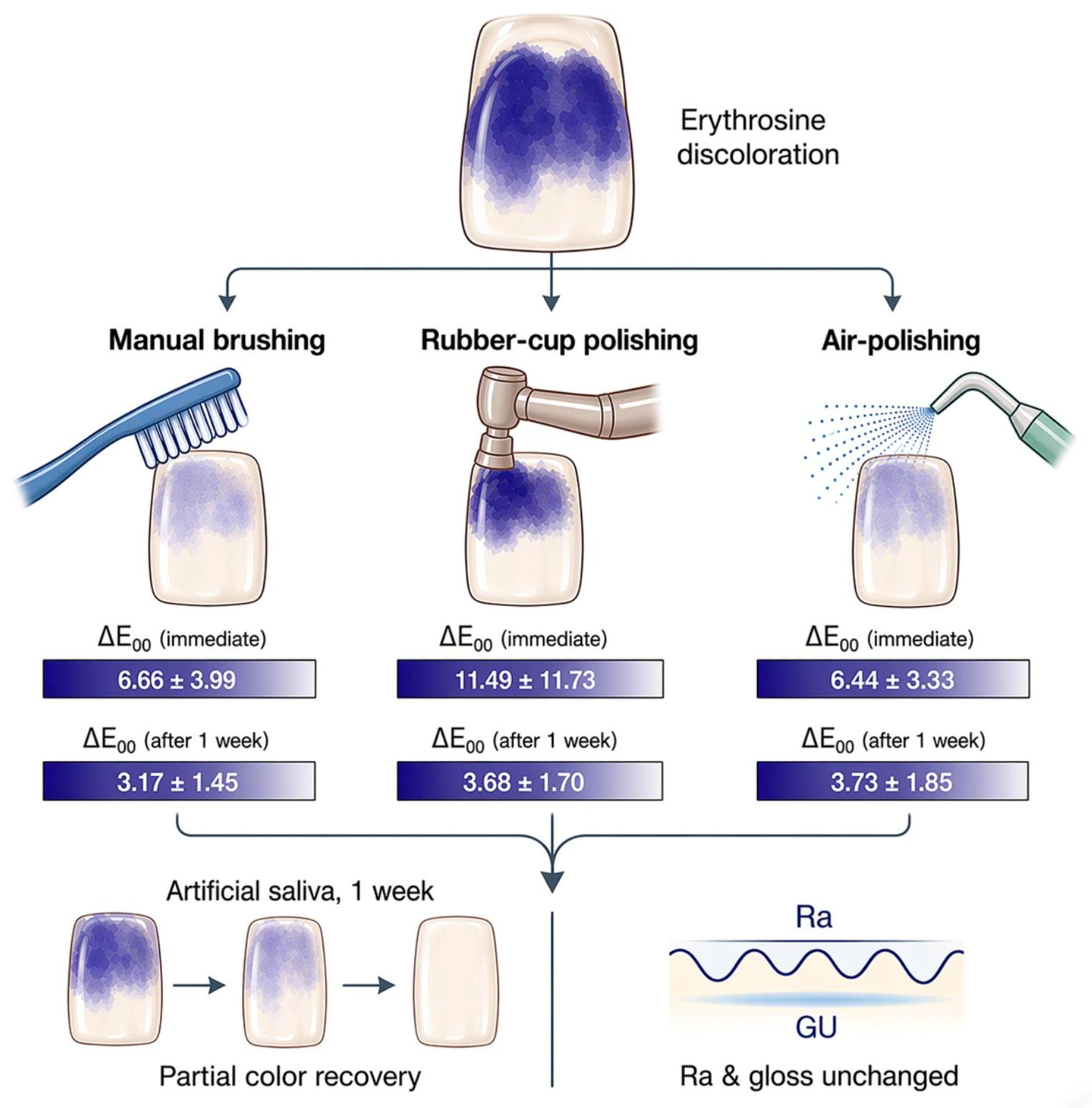
The present study investigated the effects of a plaque disclosing agent and three commonly used discoloration removal modalities (manual brushing, rubber-cup polishing, and air-polishing) on enamel surface roughness, gloss, and color stability. Overall, the findings indicate that all cleaning methods were associated with a reduction in disclosing-agent-induced discoloration under the present experimental conditions, with no statistically significant changes detected in enamel surface characteristics. The disclosing agent was associated with visually perceptible staining, as reflected by the elevated immediate ΔE00 values recorded after the removal procedures. However, following 1 week of storage in artificial saliva, color differences decreased, indicating partial short-term optical recovery under controlled conditions. This observation may be of clinical interest, as residual post-cleaning discoloration may remain perceptible immediately after plaque disclosure and removal, potentially influencing chairside esthetic assessment and patient perception. Importantly, none of the removal modalities resulted in significant changes in surface roughness or gloss, and no intergroup differences were detected across the evaluated parameters. Based on these findings, the null hypothesis that different stain removal methods would not result in significant differences in enamel surface roughness, gloss, or color change was not rejected.
Contemporary biofilm management increasingly incorporates plaque disclosure to provide immediate visual feedback and enable more selective, minimally invasive removal. In professional care, disclosure-guided mechanical plaque removal has been associated with improved biofilm elimination compared with non-guided approaches, underscoring the clinical relevance of color-guided plaque control [
6,
18]. Within this context, patient-performed brushing and professional rubber-cup polishing remain widely used methods for removing disclosed deposits. In parallel, minimally invasive professional mechanical plaque removal concepts have increasingly incorporated fine-particle air-polishing as an adjunct modality, supported by evidence of effective plaque removal at difficult-to-access sites and a favorable profile during periodontal maintenance [
19,
20]. Accordingly, the inclusion of erythritol-based, low-abrasive air-polishing alongside conventional methods provides a relevant clinical context for interpreting the present findings.
Beyond its clinical advantages, disclosure-guided biofilm control also raises esthetic considerations, as the chromogenic properties that enable visualization may facilitate pigment retention on oral surfaces. Plaque disclosers vary in delivery form and dye composition, with commonly used colorants including CI 45430 (erythrosine), CI 42090 (brilliant blue), 45350 (fluorescein), CI 16035 (allura red), CI 45410 (phloxine, red dye), CI 42051 (patent blue), and CI 42053 (fast green) [
2]. The chemical characteristics of these dyes support their potential interaction with biological substrates, providing a plausible mechanistic basis for pigment adsorption and persistence [
21,
22]. Consistent with this concern, experimental studies have shown that disclosing agents can induce measurable and sometimes clinically relevant color changes on tooth-colored restorative materials and surface sealants, with incomplete reversal after cleaning or repolishing [
2,
10,
13]. In addition, laboratory evidence indicates that disclosing solutions may spread beyond the target biofilm and stain adjacent oral tissues, supporting the practical plausibility of off-target pigment deposition during routine use [
9]. However, it is critical to emphasize that a substantial portion of this evidence derives from restorative substrates rather than natural enamel. Therefore, while these studies provide mechanistic insight into dye-substrate interactions, their findings cannot be directly extrapolated to enamel behavior, which differs in composition, permeability, and surface chemistry.
Although restorative-material studies are frequently cited to support general hypotheses regarding dye retention [
23,
24,
25], the present investigation is limited to sound natural enamel and does not evaluate restoration-bearing teeth or enamel-restoration interfaces. This distinction is clinically important, as staining dynamics in restorative margins, hybrid layers, or resin-based materials may be more pronounced and governed by fundamentally different physicochemical mechanisms. Accordingly, the current findings should be interpreted strictly within the context of intact enamel surfaces. Within enamel, evaluation of color change alone may be insufficient, as perceived tooth appearance is influenced not only by chromatic coordinates but also by surface-related optical behavior [
24]. Therefore, the present study assessed ΔE
00 together with Ra and GU to capture both discoloration and potential surface-related changes that may independently influence visual perception. Color differences were calculated using CIEDE2000 to enhance the perceptual relevance of instrumental measurements [
17], in line with dental evidence indicating closer correspondence with visually perceived differences and clinically meaningful acceptability concepts than conventional CIELAB-based metrics [
26].
While no statistically significant differences were detected among groups, the numerical differences observed (e.g., lower mean ΔE
00 and Ra values in the erythritol-based air-polishing group) should be interpreted cautiously and strictly as descriptive findings rather than evidence of superior performance. Fine-particle erythritol powders have been reported to effectively disrupt biofilm while maintaining low abrasivity under controlled conditions, a characteristic that may be advantageous in workflows involving repeated disclosure and cleaning cycles [
19]. However, in the absence of statistical significance, no claims of relative efficacy or superiority can be made based on the present dataset.
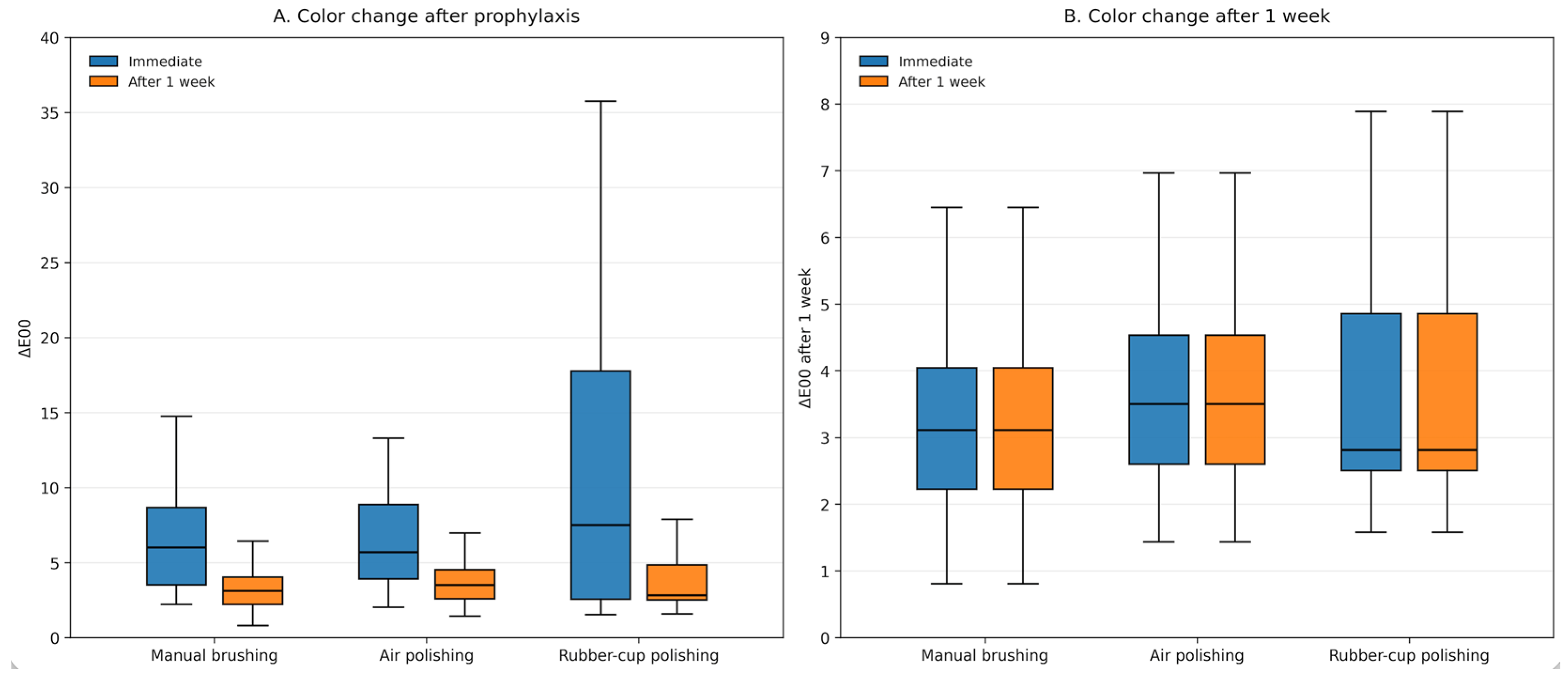
Following plaque disclosure-induced discoloration, measurable color differences relative to baseline enamel persisted after all three removal modalities, and baseline enamel color was not fully re-established either immediately after removal or after 1 week storage in artificial saliva. Importantly, the present color outcome quantified the difference between baseline enamel and the post-disclosure/post-removal condition rather than absolute discoloration elimination. Under this framework, the results indicate that all tested procedures were associated with residual color differences after cleaning, with no statistically significant differences detected among the groups under the present experimental conditions. Immediately after cleaning, mean ΔE
00 values (6.44–11.49) were clearly above widely cited CIEDE2000 acceptability values reported for dental hard tissues (often summarized around a 50:50 acceptability threshold near ΔE
00 ≈ 1.8) [
27,
28]. After 1-week artificial saliva storage, ΔE
00 values decreased in all groups (means 3.17–3.73), indicating partial short-term optical recovery. When interpreted against a more permissive clinical acceptability threshold of ΔE
00 ≈ 3.3, the residual color differences approached, but still exceeded, this boundary, suggesting that disclosure-associated discoloration may persist beyond short-term removal under standardized in vitro conditions [
27]. This pattern indicates that while optical recovery occurs, complete return to baseline color may require longer exposure to oral environmental factors. A plausible explanation is that saliva-like storage may promote partial rehydration of the enamel surface and modify its refractive behavior, thereby contributing to gradual improvement in perceived color. A cautious interpretation is warranted because longer and more complex in vivo conditions, such as continuous salivary flow, intraoral temperature fluctuations, dietary influences, and individualized oral hygiene behaviors, may contribute to further attenuation of residual discoloration. Finally, interpretation against thresholds should acknowledge that perceptibility and acceptability tolerances are not fixed and can vary with chroma and location within the dental color space, as well as methodological conditions [
28,
29,
30]. From a clinical perspective, these findings suggest that immediate post-disclosure color differences may remain noticeable despite chairside removal but may progressively diminish over time; therefore, selection among the tested cleaning methods may reasonably be guided by practical and patient-related considerations rather than color outcomes alone under the present conditions.
The enamel-based findings of this study align conceptually with previous reports on restorative materials showing that disclosing agents may induce persistent discoloration. Hino et al. [
10] and Şen et al. [
13] both reported persistent discoloration of esthetic materials following discloser exposure despite professional cleaning procedures. Expanding on these observations, Mazzitelli et al. [
2] showed that discoloration magnitude and reversibility depend on the dye composition and delivery form of plaque detectors and that repolishing may fail to fully restore baseline color, supporting a retention-driven mechanism rather than a purely superficial stain. Although derived from restorative substrates, these findings provide a mechanistic framework for interpreting the present enamel data, in which residual ΔE
00 values persisted even after cleaning and subsequent artificial saliva storage, suggesting that similar dye–substrate interaction processes may also occur on natural enamel surfaces. Nevertheless, restorative materials may exhibit distinct dye–substrate interactions (e.g., resin–matrix sorption and surface porosity) compared with enamel. Therefore, the present enamel findings should be considered mechanistically concordant but not directly interchangeable with outcomes derived from restorative-material models.
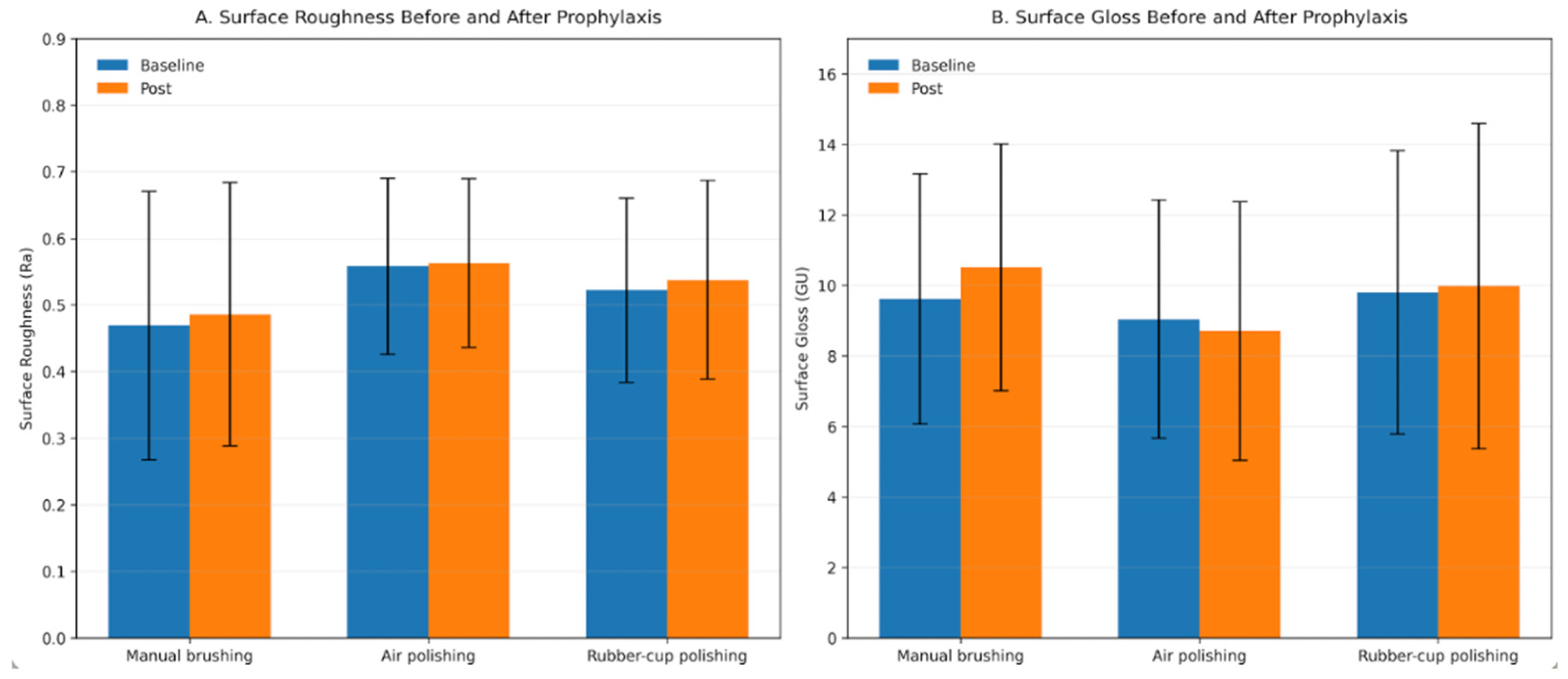
Regarding surface roughness, all three removal protocols produced only negligible Ra shifts (mean ΔRa ≈ 0.01–0.02 µm), and neither intergroup nor within-group analyses demonstrated statistically significant deviations from baseline. From a clinical mechanistic standpoint, these values remain well below the commonly cited plaque-retention threshold (Ra ≈ 0.2 µm), indicating that the applied protocols are unlikely to induce roughness changes in practical relevance for enamel biofilm retention [
25]. This surface-neutral pattern is consistent with evidence that low-abrasive air-polishing powders, particularly erythritol, exert minimal measurable impact on enamel roughness under controlled conditions [
31,
32] and are supported by periodontal maintenance literature describing favorable hard-tissue profiles when appropriately applied [
19,
20]. The negligible ΔRa observed after manual brushing aligns with reports indicating that enamel abrasion and roughness changes depend strongly on brushing force, duration, and dentifrice abrasivity, and may remain limited under standardized short-duration protocols [
33,
34]. Similarly, the absence of roughness increase after rubber-cup polishing agrees with evidence that prophylaxis polishing outcomes are highly dependent on paste abrasivity and application variables, with clinically realistic regimens often maintaining roughness values below the plaque-retention threshold [
15,
35]. Beyond plaque retention, surface texture may contribute more directly to staining and optical appearance. Experimental models have shown that deliberately roughened enamel retains more stain and requires greater effort for stain removal than polished surfaces, supporting the role of micro-irregularities in pigment retention independent of plaque-mediated effects [
36]. Moreover, previous studies have demonstrated that alterations in enamel roughness can influence optical parameters and color coordinates, indicating that surface texture may affect perceived appearance even when overall color change does not directly track roughness [
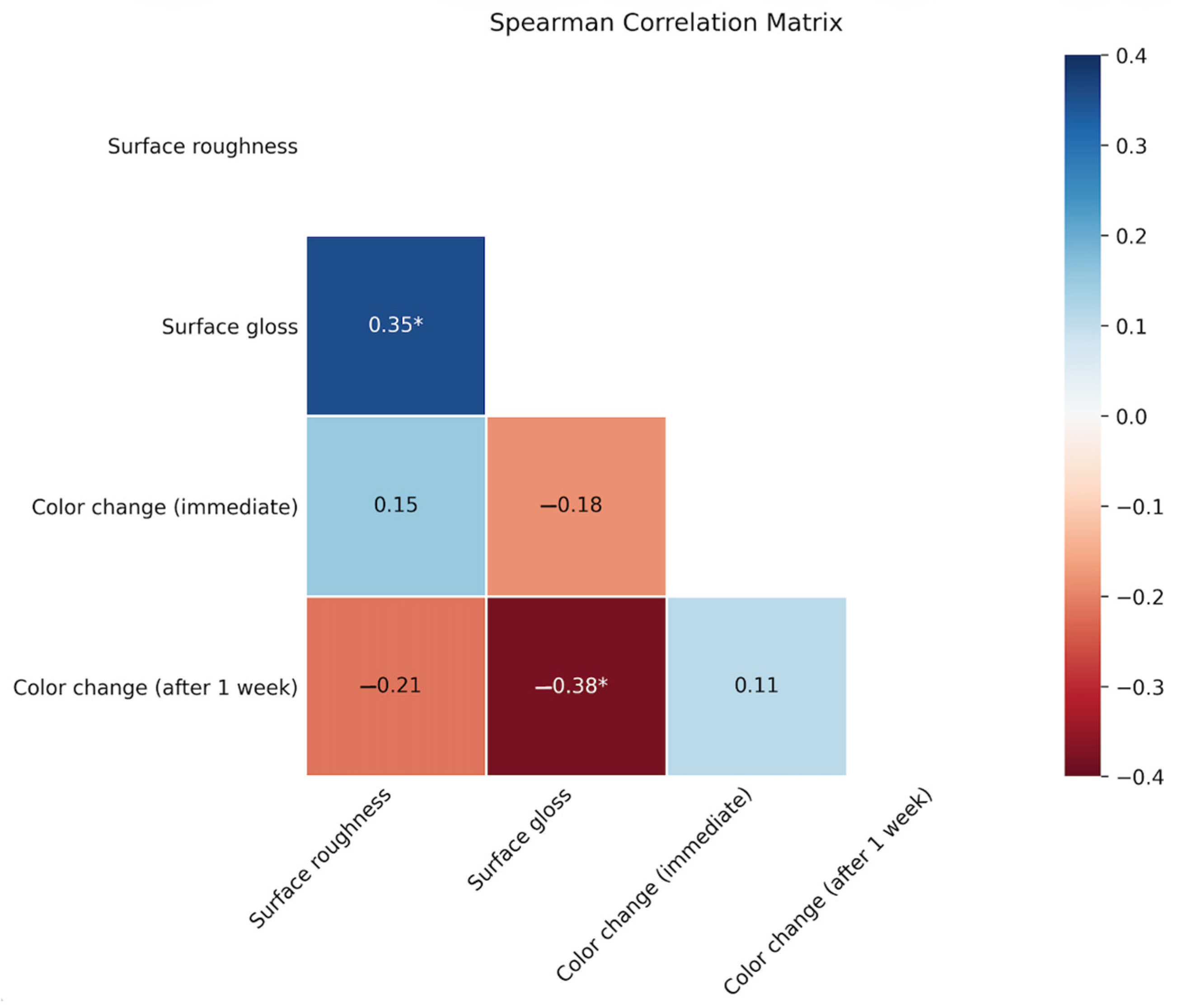
37]. In the present dataset, however, ΔRa remained minimal and did not correlate with ΔE
00, suggesting that roughness-related pathways were unlikely to be the primary driver of the residual color differences under the applied conditions. Taken together, the negligible ΔRa and modest gloss shifts suggest that these modalities did not measurably compromise enamel surface characteristics under the standardized, short-duration conditions tested.
Consistent with the minimal ΔRa observed across groups, surface gloss changes were also modest and did not differ significantly either from baseline or between removal modalities, indicating no measurable gloss deterioration under the applied conditions. This may be of clinical interest because perceived tooth appearance is not determined by color coordinates alone; surface reflectance and light-scattering behavior, which are influenced by surface texture, also contribute to the visual brightness and overall liveliness of enamel surfaces [
24]. In addition, gloss outcomes after prophylaxis polishing are known to depend on paste abrasivity and application variables, and clinically realistic regimens may not necessarily produce a clinically meaningful deterioration in surface appearance [
15,
35]. Importantly, gloss does not exhibit a simple linear relationship with roughness, as surfaces with similar average Ra values may differ in microtopography and reflectance characteristics, resulting in divergent gloss behavior [
38,
39]. These observations are consistent with the physicochemical profile of fine-particle air-polishing systems, as low-abrasive erythritol powders have been shown to exert minimal effects on enamel microtopography under controlled conditions, thereby supporting preservation of specular reflection and gloss [
31,
32].
Correlation analysis further indicated that residual optical differences following discloser removal were not primarily mediated by surface alterations. No significant association was observed between color change and changes in either surface roughness or gloss, suggesting that roughness- or gloss-dependent pathways were unlikely to account for the remaining color differences under the present conditions. In contrast, a weak but statistically significant positive correlation was detected between changes in roughness and gloss, consistent with evidence that gloss is sensitive to surface morphology and may co-vary with texture without proportionally influencing overall color-difference metrics [
24,
38]. Notably, enamel-focused models have reported mixed findings regarding roughness-color relationships. Atalay et al. [
40] observed a significant association between surface-texture changes and color outcomes under their experimental conditions, supporting a roughness-color link when surface alterations are sufficiently pronounced. By contrast, Vieira-Junior et al. [
37] did not identify a direct relationship between roughness and overall color change, a pattern more consistent with the present dataset and suggestive that color coordinates may shift independently of Ra depending on the type and magnitude of surface modification. In the present dataset, the absence of a ΔE
00 association likely reflects the limited magnitude and narrow distribution of ΔRa and ΔGU values, whereas residual discoloration is more plausibly attributable to persisting discloser-derived pigments rather than topography-driven optical changes.
This study has limitations inherent to its in vitro design, and these should be considered when interpreting the findings. The experimental conditions do not replicate key dynamic features of the oral environment, which may directly influence both discoloration behavior and its reversibility. In particular, the absence of continuous salivary flow eliminates the mechanical flushing and dilution effects that may facilitate the clearance of loosely bound pigments. Similarly, the lack of natural acquired pellicle formation and turnover may alter the interaction between disclosing dyes and the enamel surface, as the pellicle can act both as a barrier limiting direct enamel staining and as a substrate for transient pigment adsorption. Furthermore, the study design did not incorporate repeated staining and cleaning cycles, which are characteristic of clinical plaque-control regimens. Such cyclic exposure may lead to cumulative or, conversely, progressively reduced staining depending on surface conditioning and pellicle dynamics over time. The relatively short observation period also limits assessment of longer-term optical recovery, which in vivo may be enhanced by ongoing salivary remineralization, hydration changes, and routine oral hygiene practices. Importantly, this study was restricted to sound natural enamel and did not include restorative materials or enamel-restoration interfaces. Therefore, the findings cannot be extrapolated to restoration-bearing teeth, where staining behavior may be more pronounced and governed by material-specific factors such as resin matrix composition, surface porosity, and interfacial characteristics. Although the discussion refers to literature on restorative materials to provide mechanistic context, these data represent different substrates and should not be interpreted as directly comparable to the present enamel findings. Furthermore, the use of extracted teeth precludes biological processes such as pellicle renewal, biofilm dynamics, and interactions with gingival crevicular fluid, all of which may influence pigment retention and surface properties. Although the removal protocols were standardized, they may not fully capture clinical variability related to operator technique, access, angulation, pressure, and duration. Taken together, these limitations suggest that the present results represent a controlled assessment of enamel response and may provide a conservative estimate of color recovery under simplified conditions. Extrapolation to clinical scenarios should therefore be made with caution. Future studies incorporating restorative materials, particularly anterior restorations and enamel-restoration interfaces, would likely enhance clinical relevance by better reflecting staining behavior and removal dynamics in restoration-bearing teeth.