Brief Report

DHA-Rich n-3 PUFA Supplementation Improves Morning Stiffness and Quality of Life in Patients with Rheumatoid Arthritis and Downregulates TNF-α Expression in PBMCs


by
Alejandro A. Candia
1,
Victoria Torres-Galaz
1,
Camila Latapiatt
1,
Denisse Valladares-Ide
2,* and
Sebastián Jannas-Vela
1,*
1
Instituto de Ciencias de la Salud, Universidad de O’Higgins, Rancagua 2820000, Chile
2
Long Active Life Laboratory, Instituto de Ciencias de la Salud, Universidad de O’Higgins, Rancagua 2820000, Chile
*
Authors to whom correspondence should be addressed.
Appl. Sci. 2026, 16(4), 1980; https://doi.org/10.3390/app16041980
Submission received: 6 January 2026 / Revised: 3 February 2026 / Accepted: 9 February 2026 / Published: 17 February 2026
(This article belongs to the Special Issue Application of Nutrition and Clinical Exercise Physiology)

Abstract

Rheumatoid arthritis (RA) is a chronic inflammatory autoimmune disease associated with joint pain, stiffness, functional impairment, and reduced quality of life. Omega-3 (n-3) polyunsaturated fatty acids (PUFAs) have been proposed as adjunctive therapies for RA due to their anti-inflammatory and pro-resolving properties; however, evidence regarding high-dose docosahexaenoic acid (DHA) supplementation remains inconclusive. This exploratory study investigated the effects of 16 weeks of DHA-rich PUFA supplementation (2.5 g/d DHA and 0.5 g/d EPA) on clinical, functional, and inflammatory outcomes in individuals with moderate RA. A modest sample size of thirteen women (23–61 y) were randomized to receive either n-3 PUFA (n = 7) or placebo (n = 6). Clinical outcomes, quality of life, functional performance, cardiometabolic parameters, and systemic inflammatory markers were assessed before and after the intervention. Additionally, the inflammatory response of peripheral blood mononuclear cells (PBMCs) exposed to patient serum collected before and after supplementation, with and without the n-3 PUFA mediator resolvin D1 (RvD1), was evaluated. DHA-rich PUFA supplementation significantly reduced morning stiffness duration and improved RA-specific quality of life scores, without affecting systemic inflammatory or cardiometabolic markers. Serum obtained after supplementation attenuated TNF-α expression in PBMCs and produced effects comparable to RvD1 treatment. These findings suggest that DHA-rich PUFA supplementation improves clinically relevant outcomes in RA, potentially through local pro-resolving mechanisms.

1. Introduction

Rheumatoid arthritis (RA) is an autoimmune disease characterized by the production of autoantibodies (rheumatoid factor and anti-citrullinated protein antibody) and by the inflammation of the synovial membrane in the joints, resulting in a range of symptoms, including swelling, pain, stiffness, and fatigue [1,2]. Treatment for RA includes the prescription of anti-rheumatic and anti-inflammatory pharmacological and biological medications, such as nonsteroidal anti-inflammatory drugs, corticosteroids, and disease-modifying anti-rheumatic drugs. These medications have been shown to improve functionality by reducing stiffness and enhancing quality of life; however, they have the potential to exacerbate cardiometabolic risk factors associated with RA while increasing healthcare expenditures [3,4,5].
To date, there is substantial evidence supporting that the regular intake of omega-3 (n-3) polyunsaturated fatty acids (PUFA) has positive effects on RA patients, specifically on clinical and functional outcomes [6,7,8]. For instance, supplementation with fish oil leads to the incorporation of eicosapentaenoic acid (C20:5n-3, EPA) and docosahexaenoic acid (C22:6n-3, DHA) into cell membranes [9,10]. This has been shown to improve several distinct parameters of RA including morning stiffness [11], joint pain [12], number of swollen and tender joints [13], grip strength [14,15], and quality of life [8]. Furthermore, in some cases fish oil supplementation has been reported to reduce the production of pro-inflammatory cytokines, including C-reactive protein (CRP) [16] and tumor necrosis factor alpha (TNF-α) [17]. These benefits are thought to be partially mediated by the PUFA oxidation derivatives, the oxylipins. These lipid mediators are produced by the release of PUFAs from immune cell membranes after a controlled inflammatory response by the enzyme phospholipaseA2. They are further metabolized by cyclooxygenase, lipoxygenase, or the cytochrome P450 pathways to oxylipins [18]. The oxylipins derived from omega-6 (n-6) PUFA are generally associated with a pro-inflammatory effect [19], whereas oxylipins derived from n-3 PUFA decrease the production of proinflammatory molecules and, most importantly, have been purported to promote the resolution of inflammation [19,20]. Furthermore, the metabolism of n-3 PUFAs through LOX and COX pathways can give rise to the specialized pro-resolving mediators (SPMs), including the resolvins, protectins, and maresins [21], whereas the cytochrome P450 epoxygenase pathway represents another pathway that metabolizes n-3 PUFA into epoxy fatty acids [18]. Together, these lipid mediators exert anti-inflammatory and pro-resolving effects, which could be crucial for repairing damaged tissue and for restoring function in RA individuals. Interestingly, the DHA-derived oxylipin resolvin D1 (RvD1) (100 ng/day) has shown a positive anti-inflammatory effect by attenuating arthritis severity, cachexia, hind-paw edema, paw leukocyte infiltration, and cartilage degeneration [22] in a rodent model of arthritis, whereas systemic treatment with resolvin E1 in a murine collagen-induced arthritis model may be ineffective in ameliorating disease severity [23]. These findings suggest that DHA and their derived oxylipins may offer enhanced protection for individuals with RA. Despite growing interest in n-3 PUFA supplementation in RA, most studies have employed combined doses of EPA and DHA. In consequence, the efficacy of supplementing with a high dose of DHA in individuals with RA remains inconclusive. Thus, the objective of the present exploratory study was to investigate the effects of DHA-rich PUFA supplementation on RA individuals and the TNF-α response to combined RA serum and RvD1 from healthy polymorphonuclear cells.

2. Materials and Methods

2.1. Participants

RA individuals with a moderate disease activity score-28 (DAS28) > 2.6 and <5.1, and taking nonsteroidal anti-inflammatory drugs, glucocorticoids, or disease-modifying anti-rheumatic drugs were eligible. However, the dosages of these medications had to be constant—at least four weeks before study commencement—and remain stable throughout the study period. In addition, RA patients who were regular alcohol abusers, smokers, consumers of fish oil supplements or dietary fish >2 times per week were excluded. Individuals diagnosed with gastrointestinal or metabolic diseases, performed regular aerobic exercise (>150 min moderate intensity per week), or had any physical or biomechanical limitations were also excluded. The participants were fully informed of the nature and possible risks of the experimental procedures before providing their written informed consent. This study was conducted following the Declaration of Helsinki and approved by the Comité de Ética de Investigación en Seres Humanos #0219/2021 (University of Chile, Santiago, Chile). All procedures were performed at the University of O’Higgins. This work presents preliminary results from the study registered on clinicaltrials.gov #NCT05945693 (3 July 2023). A schematic diagram documenting the flow of participants is presented in Supplementary Figure S1.

2.2. Study Design

This study utilized a randomized, double-blind, placebo-controlled experimental design. The subjects were randomly assigned to one of two groups: placebo control (Placebo) or Omega-3. Experimental groups were balanced for disease activity, pharmacological treatment, sex, and age. To ensure double blinding, a randomization sequence was generated by an independent investigator using a website (https://www.sealedenvelope.com/) (accessed on 28 September 2023). A second investigator, who was not involved in participant contact or data collection, was responsible for placing the supplements into identical, coded bottles. All personnel responsible for providing the supplements, as well as the participants, remained blinded to the group assignments throughout the study. Before beginning the 16-week supplementation period with n-3 PUFA, the participants completed a battery of tests, including RA specific clinical measures (e.g., DAS28), physical function measurements (e.g., handgrip strength), maximum aerobic capacity test (VO2max), and blood sampling, as well as a dietary and physical activity assessment. These same evaluations were repeated upon completion of the intervention period.

2.3. Primary and Secondary Outcomes

Primary outcome: Duration (minutes) of early morning stiffness. This measure was chosen based on prior evidence with n-3 PUFA supplementation in RA patients [6].
Secondary outcomes: Other relevant outcomes which allow the assessment of disease progression were measured, such as the DAS28, quality of life, handgrip strength, and TNF-α blood levels.

2.4. Supplementation

The participants consumed either five daily fish oil capsules (0.5 g/capsule DHA and 0.1 g/capsule EPA), comprising a total of 2.5 g/d of DHA and 0.5 g/d of EPA (Omega Up TG DHA 600, Newscience, Santiago, Chile), or five daily placebo filled with maltodextrin capsules for 16 weeks (two in the morning and three at night). These doses have been reported to be safe, to be incorporated into cell membranes, and to produce health improvements in individuals with RA [24,25,26,27]. To ensure double blinding, capsules were placed into de-identified bottles and subsequently provided to the participants.

2.5. General Characteristics

The participant’s age, body mass index (BMI), waist circumference (WC), educational status, socioeconomic level, years of diagnosis, smoking habits, comorbidities, and medications were collected. Body mass and height were determined with a scale (Omrom Sc-150, Kyoto, Japan) and wall-mounted stadiometer (Seca 213, Hamburg, Germany).

2.6. Rheumatoid Arthritis Clinical Outcomes

2.6.1. Duration of Morning Stiffness

Was assessed by questioning “How long is your morning stiffness from waking until maximum improvement?” [28]. The duration of morning stiffness was reported in minutes, from 0 up to a maximum of 180 min.

2.6.2. Disease Activity Score-28 (DAS28)

Describes the severity of RA using clinical and laboratory data including the number of swollen joints (total 28), the number of tender joints (total 28), the Westgren erythrocyte sedimentation rate (ESR), and patients’ global disease assessment. The DAS28 is validated and it is required by the Chilean Ministry of Health [29].

2.6.3. Health Assessment Questionnaire Disability Index (HAQDI)

This tool consists of eight sections with 2 or 3 questions per section including dressing, arising, eating, walking, hygiene, reach, grip, and activities. The scoring ranges from 0 (without any difficulty) to 3 (unable to do). This questionnaire has been validated to be used in Chilean RA individuals [30].

2.6.4. Quality of Life RA (QOL-RA) Scale

It consists of an 8-item scale that measures physical ability, pain, interaction with family and friends, support from family and friends, mood, tension, arthritis, and health using a 10-point scale with very poor [1] at one end and excellent [10] at the other. The higher the score, the higher the health-related quality of life [31].

2.7. Functional Measurements

2.7.1. Timed up and Go Test

It consists of a physical function test that starts with the individual in a seated position who must stand up on command, walk 3 m, turn around, and walk back to the chair to sit down. The time starts on command and stops when the participant is seated.

2.7.2. Short Physical Performance Battery

This tests measures lower extremity performance using timed measures of standing balance (side by side, semi-tandem, tandem), a 4 m walk, and five repetitive chair stands [32].

2.7.3. Handgrip Strength

Measures the maximum isometric strength of the hand and forearm muscles. The participant pressures the dynamometer (Jamar; Sammons Preston, Rolyon, Bolingbrook, IL, USA) with maximum effort for 3–5 s and the highest value out of three contractions is used for analysis.

2.8. Maximum Aerobic Test

The VO2max was assessed during an incremental cycling test to exhaustion on a recumbent ergometer (Ergoselect 5M, Ergoline, Bitz, Germany) following ACSM guidelines [33] and utilizing a portable, validated gas analyzer (Metalyzer 3B, Cortex, Leipzig, Germany) [34].

2.9. Blood Analyses

Blood was sampled after an overnight fast (12 h) using a needle and Vacutainer kits. They were centrifuged at 1000× g for 10 min at room temperature to obtain plasma, serum, and erythrocyte fractions. Each fraction was frozen at −80 °C until further analysis. Plasma lipid profiles were determined at the laboratory of Hospital Dr. Franco Ravera Zunino and plasma TNF-α levels were measured via commercial ELISA assay kits (RayBio®, Peachtree Corners, GA, USA).

2.10. Peripheral Blood Mononuclear Cells

Blood samples were collected from three healthy young women (28.7 ± 1.5 y) for the isolation of peripheral blood mononuclear cells (PBMCs). They were incubated for 24 h with either 2% fetal bovine serum (FBS; control) or 0.5% FBS + 1.5% pooled serum by combining equal volumes (100 µL) from each patient of the Omega-3 group (before and after supplementation), with and without RvD1 (100 ng/mL), to assess TNF-α expression. Briefly, whole blood was collected into EDTA tubes and centrifuged at 1700× g for 10 min to separate the plasma and buffy coat. The buffy coat was collected, diluted with PBS to a final volume of 10 mL, and carefully layered over a Ficoll density gradient separation solution. The layered sample was then centrifuged at 550× g for 40 min at room temperature. The intermediate layer containing the PBMCs was collected, washed twice in PBS by centrifugation at 1000× g for 10 min, and finally resuspended in 2 mL of PBS [35]. After incubation, total RNA was isolated with TRIzol H reagent (Invitrogen, Waltham MA, USA). cDNA was obtained by reverse transcription reaction of 1 μg of total RNA using the MyTaq one-step RT-PCR kit (Bioline, London, UK) on a Heal Force Thermal Cycler T960 (Shanghai, China). For real-time polymerase chain reaction analyses, a SYBR Green-based Master Mix (Bioline, London, UK) with the Rotor-gene Q (Qiagen, Hilden, Germany) was used with the following primers: TNF-α 5′-GGTGCTTGTTCCTCAGCCTCT-3′ and 5′-AGGGTTTGCTACAACATGGGC-3′; 18S rRNA 5′-AAACGGCTACCACATCCAAG-3′ and 5′-CCTCCAATGGATCCTCGTTA-3′; cyclophilin A 5′-CTTCATCCTAAAGCATACGGGTC-3′ and 5′-TGCCATCCAACCACTCAGTCT-3′; and ribosomal protein L4 mRNA 5′-ATACGCCATCTGTTCTGCCCT-3′ and 5′-GCTTCCTTGGTCTTCTTGTAGCCT-3′. All samples were run in duplicate and were normalized to the mRNA expression of ribosomal protein L4. The relative level of mRNA was calculated using the 2-DDCt method [36] and reported as mean fold changes.

2.11. Statistical Analysis

Due to small sample size, all data was treated as non-parametric. Baseline results are expressed as median with interquartile range, whereas pre- and post-supplementation results are expressed as mean ± SD. Baseline characteristics were compared using the Mann–Whitney U test. To measure the effects of n-3 PUFAs, a two-way ANOVA followed by uncorrected Fisher’s least significant difference post test for multiple comparisons was used to compare between groups and time. For the assessment of TNF-α expression in PBMCs a Kruskal–Wallis test followed by Dunn’s multiple comparisons test was performed. A value of p ≤ 0.05 was considered statistically significant. Effect size for significant differences was calculated as the within-group difference between means (pre- and post-supplementation) divided by standard deviation. GraphPad Prism, version 10.0 (GraphPad 10 Software Inc., San Diego, CA, USA), was used for all statistical analyses.

3. Results

3.1. Baseline Characteristics

Table 1 summarizes the demographic and clinical characteristics of the 13 participants who completed the study. Both groups were well-matched for all body composition, physical function, cardiovascular, and quality of life variables.

3.2. Effect of n-3 PUFAs on Systemic Inflammatory and Cardiometabolic Parameters

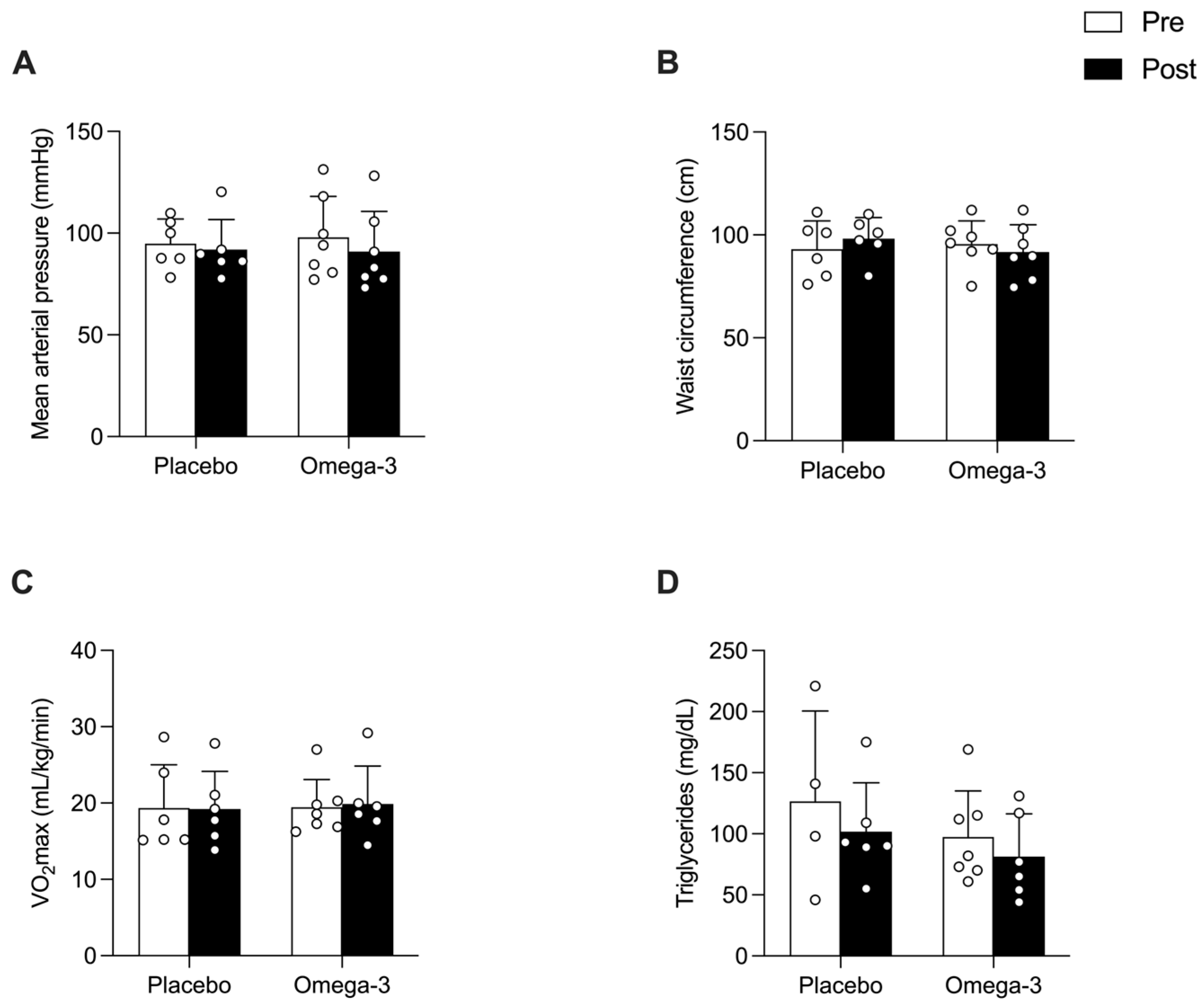
Supplementation with 2.5 g/d of DHA for 16 weeks had no effect on systemic inflammatory markers. Specifically, there were no differences in the circulating levels of CRP, erythrocyte sedimentation rate (ESR), or TNF-α between the two intervention groups (Figure 1). Additionally, no significant differences were observed between the Omega-3 and Placebo groups for any of the assessed cardiometabolic health variables, including mean arterial pressure, WC, VO2max, total cholesterol, and triglycerides (TG) (Figure 2).

3.3. Effect of n-3 PUFAs on Clinical and Functional Outcomes

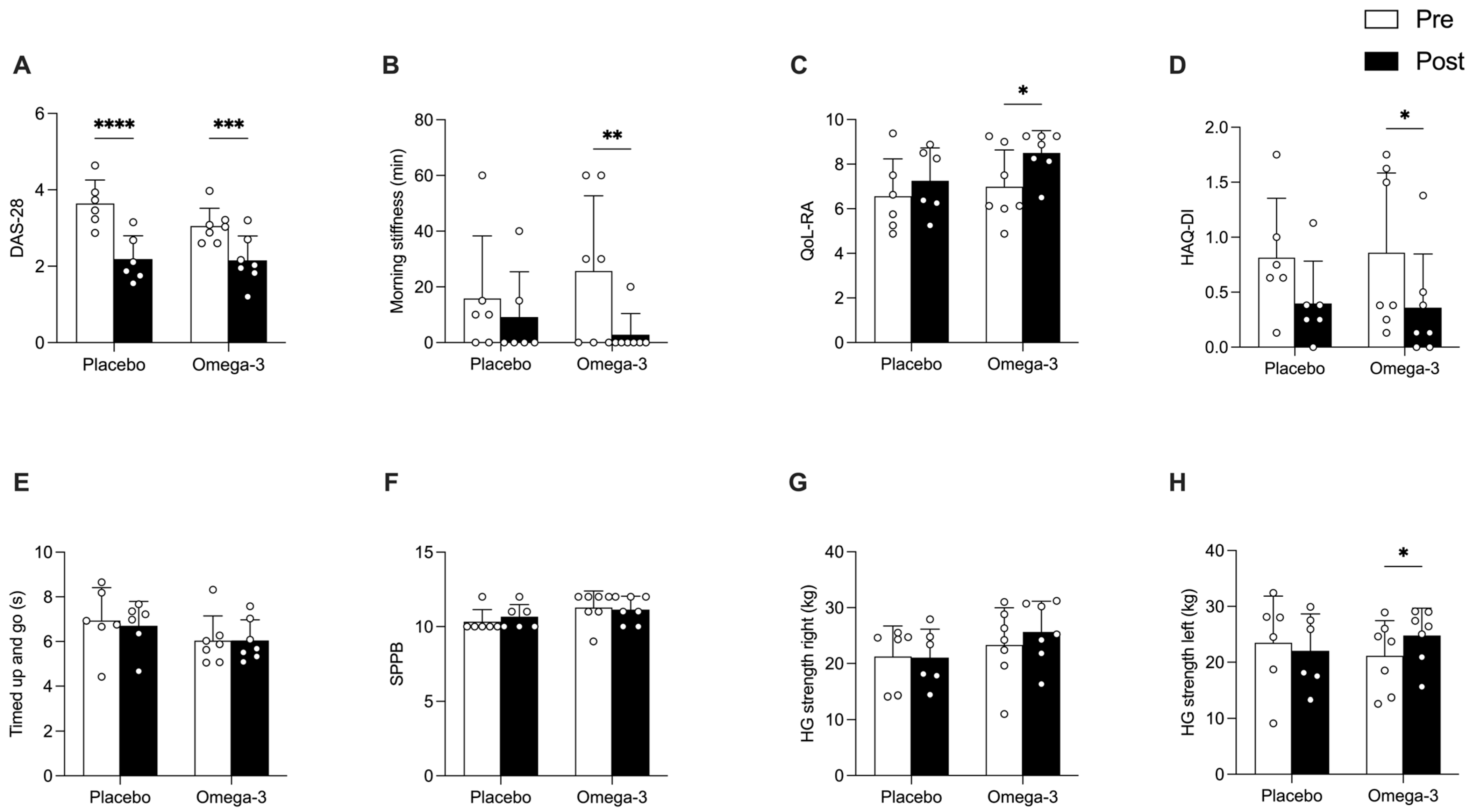
After the 16 week supplementation period, both groups had a significant improvement in disease activity, assessed by the DAS28 score (p < 0.0001); however, only the Omega-3 group exhibited a significant reduction in the duration of morning stiffness (p = 0.01; Effect Size: 1.32) after the supplementation period (Figure 3A,B). Similarly, quality of life, assessed by two questionnaires—QoL-RA and HAQ-DI—improved significantly (p < 0.05; Effect Size QoL-RA: 1.14; Effect Size HAQ-DI: 0.82) only in the Omega-3 group (Figure 3C,D). Meanwhile, functional performance measurements, including the timed up and go test, the SPPB and right handgrip strength, remained stable across time in both groups (Figure 3E–G); however, a significant increase in left-hand grip strength (p < 0.05; Effect Size: 0.38) was observed only in the Omega-3 group after supplementation (Figure 3H).

3.4. Effect of RvD1 on Inflammatory Expression in PBMCs

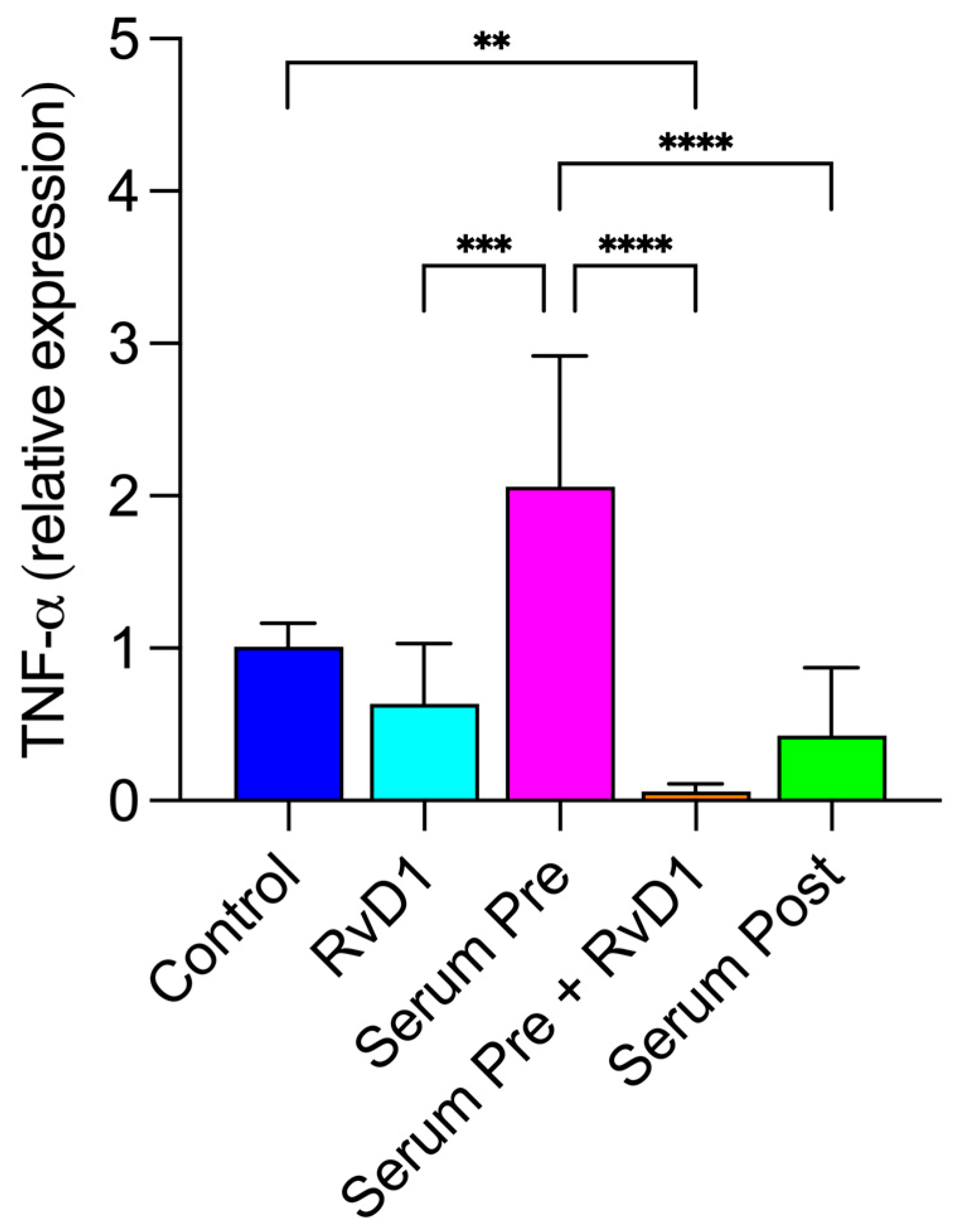
Given the role of DHA-derived resolvin-D1 (RvD1) in reducing inflammation, an ex vivo cell culture model in PBMCs from three healthy female donors was employed. PBMCs were stimulated with patient serum collected both before and after n-3 PUFA supplementation. Incubation of PBMCs with serum obtained before supplementation markedly increased TNF-α expression relative to control conditions, suggesting a pro-inflammatory effect of serum from RA patients (Figure 4). This response was substantially attenuated (p < 0.0001) when PBMCs were co-incubated with 100 ng/mL of RvD1. Furthermore, serum collected after the n-3 PUFA intervention elicited a significantly lower inflammatory response compared to before supplementation (p < 0.0001), with TNF-α expression levels comparable to those observed in the control and RvD1-only conditions (Figure 4). These findings demonstrate that both RvD1 and post-n-3 PUFA supplementation serum reduce the inflammatory activation of PBMCs ex vivo.

4. Discussion

The present study demonstrated that 16 weeks of high-dose DHA-rich fish oil supplementation (2.5 g/d DHA, 0.5 g/d EPA) significantly reduced morning stiffness duration and improved quality of life measures in patients with RA, despite showing no effects on systemic inflammatory markers or cardiometabolic parameters. Furthermore, our ex vivo experiments revealed that serum from RA patients collected after n-3 PUFA supplementation prevented the inflammatory response in PBMCs compared to pre-supplementation serum, and the anti-inflammatory effects of post-supplementation serum were comparable to those observed with the addition of the DHA-derived specialized pro-resolving mediator RvD1. The findings of the present study suggest that the observed benefits of n-3 PUFA supplementation in RA patients may be associated with the production of the DHA-derived oxylipin RvD1.
RA is characterized by chronic inflammation resulting in joint damage, pain, stiffness, and functional impairment [1,2] leading to decreased physical function and quality of life. While previous studies examining n-3 PUFA supplementation in RA have reported positive effects on systemic inflammatory markers [6,7], our findings align with some studies indicating no effects of n-3 PUFAs on circulating inflammatory biomarkers in RA patients [11,14]. Although the absence of changes in systemic inflammatory markers may appear unexpected, it does not imply a lack of biological effect, as n-3 PUFAs exert additional actions, including the modulation of pro-resolving inflammatory pathways [37] that may not be captured by conventional circulating inflammatory markers. On the other hand, the findings that n-3 PUFA supplementation did not significantly alter cardiometabolic parameters, including TG levels, was partially unexpected given the established triglyceride-lowering effects of n-3 PUFAs in the general population [38]. This finding could be explained by concomitant glucocorticoid use by our cohort, which may attenuate the lipid-modifying effects of n-3 PUFAs [39]. It is also plausible that, given that baseline TG levels in both groups were within the healthy range and that responses of TG to n-3 PUFA supplementation show substantial interindividual variability across both healthy and diseased populations [38], the likelihood of observing a clinically meaningful improvement following n-3 PUFA supplementation is reduced.
The reduction in morning stiffness observed in our study is particularly clinically relevant, as prolonged morning stiffness is a hallmark symptom of active RA and the primary result of the present study, impacting quality of life and functional capacity [28]. Furthermore, the improvement in HAQ-DI and QoL-RA scores supports the clinical significance of our findings in morning stiffness. Interestingly, these improvements occurred without concomitant changes in systemic inflammatory markers (Figure 1). This apparent dissociation is plausible, as quality of life measurements may diverge from circulating inflammatory markers, given that they reflect integrated functional, sensory, and psychosocial processes that are not fully captured by systemic biomarkers [40]. Finally, the observed increase in left-hand grip strength, a finding that has been documented in previous studies [14,15], may be attributable to several mechanisms supported by n-3 PUFAs, including and not limited to increased muscle protein synthesis [41], enhanced tissue regeneration [42,43], and improved mitochondrial function [44,45], among others.
An important finding from the present study was the prevention of TNF-α increased expression in PBMCs when incubated with serum after n-3 PUFA supplementation compared to serum from before supplementation. Interestingly, this effect mirrored that observed with exogenous RvD1 administration, a DHA-derived specialized pro-resolving mediator, which has been shown to resolve inflammation by reducing neutrophil infiltration, decreasing pro-inflammatory cytokine production, and enhancing macrophage clearance of cellular debris [18,19]. These data suggest that n-3 PUFA supplementation enhances the production of RvD1 or other DHA-related pro-resolving mediators in RA patients [46], which then act locally at the joint level to reduce morning stiffness and improve physical function without necessarily altering systemic inflammatory markers. It also aligns with the growing recognition that resolution of inflammation is an active process mediated by specialized pro-resolving mediators rather than simply the absence of pro-inflammatory stimuli [18,20]. Although RvD1 was the primary SPM assessed, other resolvins (e.g., RvE1), protectins, and maresins may also contribute to inflammatory resolution and tissue recovery in RA [47]. Future studies should employ broader lipid mediator profiling to capture the full spectrum of pro-resolving pathways influenced by n-3 PUFA supplementation.
The present study has some limitations that should be taken into consideration. Although not designed as a sex-specific study, the exclusive inclusion of women reflects the higher prevalence and burden of RA in this population [48]. In addition, the modest sample size may have constrained the statistical power to discern subtle variations in inflammatory markers and functional outcomes; however, this study was exploratory in nature, aimed at estimating biological plausibility, rather than providing definitive efficacy conclusions. The improvement in morning stiffness should be taken with caution, due to the modest baseline values and the lack of a defined minimal clinically important difference; however, morning stiffness is a key manifestation of RA, exerting significant impact on functional capacity and quality of life [49]. Additionally, we did not directly measure levels of n-3 PUFA, nor did we measure plasma RvD1 or other specialized pro-resolving mediators; however, we and others have previously shown that >8 weeks of n-3 PUFA supplementation using a similar dose results in incorporation of EPA and DHA into red blood cell and skeletal muscle phospholipids [10,50]. This suggests that the 16-week period was sufficient for incorporation into cell membranes. Finally, we acknowledge that the PBMCs responses assessed in the present study may not fully recapitulate the disease-specific inflammatory milieu of RA. Thus, the present results should be interpreted as a proof-of-concept rather than a definitive representation of immune behavior of RA patients. Future studies incorporating PBMCs directly derived from RA patients are essential to accurately characterize disease-specific responses to n-3 PUFA supplementation and their lipid derivatives.

5. Conclusions and Future Directions

In conclusion, our findings demonstrate that high-dose DHA-rich fish oil supplementation improves clinically relevant outcomes in RA patients despite minimal effects on systemic inflammatory markers. These benefits may be mediated through enhanced production of specialized pro-resolving mediators like RvD1, which act locally to resolve inflammation at the joint level. Altogether, these results support the inclusion of high-dose n-3 PUFA supplementation as an adjunctive therapy for managing morning stiffness and improving quality of life in RA patients. Future studies should integrate patient-reported outcomes such as quality-of-life measures with functional and mechanistic assessments to better capture clinically meaningful responses, and employ targeted lipidomics approaches to quantify lipid mediators in both circulation and synovial fluid to further elucidate the local anti-inflammatory mechanisms of n-3 PUFAs in RA.

Supplementary Materials

The following supporting information can be downloaded at: https://www.mdpi.com/article/10.3390/app16041980/s1, Figure S1: Study flow chart.

Author Contributions

Conceptualization, D.V.-I. and S.J.-V.; Investigation, A.A.C., V.T.-G. and C.L.; methodology, A.A.C., V.T.-G., C.L., D.V.-I. and S.J.-V.; writing—original draft preparation, S.J.-V.; writing—review and editing, A.A.C., V.T.-G., C.L., D.V.-I. and S.J.-V.; funding acquisition, DV-I and S.J.-V. All authors have read and agreed to the published version of the manuscript.

Funding

This research was funded by Fondo Nacional de Desarrollo Científico y Tecnológico (FONDECYT), grant numbers 1241959 (DV-I) and 11220333 (SJ-V).

Institutional Review Board Statement

The study was conducted in accordance with the Declaration of Helsinki, and approved by the Comité de Ética de Investigación en Seres Humanos #0219/2021 (Universidad de Chile, Santiago, Chile) for studies involving humans (19 April 2022).

Informed Consent Statement

Informed consent was obtained from all subjects involved in the study.

Data Availability Statement

The original contributions presented in the study are included in the article, further inquiries can be directed to the corresponding authors.

Acknowledgments

We thank all the staff from Hospital Franco Ravera Zunino, Paola Barrios-Troncoso, Jeremy Zapata-Urzúa, Joanny Rey-Puente, Rodrigo González-Aranda, Jocelyn Urrutia-Pino, Silvana Donoso-Acevedo, Camila Riquelme-Palacios, Aurora Palominos-González, and Dixon Cid-Hidalgo for their support in study coordination, patient recruitment, and blood draws and analysis. We also thank the participants for their time and commitment to this study and Farmacia Sibonat for providing the maltodextrin capsules.

Conflicts of Interest

The authors declare no conflicts of interest.

Abbreviations

The following abbreviations are used in this manuscript:
RARheumatoid arthritis
n-3Omega-3
PUFAPolyunsaturated fatty acids
EPAEicosapentaenoic acid
DHADocosahexaenoic acid
CRPC-reactive protein
TNF-αTumor necrosis factor Alpha
n-6Omega-6
RvD1Resolvin D1
DAS28Disease Activity Score-28
VO2maxMaximum aerobic capacity
BMIBody mass index
WCWaist circumference
HAQDIHealth assessment questionnaire disability index
QOL-RAQuality of life RA
SPPBShort Physical Performance Battery
PBMCsPeripheral blood mononuclear cells
FBSFetal bovine serum
ESRErythrocyte sedimentation rate

References

  1. Walsmith, J.; Roubenoff, R. Cachexia in rheumatoid arthritis. Int. J. Cardiol. 2002, 85, 89–99. [Google Scholar] [CrossRef] [Scilit] [PubMed]
  2. Pucino, V.; Certo, M.; Varricchi, G.; Marone, G.; Ursini, F.; Rossi, F.W.; De Paulis, A.; Mauro, C.; Raza, K.; Buckley, C.D. Metabolic checkpoints in rheumatoid arthritis. Front. Physiol. 2020, 11, 347. [Google Scholar] [CrossRef] [Scilit] [PubMed]
  3. Nikiphorou, E.; de Lusignan, S.; Mallen, C.D.; Khavandi, K.; Bedarida, G.; Buckley, C.D.; Galloway, J.; Raza, K. Cardiovascular risk factors and outcomes in early rheumatoid arthritis: A population-based study. Heart 2020, 106, 1566–1572. [Google Scholar] [CrossRef] [Scilit]
  4. Metsios, G.S.; Kitas, G.D. Physical activity, exercise and rheumatoid arthritis: Effectiveness, mechanisms and implementation. Best Pract. Res. Clin. Rheumatol. 2018, 32, 669–682. [Google Scholar]
  5. Hernández-Hernández, V.; Ferraz-Amaro, I.; Díaz-González, F. Influence of disease activity on the physical activity of rheumatoid arthritis patients. Rheumatology 2014, 53, 722–731. [Google Scholar]
  6. Gioxari, A.; Kaliora, A.C.; Marantidou, F.; Panagiotakos, D.P. Intake of ω-3 polyunsaturated fatty acids in patients with rheumatoid arthritis: A systematic review and meta-analysis. Nutrition 2018, 45, 114–124. [Google Scholar] [CrossRef] [Scilit]
  7. Philippou, E.; Petersson, S.D.; Rodomar, C.; Nikiphorou, E. Rheumatoid arthritis and dietary interventions: Systematic review of clinical trials. Nutr. Rev. 2021, 79, 410–428. [Google Scholar]
  8. Tedeschi, S.K.; Bathon, J.M.; Giles, J.T.; Lin, T.-C.; Yoshida, K.; Solomon, D.H. Relationship between fish consumption and disease activity in rheumatoid arthritis. Arthritis Care Res. 2018, 70, 327–332. [Google Scholar] [CrossRef] [Scilit] [PubMed]
  9. Moghaddami, M.; James, M.; Proudman, S.; Cleland, L.G. Synovial fluid and plasma n3 long chain polyunsaturated fatty acids in patients with inflammatory arthritis. Prostaglandins Leukot. Essent. Fat. Acids 2015, 97, 7–12. [Google Scholar] [CrossRef] [Scilit]
  10. Gerling, C.J.; Mukai, K.; Chabowski, A.; Heigenhauser, G.J.F.; Holloway, G.P.; Spriet, L.L.; Jannas-Vela, S. Incorporation of omega-3 fatty acids into human skeletal muscle sarcolemmal and mitochondrial membranes following 12 weeks of fish oil Supplementation. Front. Physiol. 2019, 10, 348. [Google Scholar] [CrossRef] [Scilit]
  11. Sköldstam, L.; Börjesson, O.; Kjällman, A.; Seiving, B.; Akesson, B. Effect of six months of fish oil supplementation in stable rheumatoid arthritis. A double-blind, controlled study. Scand. J. Rheumatol. 1992, 21, 178–185. [Google Scholar] [CrossRef] [Scilit]
  12. Tulleken, J.E.; Limburg, P.C.; Muskiet, F.A.; van Rijswijk, M.H. Vitamin E status during dietary fish oil supplementation in rheumatoid arthritis. Arthritis Rheum. 1990, 33, 1416–1419. [Google Scholar] [CrossRef] [Scilit] [PubMed]
  13. Kremer, J.M.; Lawrence, D.A.; Jubiz, W.; DiGiacomo, R.; Rynes, R.; Bartholomew, L.E.; Sherman, M. Dietary fish oil and olive oil supplementation in patients with rheumatoid arthritis clinical and immunologic effects. Arthritis Rheum. 1990, 33, 810–820. [Google Scholar] [CrossRef] [Scilit]
  14. Kremer, J.M.; Lawrence, D.A.; Petrillo, G.F.; Litts, L.L.; Mullaly, P.M.; Rynes, R.I.; Stocker, R.P.; Parhami, N.; Greenstein, N.S.; Fuchs, B.R. Effects of high-dose fish oil on rheumatoid arthritis after stopping nonsteroidal antiinflammatory drugs clinical and immune correlates. Arthritis Rheum. 1995, 38, 1107–1114. [Google Scholar] [CrossRef] [Scilit]
  15. Cleland, L.G.; French, J.K.; Betts, W.H.; Murphy, G.A.; Elliott, M.J. Clinical and biochemical effects of dietary fish oil supplements in rheumatoid arthritis. J. Rheumatol. 1988, 15, 1471–1475. [Google Scholar]
  16. Deutsch, L. Evaluation of the effect of Neptune Krill Oil on chronic inflammation and arthritic symptoms. J. Am. Coll. Nutr. 2007, 26, 39–48. [Google Scholar] [CrossRef] [Scilit] [PubMed]
  17. Sundrarjun, T.; Komindr, S.; Archararit, N.; Dahlan, W.; Puchaiwatananon, O.; Angthararak, S.; Udomsuppayakul, U.; Chuncharunee, S. Effects of n-3 fatty acids on serum interleukin-6, tumour necrosis factor-alpha and soluble tumour necrosis factor receptor p55 in active rheumatoid arthritis. J. Int. Med. Res. 2004, 32, 443–454. [Google Scholar] [CrossRef] [Scilit] [PubMed]
  18. Gabbs, M.; Leng, S.; Devassy, J.G.; Monirujjaman, M.; Aukema, H.M. Advances in our understanding of oxylipins derived from dietary PUFAs. Adv. Nutr. 2015, 6, 513–540. [Google Scholar]
  19. Levy, B.D.; Clish, C.B.; Schmidt, B.; Gronert, K.; Serhan, C.N. Lipid mediator class switching during acute inflammation: Signals in resolution. Nat. Immunol. 2001, 2, 612–619. [Google Scholar] [CrossRef] [Scilit]
  20. Markworth, J.F.; Maddipati, K.R.; Cameron-Smith, D. Emerging roles of pro-resolving lipid mediators in immunological and adaptive responses to exercise-induced muscle injury. Exerc. Immunol. Rev. 2016, 22, 110–134. [Google Scholar]
  21. Serhan, C.N. Treating inflammation and infection in the 21st century: New hints from decoding resolution mediators and mechanisms. FASEB J. Off. Publ. Fed. Am. Soc. Exp. Biol. 2017, 31, 1273–1288. [Google Scholar] [CrossRef] [Scilit]
  22. Norling, L.V.; Headland, S.E.; Dalli, J.; Arnardottir, H.H.; Haworth, O.; Jones, H.R.; Irimia, D.; Serhan, C.N.; Perretti, M. Proresolving and cartilage-protective actions of resolvin D1 in inflammatory arthritis. JCI Insight 2016, 1, e85922. [Google Scholar] [CrossRef] [Scilit] [PubMed]
  23. de Molon, R.S.; Thurlings, R.M.; Walgreen, B.; Helsen, M.M.; van der Kraan, P.M.; Cirelli, J.A.; Koenders, M.I. Systemic Resolvin E1 (RvE1) Treatment Does Not Ameliorate the Severity of Collagen-Induced Arthritis (CIA) in Mice: A Randomized, Prospective, and Controlled Proof of Concept Study. Mediat. Inflamm. 2019, 2019, 5689465. [Google Scholar] [CrossRef] [Scilit] [PubMed]
  24. Products, N. Scientific opinion on the tolerable upper intake level of eicosapentaenoic acid (EPA), docosahexaenoic acid (DHA) and docosapentaenoic acid (DPA). EFSA J. 2012, 10, 2815. [Google Scholar] [CrossRef] [Scilit]
  25. Jannas-Vela, S.; Klingel, S.L.; Mutch, D.M.; Spriet, L.L. DHA supplementation decreases resting metabolic rate in healthy young females. Appl. Physiol. Nutr. Metab. 2020, 45, 221–225. [Google Scholar] [CrossRef] [Scilit]
  26. Jannas-Vela, S.; Klingel, S.L.; Cervone, D.T.; Wickham, K.A.; Heigenhauser, G.J.F.; Mutch, D.M.; Holloway, G.P.; Spriet, L.L. Resting metabolic rate and skeletal muscle SERCA and Na+/K+ ATPase activities are not affected by fish oil supplementation in healthy older adults. Physiol. Rep. 2020, 8, e14408. [Google Scholar] [CrossRef] [Scilit]
  27. Videla, L.A.; Hernandez-Rodas, M.C.; Metherel, A.H.; Valenzuela, R. Influence of the nutritional status and oxidative stress in the desaturation and elongation of n-3 and n-6 polyunsaturated fatty acids: Impact on non-alcoholic fatty liver disease. Prostaglandins Leukot. Essent. Fat. Acids 2022, 181, 102441. [Google Scholar] [CrossRef] [Scilit] [PubMed]
  28. van Nies, J.A.B.; Alves, C.; Radix-Bloemen, A.L.S.; Gaujoux-Viala, C.; Huizinga, T.W.J.; Hazes, J.M.W.; Brouwer, E.; Fautrel, B.; van der Helm-van Mil, A.H.M. Reappraisal of the diagnostic and prognostic value of morning stiffness in arthralgia and early arthritis: Results from the Groningen EARC, Leiden EARC, ESPOIR, Leiden EAC and REACH. Arthritis Res. Ther. 2015, 17, 108. [Google Scholar] [CrossRef] [Scilit]
  29. Ministerio de Salud (MINSAL). Guía Clínica AUGE: Artritis Reumatoide; Ministerio de Salud (MINSAL): Santiago, Chile, 2014; pp. 1–80. [Google Scholar]
  30. Durán, J.; Domínguez, A.; Espinoza, M. Evaluation of the Health Assessment Questionnaire Disability Index in Chilean patients with rheumatoid arthritis. Rev. Medica Chile 2019, 147, 612–617. [Google Scholar] [CrossRef] [Scilit]
  31. Danao, L.L.; Padilla, G.V.; Johnson, D.A. An english and spanish quality of life measure for rheumatoid arthritis. Arthritis Rheum. 2001, 45, 167–173. [Google Scholar] [CrossRef]
  32. Guralnik, J.M.; Simonsick, E.M.; Ferrucci, L.; Glynn, R.J.; Berkman, L.F.; Blazer, D.G.; Scherr, P.A.; Wallace, R.B. A short physical performance battery assessing lower extremity function: Association with self-reported disability and prediction of mortality and nursing home admission. J. Gerontol. 1994, 49, M85–M94. [Google Scholar] [CrossRef] [Scilit]
  33. The American College of Sports Medicine (ACSM). ACSM’s Guidelines for Exercise Testing and Prescription; Lippincott Williams & Wilkins: Philadelphia, PA, USA, 2013. [Google Scholar]
  34. Jannas-Vela, S.; Roke, K.; Boville, S.; Mutch, D.M.; Spriet, L.L. Lack of effects of fish oil supplementation for 12 weeks on resting metabolic rate and substrate oxidation in healthy young men: A randomized controlled trial. PLoS ONE 2017, 12, e0172576. [Google Scholar] [CrossRef] [Scilit]
  35. Grievink, H.W.; Luisman, T.; Kluft, C.; Moerland, M.; Malone, K.E. Comparison of Three Isolation Techniques for Human Peripheral Blood Mononuclear Cells: Cell Recovery and Viability, Population Composition, and Cell Functionality. Biopreservation Biobanking 2016, 14, 410–415. [Google Scholar] [CrossRef] [Scilit]
  36. Livak, K.J.; Schmittgen, T.D. Analysis of Relative Gene Expression Data Using Real-Time Quantitative PCR and the 2−ΔΔCT Method. Methods 2001, 25, 402–408. [Google Scholar] [CrossRef] [Scilit]
  37. Spite, M.; Clària, J.; Serhan, C.N. Resolvins, Specialized Proresolving Lipid Mediators, and Their Potential Roles in Metabolic Diseases. Cell Metab. 2014, 19, 21–36. [Google Scholar] [CrossRef] [Scilit] [PubMed]
  38. Abdelhamid, A.S.; Martin, N.; Bridges, C.; Brainard, J.S.; Wang, X.; Brown, T.J.; Hanson, S.; Jimoh, O.F.; Ajabnoor, S.M.; Deane, K.H.; et al. Polyunsaturated fatty acids for the primary and secondary prevention of cardiovascular disease. Cochrane Database Syst. Rev. 2018, 7, CD012345. [Google Scholar] [PubMed]
  39. Park, Y.; Lee, A.; Shim, S.-C.; Lee, J.H.; Choe, J.-Y.; Ahn, H.; Choi, C.B.; Sung, Y.K.; Bae, S.C. Effect of n-3 polyunsaturated fatty acid supplementation in patients with rheumatoid arthritis: A 16-week randomized, double-blind, placebo-controlled, parallel-design multicenter study in Korea. J. Nutr. Biochem. 2013, 24, 1367–1372. [Google Scholar] [CrossRef] [Scilit] [PubMed]
  40. Sunar, İ.; Ataman, Ş. Serum C-Reactive Protein/Albumin Ratio in Rheumatoid Arthritis and its Relationship with Disease Activity, Physical Function, and Quality of Life. Arch. Rheumatol. 2020, 35, 247–253. [Google Scholar] [CrossRef] [Scilit]
  41. Smith, G.I.; Atherton, P.; Reeds, D.N.; Mohammed, B.S.; Rankin, D.; Rennie, M.J.; Mittendorfer, B. Dietary omega-3 fatty acid supplementation increases the rate of muscle protein synthesis in older adults: A randomized controlled trial. Am. J. Clin. Nutr. 2011, 93, 402–412. [Google Scholar] [CrossRef] [Scilit]
  42. Jannas-Vela, S.; Espinosa, A.; Candia, A.A.; Flores-Opazo, M.; Peñailillo, L.; Valenzuela, R. The role of omega-3 polyunsaturated fatty acids and their lipid mediators on skeletal muscle regeneration: A narrative review. Nutrients 2023, 15, 871. [Google Scholar] [CrossRef] [Scilit]
  43. Markworth, J.F.; Brown, L.A.; Lim, E.; Floyd, C.; Larouche, J.; Castor-Macias, J.A.; Sugg, K.B.; Sarver, D.C.; Macpherson, P.C.; Davis, C.; et al. Resolvin D1 supports skeletal myofiber regeneration via actions on myeloid and muscle stem cells. JCI Insight 2020, 5, e137713. [Google Scholar] [CrossRef] [Scilit]
  44. Herbst, E.F.; Paglialunga, S.; Gerling, C.; Whitfield, J.; Mukai, K.; Chabowski, A.; Heigenhauser, G.J.F.; Spriet, L.L.; Holloway, G.P. Omega-3 supplementation alters mitochondrial membrane composition and respiration kinetics in human skeletal muscle. J. Physiol. 2014, 592, 1341–1352. [Google Scholar] [CrossRef] [Scilit] [PubMed]
  45. McGlory, C.; Calder, P.C.; Nunes, E.A. The Influence of Omega-3 Fatty Acids on Skeletal Muscle Protein Turnover in Health, Disuse, and Disease. Front. Nutr. 2019, 6, 144. [Google Scholar] [CrossRef] [Scilit] [PubMed]
  46. Dawczynski, C.; Dittrich, M.; Neumann, T.; Goetze, K.; Welzel, A.; Oelzner, P.; Völker, S.; Schaible, A.M.; Troisi, F.; Thomas, L.; et al. Docosahexaenoic acid in the treatment of rheumatoid arthritis: A double-blind, placebo-controlled, randomized cross-over study with microalgae vs. sunflower oil. Clin. Nutr. 2018, 37, 494–504. [Google Scholar] [CrossRef] [Scilit]
  47. Dyall, S.C.; Balas, L.; Bazan, N.G.; Brenna, J.T.; Chiang, N.; Da Costa Souza, F.; Dalli, J.; Durand, T.; Galano, J.-M.; Lein, P.J.; et al. Polyunsaturated fatty acids and fatty acid-derived lipid mediators: Recent advances in the understanding of their biosynthesis, structures, and functions. Prog. Lipid Res. 2022, 86, 101165. [Google Scholar] [CrossRef] [Scilit] [PubMed]
  48. Durán, J.; Massardo, L.; Llanos, C.; Iacobelli, S.; Burgos, P.I.; Cisternas, M.; Iruretagoyena, M.; Armstrong, M.; Aguilera, R.; Radrigán, F.; et al. The prevalence of rheumatoid arthritis in Chile: A nationwide study performed as part of the national health survey. J. Rheumatol. 2020, 47, 951–958. [Google Scholar] [CrossRef] [Scilit]
  49. Van Tuyl, L.H.; Lems, W.F.; Boers, M. Measurement of stiffness in patients with rheumatoid arthritis in low disease activity or remission: A systematic review. BMC Musculoskelet. Disord. 2014, 15, 28. [Google Scholar] [CrossRef] [Scilit]
  50. McGlory, C.; Galloway, S.D.R.; Hamilton, D.L.; McClintock, C.; Breen, L.; Dick, J.R.; Bell, J.G.; Tipton, K.D. Temporal changes in human skeletal muscle and blood lipid composition with fish oil supplementation. Prostaglandins Leukot. Essent. Fat. Acids 2014, 90, 199–206. [Google Scholar] [CrossRef] [Scilit]
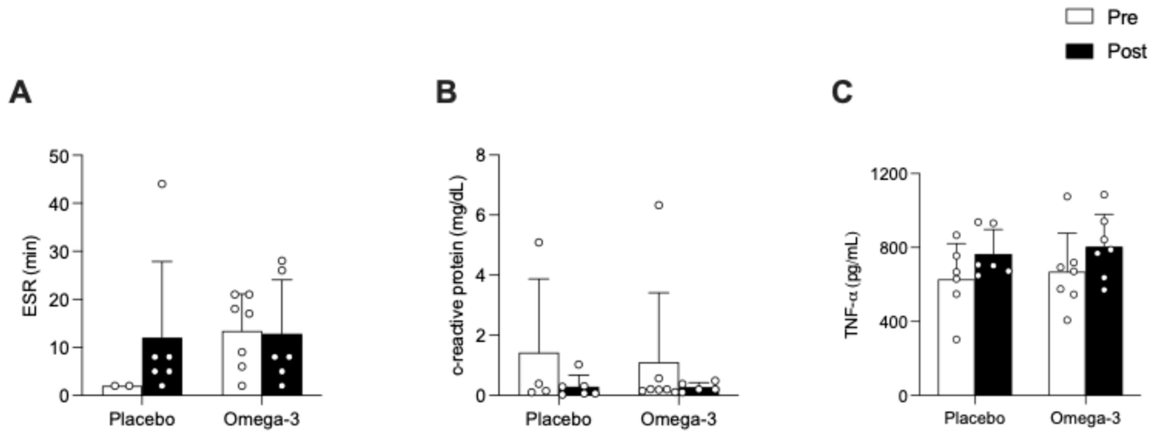
Figure 1. Effect of n-3 PUFA on blood inflammatory markers in patients with rheumatoid arthritis. (A) Erythrocyte sedimentation rate (ESR); (B) C-reactive protein; and (C) tumor necrosis alpha (TNF-α). Values are presented as individual data, means ± SD. No significant differences within and between groups.
Applsci 16 01980 g001
Figure 2. Effect of n-3 PUFA on cardiometabolic health in patients with rheumatoid arthritis. (A) Mean arterial pressure; (B) waist circumference; (C) maximum oxygen capacity (VO2max); (D) triglycerides. Values are presented as individual data, means ± SD. No significant differences within and between groups.
Applsci 16 01980 g002
Figure 3. Effect of omega-3 fatty acids on physical function and quality of life in patients with rheumatoid arthritis. (A) Disease activity score-28 (DAS-28); (B) morning stiffness; (C) quality of life—rheumatoid arthritis (QoL-RA); (D) health assessment questionnaire disability index (HAQ-DI); (E) timed up and go; (F) short physical-performance test (SPPB); (G) handgrip (HG) strength right hand, (H) handgrip (HG) strength left hand. Values are presented as individual data, means ± SD. **** p < 0.0001; *** p = 0.0001; ** p = 0.01; * p < 0.05.
Applsci 16 01980 g003
Figure 4. TNF-alpha expression in polymorphonuclear cells (PBMCs) from healthy young women (n = 3). PBMCs were incubated with 2% fetal bovine serum (Control—blue), with 2% fetal bovine serum plus 100 ng/mL resolvin-D1 (RvD1—cyan); with 0.5% fetal bovine serum plus 1.5% serum from patients with rheumatoid arthritis before omega-3 supplementation (Serum Pre—magenta); with 0.5% fetal bovine serum plus 1.5% serum from patients with rheumatoid arthritis before omega-3 supplementation plus 100 ng/mL resolvin-D1 (Serum Pre + RvD1—orange); and with 0.5% fetal bovine serum plus 1.5% serum from patients with rheumatoid arthritis after omega-3 supplementation (Serum Post—green). Values are presented as means ± SD. ** p = 0.0032; *** p = 0.0008; **** p < 0.0001.
Applsci 16 01980 g004
Table 1. Baseline subject characteristics.
Placebo (n = 6)Omega-3 (n = 7)p Value
Age (y)54 [35–58]51 [36–57]0.9231
Weight (kg)75 [66–86]71 [65–80]0.8357
Height (m)1.57 [1.52–1.59]1.54 [1.49–1.57]0.3071
BMI (kg/m2)30.6 [26.6–36.7]30.1 [28.1–32.0]0.7308
WC (cm)95 [79–104]96 [66–86]0.8666
VO2max (mL/min)1395 [1190–1720]1390 [1200–1545]0.5991
Resting heart rate (b/min)74 [69–77]71 [66–78]0.6573
Mean arterial pressure (mmHg)94 [85–107]94 [81–118]0.9452
Handgrip strength right (kg)24.5 [14.3–24.9]24.2 [19.6–28.8]0.7308
Handgrip strength left (kg)26.3 [16.3–29.2]23.7 [13.7–26]0.5338
Morning stiffness (min)10.0 [0.0–26.3]30.0 [0.0–60.0]0.6882
DAS-283.60 [3.14–4.11]3.03 [2.60–3.21]0.0967
HAQ-Di0.69 [0.51–1.19]0.38 [0.25–1.63]0.8094
QoL-RA6.19 [5.16–7.97]6.13 [6.00–9.00]0.7051
TG (mg/dL)119 [59–201]82 [70–115]0.6485
Total cholesterol (mg/dL)168 [127–195]175 [152–203]0.6485
C-reactive protein (mg/dL)0.27 [0.11–3.92]0.20 [0.15–0.57]0.8242
ESR (min)2.0 [2.0–2.0]17.0 [6.0–21.0]0.1111
Data are expressed as median [IQR]. BMI: body mass index; WC: waist circumference; VO2max: maximum oxygen uptake; DAS-28: disease activity score-28; HAQ-Di: health assessment questionnaire disability index; QoL-RA: quality of life—rheumatoid arthritis; TG: triglycerides ESR: erythrocyte sedimentation rate. No significant differences in baseline characteristics.
Disclaimer/Publisher’s Note: The statements, opinions and data contained in all publications are solely those of the individual author(s) and contributor(s) and not of MDPI and/or the editor(s). MDPI and/or the editor(s) disclaim responsibility for any injury to people or property resulting from any ideas, methods, instructions or products referred to in the content.