Review

Exploring the Therapeutic Potential of Calendula and Chamomile in Dermatology: From Anti-Inflammatory and Antioxidant Properties to New Therapeutic Perspectives


by
Luca Gammeri
1,†,
Federica Li Pomi
2,†,
Francesco Borgia
3,
Eleonora Di Salvo
1,*,
Antonino Nazareno Virga
4,5,* and
Sebastiano Gangemi
6
1
Department of Biomedical and Dental Science and Morphofunctional Imaging, University of Messina, 98125 Messina, Italy
2
Department of Precision Medicine in Medical, Surgical and Critical Care (Me.Pre.C.C.), University of Palermo, 90127 Palermo, Italy
3
Section of Dermatology, Department of Clinical and Experimental Medicine, University of Messina, 98125 Messina, Italy
4
Meat and Agribusiness Chain Research Consortium, Polo Universitario dell’Annunziata, Viale Giovanni Palatucci 13, 98168 Messina, Italy
5
Department of Agricultural, Food and Forestry Sciences, University of Palermo, 90121 Palermo, Italy
6
School and Operative Unit of Allergy and Clinical Immunology, Department of Clinical and Experimental Medicine, University of Messina, 98125 Messina, Italy
*
Authors to whom correspondence should be addressed.
These authors contributed equally and share first authorship.
Appl. Sci. 2026, 16(4), 1965; https://doi.org/10.3390/app16041965
Submission received: 20 January 2026 / Revised: 11 February 2026 / Accepted: 13 February 2026 / Published: 16 February 2026

Abstract

Oxidative stress (OS) plays a central role in the pathogenesis of several cutaneous disorders, including inflammatory dermatoses, photoaging, and carcinogenesis. The imbalance between reactive oxygen species (ROS) production and endogenous antioxidant defenses contributes to inflammation, cellular senescence, and barrier dysfunction. Phytochemicals with antioxidant and anti-inflammatory properties have therefore gained attention as potential therapeutic agents in dermatology. Calendula officinalis (CO) and Matricaria chamomilla (MC) contain bioactive compounds, including carotenoids, flavonoids, terpenoids, and phenolic acids, that modulate redox homeostasis and inflammatory pathways. Evidence from preclinical and clinical studies indicates that CO and MC exert photoprotective effects by reducing UV-induced ROS generation and preserving dermal collagen. Both extracts promote wound healing through fibroblast stimulation, collagen deposition, and antimicrobial activity. In chronic inflammatory dermatoses, including atopic dermatitis (AD) and psoriasis (Pso), CO and MC downregulate pro-inflammatory cytokines, thereby restoring immune balance. Emerging delivery systems have enhanced their skin bioavailability and clinical effectiveness. Collectively, current data support the antioxidant, anti-inflammatory, and regenerative properties of CO and MC, underscoring their potential in maintaining skin homeostasis and protecting against oxidative damage. Further standardized, large-scale clinical studies are warranted to validate their efficacy, safety, and optimal formulations for dermatological use.

1. Introduction

Skin diseases represent a growing global burden, affecting patients’ quality of life and often requiring long-term management strategies [1]. The complexity of these conditions, frequently associated with oxidative stress (OS), inflammation, and impaired skin barrier function, highlights the need for safe and effective therapeutic options [2]. In recent years, attention towards OS has increasingly grown, opening up new potential therapeutic avenues, with fewer side effects and greater efficacy. Persistent Th2 inflammation, as well as exposure to solar radiation, infections, pollution, and stress, contributes to the overproduction of reactive oxygen species (ROS), including superoxide (O2•−) and hydrogen peroxide (H2O2). Normally, systems exist that counteract the production of ROS, namely the antioxidant system (AOS) [3]. Enzymatic and non-enzymatic defenses characterize AOS. Enzymatic antioxidants, such as superoxide dismutase (SOD), catalase (CAT), and glutathione peroxidase (GPx), are synthesized intracellularly. These antioxidants act by directly neutralizing ROS through catalytic conversion. For instance, SOD catalyzes the dismutation of superoxide radicals into hydrogen peroxide, which is then further reduced to water by CAT or GPx. Non-enzymatic antioxidants include low-molecular-weight compounds such as glutathione (GSH), as well as exogenous nutrients such as vitamins C and E and carotenoids. These factors protect the skin by acting as “scavengers” that donate electrons to stabilize free radicals, thereby preventing chain reactions that lead to cellular damage [3,4].
The imbalance between ROS production and the activity of this system determines the onset of OS. OS contributes to the perpetuation and worsening of inflammation and is, in turn, maintained by cellular aging, thus contributing to the phenomenon now known as inflammaging [4]. In chronic inflammatory skin diseases such as atopic dermatitis (AD) or psoriasis (Pso), an increase in OS biomarkers and a reduction in AOS biomarkers are observed [5]. Dermatological conditions vary significantly across the lifespan and in clinical severity. For example, AD is primarily a pediatric concern, often characterized by a prevalent Th2 immune profile and an impaired skin barrier, which can persist into adulthood as chronic, severe lesions [5]. Instead, Pso typically presents in adults as a chronic autoimmune condition involving keratinocyte hyperproliferation and systemic inflammation. Furthermore, the aging process produces pro-inflammatory cytokines and ROS, leading to heightened susceptibility to skin cancer and environmental damage in elderly populations [4]. The severity of these conditions is often assessed using standardized scales that reflect the degree of erythema, infiltration, and desquamation.
One of the primary mechanisms by which OS exerts its harmful effects on the body is lipid peroxidation (LPO). LPO is a primary driver of skin damage because it targets the polyunsaturated fatty acids (PUFAs) within cell membranes and is a biochemical process through which these lipids are peroxidized in the presence of free radicals, causing alterations in biological membranes and the formation of harmful substances such as malondialdehyde (MDA) and 4-hydroxynonenal (4-HNE). These compounds possess cytotoxic and genotoxic properties and have been identified as playing a role in the pathogenesis of various diseases, neoplasms [6], and skin diseases like Pso, vitiligo, and alopecia. Furthermore, LPO may also play a role in the pathogenesis of other skin diseases (AD, urticaria, pemphigus, and skin cancer) [7]. MDA is primarily used as a biomarker of systemic oxidative damage. At the same time, 4-HNE is particularly cytotoxic, capable of forming covalent adducts with proteins and DNA, which disrupt signal transduction and lead to apoptosis or malignant transformation. In the skin, this process degrades the lipid barrier, increasing transepidermal water loss and facilitating the entry of environmental allergens [7].
The role of antioxidants is to counteract OS, which can be mediated by endogenous enzyme systems or exogenous substances ingested through diet. Plant-based foods in particular have strong antioxidant effects. Many varieties of fruits and vegetables contain powerful antioxidants, including alkaloids, polyphenols, and terpenoids. Today, it is widely demonstrated that the administration of these substances through the use of derivatives or food supplements can be effective in combating many inflammatory diseases [8]. The branch that deals with this is phytotherapy [9], which has been the basis of traditional medicine for centuries. The use of phytotherapeutics is particularly employed in the treatment of skin conditions, often with remarkable results [10].
Standard management of inflammatory dermatoses (such as AD and Pso) is based on emollients and topical anti-inflammatory agents (topical corticosteroids and calcineurin inhibitors), with phototherapy or systemic therapies for more severe disease. Wounds and radiodermatitis are usually addressed through supportive care, including cleansing, dressings, and barrier creams/emollients (e.g., aqueous moisturizers, urea-based products, or trolamine). In this context, Calendula officinalis (CO) and Matricaria chamomilla (MC) have predominantly been investigated as adjunctive topical options or supportive-care alternatives, primarily for their anti-inflammatory and antioxidant properties. CO has been used for its pharmacological properties since the 12th century [11]. Today, it is recognized for its important therapeutic properties, including anti-inflammatory, antimicrobial, and hepatoprotective effects, while also promoting wound healing [12]. CO also has significant antioxidant activity, especially due to its high content of carotenoids, flavonoids, terpenoids, coumarins, and phenolic acids [12].
MC is another plant found worldwide and widely used in traditional medicine to treat various conditions, including infections and respiratory, gastrointestinal, and neuropsychiatric disorders. This plant has also demonstrated several biological properties: antioxidant, antimicrobial, antidiabetic, anticancer, and anti-inflammatory effects [13]. Its traditional use in poultices, infusions, and topical formulations is supported by reports of beneficial effects on irritated and fragile skin [14].
The scientific literature increasingly suggests that the antioxidant potential of these plants is a key mechanism underlying their skin benefits [14]. By modulating redox balance and mitigating OS, both MC and CO may help to protect the skin from external aggressors, including ultraviolet (UV) radiation and environmental pollutants [1]. In addition, their anti-inflammatory and soothing properties contribute to maintaining skin homeostasis and health [2].
Beyond their individual actions, there is growing interest in the synergistic effects when MC and CO are used in combination [15]. Their complementary phytochemical compositions suggest that joint formulations could provide broader protection and enhanced activity compared to single-plant preparations [14]. This perspective is particularly relevant in the context of modern cosmeceutical and dermatological research, which seeks to integrate natural compounds into innovative strategies for skin care and treatment [2].
In light of these considerations, this work aims to explore the beneficial actions of MC and CO in the field of skin diseases, with particular attention to their antioxidant and skin-protective properties [1]. By reviewing current evidence, we will discuss how these two plants, individually and in combination, may contribute to the prevention and management of OS-related skin conditions [15].

2. Calendula officinalis (CO)

Calendula officinalis is a medicinal plant belonging to the Asteraceae family, also known as English marigold and pot marigold [16]. The primary and most widespread varieties of CO are C. officinalis, C. arvensis, C. tripterocarpa, C. stellata, and C. suffruticosa, but there are currently about 25 species [17]. Its extracts have long been used in traditional medicine and are now utilized in the pharmaceutical industry due to the plant’s numerous properties. These pharmacological activities are guaranteed by the rich chemical composition of CO extracts: flavonoids, terpenoids, triterpene esters, triterpenoids, phenolic compounds, carotenes, quinones, tocopherol, steroids, essential oils, fatty acids, minerals, saponins, carbohydrates, and sterols [12].
CO extracts have demonstrated not only anti-inflammatory, antioxidant, and antimicrobial activity, but also antidiabetic, hepatoprotective, and antitumorigenic effects [12].
The anti-inflammatory effects of CO could be attributed to the extracts’ ability to inhibit the production of proinflammatory cytokines and the cyclooxygenase Cox-2 [18]. Furthermore, CO appears to have a dose-dependent inhibitory effect on the nitric oxide (NO) cascade [19].
The antioxidant activity of CO extracts is closely linked to their polyphenolic content, particularly flavonoids. These compounds are among those with the most active antioxidant properties due to their high content of hydroxyl groups [12].
These effects, combined with its ability to promote wound healing, make this plant very useful in the treatment of many skin disorders.

3. Matricaria chamomilla L. (MC)

Matricaria chamomilla L. is one of the most widely used medicinal plants in dermatology, traditionally valued for its soothing and anti-inflammatory properties. MC extracts contain over 120 pharmacologically active compounds, of which terpenoids represent the most significant group. Other constituents include phenolic compounds, such as phenolic acids, flavonoids, coumarins, and amino acids [13]. MC and its extracts exert various biological activities and pharmacological effects. This plant contains compounds with high antioxidant potential [20] and has been shown to have significant antimicrobial effects, especially against Gram-positive and Gram-negative bacteria [21]. Flavonoids are major immunomodulators and, as such, also exert anti-inflammatory effects, as well as the ability to promote Th1/Th2 balance through upregulation of Th1 [22]. Contemporary research has begun to clarify how flavonoids, such as apigenin and luteolin, together with sesquiterpenes, like α-bisabolol and chamazulene, modulate inflammation and restore cutaneous homeostasis [23]. Plant oils containing these constituents have been demonstrated to repair the skin barrier, reduce transepidermal water loss, and attenuate cytokine-driven inflammation, thereby providing a rationale for their use in inflammatory dermatoses [24].

4. Potential Use of Calendula officinalis and Matricaria chamomilla in Skin Diseases

4.1. Prevention of UV-Induced Damage and Potential Implications for Skin Cancer

UV radiation acts by stimulating ROS production, leading to DNA damage, OS, and an increase in inflammatory cytokines, thus triggering various signaling pathways [25]. Antioxidants, such as flavonoids and phenolic acids, present in extracts of medicinal plants like CO and MC, have the ability to reduce the damage resulting from exposure to this radiation.
In clinical practice, various topical phytotherapeutic preparations based on natural substances that shield against UV radiation and have an antioxidant effect are currently used for radiation protection. These preparations reduce the incidence of skin diseases related to photoexposure, such as skin cancer, to varying degrees of effectiveness. Among the various extracts used to prepare topical shielding formulations, several commercially available formulations based on CO or MC are available, and several studies in the literature have demonstrated their effectiveness.
The first studies on the photoprotective effect of CO date back to 2010. Fonseca et al. [26] examined the efficacy of orally administered CO extract in preventing UVB-induced OS on the skin. This study demonstrated not only the in vitro antioxidant efficacy of the extract but also its photoprotective effect in murine models. Oral treatment with CO extract, at the dose of 150 or 300 mg/kg in irradiated glabrous mice, maintained GSH levels close to those of non-irradiated controls. Indeed, GSH levels are a good early sensor of UVB-induced epidermal OS. However, using higher doses of CO extract (600 mg/kg), a depletion of approximately 43% in GSH levels was observed [26].
In light of the therapeutic potential demonstrated by this study, the same authors subsequently evaluated the possible efficacy of topical formulations containing CO extract [27]. This in vivo study on glabrous mouse models demonstrated that all tested formulations had good efficacy and optimal stability. Specifically, the gel (F3) appeared to be the most effective formulation in maintaining GSH levels similar to those of the control cases. The same study demonstrates that the therapeutic effect is not linked to the possible impact of the gel on the activity of UVB-induced enzymes (gelatinase-9 or myeloperoxidase), but rather to a potential positive action on collagen synthesis [27].
The sunscreen effect, measured as sun protection factor (SPF), was subsequently evaluated. Mishra et al. [28] studied the in vitro SPF of CO oil using ultraviolet spectrometry. The CO oil-based cream formulation showed a good SPF (14.84 ± 0.16), thus demonstrating a valid photoprotective effect. Several cosmetic preparations use CO essential oil in combination with geranium essential oil. Indeed, the two oils appear to enhance their respective antioxidant and photoprotective effects, also providing an anti-aging benefit for the skin. In 2019, Lohani and her team [29] studied the antioxidant effect and SPF of these two oils. The study demonstrated the antioxidant efficacy of the combination of CO oil and geranium oil, which had SPF values of 8.36 and 6.45, respectively.
The anti-photoaging effect of a blend of CO and rosemary extracts was subsequently studied [30]. This study, on mouse models of UV-induced photoaging, demonstrated how this blend induced a reduction in photoaging-related biomarkers such as MMPs, cytokines, tumor necrosis factor-alpha, type I procollagen, 8-hydroxydeoxyguanosine, SOD, GPx, and catalase. In the same study, the photoprotective effects were also demonstrated histologically.
Exposure to UV radiation is a significant risk factor for the development of skin cancers, primarily melanoma [31]. Therefore, skin protection mechanisms and strategies to enhance photoprotection are essential for reducing the risk of skin cancer. Ukiya et al. [32] in 2006 highlighted that some bioactive compounds extracted from CO exhibited marked anti-inflammatory and antitumor effects. This study on tumor cell models showed that two triterpene glycosides extracted from CO flowers exhibited potent cytotoxic activity against melanoma cells. In the same year, several authors evaluated the in vitro cytotoxic, antitumor, and immunomodulatory activities, as well as the in vivo antitumor effect, of the CO extract, with encouraging results. The extract inhibited tumor cell proliferation in vitro by blocking the cell cycle in the G0/G1 phase and inducing apoptosis. The same extract in murine models inhibited tumor growth of Ando-2 melanoma cells [33]. Matic et al. [34] in their 2013 study observed that CO tea exerted a highly selective antitumor effect against Fem-x melanoma cells.
CO derivatives also appear to have an effect on tumor spread. Studies using mouse models of lung metastasis induced by B16F-10 melanoma cells have demonstrated that treatment with CO extracts exerts anti-metastatic effects. Administration of the extract reduced lung tumor nodules by 74% thanks to its inhibitory effect on key enzymes involved in metastasis and the reduction of proinflammatory cytokines [35]. Recently, Xuan et al. [36] studied the antimelanogenic and antimigratory activities of ethyl acetate fraction (EFC) from CO flowers. EFC reduced α-MSH-induced melanin production and inhibited the cell migration ability of melanoma cells in a dose-dependent manner.
MC is increasingly studied for its potential to contribute to skin photoprotection, with its beneficial properties primarily deriving from a rich blend of flavonoids and volatile terpenoids, especially apigenin, bisabolol compounds, and chamazulene. Together, they offer antioxidant, anti-inflammatory, and UV-absorbing activities. In particular, apigenin is the most intensively studied flavonoid in MC extracts for UV protection [37].
Apigenin’s protective activity relies on the ability to inhibit multiple biological processes activated by UV radiation. By reducing OS and limiting DNA damage, apigenin prevents the accumulation of photoproducts that underlie mutagenesis and photoaging [38]. In keratinocytes, it has been shown to restore autophagic activity and maintain protein homeostasis following UVB exposure, thereby preserving cell viability under conditions of environmental stress [39]. These effects are complemented by its antioxidant properties, which counteract ROS generation and mitigate downstream inflammatory signaling [40]. Importantly, apigenin exerts these actions without phototoxicity, with experimental models confirming its capacity to attenuate erythema in UV-exposed skin, linking molecular mechanisms to tangible clinical protection [41,42].
Beyond isolated compounds, the essential oils of MC have been evaluated for intrinsic UV-absorbing capacity. A recent study comparing German chamomile (M. recutita) and Roman chamomile (Chamaemelum nobile) revealed that the former’s essential oils, when tested in vitro, achieved SPF estimates in the range of ~26–27, while Roman chamomile oils produced lower SPF values (~9–16) under the same assay [43]. This result suggested that species, chemotype, and cultivation practices influence photoprotective potential.
The photostability of MC’s volatile components remains a relevant concern. Chamazulene, an aromatic chemical compound of MC, is known to degrade under UVA/UVB exposure, particularly in pro-oxidant conditions. Stabilizing agents—such as antioxidants or UV filters—are thus critical in formulation to preserve MC’s efficacy over time. The species and processing also affect antioxidant content, influencing the resilience of the extract under light stress [43]. However, the available data are limited to preclinical models, and no large-scale human trials have yet demonstrated that MC-containing formulations effectively prevent erythema or actinic skin damage in vivo. Table 1 reports the data on the photoprotective effect of the bioactive compounds found in CO and MC.

4.2. Effects on Wound Healing

A narrative review of the use of CO in wound healing in human clinical trials was published in 2008 [44], demonstrating that extracts of this flower possessed several beneficial properties for wound healing, including anti-inflammatory, antibacterial, antioxidant, and analgesic activity. In 2019, Givol et al. [45] published a new systematic review evaluating the healing efficacy of CO extract as monotherapy, analyzing all in vivo studies in animal and human models. The authors highlighted how, in animal models, treating acute wounds with CO extract led to faster healing from the inflammatory phase and increased production of granulation tissue. These phenomena are likely related to the anti-inflammatory properties of CO and its ability to activate and promote fibroblast migration [45]. The extract is also effective in treating chronic wounds (such as chronic venous ulcers) due to the same effects mentioned above.
Furthermore, the review shows that CO extract can promote skin healing after burns and provide protective effects against radiation damage, likely due to its antioxidant and proangiogenic effects. Since 2019, several authors have confirmed the role of CO extract in accelerating wound healing.
Giostri et al. [46] published a randomized controlled clinical trial in 2022 analyzing the progression of secondary intention healing in acute hand wounds treated with CO extract. The study in human models demonstrated that the time to epithelialization in the treated group was shorter, with faster healing rates, compared to the control group. In a recent study, the authors investigated the efficacy of topical 5% aqueous CO extract on full-thickness skin wound healing in male BALB/c mice.
The authors observed that topical use of the extract significantly reduced wound size as early as day seven, also resulting in increased levels of fibroblasts, fibroblast growth factor, and hydroxyproline. Simultaneously, reduced levels of macrophages and inflammatory biomarkers, including MMP-2 and MMP-9, were observed in the wounds of treated mice [47].
One of the main problems with the application of CO extracts in treating chronic skin wounds is the lack of adhesive properties, which prevents the extract from remaining in place for prolonged periods. For this reason, some authors have considered extract-loaded scaffolds that could overcome this problem. In 2020, a hydrogel delivery system loaded with lyophilized CO extract was developed for the treatment of chronic wounds [48]. The authors of the study used chitosan as the carrier. This carrier enabled the better diffusion of the active principles in the extract. The combination also facilitated a synergistic action, allowing the creation of formulations with 3% CO and 80/500 chitosan in a 1:1 weight ratio, as well as 2% or 3% hypromellose [48]. In 2021, Kharat et al. [49] studied devices loaded with CO extract by electrospinning onto chitosan/polyethylene oxide (CS/PEO) scaffolds. The study of these devices demonstrated that the incorporation of CO extract improved the mechanical properties of the CS/PEO nanofibers while also displaying strong antibacterial properties (96% and 94% reduction in Gram-positive and Gram-negative bacteria, respectively). In vitro studies performed by Kharat’s group confirmed that the nanofibers induced increased cell proliferation, growth, and adhesion. In vivo studies in murine models have also demonstrated the excellent wound-healing ability of CS/PEO/CO dressings [49].
In 2022, Naseriyeh and his team developed and studied a chitosan/aloe vera hydrogel loaded with high-CO nanoliposomes (83% encapsulation rate). The hydrogel’s efficacy in releasing CO was both tested and demonstrated in vitro, using the French diffusion cell and rat abdominal skin as a biological membrane. The authors showed that within 24 h, 90% of the total CO was absorbed into the skin [50]. Over the years, several authors have continued to use hydrogel as a biomaterial. Recently, a polyacrylamide hydrogel containing CO extract was synthesized. In vivo tests conducted on male Wistar rats demonstrated increased collagen fiber production and enhanced skin repair, without dermal toxicity [51]. In recent years, nanoscience and the development of nanoparticles have become increasingly relevant in the pharmaceutical and cosmetic industries. Zinc oxide nanoparticles have potent antimicrobial and antioxidant properties. Recently, some authors developed a cream containing zinc oxide nanoparticles loaded with CO extract and evaluated its efficacy in healing skin burns. The study demonstrated that the nanoparticles increased fibroblast proliferation, promoting (in vivo in animal models) granulation and epithelialization of tissues without signs of hemorrhage or infectious complications. This effect is thought to be linked to increased Bcl-2 gene expression and decreased Bax expression [52].
MC has been used for centuries in traditional medicine, and recent studies have also highlighted its efficacy in wound healing through several mechanisms. The dried flowers contain terpenoids, flavonoids, coumarins, and phenolic acids, which have been shown to exert antioxidant, antimicrobial, and immunomodulatory effects, all of which are essential for repair processes [53]. One of the earliest observations in experimental models was that MC promoted faster ulcer healing than corticosteroids, with mice treated with MC showing complete repair up to nine days earlier and even full closure within five days, thus showing its superior regenerative potential [54]. Clinical data corroborate these findings, as patients treated topically after dermabrasion experienced significantly accelerated wound drying and re-epithelialization when compared with controls [53].
At the mechanistic level, MC modulates several biological targets relevant to tissue repair. Antagonistic effects on histamine H2 receptors contribute to barrier restoration, while action on 5-hydroxytryptamine-2 (5-HT2) receptors promotes re-epithelialization [55]. In addition, opioid-like properties have been reported, consistent with the observation that topical opioids stimulate platelet aggregation and activate the cascade of wound healing [56].
MC has also shown notable potential in wound-healing applications when integrated into nanofibrous scaffolds. Motealleh et al. [57] reported that chamomile-loaded electrospun fibers enhanced cell viability and achieved almost complete wound closure in vivo within 14 days, with well-formed epithelial tissue. Similarly, Nezhadmokhtari et al. [58] demonstrated that MC essential oil incorporated into hybrid nanofibers improved scaffold hydrophilicity and stability, promoted fibroblast compatibility, and provided vigorous antibacterial activity. This evidence suggests that MC may contribute to wound repair by supporting tissue regeneration while also providing antimicrobial protection and favorable scaffold properties.
Building on this approach, more recent studies developed innovative wound dressings by incorporating MC essential oil into electrospun nanofibers together with zeolite imidazolate framework nanoparticles. These composite dressings demonstrated excellent compatibility with human fibroblasts, strong mechanical and thermal stability, and potent antibacterial activity against both Staphylococcus aureus and Escherichia coli, highlighting their potential as advanced biomaterials for clinical wound management [58].
In fact, another crucial aspect of MC in wound healing is its antimicrobial properties, which prevent infection and promote tissue regeneration. Extracts of C. nobile demonstrated significant antibacterial activity against Pseudomonas aeruginosa in infected wounds, with healing outcomes superior to tetracycline ointment, supporting its potential use either alone or in synergy with antibiotics [59]. Beyond bacteria, MC extracts incorporated into polymeric nanofibers inhibited Candida albicans, further expanding their application spectrum [57].
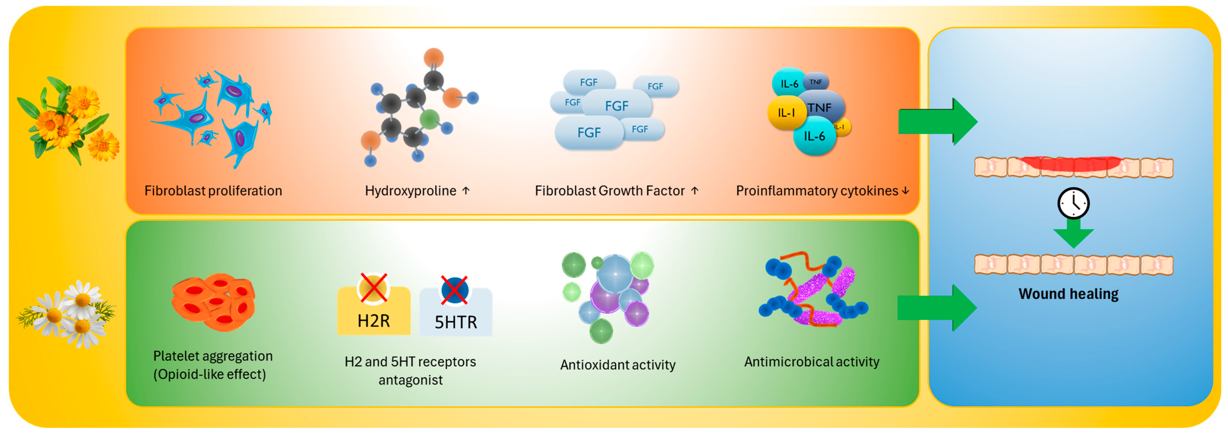
Finally, MC’s wound-healing potential can be amplified through combination with other natural remedies. In experimental models, methanolic extracts of Punica granatum flowers (phenolic-rich) and MC (flavonoid-rich) showed potent antioxidant activity and promoted faster wound contraction. The combined formulation outperformed single extracts, especially at early stages, with histology confirming enhanced collagen deposition and re-epithelialization, underscoring the promise of polyherbal approaches for effective healing at lower doses [60] (Figure 1). All the data are reported in Table 2.

4.3. Anti-Inflammatory Activity

CO was and still is widely used in the preparation of phytotherapeutic creams with anti-inflammatory action. However, there is a lack of studies in the literature. As early as 1994, an in vitro study demonstrated the anti-inflammatory properties of CO extracts, which were attributed to the triterpenoid content and, in particular, to faradiol esters [61]. An Indian study on mouse models suggested that the anti-inflammatory effects of CO could be attributed to the extracts’ ability to inhibit the production of TNF-α and other proinflammatory cytokines such as IL-1-β, IL-6, and IFN-γ, as well as to CO’s ability to inhibit the cyclooxygenase Cox-2 [18]. Furthermore, CO appears to have a dose-dependent inhibitory effect on the NO cascade, a potent proinflammatory agent produced by induced nitric oxide synthase (iNOS) [19].
Given the potential effects of CO and the lack of evidence in the literature, Fuchs et al. [62] investigated the use of CO and rosemary preparations for treating irritant contact dermatitis (ICD) in 2005. Seven different types of extracts were tested on healthy volunteers with ICD induced by sodium lauryl sulfate. The extracts, applied during the same period as the induction of ICD, demonstrated a statistically significant protective effect.
CO ointment has also been used to treat diaper dermatitis (DD). This formulation has even been compared to other topical products, such as aloe vera creams, with superior results. In a randomized, double-blind clinical study of sixty-six infants with DD, the group treated with CO ointment experienced significantly fewer rashes than the group treated with aloe [63].
Wu et al. [64] conducted in vitro and in vivo studies to evaluate the efficacy of CO, combined with two other herbs (Herba Menthae and Cortex Moutan), in the treatment of skin lesions in AD. The in vitro study evaluated the effects of the individual herbs and the mixture, demonstrating that the anti-inflammatory effects of the mix were primarily attributed to Herba Menthae and Cortex Moutan. These two herbs induced the reduction of IL-6 and TNF-α in HMC-1 cells; inhibited the expression of IL-6, IL-8, and CCL2 in TNF-α/IFN-γ-stimulated HaCaT cells; and suppressed NO production in RAW 264.7 cells. Conversely, CO affected skin wound healing processes more than the inflammatory microenvironment. However, the combination of these three herbs also proved effective in mouse models of AD, thanks to the synergistic effect between the three herbs [64]. A recent study developed and evaluated the efficacy of an emulsifying gel containing hyaluronic acid, glycerol, CO, aloe vera, polyphenols, and epidermal growth factor (EGF) for the treatment of AD. The study, conducted on human subjects over 14 years of age with AD, assessed the response to topical therapy by evaluating objective parameters of skin barrier function (transepidermal water loss, skin temperature, pH, stratum corneum hydration, skin elasticity, and degree of erythema) and subjective parameters. The product not only induced an improvement in objective parameters but also achieved 100% positive response among patients [65].
MC has been highlighted to interfere with key signaling pathways that orchestrate skin inflammation. Comparative studies of different plant fractions—whole extracts, flowers, and roots—demonstrated differential activity on human T cells, suggesting that MC’s broad immunomodulatory effects can be refined by selecting specific fractions or compounds [66]. The role of MC in AD, characterized by impaired barrier function, itch, xerosis, and immune imbalance, illustrates how these molecular properties translate into therapeutic benefits. Innovative formulations have demonstrated that nanoemulsions of MC volatile oil, combined with Bletilla striata polysaccharides, can significantly reduce dermatitis-like lesions, thereby improving skin hydration and reducing inflammatory mediators [67]. Likewise, MC nanoemulgels restored skin architecture and reduced inflammatory infiltrates in preclinical models, highlighting the importance of delivery systems in enhancing efficacy [68]. Earlier studies with German chamomile oil also confirmed immunological benefits, normalizing Th1/Th2 imbalance and lowering IgE levels in murine models [69], while aqueous extracts improved dermatitis-like lesions and erythema in animals. These preclinical observations are complemented by clinical reports that consistently list MC among the most effective and best-tolerated botanicals for AD, particularly when patients seek steroid-sparing alternatives [70].
Pso represents another field in which MC’s anti-inflammatory effects have gained attention. The chronic nature of pso, with its combination of keratinocyte hyperproliferation and sustained immune activation, aligns well with the pathways inhibited by MC [71]. MC essential oil managed to alleviate psoriatic-like skin inflammation by inhibiting the PI3K/Akt/mTOR and p38 MAPK pathways, both of which are central to keratinocyte hyperproliferation and inflammatory cytokine production [72].
A randomized, double-blind, placebo-controlled clinical trial compared the efficacy of a topical oleogel containing MC oil and Cucurbita pepo seed oil to that of a placebo formulation in 40 adults with plaque pso. At the end of the treatment period, the lesions treated with the MC–pumpkin oleogel showed a significantly greater reduction in Psoriasis Severity Index scores compared with those treated with placebo [73].
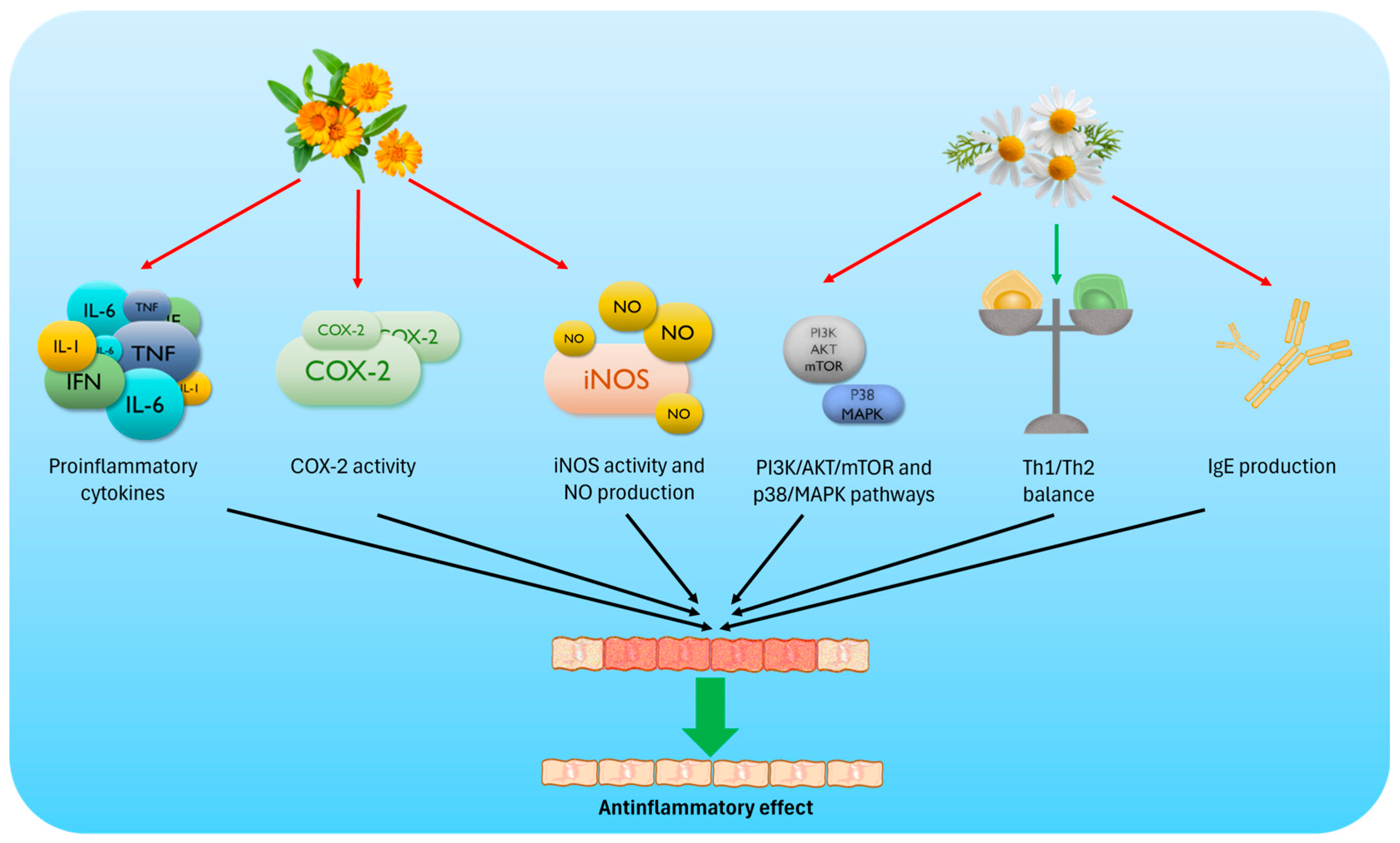
Nanoformulations can substantially modify the pharmacodynamic profile of botanical extracts by increasing local cutaneous exposure and optimizing deposition across skin compartments, thereby amplifying and prolonging anti-inflammatory/antioxidant effects compared with conventional vehicles. However, the magnitude of pharmacodynamic modulation is formulation- and extract-dependent, and enhanced penetration may affect tolerability; therefore, vehicle-controlled, dose-equivalent comparisons with skin deposition/retention characterization are warranted. Together, these findings suggest that MC is a botanically derived, multi-target anti-inflammatory agent with applications across various chronic inflammatory skin diseases (Figure 2). Table 3 summarizes the main clinical studies on the potential antinflammatory effects of CO and MC extracts.

4.4. Radioprotection

The search for effective, safe, and affordable agents to mitigate cutaneous radiation-induced toxicities remains a significant priority in oncology. A substantial body of both clinical and preclinical evidence supports the use of CO and MC as radioprotective agents, either alone or in combination with other natural compounds.
The first studies on the efficacy of topical CO preparations in treating acute radiodermatitis date back to the early 2000s. Pommier et al. [74], in a phase III study, demonstrated that CO was effective in preventing acute radiodermatitis of grade 2 or higher compared to trolamine, considered the reference topical treatment. The study showed that the use of CO cream reduced the onset of radiodermatitis to a greater extent than trolamine (41% versus 63%; p < 0.001). In a subsequent study dating back to 2013, the ineffectiveness of topical CO preparation compared to placebo was observed [75]. The incidence of severe acute radiodermatitis between the two groups was almost identical (23% in the CO group vs. 19% among controls). However, a review by Simões et al. [76] in 2020 confirmed the efficacy of CO in the prevention and treatment of acute radiodermatitis. Subsequently, Siddiquee et al. [77] in a randomized controlled clinical trial compared the efficacy of topical CO with Sorbolene (10% glycerin in cetomacrogol cream), the standard of care drug for the prevention of radiodermatitis. In their study, the authors screened 271 women, recruiting 81 participants (n = 40 Calendula; n = 41 Sorbolene). They found no substantial differences in the prevalence of radiation-induced dermatitis between the two groups. CO was therefore not shown to be inferior to Sorbolene in terms of efficacy. Still, the study was limited by difficulties in recruiting patients.
Regarding MC, one of the earliest clinical investigations was conducted by Maiche et al. [78], who compared MC cream with almond ointment in patients undergoing radiotherapy. Their findings showed that MC reduced the severity of acute radiation skin reactions, laying the groundwork for later controlled trials. More recent studies have refined these early observations, adopting improved trial designs and developing innovative formulations.
The clinical relevance of MC has been particularly explored in head and neck cancer, where radiation dermatitis and mucositis represent significant therapeutic challenges. A randomized trial comparing MC gel with urea cream demonstrated that MC was more effective in reducing both the incidence and severity of acute radiation dermatitis [79]. A subsequent preliminary trial confirmed these findings, reporting improved tolerability and lower skin toxicity in the MC arm compared to urea [80]. More recently, MC infusion was evaluated as a topical approach to mitigate radiotherapy-induced dry desquamation, with promising results in reducing both the extent and intensity of skin peeling in patients with head and neck cancers [81].
Parallel progress has been made in optimizing MC delivery through advances in formulation technology. A phase II clinical trial testing a chitosan-coated MC microparticle gel in breast cancer patients showed significant prophylactic effects against radiodermatitis, highlighting the potential of bioengineered carriers to enhance MC’s therapeutic activity [82]. Similarly, recent randomized trials have reinforced the value of advanced delivery systems in maximizing MC’s clinical activity. In patients with head and neck cancer, a liposomal gel containing MC proved superior to the liposomal base alone, particularly in reducing erythema and desquamation [83]. A parallel investigation in breast cancer patients yielded consistent results, demonstrating that MC-enriched liposomal gels outperformed controls in preventing radiation dermatitis [83].
Combination strategies have also been investigated to enhance the radioprotective potential of MC through its association with other botanical extracts. A cream containing aloe vera, MC, and thyme demonstrated efficacy in breast cancer patients, showing benefits in both the prevention and treatment of mild radiation dermatitis [84] as well as in improving patient-reported outcomes [85]. Conversely, Gomes de Meneses and his group found no statistically significant differences between MC-based liposomal gel and MC-free gel with regard to the incidence of radiodermatitis [86].
The protective effects of MC extend beyond the skin. Preclinical studies have shown that MC extracts may attenuate radiation-induced intestinal mucositis through anti-inflammatory and antioxidant mechanisms [87]. Mixtures of natural agents, including MC, have also been evaluated in patients undergoing chemoradiotherapy for head and neck cancer. However, not all endpoints achieved statistical significance; trends suggested a potential benefit in reducing the severity of mucositis [88].
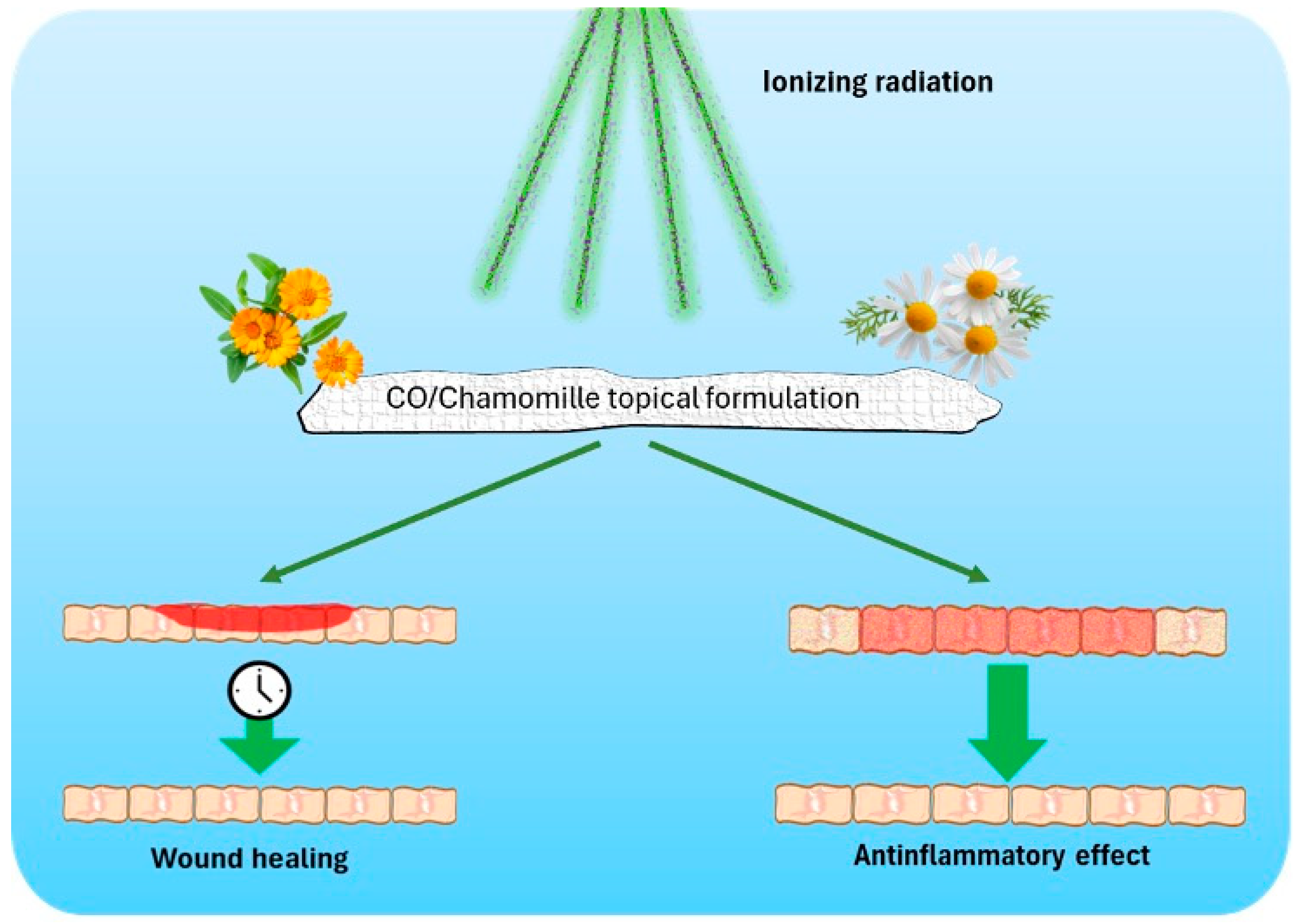
Mechanistic studies provide further support for these clinical findings. The radioprotective effects of MC are linked to its phytochemical composition. Experimental evidence has shown that radiation exposure induces the production of antioxidant compounds in the plant itself—particularly flavonoids and phenolic derivatives—which are known to counteract genomic instability. These bioactive metabolites are preserved in MC-based preparations used clinically, thereby offering a biological rationale for their protective activity [89] (Figure 3). Detailed study characteristics are reported in Table 4.
Overall, the reported radioprotection trials are heterogeneous in sample size, radiotherapy regimens, comparators, and dermatitis grading/endpoints, which may limit power and precision and complicate cross-study synthesis. As a result, apparent benefits may be imprecisely estimated, whereas negative findings may reflect insufficient power rather than a true lack of efficacy. Therefore, current evidence is encouraging but not uniformly sufficient for firm conclusions, supporting the need for adequately powered, multi-center RCTs with standardized outcomes.

5. Discussion

The current findings underscore the therapeutic promise of CO and MC in dermatology, with a particular emphasis on their antioxidant mechanisms. Both plants have a long history of traditional use, yet modern research is beginning to elucidate the molecular and clinical underpinnings of their cutaneous effects. A critical emerging point is that OS represents a unifying pathogenic mechanism across several cutaneous diseases, including inflammatory ones such as AD, Pso, and DD; photodamage; and carcinogenesis. The imbalance between ROS generation and the skin’s endogenous antioxidant defenses sustains chronic inflammation, accelerates cellular senescence, and disrupts barrier function, thereby contributing to the chronicity of dermatoses [3,4,5]. The phytochemicals of CO and MC—principally carotenoids, flavonoids, terpenoids, coumarins, and phenolic acids—appear strongly involved in modulating redox homeostasis and mitigating OS [12,13].
One of the most compelling applications of these botanicals is in photoprotection, a field where antioxidant activity intersects with skin cancer prevention. CO extracts exert photoprotective effects against UVB-induced oxidative damage, primarily by preserving intracellular antioxidant defenses, while also promoting structural support through enhanced collagen metabolism. These dual antioxidant and dermo-regenerative properties highlight its potential in counteracting photoaging and UV-mediated skin injury [26,27]. Similarly, MC’s flavonoid apigenin has been shown to reduce ROS generation, attenuate DNA photoproduct formation, and restore autophagy in keratinocytes following UVB exposure, thereby preventing photoaging and mutagenesis, which can ultimately lead to carcinogenesis [38,39]. These mechanistic data provide biological plausibility for clinical photoprotection; however, translation into human trials remains incomplete. While in vitro SPF evaluation for MC and CO oils is promising, the absence of large-scale controlled trials limits firm conclusions about their role in preventing actinic damage in vivo [43].
Beyond photoprotection, both CO and MC appear to accelerate wound healing by orchestrating a redox-sensitive interplay between inflammatory resolution and tissue regeneration. CO extract facilitated the transition from the inflammatory to the proliferative phase, enhanced fibroblast migration, and improved collagen synthesis [45,47]. MC likewise accelerated wound contraction and re-epithelialization, in some models even outperforming corticosteroids and promoting earlier ulcer closure [53,54]. Advances in nanotechnology have enabled the incorporation of these extracts into electrospun nanofibers and hydrogel-based scaffolds, which significantly improved antioxidant stability, cellular proliferation, and antimicrobial protection, leading to enhanced healing outcomes [49,57,58]. This body of evidence highlights how antioxidant activity is not an isolated property but rather embedded within a broader regenerative and antimicrobial framework.
Chronic inflammatory dermatoses such as AD and Pso represent another relevant field for phytotherapeutic exploration. CO’s triterpenoid fraction has demonstrated efficacy against irritant contact dermatitis, while ointments showed superiority over aloe vera formulations in reducing DD severity [61,63]. Recent formulations combining CO with other natural agents, such as hyaluronic acid or aloe vera, have further enhanced hydration, reduced erythema, and improved patient-reported outcomes, suggesting synergistic effects beyond single-plant preparations [33]. MC, by contrast, has demonstrated deeper molecular immunomodulation. By targeting PI3K/Akt/mTOR and MAPK signaling, MC extracts effectively reduced keratinocyte hyperproliferation and cytokine production, central features of Pso and AD [71,72]. Clinical trials using MC–pumpkin oil oleogels in Pso further support these mechanistic insights, reporting significant reductions in Psoriasis Severity Index (PASI) scores [73]. Together, these findings suggest that the antioxidant activity of CO and MC serves as a gateway to broader immunological and barrier-restorative benefits.
Radioprotection studies provide a further dimension to CO and MC effects on the skin. CO has shown variable efficacy in clinical trials, sometimes outperforming conventional treatments yet demonstrating outcomes equivalent to placebo in other studies, raising questions about formulation, dose, and trial design [74,75]. MC, however, has shown more consistent benefits in mitigating radiation dermatitis, with liposomal or chitosan-coated formulations reducing erythema and desquamation in breast and head and neck cancer patients [83,86]. These data support the pivotal role of delivery systems, as flavonoids and terpenoids are prone to oxidative degradation, and their stabilization appears to be critical for clinical efficacy [82].

6. Major Open Questions

While encouraging, the current evidence also underscores significant limitations. First, heterogeneity in extraction methods, plant chemotypes, and formulations complicates cross-study comparisons and limits standardization. The antioxidant potential of CO and MC is highly dependent on species, cultivation practices, and processing, which can lead to variability in both efficacy and reproducibility [43]. Second, attributing clinical outcomes solely to antioxidant activity may lead to oversimplification, given that both plants contain complex mixtures of bioactive compounds with overlapping effects. Finally, safety data for long-term use remain limited, particularly in chronic dermatoses where patients often require prolonged therapy. Collectively, these limitations define clear translational priorities for future research, spanning extract standardization, mechanism-linked endpoints, formulation-dependent pharmacodynamics, trial robustness, and long-term safety.
Looking forward, advances in nanotechnology, bioengineered scaffolds, and polyherbal formulations appear to offer promising solutions for enhancing the clinical utility of CO and MC. Nanocarriers not only stabilize antioxidants but also improve dermal penetration and bioavailability, as demonstrated in preclinical models of wound healing and radioprotection [49,82]. Polyherbal approaches, leveraging synergistic antioxidant and anti-inflammatory actions, have already shown superior outcomes in wound healing compared with single extracts, suggesting a future path for combination therapies in dermatology [60]. To support robust clinical implementation, future research should prioritize: (i) standardized, reproducible extracts (botanical authentication, chemotype control, quantified marker compounds); (ii) mechanism-to-clinic linkage using redox/LPO biomarkers alongside validated severity outcomes; (iii) vehicle-controlled comparisons of conventional formulations versus nano/advanced carriers, with evidence of skin deposition/retention and release behavior; (iv) adequately powered, multi-center RCTs with prespecified primary endpoints and standard-of-care comparators; and (v) systematic safety evaluation for repeated/chronic use (irritancy, sensitization, phototoxicity, and interactions with concomitant therapies).

7. Conclusions and Perspectives

Calendula and chamomile exemplify the promise of phytotherapy in the management of skin diseases. Their antioxidant properties not only counteract OS but also integrate into complex biological networks that modulate inflammation, tissue repair, and barrier function. While preclinical and early clinical data are compelling, rigorous large-scale randomized trials remain essential to confirm efficacy, define standardized preparations, and ensure safety in long-term use. Only through well-designed translational studies can these phytotherapeutic agents be fully integrated into evidence-based dermatological practice.

Author Contributions

Conceptualization, F.B. and S.G.; investigation, L.G. and F.L.P.; resources, A.N.V.; data curation, L.G. and F.L.P.; writing—original draft preparation, L.G. and F.L.P.; writing—review and editing, L.G., F.L.P., A.N.V. and E.D.S.; visualization, L.G., F.L.P., F.B., E.D.S., A.N.V. and S.G.; supervision, F.B. and S.G.; project administration, S.G.; funding acquisition, A.N.V. All authors have read and agreed to the published version of the manuscript.

Funding

This research received no external funding.

Informed Consent Statement

Not applicable.

Data Availability Statement

No new data were created or analyzed in this study. Data sharing is not applicable to this article.

Conflicts of Interest

The authors declare no conflicts of interest.

References

  1. Gonçalves, S.; Fernandes, L.; Caramelo, A.; Martins, M.; Rodrigues, T.; Matos, R.S. Soothing the Itch: The Role of Medicinal Plants in Alleviating Pruritus in Palliative Care. Plants 2024, 13, 3515. [Google Scholar] [CrossRef]
  2. Olivero-Verbel, J.; Quintero-Rincón, P.; Caballero-Gallardo, K. Aromatic Plants as Cosmeceuticals: Benefits and Applications for Skin Health. Planta 2024, 260, 132. [Google Scholar] [CrossRef]
  3. Bertino, L.; Guarneri, F.; Cannavò, S.P.; Casciaro, M.; Pioggia, G.; Gangemi, S. Oxidative Stress and Atopic Dermatitis. Antioxidants 2020, 9, 196. [Google Scholar] [CrossRef] [PubMed]
  4. Zuo, L.; Prather, E.R.; Stetskiv, M.; Garrison, D.E.; Meade, J.R.; Peace, T.I.; Zhou, T. Inflammaging and Oxidative Stress in Human Diseases: From Molecular Mechanisms to Novel Treatments. Int. J. Mol. Sci. 2019, 20, 4472. [Google Scholar] [CrossRef]
  5. Borgia, F.; Li Pomi, F.; Vaccaro, M.; Alessandrello, C.; Papa, V.; Gangemi, S. Oxidative Stress and Phototherapy in Atopic Dermatitis: Mechanisms, Role, and Future Perspectives. Biomolecules 2022, 12, 1904. [Google Scholar] [CrossRef]
  6. Morabito, F.; Cristani, M.; Saija, A.; Stelitano, C.; Callea, V.; Tomaino, A.; Minciullo, P.L.; Gangemi, S. Lipid Peroxidation and Protein Oxidation in Patients Affected by Hodgkin’s Lymphoma. Mediat. Inflamm. 2004, 13, 381–383. [Google Scholar] [CrossRef]
  7. Li Pomi, F.; Gammeri, L.; Borgia, F.; Di Gioacchino, M.; Gangemi, S. Oxidative Stress and Skin Diseases: The Role of Lipid Peroxidation. Antioxidants 2025, 14, 555. [Google Scholar] [CrossRef]
  8. Mannucci, C.; Casciaro, M.; Sorbara, E.E.; Calapai, F.; Di Salvo, E.; Pioggia, G.; Navarra, M.; Calapai, G.; Gangemi, S. Nutraceuticals against Oxidative Stress in Autoimmune Disorders. Antioxidants 2021, 10, 261. [Google Scholar] [CrossRef] [PubMed]
  9. Falzon, C.C.; Balabanova, A. Phytotherapy: An Introduction to Herbal Medicine. Prim. Care Clin. Off. Pract. 2017, 44, 217–227. [Google Scholar] [CrossRef]
  10. Li Pomi, F.; Papa, V.; Borgia, F.; Vaccaro, M.; Allegra, A.; Cicero, N.; Gangemi, S. Rosmarinus Officinalis and Skin: Antioxidant Activity and Possible Therapeutical Role in Cutaneous Diseases. Antioxidants 2023, 12, 680. [Google Scholar] [CrossRef] [PubMed]
  11. Basch, E.; Bent, S.; Foppa, I.; Haskmi, S.; Kroll, D.; Mele, M.; Szapary, P.; Ulbricht, C.; Vora, M.; Yong, S. Marigold (Calendula Officinalis L.): An Evidence-Based Systematic Review by the Natural Standard Research Collaboration. J. Herb. Pharmacother. 2006, 6, 135–159. [Google Scholar] [CrossRef]
  12. Shahane, K.; Kshirsagar, M.; Tambe, S.; Jain, D.; Rout, S.; Ferreira, M.K.M.; Mali, S.; Amin, P.; Srivastav, P.P.; Cruz, J.; et al. An Updated Review on the Multifaceted Therapeutic Potential of Calendula officinalis L. Pharmaceuticals 2023, 16, 611. [Google Scholar] [CrossRef]
  13. El Mihyaoui, A.; Esteves Da Silva, J.C.G.; Charfi, S.; Castillo, M.E.C.; Lamarti, A.; Arnao, M.B. Chamomile (Matricaria chamomilla L.): A Review of Ethnomedicinal Use, Phytochemistry and Pharmacological Uses. Life 2022, 12, 479. [Google Scholar] [CrossRef] [PubMed]
  14. Jadoon, S.; Karim, S.; Bin Asad, M.H.H.; Akram, M.R.; Kalsoom Khan, A.; Malik, A.; Chen, C.; Murtaza, G. Anti-Aging Potential of Phytoextract Loaded-Pharmaceutical Creams for Human Skin Cell Longetivity. Oxid. Med. Cell Longev. 2015, 2015, 709628. [Google Scholar] [CrossRef] [PubMed]
  15. Albahri, G.; Badran, A.; Hijazi, A.; Daou, A.; Baydoun, E.; Nasser, M.; Merah, O. The Therapeutic Wound Healing Bioactivities of Various Medicinal Plants. Life 2023, 13, 317. [Google Scholar] [CrossRef] [PubMed]
  16. Ashwlayan, V.D.; Kumar, A.; Verma, M.; Garg, V.K.; Gupta, S. Therapeutic Potential of Calendula officinalis. Pharm. Pharmacol. Int. J. 2018, 6, 149–155. [Google Scholar] [CrossRef]
  17. Fallahi, M.; Mohammadi, A.; Miri, S.M. The Natural Variation in Six Populations of Calendula officinalis L.: A Karyotype Study. J. Genet. Resour. 2020, 6, 34–40. [Google Scholar] [CrossRef]
  18. Chandran Preethi, K.; Kuttan, G.; Kuttan, R. Anti-Inflammatory Activity of Flower Extract of Calendula officinalis Linn. and Its Possible Mech. Action. Indian. J. Exp. Biol. 2009, 47, 113–120. [Google Scholar]
  19. Silva, D.; Ferreira, M.S.; Sousa-Lobo, J.M.; Cruz, M.T.; Almeida, I.F. Anti-Inflammatory Activity of Calendula officinalis L. Flower Extract. Cosmetics 2021, 8, 31. [Google Scholar] [CrossRef]
  20. Hassanpour, H.; Ghanbarzadeh, M. Induction of Cell Division and Antioxidative Enzyme Activity of Matricaria chamomilla L. Cell Line Under Clino-Rotation. Plant Cell Tissue Organ. Cult. 2021, 146, 215–224. [Google Scholar] [CrossRef]
  21. Lavanya, J.; Periyar Selvam, S.; Jeevitha Priya, M.; Jacintha, P.; Aradana, M. Antioxidant and antimicrobial activity of selected medicinal plants against human oral pathogens. Int. J. Pharm. Pharm. Sci. 2016, 8, 71–78. [Google Scholar] [CrossRef]
  22. Asadi, Z.; Ghazanfari, T.; Hatami, H. Anti-Inflammatory Effects of Matricaria chamomilla Extracts on BALB/c Mice Macrophages and Lymphocytes. Iran. J. Allergy Asthma Immunol. 2020, 19, 63–73. [Google Scholar] [CrossRef] [PubMed]
  23. Pezantes-Orellana, C.; German Bermúdez, F.; Montalvo, J.; Packer, T.; Orellana-Manzano, A. Evaluating Efficacy, Safety, and Innovation in Skin Care Applications of Essential Oils: A Systematic Review. Front. Med. 2025, 12, 1589691. [Google Scholar] [CrossRef]
  24. Lin, T.K.; Zhong, L.; Santiago, J.L. Anti-Inflammatory and Skin Barrier Repair Effects of Topical Application of Some Plant Oils. Int. J. Mol. Sci. 2017, 19, 70. [Google Scholar] [CrossRef]
  25. Zhao, C.; Wu, S.; Wang, H. Medicinal Plant Extracts Targeting UV-Induced Skin Damage: Molecular Mechanisms and Therapeutic Potential. Int. J. Mol. Sci. 2025, 26, 2278. [Google Scholar] [CrossRef] [PubMed]
  26. Fonseca, Y.M.; Catini, C.D.; Vicentini, F.T.M.C.; Nomizo, A.; Gerlach, R.F.; Fonseca, M.J.V. Protective Effect of Calendula officinalis Extract against UVB-Induced Oxidative Stress in Skin: Evaluation of Reduced Glutathione Levels and Matrix Metalloproteinase Secretion. J. Ethnopharmacol. 2010, 127, 596–601. [Google Scholar] [CrossRef] [PubMed]
  27. Fonseca, Y.M.; Catini, C.D.; Vicentini, F.T.M.C.; Cardoso, J.C.; Cavalcanti De Albuquerque Junior, R.L.; Vieira Fonseca, M.J. Efficacy of Marigold Extract-Loaded Formulations against UV-Induced Oxidative Stress. J. Pharm. Sci. 2011, 100, 2182–2193. [Google Scholar] [CrossRef]
  28. Mishra, A.; Mishra, A.; Chattopadhyay, P. Assessment of In Vitro Sun Protection Factor of Calendula officinalis L. (Asteraceae) Essent. Oil Formulation. J. Young Pharm. 2012, 4, 17–21. [Google Scholar] [CrossRef]
  29. Lohani, A.; Mishra, A.K.; Verma, A. Cosmeceutical Potential of Geranium and Calendula essential Oil: Determination of Antioxidant Activity and in Vitro Sun Protection Factor. J. Cosmet. Dermatol. 2019, 18, 550–557. [Google Scholar] [CrossRef]
  30. Auh, J.H.; Madhavan, J. Protective Effect of a Mixture of Marigold and Rosemary Extracts on UV-Induced Photoaging in Mice. Biomed. Pharmacother. 2021, 135, 111178. [Google Scholar] [CrossRef]
  31. Sample, A.; He, Y.Y. Mechanisms and Prevention of UV-Induced Melanoma. Photodermatol. Photoimmunol. Photomed. 2018, 34, 13–24. [Google Scholar] [CrossRef]
  32. Ukiya, M.; Akihisa, T.; Yasukawa, K.; Tokuda, H.; Suzuki, T.; Kimura, Y. Anti-Inflammatory, Anti-Tumor-Promoting, and Cytotoxic Activities of Constituents of Marigold (Calendula officinalis) Flowers. J. Nat. Prod. 2006, 69, 1692–1696. [Google Scholar] [CrossRef] [PubMed]
  33. Jiménez-Medina, E.; Garcia-Lora, A.; Paco, L.; Algarra, I.; Collado, A.; Garrido, F. A New Extract of the Plant Calendula officinalis Produces a Dual in Vitro Effect: Cytotoxic Anti-Tumor Activity and Lymphocyte Activation. BMC Cancer 2006, 6, 119. [Google Scholar] [CrossRef] [PubMed]
  34. Matić, I.Z.; Juranić, Z.; Šavikin, K.; Zdunić, G.; Nadvinski, N.; Goddevac, D. Chamomile and Marigold Tea: Chemical Characterization and Evaluation of Anticancer Activity. Phytother. Res. 2013, 27, 852–858. [Google Scholar] [CrossRef]
  35. Preethi, K.; Siveen, K.; Kuttan, R.; Kuttan, G. Inhibition of Metastasis of B16F-10 Melanoma Cells in C57BL/6 Mice by an Extract of Calendula officinalis L Flowers. Asian Pac. J. Cancer Prev 2010, 11, 1773–1779. [Google Scholar]
  36. Xuan, S.H.; Park, Y.M.; Park, S.N. Antimelanogenic and Antimigration Properties of the Ethyl Acetate Fraction of Calendula Officinalis Flowers on Melanoma Cells. Photochem. Photobiol. 2019, 95, 860–866. [Google Scholar] [CrossRef] [PubMed]
  37. Wang, M.; Firrman, J.; Liu, L.S.; Yam, K. A Review on Flavonoid Apigenin: Dietary Intake, ADME, Antimicrobial Effects, and Interactions with Human Gut Microbiota. Biomed. Res. Int. 2019, 2019, 7010467. [Google Scholar] [CrossRef]
  38. García Forero, A.; Villamizar Mantilla, D.A.; Núñez, L.A.; Ocazionez, R.E.; Stashenko, E.E.; Fuentes, J.L. Photoprotective and Antigenotoxic Effects of the Flavonoids Apigenin, Naringenin and Pinocembrin. Photochem. Photobiol. 2019, 95, 1010–1018. [Google Scholar] [CrossRef]
  39. Li, L.; Li, M.; Xu, S.; Chen, H.; Chen, X.; Gu, H. Apigenin Restores Impairment of Autophagy and Downregulation of Unfolded Protein Response Regulatory Proteins in Keratinocytes Exposed to Ultraviolet B Radiation. J. Photochem. Photobiol. B 2019, 194, 84–95. [Google Scholar] [CrossRef] [PubMed]
  40. Sánchez-Marzo, N.; Pérez-Sánchez, A.; Ruiz-Torres, V.; Martínez-Tébar, A.; Castillo, J.; Herranz-López, M.; Barrajón-Catalán, E. Antioxidant and Photoprotective Activity of Apigenin and Its Potassium Salt Derivative in Human Keratinocytes and Absorption in Caco-2 Cell Monolayers. Int. J. Mol. Sci. 2019, 20, 2148. [Google Scholar] [CrossRef]
  41. Freitas, J.V.; Gaspar, L.R. In Vitro Photosafety and Efficacy Screening of Apigenin, Chrysin and Beta-Carotene for UVA and VIS Protection. Eur. J. Pharm. Sci. 2016, 89, 146–153. [Google Scholar] [CrossRef] [PubMed]
  42. Villamizar-Mantilla, D.A.; Stashenko, E.E.; Fuentes Lorenzo, J.L. Plant Compounds Inhibit UV Radiation-Induced Erythema in Mice Skin. Nat. Prod. Res. 2025, 1–8. [Google Scholar] [CrossRef]
  43. Batovska, D.; Panova, N.; Gerasimova, A.; Tumbarski, Y.; Ivanov, I.; Dincheva, I.; Yotkovska, I.; Gentscheva, G.; Nikolova, K. Chamomile Matters: Species- and Producer-Dependent Variation in Bulgarian Matricaria recutita L. and Chamaemelum nobile L. Essential Oils and Their Cosmetic Potential. Cosmetics 2025, 12, 123. [Google Scholar] [CrossRef]
  44. Leach, M.L. Calendula officinalis and Wound Healing: A Systematic Review. Wounds 2008, 20, 236–243. [Google Scholar]
  45. Givol, O.; Kornhaber, R.; Visentin, D.; Cleary, M.; Haik, J.; Harats, M. A Systematic Review of Calendula officinalis Extract for Wound Healing. Wound Repair. Regen. 2019, 27, 548–561. [Google Scholar] [CrossRef]
  46. Giostri, G.S.; Novak, E.M.; Buzzi, M.; Guarita-Souza, L.C. Treatment of Acute Wounds in Hand with Calendula officinalis L.: A Randomized Trial. Tissue Barriers 2022, 10, 1994822. [Google Scholar] [CrossRef] [PubMed]
  47. Ozturan, Y.A.; Akin, I. Calendula Officinalis Extract Enhances Wound Healing by Promoting Fibroblast Activity and Reducing Inflammation in Mice. Cutan. Ocul. Toxicol. 2025, 44, 161–171. [Google Scholar] [CrossRef] [PubMed]
  48. Chanaj-Kaczmarek, J.; Paczkowska, M.; Osmałek, T.; Kaproń, B.; Plech, T.; Szymanowska, D.; Karaźniewicz-łada, M.; Kobus-Cisowska, J.; Cielecka-Piontek, J. Hydrogel Delivery System Containing Calendulae Flos Lyophilized Extract with Chitosan as a Supporting Strategy for Wound Healing Applications. Pharmaceutics 2020, 12, 634. [Google Scholar] [CrossRef] [PubMed]
  49. Kharat, Z.; Amiri Goushki, M.; Sarvian, N.; Asad, S.; Dehghan, M.M.; Kabiri, M. Chitosan/PEO Nanofibers Containing Calendula officinalis Extract: Preparation, Characterization, in Vitro and in Vivo Evaluation for Wound Healing Applications. Int. J. Pharm. 2021, 609, 121132. [Google Scholar] [CrossRef]
  50. Naseriyeh, T.; Kahrizi, D.; Alvandi, H.; Rajati, H.; Behbood, L.; Khodabandeh Shahraky, M.; Arkan, E. Preparation of Liposomal Hydrogel Containing Calendula and Application as a Wound Dressing. Cell. Mol. Biol. 2022, 68, 1–7. [Google Scholar] [CrossRef]
  51. de Meneses Costa Ferreira, L.M.; de Sousa Bandeira, E.; Gomes, M.F.; Lynch, D.G.; Bastos, G.N.T.; Silva-Júnior, J.O.C.; Ribeiro-Costa, R.M. Polyacrylamide Hydrogel Containing Calendula Extract as a Wound Healing Bandage: In Vivo Test. Int. J. Mol. Sci. 2023, 24, 3806. [Google Scholar] [CrossRef] [PubMed]
  52. Hashemi, S.S.; Pakdin, A.; Mohammadi, A.; Keshavarzi, A.; Mortazavi, M.; Sanati, P. Study the Effect of Calendula officinalis Extract Loaded on Zinc Oxide Nanoparticle Cream in Burn Wound Healing. ACS Appl. Mater. Interfaces 2023, 15, 59269–59279. [Google Scholar] [CrossRef]
  53. Pazyar, N.; Yaghoobi, R.; Rafiee, E.; Mehrabian, A.; Feily, A. Skin Wound Healing and Phytomedicine: A Review. Ski. Pharmacol. Physiol. 2014, 27, 303–310. [Google Scholar] [CrossRef]
  54. Martins, M.D.; Marques, M.M.; Bussadori, S.K.; Martins, M.A.T.; Pavesi, V.C.S.; Mesquita-Ferrari, R.A.; Fernandes, K.P.S. Comparative Analysis between Chamomilla recutita and Corticosteroids on Wound Healing. An in Vitro and in Vivo Study. Phytother. Res. 2009, 23, 274–278. [Google Scholar] [CrossRef]
  55. Shivananda Nayak, B.; Sivachandra Raju, S.; Chalapathi Rao, A.V. Wound Healing Activity of Matricaria recutita L. Extract. J. Wound Care 2007, 16, 298–302. [Google Scholar] [CrossRef]
  56. Srivastava, J.K.; Shankar, E.; Gupta, S. Chamomile: A Herbal Medicine of the Past with Bright Future. Mol. Med. Rep. 2010, 3, 895–901. [Google Scholar] [CrossRef]
  57. Motealleh, B.; Zahedi, P.; Rezaeian, I.; Moghimi, M.; Abdolghaffari, A.H.; Zarandi, M.A. Morphology, Drug Release, Antibacterial, Cell Proliferation, and Histology Studies of Chamomile-Loaded Wound Dressing Mats Based on Electrospun Nanofibrous Poly(Ɛ-Caprolactone)/Polystyrene Blends. J. Biomed. Mater. Res. B Appl. Biomater. 2014, 102, 977–987. [Google Scholar] [CrossRef] [PubMed]
  58. Nezhad-Mokhtari, P.; Kazeminava, F.; Abdollahi, B.; Gholizadeh, P.; Heydari, A.; Elmi, F.; Abbaszadeh, M.; Kafil, H.S. Matricaria chamomilla Essential Oil-Loaded Hybrid Electrospun Nanofibers Based on Polycaprolactone/Sulfonated Chitosan/ZIF-8 Nanoparticles for Wound Healing Acceleration. Int. J. Biol. Macromol. 2023, 247, 125718. [Google Scholar] [CrossRef] [PubMed]
  59. Kazemian, H.; Ghafourian, S.; Sadeghifard, N.; Houshmandfar, R.; Badakhsh, B.; Taji, A.; Shavalipour, A.; Mohebi, R.; Ebrahim-Saraie, H.S.; Houri, H.; et al. In Vivo Antibacterial and Wound Healing Activities of Roman Chamomile (Chamaemelum nobile). Infect. Disord. Drug Targets 2018, 18, 41–45. [Google Scholar] [CrossRef]
  60. Niknam, S.; Tofighi, Z.; Faramarzi, M.A.; Abdollahifar, M.A.; Sajadi, E.; Dinarvand, R.; Toliyat, T. Polyherbal Combination for Wound Healing: Matricaria chamomilla L. and Punica granatum L. Daru 2021, 29, 133–145. [Google Scholar] [CrossRef]
  61. Della Loggia, R.; Tubaro, A.; Sosa, S.; Becker, H.; Saar, S.; Isaac, O. The Role of Triterpenoids in the Topical Anti-Inflammatory Activity of Calendula officinalis Flowers. Planta Med. 1994, 60, 516–520. [Google Scholar] [CrossRef] [PubMed]
  62. Fuchs, S.M.; Schliemann-Willers, S.; Fischer, T.W.; Elsner, P. Protective Effects of Different Marigold (Calendula officinalis L.) and Rosemary Cream Preparations against Sodium-Lauryl-Sulfate-Induced Irritant Contact Dermatitis. Ski. Pharmacol. Physiol. 2005, 18, 195–200. [Google Scholar] [CrossRef]
  63. Panahi, Y.; Sharif, M.R.; Sharif, A.; Beiraghdar, F.; Zahiri, Z.; Amirchoopani, G.; Marzony, E.T.; Sahebkar, A. A Randomized Comparative Trial on the Therapeutic Efficacy of Topical Aloe Vera and Calendula officinalis on Diaper Dermatitis in Children. Sci. World J. 2012, 2012, 810234. [Google Scholar] [CrossRef] [PubMed]
  64. Wu, X.X.; Siu, W.S.; Wat, C.L.; Chan, C.L.; Koon, C.M.; Li, X.; Cheng, W.; Ma, H.; Tsang, M.S.M.; Lam, C.W.K.; et al. Effects of Topical Application of a Tri-Herb Formula on Inflammatory Dry-Skin Condition in Mice with Oxazolone-Induced Atopic Dermatitis. Phytomedicine 2021, 91, 153691. [Google Scholar] [CrossRef]
  65. Gómez-Farto, A.; Jiménez-Escobar, A.L.; Pérez-González, N.; Castán, H.; Clares, B.; Arias-Santiago, S.; Montero-Vílchez, T. Development of an Emulgel for the Effective Treatment of Atopic Dermatitis: Biocompatibility and Clinical Investigation. Gels 2024, 10, 370. [Google Scholar] [CrossRef]
  66. Lairikyengbam, D.; Wetterauer, B.; Schmiech, M.; Jahraus, B.; Kirchgessner, H.; Wetterauer, P.; Berschneider, K.; Beier, V.; Niesler, B.; Balta, E.; et al. Comparative Analysis of Whole Plant, Flower and Root Extracts of Chamomilla recutita L. and Characteristic Pure Compounds Reveals Differential Anti-Inflammatory Effects on Human T Cells. Front. Immunol. 2024, 15, 1388962. [Google Scholar] [CrossRef]
  67. Xu, J.; Hu, H.; Qian, X.; Zhang, D.; Chen, G.; Zhang, F.; Huang, X.; Ma, S.; Chen, B.; Zhou, Q.; et al. Therapeutic Effects of Chamomile Volatile Oil Nanoemulsion/Bletilla Striata Polysaccharides Gels on Atopic Dermatitis. Int. J. Biol. Macromol. 2024, 277, 134404. [Google Scholar] [CrossRef] [PubMed]
  68. El-Salamouni, N.S.; Ali, M.M.; Abdelhady, S.A.; Kandil, L.S.; Elbatouti, G.A.; Farid, R.M. Evaluation of Chamomile Oil and Nanoemulgels as a Promising Treatment Option for Atopic Dermatitis Induced in Rats. Expert. Opin. Drug Deliv. 2020, 17, 111–122. [Google Scholar] [CrossRef]
  69. Lee, S.H.; Heo, Y.; Kim, Y.C. Effect of German Chamomile Oil Application on Alleviating Atopic Dermatitis-like Immune Alterations in Mice. J. Vet. Sci. 2010, 11, 35–41. [Google Scholar] [CrossRef]
  70. Natural Ingredients in Atopic Dermatitis and Other Inflammatory Skin Disease—JDDonline—Journal of Drugs in Dermatology. Available online: https://jddonline.com/articles/natural-ingredients-in-atopic-dermatitis-and-other-inflammatory-skin-disease-S1545961613S0128X/ (accessed on 8 October 2025).
  71. Anheyer, M.; Cramer, H.; Ostermann, T.; Längler, A.; Anheyer, D. Herbal Medicine for Treating Psoriasis: A Systematic Review. Complement. Ther. Med. 2025, 90, 103173. [Google Scholar] [CrossRef]
  72. Chen, G.; Lv, C.; Nie, Q.; Li, X.; Lv, Y.; Liao, G.; Liu, S.; Ge, W.; Chen, J.; Du, Y. Essential Oil of Matricaria chamomilla Alleviate Psoriatic-Like Skin Inflammation by Inhibiting PI3K/Akt/MTOR and P38MAPK Signaling Pathway. Clin. Cosmet. Investig. Dermatol 2024, 17, 59–77. [Google Scholar] [CrossRef] [PubMed]
  73. Kolahdooz, S.; Karimi, M.; Esmaili, N.; Zargaran, A.; Kordafshari, G.; Mozafari, N.; Ayati, M.H. Evaluation of the Efficacy of a Topical Chamomile-Pumpkin Oleogel for the Treatment of Plaque Psoriasis: An Intra-Patient, Double-Blind, Randomized Clinical Trial. Biomed. Res. Ther. 2018, 5, 2811–2819. [Google Scholar] [CrossRef]
  74. Pommier, P.; Gomez, F.; Sunyach, M.P.; D’Hombres, A.; Carrie, C.; Montbarbon, X. Phase III Randomized Trial of Calendula officinalis Compared with Trolamine for the Prevention of Acute Dermatitis during Irradiation for Breast Cancer. J. Clin. Oncol. 2004, 22, 1447–1453. [Google Scholar] [CrossRef]
  75. Sharp, L.; Finnilä, K.; Johansson, H.; Abrahamsson, M.; Hatschek, T.; Bergenmar, M. No Differences between Calendula Cream and Aqueous Cream in the Prevention of Acute Radiation Skin Reactions—Results from a Randomised Blinded Trial. Eur. J. Oncol. Nurs. 2013, 17, 429–435. [Google Scholar] [CrossRef]
  76. Simões, F.V.; Santos, V.O.; da Silva, R.N.; da Silva, R.C. Effectiveness of Skin Protectors and Calendula officinalis for Prevention and Treatment of Radiodermatitis: An Integrative Review. Rev. Bras. Enferm. 2020, 73, e20190815. [Google Scholar] [CrossRef]
  77. Siddiquee, S.; McGee, M.A.; Vincent, A.D.; Giles, E.; Clothier, R.; Carruthers, S.; Penniment, M. Efficacy of Topical Calendula officinalis on Prevalence of Radiation-Induced Dermatitis: A Randomised Controlled Trial. Australas. J. Dermatol. 2021, 62, e35–e40. [Google Scholar] [CrossRef] [PubMed]
  78. Maiche, A.G.; Gröhn, P.; Mäki-Hokkonen, H. Effect of Chamomile Cream and Almond Ointment on Acute Radiation Skin Reaction. Acta Oncol. 1991, 30, 395–397. [Google Scholar] [CrossRef]
  79. Ferreira, E.B.; Ciol, M.A.; Vasques, C.I.; de Souza Maggi Bontempo, P.; Vieira, N.N.P.; Silva, L.F.O.E.; Avelino, S.R.; Dos Santos, M.A.; Dos Reis, P.E.D. Gel of Chamomile vs. Urea Cream to Prevent Acute Radiation Dermatitis in Patients with Head and Neck Cancer: A Randomized Controlled Trial. J. Adv. Nurs. 2016, 72, 1926–1934. [Google Scholar] [CrossRef] [PubMed]
  80. Ferreira, E.B.; Ciol, M.A.; de Meneses, A.G.; de Souza Maggi Bontempo, P.; Hoffman, J.M.; Reis, P.E.D. dos Chamomile Gel versus Urea Cream to Prevent Acute Radiation Dermatitis in Head and Neck Cancer Patients: Results from a Preliminary Clinical Trial. Integr. Cancer Ther. 2020, 19, 1534735420962174. [Google Scholar] [CrossRef]
  81. Gomes de Menêses, A.; Ferreira, E.B.; de Souza Maggi Bontempo, P.; Guerra, E.N.S.; Reis, P.E.D. dos Use of Chamomile Infusion to Mitigate Radiotherapy-Induced Dry Desquamation in Head and Neck Cancer Patients. Integr. Cancer Ther. 2022, 21, 15347354221105491. [Google Scholar] [CrossRef]
  82. Garbuio, D.C.; Ribeiro, V.D.S.; Hamamura, A.C.; Faustino, A.; de Freitas, L.A.P.; Viani, G.; Carvalho, E.C. De A Chitosan-Coated Chamomile Microparticles Formulation to Prevent Radiodermatitis in Breast: A Double-Blinded, Controlled, Randomized, Phase II Clinical Trial. Am. J. Clin. Oncol. 2022, 45, 183–189. [Google Scholar] [CrossRef]
  83. Menêses, A.G.; Ferreira, E.B.; Vieira, L.A.C.; de Souza Maggi Bontempo, P.; Guerra, E.N.S.; Ciol, M.A.; Reis, P.E.D. Comparison of Liposomal Gel with and without Addition of Chamomile for Prevention of Radiation Dermatitis in Head and Neck Cancer Patients: A Randomized Controlled Trial. Radiother. Oncol. 2024, 199, 110440. [Google Scholar] [CrossRef] [PubMed]
  84. Jimenez-Garcia, C.; Perula-de Torres, L.A.; Villegas-Becerril, E.; Muñoz-Gavilan, J.J.; Espinosa-Calvo, M.; Montes-Redondo, G.; Romero-Rodriguez, E. Efficacy of an Aloe Vera, Chamomile, and Thyme Cosmetic Cream for the Prophylaxis and Treatment of Mild Dermatitis Induced by Radiation Therapy in Breast Cancer Patients: A Controlled Clinical Trial (Alantel Trials). Trials 2024, 25, 84. [Google Scholar] [CrossRef] [PubMed]
  85. Villegas-Becerril, E.; Jimenez-Garcia, C.; Perula-de Torres, L.A.; Espinosa-Calvo, M.; Bueno-Serrano, C.M.; Romero-Ruperto, F.; Gines-Santiago, F.; Moreno-Manzanaro, M.C.; Muñoz-Gavilan, J.J.; Montes-Redondo, G.; et al. Efficacy of an Aloe Vera, Chamomile, and Thyme Cosmetic Cream for the Prophylaxis and Treatment of Mild Dermatitis Induced by Radiation Therapy in Breast Cancer Patients (the Alantel Study). Contemp. Clin. Trials Commun. 2024, 39, 101288. [Google Scholar] [CrossRef]
  86. Gomes de Meneses, A.; Ferreira, E.B.; Vieira, L.A.C.; de Souza Maggi Bontempo, P.; Guerra, E.N.S.; Ciol, M.A.; Reis, P.E.D. dos Comparison of Liposomal Gel with and without Chamomile to Prevent Radiation Dermatitis in Breast Cancer Patients: A Randomized Controlled Trial. Strahlenther. Onkol. 2025, 201, 115–125. [Google Scholar] [CrossRef]
  87. Khayyal, M.T.; Kreuter, M.H.; Kemmler, M.; Altmann, P.; Abdel-Naby, D.H.; El-Ghazaly, M.A. Effect of a Chamomile Extract in Protecting against Radiation-Induced Intestinal Mucositis. Phytother. Res. 2019, 33, 728–736. [Google Scholar] [CrossRef] [PubMed]
  88. Marucci, L.; Farneti, A.; Di Ridolfi, P.; Pinnaro, P.; Pellini, R.; Giannarelli, D.; Vici, P.; Conte, M.; Landoni, V.; Sanguineti, G. Double-Blind Randomized Phase III Study Comparing a Mixture of Natural Agents versus Placebo in the Prevention of Acute Mucositis during Chemoradiotherapy for Head and Neck Cancer. Head. Neck 2017, 39, 1761–1769. [Google Scholar] [CrossRef]
  89. Sokolova, D.A.; Halych, T.V.; Zhuk, V.V.; Kravets, A.P. Relationship of Radiation-Induced Genomic Instability and Antioxidant Production in the Chamomile Plant. Int. J. Radiat. Biol. 2023, 99, 1631–1638. [Google Scholar] [CrossRef]
Figure 1. Effects of Calendula officinalis and chamomile extracts on wound healing. CO compounds promote fibroblast proliferation, increase skin regeneration biomarkers (hydroxyproline, FGF), and reduce proinflammatory cytokines. MC has antioxidant and antimicrobial properties, which reduce symptoms such as itching, thanks to its antihistaminic and antiserotonergic effects. Furthermore, MC appears to have opioid-like effects, stimulating platelet aggregation.
Applsci 16 01965 g001
Figure 2. Antinflammatory Effects of Calendula officinalis and chamomile. CO exerts an anti-inflammatory effect by reducing key cytokines associated with inflammation (IL-1-β, TNF-α, IFN-γ, IL-6) and inhibiting enzymes implicated in inflammatory processes, such as COX-2 and iNOS. MC, on the other hand, inhibits key pathways involved in skin inflammatory processes (e.g., Pso) and exerts immunomodulatory effects.
Applsci 16 01965 g002
Figure 3. Radioprotective effects of Calendula officinalis and chamomile extracts. Topical application of creams containing extracts of these two plants has been shown to reduce the severity of radiodermatitis and speed healing. These effects are linked to the previously described abilities of CO and MC to promote wound healing and reduce inflammation.
Applsci 16 01965 g003
Table 1. Main clinical studies regarding the potential photoprotective effect of bioactive compounds contained in CO and MC.
AuthorStudy ModelAimResults
Fonseca et al. [26], 2010In vivo
(mice)
To investigate the potential use of CO extract to prevent UV-induced cutaneous OSOral treatment with 150 and 300 mg/kg of CO extract maintained GSH levels similar to those in controls
Fonseca et al. [27], 2011In vivo
(mice)
To investigate the potential use of topical formulations containing CO extract against UVB irradiation-induced skin damageThe topical formulation can maintain GSH levels close to control levels and reduces histological skin changes induced by UVB rays
Mishra et al. [28], 2012In vitroTo evaluate the sunscreen activity of a formulation based on the essential oil of marigold flowersThe SPF of CO oil, measured by spectrophotometer, showed good activity
Lohani et al. [29], 2019In vitroTo find out the SPF and antioxidant potential of geranium essential oil and CO essential oilThe SPF of both essential oils was found to be 6.45 (geranium) and 8.36 (CO)
Auh et al. [30], 2021In vivo (mice)To evaluate the anti-photoaging effects of CO and rosemary extractsA reduction of various biomarkers related to photoaging was observed due to the suppression of the inflammatory response.
Ukija et al. [32], 2006In vitroTo evaluate the anti-inflammatory and cytotoxic effects of CO extractsTriterpene glycosides have shown potent cytotoxic effects against colon cancer, leukemia, and melanoma cells
Jimenez-Medina et al. [33], 2006In vitro, In vivo (mice)To evaluate the cytotoxic anti-tumor and immunomodulatory activities of LACEThe aqueous extract of LACE exhibited cytotoxic activity on tumor cells and stimulated lymphocyte activation
Matic et al. [34], 2013In vitroTo evaluate the antitumor action and the cytotoxic activity of CO and MC tea against various malignant cell lines and against healthy immunocompetent PBMCCO infusion exerts a highly selective antitumor effect through the activity of phenolic compound
Preethi et al. [35], 2010In vivo (mice)To determine the effect of a CO flower extract on lung metastasis by B16F-10 melanoma cells in C57BL/6 miceAdministration of CO reduced lung tumor nodules by 74%, with a 43.3% increase in lifespan.
Xuan et al. [36], 2019In vitroTo investigate the chemical characterization, antimelanogenic and antimigration activities of the EFC of CO flowersEFC decreased α-MSH-induced melanin production and the cell migration ability of melanoma cells in a dose-dependent manner
Forero et al. [38], 2019In vitroTo evaluate the photoprotective and antigenotoxic effects of the flavonoid compounds apigenin, naringenin, and pinocembrinThese compounds acted as UV filters reducing UV-induced genotoxicity
Li et al. [39], 2019In vitroTo study the function of apigenin in UV-damaged keratinocytesApigenin treatment induces cell death in the COLO-16 cutaneous squamous cell carcinoma cell line, reduces microtubule-associated protein 1 LC3-II turnover, and inhibits autophagy
Sànchez-Marzo et al. [40], 2019In vitroTo compare the absorption and biological activity of apigenin and apigenin-K in terms of antioxidant and photoprotective activityApigenins protected cell viability by approximately 50% at 5 J/m2 UVA radiation and by 90% at 500 J/m2 UVB radiation
Freitas et al. [41], 2016In vitroTo evaluate the photostability, photoreactivity and phototoxicity of apigenin, chrysin and beta-caroteneThe products were found to be stable under UVA/VIS and VIS light
Villamizar-Mantilla et al. [42], 2025In vivo (mice)To study the protective activity of plant compounds against UVB-induced skin erythema in BALB/c albino miceApigenin inhibited UVB-induced skin erythema in mice
CO: Calendula officinalis; EFC: Ethyl Acetate Fraction; GSH: Reduced Glutathione; LACE: Laser Activated Calendula Extract; LC3-II: Light Chain 3; MC: Matricaria chamomilla; PBMC: Peripheral Blood Mononuclear Cells; SPF: Sun Protection Factor.
Table 2. Main clinical studies regarding the potential use of CO and MC extract in wound healing.
AuthorStudy ModelAimResults
Silveira-Giostri et al. [46], 2022Randomized controlled trialTo analyze by photoplanimetry the progress of the healing process by secondary intention in acute wounds of the hand using the standardized CO extractEpithelialization time was shorter and healing rate was faster in subjects treated with CO extracts
Ozturan et al. [47], 2025In vivo (mice)To evaluate the efficacy of topically administered 5% aqueous CO extract on full-thickness skin wound healing in male BALB/c mice5% aqueous CO extract enhances wound healing
Chanaj-Kaczmarek et al. [48], 2020In vitroTo evaluate the efficacy of a combination of CO extract and chitosan used as a carrier for wound healingThe combination of freeze-dried CO extract and chitosan inhibits hyaluronidase
Kharat et al. [49], 2021In vivo (mice)To investigate the efficacy of a CS/PEO scaffold loaded with CO extract by electrospinning on wound healingIn vivo and histological analysis showed that CS/PEO/CO dressings improved collagen synthesis, re-epithelialization, and tissue remodeling
Naseriyeh et al. [50], 2022In vitro, In vivo (mice)To study the efficacy and security of a liposomal hydrogel containing CO extractsIn vitro studies have shown no cytotoxicity in nanoliposomes. In vivo, the addition of liposomes to the hydrogel increased its absorption capacity
de Meneses Costa Ferreira et al. [51], 2023In vivo (mice)To demonstrate the efficacy of a polyacrylamide hydrogel containing CO extract used as a wound healing dressingThe hydrogel showed an efficient collagen fiber production and an improved skin repair without skin toxicity
Hashemi et al. [52], 2023In vitro, In vivo (mice)To study the efficacy of CO extract-loaded zinc oxide nanoparticles on burn healingZinc oxide nanoparticles loaded with CO extract demonstrated a practical effect in healing burn wounds
Martins et al. [54], 2009Comparative studyTo compare the efficacy of MC versus corticosteroids in the topical treatment of skin ulcersAnimals treated with chamomile presented significantly faster wound healing in comparison to those treated with corticosteroids
Nayak et al. [55], 2007In vivo (mice)To evaluate the wound healing activity of MC in ratsThe test group exhibited a greater reduction in the wound area when compared with the controls
Motealleh et al. [57], 2014In vitro, In vivo (mice)To study the efficacy of electrospun nanofibrous mats based on PCL/PS (65/35) and loaded with MC for use as wound dressings)In vitro studies showed that MC-based drugs exhibited exceptional properties as a cell growth agent. In vivo studies on rat wounds, MC-loaded dressings demonstrated efficacy in inducing an increase in the rate of re-epithelialization
Nezhad-Mokhtari et al. [58], 2023In vitroTo evaluate the efficacy of eco-friendly and biodegradable nanofibers based on N-(3-sulfopropyl)chitosan/poly(ε-caprolactone) incorporated by zeolite imidazolate framework-8 nanoparticles (ZIF-8 NPs) and MC essential oilThe novel nanofibers showed improved cytocompatibility, proliferation, and physicochemical properties
Kazemian et al. [59], 2018In vivo (mice)To investigate the antimicrobial and wound healing properties of C. nobile against Pseudomonas aeruginosa using in vivo conditionsThe antibacterial and healing activity of C. nobile ointment is significantly higher than that of tetracycline ointment
Niknam et al. [60], 2021In vitro, In vivo (mice)to evaluate the individual and combined wound healing activity of the methanol extracts of pomegranate and MC flowersPhytochemical studies have shown that both fractions, exhibit potent antioxidant activity. The demonstrated improved wound healing properties.
Table 3. Main clinical studies regarding the potential antinflammatory effects of CO and MC extracts.
AuthorStudy ModelAimResults
Fuchs et al. [62], 2005In vivo (Human)To evaluate the protective action of cream preparations containing CO and rosemary extracts in healthy volunteers with experimentally induced ICDA statistically significant protective effect was observed for all cream preparations.
Panahi et al. [63], 2012Randomized controlled trialTo compare the therapeutic efficacies of aloe vera cream and CO ointment on the frequency and severity of diaper dermatitis in childrenPatients treated with CO ointment had significantly fewer flare-ups
Wu et al. [64], 2021In vitro, In vivo (mice)To study the efficacy in the treatment of atopic dermatitis of a three-herb formula comprising Cortex Moutan, Herba Menthae and CO in a weight ratio of 1:1:1The herbal blend showed anti-inflammatory effect, promoting cell migration in vitro and alleviating dermatitis symptoms in mouse models
Gomez-Farto et al. [65], 2024In vivo (human)To develop and evaluate the efficacy of a topical emulgel containing hyaluronic acid, glycerol, CO, and Aloe vera for the treatment of ADAn improvement in transepidermal water loss, erythema and skin hydration was observed
Lairikyengbam et al. [66], 2024In vitroTo compare the effects of extracts of various MCs on the redox environment of T cells, as well as on the migration, activation, proliferation and cytokine production of primary human T cellsApigenin significantly reduced granzyme B induction and cytotoxic T cell activity
Xu et al. [67], 2024In vivo (mice)To demonstrate the therapeutic effects of CVO-NEGs for the treatment of ADCVO-NEGs reduce skin damage, epidermal thickness, and mast cell infiltration by suppressing the production of IgG and cytokines, including TNF-α, IL-4, and IFN-γ. Furthermore, CVO-NEGs may regulate the differentiation of CD4+ T cell subsets
El-Salamouni et al. [68], 2020In vivo (mice)To evaluate the efficacy of MC oil and nanoemulgel formulations as a natural alternative therapeutic option for ADTreatment with MC nanoemulgel resulted in a shorter duration of skin healing and no spongiosis. Biomarker levels were reduced after topical application of both the nanoemulgel and MC oil
Lee et al. [69], 2010In vivo (mice)To evaluate the effect of MC oil on alleviating AD-like immune alterationsApplication of MC oil for 4 weeks resulted in a significant reduction in serum IgG1 and histamine levels compared to the control group as early as 2 weeks after application
Chen et al. [72], 2024In vitro, In vivo (mice)To evaluate the efficacy and mechanisms of action of MC essential oils on psoriatic-like skin inflammationMC oils improve skin lesions in psoriatic inflammation in mouse models by reducing inflammatory cytokine levels
Kolahdooz et al. [73], 2018In vivo (human)To evaluate the efficacy and safety of topical chamomile-pumpkin oleogel (ChP) in treating plaque psoriasisMean decreases in PSI score in the ChP group were significantly (p = 0.000) greater than in the placebo group. Thirty-five percent of plaques treated with ChP improved in the treatment group (0% in the control group).
AD: Atopic Dermatitis; CO: Calendula officinalis; CVO-NEG: Chamomile Volatile Oil Nanoemulsion Gels; ICD: Irritant Contact Dermatitis; MC: Matricaria chamomilla; PSI: Psoriasis Symptom Inventory.
Table 4. Main clinical studies regarding the potential use of CO and MC extracts to treating radiation dermatitis.
AuthorStudy ModelAimResults
Pommier et al. [74], 2004Clinical trialTo compare the efficacy of CO with that of trolamine in the treatment of RDThe incidence of acute dermatitis of grade 2 or higher was significantly lower in patients treated with CO.
Sharp et al. [75], 2013Randomized controlled trialTo compare the efficacy of two topical CO-based agents (aqueous cream or cream) in reducing the risk of severe acute skin radiation reactions secondary to adjuvant RTBoth formulations were shown to be effective, with no statistically significant differences
Siddiquee et al. [77], 2021Randomized controlled trialTo compare the efficacy of topical CO versus standard of care (Sorbolene: 10% glycerine in cetomacrogol cream) in reducing the prevalence of radiation-induced dermatitis in women undergoing breast cancer RTNo differences were observed between CO and Sorbolene in the prevention of radiation-induced dermatitis.
Maiche et al. [78], 1991Clinical trialTo evaluate the effects of MC cream and almond ointment in the treatment of acute RDNeither formulation prevented the onset of dermatitis. However, a reduction in grade 2 dermatitis was observed
Ferreira et al. [80], 2020Randomized controlled trialTo compare a gel made with MC with a cream of urea as an intervention to delay the occurrence of RDPreliminary results demonstrate a delayed onset of dermatitis, with Grade 2 dermatitis occurring at 5.1 weeks in the MC group and 4.5 weeks in the urea group.
Menêses et al. [81], 2022Clinical trialTo evaluate the effect of MC infusion in the regression of dry desquamation and in the prevention of moist desquamation in head and neck cancer patients undergoing RTAll participants experienced regression of dry desquamation, with a total regression of 65.1% and a mean regression time of 9 days.
Garbuio et al. [82], 2022Clinical trialTo evaluate the effect of a topical formulation containing chitosan-coated MC rauschert microparticles on the incidence and grade of RD in women with breast cancerNo significant difference was observed between the groups in the incidence or time to development of any grade of dermatitis.
Menêses et al. [83], 2024Randomized controlled trialTo compare liposomal gel with and without MC extract for the prevention and management of RD in head and neck cancer patients undergoing RTThe group treated with MC liposomal gel showed lower levels of RD than the group treated with liposomal gel
Villegas-Becerril et al. [85], 2024Clinical trialTo evaluate the efficacy of a NPs-based cream (Alantel®) to reduce the incidence of RD in women with breast cancer undergoing RT treatmentThe incidence of RD was lower in the treated group (71.4%) than in the control group (91.4%) after 4 weeks of follow-up
Meneses et al. [86], 2025Randomized controlled trialTo compare a liposomal gel with and without MC extract for the prevention of radiation dermatitis in breast cancer patients undergoing RTNo statistically significant differences between the two gels were found in RD occurrence
Marucci et al. [88], 2017Clinical trialTo evaluate the potential use of propolis, aloe vera, CO and MC for the prevention of acute mucositis during chemoradiotherapy for head and neck cancerThe four natural agents do not show ability to prevent mucositis
CO: Calendula officinalis; MC: Matricaria chamomilla; NPs: Natural Products; RD: Radiation Dermatitis; RT: Radiotherapy.
Disclaimer/Publisher’s Note: The statements, opinions and data contained in all publications are solely those of the individual author(s) and contributor(s) and not of MDPI and/or the editor(s). MDPI and/or the editor(s) disclaim responsibility for any injury to people or property resulting from any ideas, methods, instructions or products referred to in the content.