Relationships Between Postural Quality and Postural Stability Among Healthy Young Adults
 ,
,  , , , , , , , and
Abstract
1. Introduction
2. Materials and Methods
2.1. Study Design
2.2. Setting
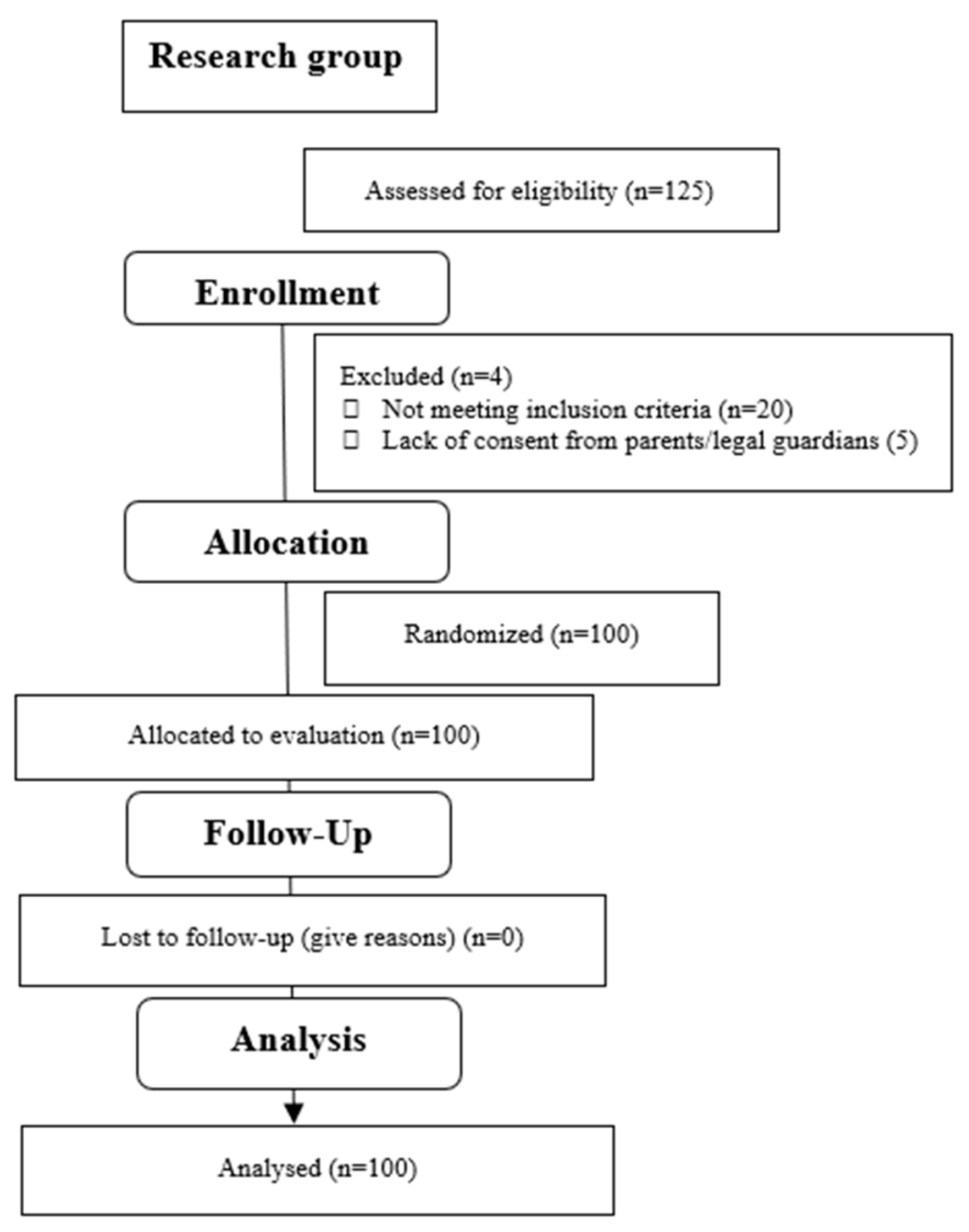
2.3. Participants
2.4. Intervention
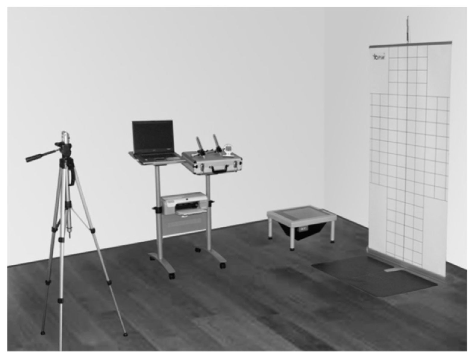
- digital camera equipped with a tripod;
- fold-out board with a printed posturographic grid and a marked mini platform;
- podoscope with a built-in webcam;
- computer with a printer;
- Posture_Screening computer programme. (Figure 1).
2.5. Outcome Measures
- ALPHA—lumbosacral tilt angle;
- BETA—thoracolumbar tilt angle;
- GAMMA—upper kyphosis tilt angle;
- PI—Frohner posture index;
- LRD—angle of lumbar lordosis;
- KPH—angle of thoracic kyphosis;
- PTL—pelvic tilt in the sagittal plane.
- L—percentage load on the left foot;
- P—percentage load on the right foot;
- —percentage load on the left forefoot;
- —percentage load on the right forefoot;
- —percentage load on the left rear foot;
- —percentage load on the right rear foot.
2.6. Statistical Analysis
3. Results
4. Discussion
Study Limitation
5. Conclusions
Author Contributions
Funding
Institutional Review Board Statement
Informed Consent Statement
Data Availability Statement
Conflicts of Interest
References
- Sousa, A.S.; Silva, A.; Tavares, J.M. Biomechanical and neurophysiological mechanisms related to postural control and efficiency of movement: A review. Somatosens. Mot. Res. 2012, 29, 131–143. [Google Scholar] [CrossRef] [Scilit]
- Lee, J.H. Effects of forward head posture on static and dynamic balance control. J. Phys. Ther. Sci. 2016, 28, 274–277. [Google Scholar] [CrossRef] [Scilit]
- Bibrowicz, K.; Szurmik, T.; Ogrodzka-Ciechanowicz, K.; Hudakova, Z.; Gąsienica-Walczak, B.; Kurzeja, P. Asymmetry of the pelvis in Polish young adults. Front. Psychol. 2023, 14, 1148239. [Google Scholar] [CrossRef] [Scilit] [PubMed]
- Lipowicz, A.; Szurmik, T.; Bugdol, M.N.; Graja, K.; Kurzeja, P.; Mitas, A.W. Relationship between body sway and body building in girls and boys in developmental age. In Information Technology in Biomedicine ITIB; Pietka, E., Badura, P., Kawa, J., Wieclawek, W., Eds.; AISC; Springer: Cham, Switzerland, 2019; Volume 1011, pp. 361–370. [Google Scholar] [CrossRef] [Scilit]
- Ludwig, O.; Mazet, C.; Mazet, D.; Hammes, A.; Schmitt, E. Changes in Habitual and Active Sagittal Posture in Children and Adolescents with and without Visual Input—Implications for Diagnostic Analysis of Posture. J. Clin. Diagn. Res. 2016, 10, SC14–SC17. [Google Scholar] [CrossRef] [Scilit]
- Dolphens, M.; Vansteelandt, S.; Cagnie, B.; Nijs, J.; Danneels, L. Factors associated with low back and neck pain in young adolescence: A multivariable modeling study. Physiotherapy 2015, 101, E1090–E1091. [Google Scholar] [CrossRef] [Scilit]
- Michnik, R.; Jurkojć, J.; Wodarski, P.; Gzik, M.; Jochymczyk-Woźniak, K.; Bieniek, A. The influence of frequency of visual disorders on stabilographic parameters. Acta Bioeng. Biomech. 2016, 18, 25–33. [Google Scholar]
- Peterka, R.J.; Loughlin, P.J. Dynamic regulation of sensorimotor integration in human postural control. J. Neurophysiol. 2004, 91, 410–423. [Google Scholar] [CrossRef] [Scilit]
- Maurer, C.; Mergner, T.; Peterka, R.J. Multisensory control of human upright stance. Exp. Brain Res. 2006, 171, 231–250. [Google Scholar] [CrossRef] [Scilit]
- Verbecque, E.; Vereeck, L.; Hallemans, A. Postural sway in children: A literature review. Gait Posture 2016, 49, 402–410. [Google Scholar] [CrossRef] [Scilit] [PubMed]
- Wallden, M. The neutral spine principle. J. Bodyw. Mov. Ther. 2009, 13, 350–361. [Google Scholar] [CrossRef] [Scilit] [PubMed]
- do Rosário, J.L.; Nakashima, I.Y.; Rizopoulos, K.; Kostopoulos, D.; Marques, A.P. Improving posture: Comparing Segmental Stretch and Muscular Chains Therapy. Clin. Chiropr. 2012, 15, 121–128. [Google Scholar] [CrossRef] [Scilit]
- Nashner, L.M.; McCollum, G. The organization of human postural movements: A formal basis and experimental synthesis. Behav. Brain Sci. 1985, 8, 135–150. [Google Scholar] [CrossRef] [Scilit]
- Watson, A.W.; Mac Donncha, C. A reliable technique for the assessment of posture: Assessment criteria for aspects of posture. J. Sports Med. Phys. Fit. 2000, 40, 260–270. [Google Scholar]
- Fortin, C.; Feldman, D.E.; Cheriet, F.; Labelle, H. Clinical methods for quantifying body segment posture: A literature review. Disabil. Rehabil. 2011, 33, 367–383. [Google Scholar] [CrossRef] [Scilit] [PubMed]
- do Rosário, J.L. Photographic analysis of human posture: A literature review. J. Bodyw. Mov. Ther. 2014, 18, 56–61. [Google Scholar] [CrossRef] [Scilit] [PubMed]
- Ferreira, E.A.; Duarte, M.; Maldonado, E.P.; Bersanetti, A.A.; Marques, A.P. Quantitative assessment of postural alignment in young adults based on photographs of anterior, posterior, and lateral views. J. Manip. Physiol. Ther. 2011, 34, 371–380. [Google Scholar] [CrossRef] [Scilit]
- Shaheen, A.A.; Basuodan, R. Quantitative assessment of head posture of young adults based on lateral view photographs. J. Phys. Ther. Sci. 2012, 24, 391–394. [Google Scholar] [CrossRef] [Scilit]
- Hazar, Z.; Karabicak, G.O.; Tiftikci, U. Reliability of photographic posture analysis of adolescents. J. Phys. Ther. Sci. 2015, 27, 3123–3126. [Google Scholar] [CrossRef] [Scilit]
- Jochymczyk-Woźniak, K.; Nowakowska, K.; Michnik, R.; Nawrat-Szołtysik, A.; Górka, W. Assessment of balance of older people living at a social welfare home. In Innovation in Biomedical Engineering; Gzik, M., Tkacz, E., Paszenda, Z., Piętka, E., Eds.; Advances in Intelligent System and Computing; Springer International Publishing: Cham, Switzerland, 2018; Volume 623, pp. 217–224. [Google Scholar] [CrossRef] [Scilit]
- Jurkojć, J. Balance disturbances coefficient as a new value to assess ability to maintain balance on the basis of FFT curves. Acta Bioeng. Biomech. 2018, 20, 143–151. [Google Scholar] [CrossRef] [Scilit]
- Nowakowska, K.; Gzik, M.; Michnik, R.; Myśliwiec, A.; Jurkojć, J.; Suchoń, S.; Burkacki, M. The loads acting on lumbar spine during sitting down and standing up. In Innovation in Biomedical Engineering; Gzik, M., Tkacz, E., Paszenda, Z., Piętka, E., Eds.; Advances in Intelligent System and Computing; Springer International Publishing: Cham, Switzerland, 2017; Volume 526, pp. 169–176. ISBN 978-3-319-47153-2. [Google Scholar] [CrossRef] [Scilit]
- Błaszczyk, J.W.; Beck, M.; Szczepańska, J.; Sadowska, D.; Bacik, B.; Juras, G.; Słomka, K.J. Directional Measures of Postural Sway as Predictors of Balance Instability and Accidental Falls. J. Hum. Kinet. 2016, 52, 75–83. [Google Scholar] [CrossRef] [Scilit]
- von Elm, E.; Altman, D.G.; Egger, M.; Pocock, S.J.; Gøtzsche, P.C.; Vandenbroucke, J.P. STROBE Initiative. The Strengthening the Reporting of Observational Studies in Epidemiology (STROBE) statement: Guidelines for reporting observational studies. J. Clin. Epidemiol. 2008, 61, 344–349. [Google Scholar] [CrossRef] [Scilit] [PubMed]
- Dolphens, M.; Cagnie, B.; Coorevits, P.; Vleeming, A.; Danneels, L. Classification system of the normal variation in sagittal standing plane alignment. Spine 2013, 38, E1003–E1012. [Google Scholar] [CrossRef] [Scilit]
- Bibrowicz, K.; Szurmik, T.; Wodarski, P.; Michnik, R.; Myśliwiec, A.; Barszcz, J.; Mikołajowski, G.; Mitas, A. Quality of body posture and postural stability in people with intellectual disability playing volleyball. Acta Bioeng. Biomech. 2019, 21, 23–30. [Google Scholar]
- Fröhner, G. Objectification of posture and trunk flexibility in children and adolescents. Haltung Beweg. 1998, 2, 5–13. [Google Scholar]
- Bibrowicz, K.; Szurmik, T.; Lipowicz, A.; Walaszek, R.; Mitas, A. Tilt and mobility of the hip girdle in the sagittal and frontal planes in healthy subjects aged 19–30 years. J. Back Musculoskelet. Rehabil. 2022, 35, 1203–1210. [Google Scholar] [CrossRef] [Scilit]
- Robinson, R.O.; Herzog, W.; Nigg, B.M. Use of force platform variables to quantify the effects of chiropractic manipulation on gait symmetry. J. Manip. Physiol. Ther. 1987, 10, 172–176. [Google Scholar]
- Błażkiewicz, M.; Wiszomirska, I.; Wit, A. Comparison of four methods of calculating the symmetry of spatial-temporal parameters of gait. Acta Bioeng. Biomech. 2014, 16, 29–35. [Google Scholar]
- Lamartina, C.; Berjano, P. Classification of sagittal imbalance based on spinal alignment and compensatory mechanisms. Eur. Spine J. 2014, 23, 1177–1189. [Google Scholar] [CrossRef] [Scilit] [PubMed]
- Nault, M.L.; Allard, P.; Hinse, S.; Le Blanc, R.; Caron, O.; Labelle, H.; Sadeghi, H. Relations between standing stability and body posture parameters in adolescent idiopathic scoliosis. Spine 2002, 27, 1911–1917. [Google Scholar] [CrossRef] [Scilit] [PubMed]
- Pauk, J.; Daunoraviciene, K.; Ihnatouski, M.; Griskevicius, J.; Raso, J.V. Analysis of the plantar pressure distribution in children with foot deformities. Acta Bioeng. Biomech. 2010, 12, 29–34. [Google Scholar]
- Jackson, R.P.; Kanemura, T.; Kawakami, N.; Hales, C. Lumbopelvic lordosis and pelvic balance on repeated standing lateral radiographs of adult volunteers and untreated patients with constant low back pain. Spine 2000, 25, 575–586. [Google Scholar] [CrossRef] [Scilit]
- Rajnics, P.; Templier, A.; Skalli, W.; Lavaste, F.; Illés, T. The association of sagittal spinal and pelvic parameters in asymptomatic persons and patients with isthmic spondylolisthesis. J. Spinal Disord. Tech. 2002, 15, 24–30. [Google Scholar] [CrossRef] [Scilit]
- Vaz, G.; Roussouly, P.; Berthonnaud, E.; Dimnet, J. Sagittal morphology and equilibrium of pelvis and spine. Eur. Spine J. 2002, 11, 80–87. [Google Scholar] [CrossRef] [Scilit] [PubMed]
- Kostiukow, A.; Rostkowska, E.; Samborski, W. Assessment of postural balance function. Ann. Acad. Medicae Stetin. 2009, 55, 102–109. [Google Scholar]
- Lee, R.S.; Reed, D.W.; Saifuddin, A. The correlation between coronal balance and neuroaxial abnormalities detected on MRI in adolescent idiopathic scoliosis. Eur. Spine J. 2012, 21, 1106–1110. [Google Scholar] [CrossRef] [Scilit] [PubMed]
- Liao, K.; Walker, M.F.; Joshi, A.C.; Reschke, M.; Strupp, M.; Wagner, J.; Leigh, R.J. The linear vestibulo-ocular reflex, locomotion and falls in neurological disorders. Restor. Neurol. Neurosci. 2010, 28, 91–103. [Google Scholar] [CrossRef] [Scilit]
- Carlsöö, S. The static muscle load in different work positions: An electromyographic study. Ergonomics 1961, 4, 193–211. [Google Scholar] [CrossRef] [Scilit]
- Winter, D.A. Human balance and posture control during standing and walking. Gait Posture 1995, 3, 193–214. [Google Scholar] [CrossRef] [Scilit]
- Wilczyński, J. Body posture defects and mean sway X and mean sway Y in girls and boys of schoolvage. Child Neurol. 2014, 23, 27–33. [Google Scholar]
- Wilczyński, J.; Karolak, P.; Paprocki, M.J.; Rychter, P.; Wilczyński, I. Correlations of Charles Spearman rank order between changes of posture in a sagittal plane and postural reactions in girls aged 12–15 years. J. Educ. Health Sport 2016, 6, 38–54. [Google Scholar]
- Ishikawa, Y.; Miyakoshi, N.; Kasukawa, Y.; Hongo, M.; Shimada, Y. Spinal curvature and postural balance in patients with osteoporosis. Osteoporos. Int. 2009, 20, 2049–2053. [Google Scholar] [CrossRef] [Scilit] [PubMed]
- Jouira, G.; Alexe, D.I.; Moraru, C.E.; Rekik, G.; Alexe, C.I.; Marinău, M.A.; Sahli, S. The influence of cognitive load and vision variability on postural balance in adolescents with intellectual disabilities. Front. Neurol. 2024, 15, 1385286. [Google Scholar] [CrossRef] [Scilit] [PubMed]



| Type of Posture | Bal | Lord | Kyph |
|---|---|---|---|
| All | 60 | 30 | 10 |
| Women | 25 | 25 | 3 |
| Men | 35 | 5 | 7 |
| Type of Posture | N | I (SB+L) |
|---|---|---|
| All | 45 | 55 |
| Women | 27 | 26 |
| Men | 18 | 29 |
| Type of pelvis | AP (>12°) | BP (≥12°≤20°) | PP (<19°) | |
| Women | n | 15 | 36 | 2 |
| Type of pelvis | AP(>12°) | BP (≥12°≤19°) | PP (<19°) | |
| Men | n | 3 | 35 | 9 |
| Variable | Women n = 53 | Men n = 47 |
|---|---|---|
| Mean ± Std. | Mean ± Std. | |
| Weight (kg) | 66.00 ± 11.00 | 82.00 ± 13.00 |
| Height (cm) | 166.69 ± 6.02 | 179.5 ± 5.61 |
| Entire group (women and men; n = 100) | ||||||||
| Bal. (n = 60) | Lord. (n = 30) | Kyph. (n = 10) | p-value, Kruskal–Wallis | Post hoc test | p-value 1 vs. 2 | p-value 1 vs. 3 | p-value 2 vs. 3 | |
| Mean ± Std (median) | Mean ± Std (median) | Mean ± Std (median) | ||||||
| PL | 350.5 ± 102.49 (332.15) | 324.75 ± 55.15 (325.9) | 332.88 ± 66.61 (324.9) | 0.5804 | N/A | N/A | N/A | N/A |
| EA | 107.76 ± 141.85 (73.8) | 107.91 ± 68.01 (91.4) | 84.84 ± 65.80 (78.4) | 0.4803 | N/A | N/A | N/A | N/A |
| SI | 11.51 ± 9.62 (9.70) | 10.87 ± 6.98 (9.34) | 13.58 ± 6.40 (14.81) | 0.6506 | N/A | N/A | N/A | N/A |
| FORE | 47.19 ± 11.41 (47.05) | 44.17 ± 8.48 (45.95) | 51.44 ± 10.52 (52.4) | 0.1111 | N/A | N/A | N/A | N/A |
| BACK | 52.81 ± 11.41 (52.95) | 55.83 ± 8.48 (54.05) | 48.56 ± 10.52 (47.6) | 0.1111 | N/A | N/A | N/A | N/A |
| PI | 1.33 ± 0.11 (1.34) | 1.26 ± 0.12 (1.23) | 1.42 ± 0.14 (1.49) | 0.0006 * | Wilcoxon | 0.0047 * | 0.0611 | 0.0073 * |
| PTL | 16.81 ± 4.36 (17) | 20.14 ± 4.18 (20.5) | 13.39 ± 3.51 (14) | 0 * | Wilcoxon | 0.001 * | 0.016 * | 0.0005 * |
| ALPHA | 19.77 ± 5.39 (19) | 23.10 ± 4.87 (24) | 14.54 ± 5.27 (15.45) | 0.0001 * | Wilcoxon | 0.0072 * | 0.0072 * | 0.0009 * |
| BETA | 8.6 ± 4.64 (8.25) | 8.60 ± 4.56 (8) | 8.07 ± 3.55 (8.5) | 0.9988 | N/A | N/A | N/A | N/A |
| GAMMA | 25.52 ± 5.11 (25) | 22.38 ± 4.31 (22.5) | 25.9 ± 6.81 (26.5) | 0.0231 * | Wilcoxon | 0.0291 * | 0.6741 | 0.1384 |
| LRD | 28.38 ± 7.39 (28.7) | 31.7 ± 6.09 (32) | 22.61 ± 5.64 (24.1) | 0.0021 * | Wilcoxon | 0.0645 | 0.0645 | 0.0014 * |
| KPH | 34.14 ± 7.12 (33) | 30.98 ± 6.39 (31) | 33.96 ± 7.13 (33.4) | 0.2335 | N/A | N/A | N/A | N/A |
| Women (n = 53) | ||||||||
| Bal. (n = 25) | Lord. (n = 25) | Kyph. (n = 3) | p-value, Kruskal–Wallis | Post hoc test | p-value 1 vs. 2 | p-value 1 vs. 3 | p-value 2 vs. 3 | |
| Mean ± Std (median) | Mean ± Std (median) | Mean ± Std (median) | ||||||
| PL | 360.39 ± 81.11 (354) | 331.51 ± 57.10 (334.9) | 337.03 ± 62.77 (317.7) | 0.2768 | N/A | N/A | N/A | N/A |
| EA | 93.38 ± 68.09 (70.4) | 104.83 ± 69.16 (89.6) | 44.93 ± 27.32 (36) | 0.2175 | N/A | N/A | N/A | N/A |
| SI | 12.99 ± 8.79 (13.43) | 10.86 ± 6.97 (9.52) | 13.14 ± 6.64 (15.11) | 0.1615 | N/A | N/A | N/A | N/A |
| FORE | 42.40 ± 10.77 (40.75) | 42.93 ± 7.51 (44.15) | 55.23 ± 4.96 (53.15) | 0.0711 | N/A | N/A | N/A | N/A |
| BACK | 57.6 ± 10.77 (59.25) | 57.07 ± 7.51 (55.85) | 44.77 ± 4.96 (46.85) | 0.0711 | N/A | N/A | N/A | N/A |
| PI | 1.34 ± 0.09 (1.36) | 1.25 ± 0.12 (1.23) | 1.44 ± 0.15 (1.51) | 0.0017 * | Wilcoxon | 0.0041 * | 0.2195 | 0.0894 * |
| PTL | 18.14 ± 5.2 (18.5) | 20.34 ± 3.86 (20) | 13 ± 10 (13) | 0.0169 * | Wilcoxon | 0.1087 | 0.0968 | 0.0333 * |
| ALPHA | 22.98 ± 5.31 (23) | 23.52 ± 4.43 (24) | 15.33 ± 2.08 (16) | 0.0436 * | Wilcoxon | 0.6196 | 0.056 | 0.0416 * |
| BETA | 9.98 ± 4.70 (9) | 8.69 ± 4.85 (8) | 8.6 ± 2.42 (10) | 0.6508 | N/A | N/A | N/A | N/A |
| GAMMA | 24.36 ± 4.77 (24) | 22.18 ± 4.47 (22) | 27.67 ± 1.53 (28) | 0.056 | N/A | N/A | N/A | N/A |
| LRD | 32.96 ± 6.75 (32) | 32.21 ± 5.32 (32) | 23.93 ± 1.79 (23) | 0.0359 * | Wilcoxon | 0.648 | 0.0424 * | 0.0424 * |
| KPH | 34.34 ± 6.88 (32.9) | 30.87 ± 6.56 (31) | 36.27 ± 2.61 (36) | 0.1258 | N/A | N/A | N/A | N/A |
| Men (n = 47) | ||||||||
| Bal. (n = 35) | Lord. (n = 5) | Kyph. (n = 7) | p-value, Kruskal–Wallis | Post hoc test | p-value 1 vs. 2 | p-value 1 vs. 3 | p-value 2 vs. 3 | |
| Mean ± Std (median) | Mean ± Std (median) | Mean ± Std (median) | ||||||
| PL | 343.43 ± 116.02 (318.2) | 290.96 ± 27.87 (295.3) | 331.1 ± 73.01 (332.1) | 0.483 | N/A | N/A | N/A | N/A |
| EA | 118.03 ± 177.16 (74.4) | 123.28 ± 66.93 (112) | 101.94 ± 71.47 (104.8) | 0.467 | N/A | N/A | N/A | N/A |
| SI | 10.46 ± 10.06 (6.95) | 10.91 ± 7.87 (6.58) | 12.06 ± 6.14 (13.78) | 0.2265 | N/A | N/A | N/A | N/A |
| FORE | 50.61 ± 10.74 (50.45) | 50.38 ± 11.21 (47.5) | 49.81 ± 12.14 (50.9) | 0.9733 | N/A | N/A | N/A | N/A |
| BACK | 49.39 ± 10.74 (49.55) | 49.62 ± 11.21 (52.5) | 50.19 ± 12.14 (49.1) | 0.9733 | N/A | N/A | N/A | N/A |
| PI | 1.32 ± 0.12 (1.32) | 1.30 ± 0.15 (1.26) | 1.41 ± 0.14 (1.49) | 0.2691 | N/A | N/A | N/A | N/A |
| PTL | 15.86 ± 3.41 (17) | 19.14 ± 6.01 (21) | 13.56 ± 4.25 (14) | 0.1019 | N/A | N/A | N/A | N/A |
| ALPHA | 17.48 ± 4.18 (18) | 21.00 ± 6.86 (24) | 14.20 ± 6.31 (14.9) | 0.1515 | N/A | N/A | N/A | N/A |
| BETA | 7.62 ± 4.40 (8) | 8.14 ± 3.03 (8) | 7.84 ± 4.09 (7) | 0.9357 | N/A | N/A | N/A | N/A |
| GAMMA | 26.35 ± 5.25 (26) | 23.4 ± 3.58 (24) | 25.14 ± 8.15 (26) | 0.4682 | N/A | N/A | N/A | N/A |
| LRD | 25.10 ± 6.02 (24) | 29.14 ± 9.43 (32) | 22.04 ± 6.74 (24.2) | 0.5855 | N/A | N/A | N/A | N/A |
| KPH | 34.00 ± 7.38 (34) | 31.54 ± 6.09 (31) | 32.97 ± 8.38 (30.1) | 0.7543 | N/A | N/A | N/A | N/A |
| Entire group (women and men; n = 100) | |||
| N (n = 45) | I (n = 55) | Student’s t-test for independent samples or Mann–Whitney U test | |
| Mean ± Std (median) | Mean ± Std (median) | ||
| PL | 336.41 ± 105.33 (317.7) | 344.78 ± 70.98 (335.9) | 0.108 |
| EA | 118.56 ± 161.36 (76.8) | 94.84 ± 61.69 (78.8) | 0.964 |
| SI | 11.54 ± 7.23 (12.73) | 11.52 ± 9.63 (9.52) | 0.547 |
| FORE | 45.58 ± 10.65 (45.9) | 47.63 ± 10.63 (48.8) | 0.228 |
| BACK | 54.42 ± 10.65 (54.1) | 52.37 ± 10.63 (51.2) | 0.228 |
| PTL | 17.47 ± 5.04 (17.5) | 17.46 ± 4.36 (18) | 0.994 |
| ALPHA | 20.34 ± 5.71 (20) | 20.18 ± 5.77 (19) | 0.51 |
| BETA | 8.06 ± 4.3 (8) | 8.95 ± 4.62 (8) | 0.328 |
| GAMMA | 24.93 ± 5.45 (26) | 24.36 ± 5.08 (24) | 0.591 |
| LRD | 28.4 ± 7.29 (30.5) | 29.12 ± 7.31 (28.6) | 0.621 |
| KPH | 33.22 ± 7.21 (32.2) | 33.14 ± 6.88 (32) | 0.958 |
| Women (n = 53) | |||
| N (n = 27) | I (n = 26) | Student’s t-test for independent samples or Mann–Whitney U test | |
| Mean ± Std (median) | Mean ± Std (median) | ||
| PL | 336.98 ± 72.16 (334.9) | 354.23 ± 67.93 (352.15) | 0.375 |
| EA | 97.63 ± 68.83 (79.2) | 94.39 ± 67.59 (73) | 0.957 |
| SI | 12.03 ± 6.66 (13.08) | 12.42 ± 9.31 (11.85) | 0.972 |
| FORE | 43.28 ± 7.97 (45.8) | 43.48 ± 10.92 (40.95) | 0.938 |
| BACK | 56.72 ± 7.97 (54.2) | 56.52 ± 10.92 (59.05) | 0.938 |
| PTL | 18.91 ± 4.87 (18.5) | 18.87 ± 4.74 (19) | 0.977 |
| ALPHA | 22.91 ± 4.06 (24) | 22.7 ± 6.03 (23) | 0.879 |
| BETA | 8.98 ± 4.66 (8) | 9.61 ± 4.72 (9) | 0.627 |
| GAMMA | 23.93 ± 5.26 (23) | 23.1 ± 4.09 (24) | 0.527 |
| LRD | 31.89 ± 4.96 (32) | 32.31 ± 7.37 (32) | 0.81 |
| KPH | 32.91 ± 7.32 (32.2) | 32.71 ± 6.26 (32) | 0.916 |
| Men (n = 47) | |||
| N (n = 18) | I (n = 29) | Student’s t-test for independent samples or Mann–Whitney U test | |
| Mean ± Std (median) | Mean ± Std (median) | ||
| PL | 335.55 ± 144.05 (294.55) | 336.3 ± 73.74 (324.7) | 0.078 |
| EA | 149.93 ± 241.68 (76.8) | 95.26 ± 57.08 (86) | 1 |
| SI | 10.82 ± 8.16 (9.84) | 10.7 ± 9.99 (8.8) | 0.638 |
| FORE | 49.04 ± 13.25 (48.05) | 51.35 ± 9 (50.9) | 0.479 |
| BACK | 50.96 ± 13.25 (51.95) | 48.65 ± 9 (49.1) | 0.479 |
| PTL | 15.32 ± 4.61 (16) | 16.2 ± 3.63 (17) | 0.467 |
| ALPHA | 16.47 ± 5.73 (16.5) | 17.92 ± 4.54 (18) | 0.342 |
| BETA | 6.68 ± 3.34 (7) | 8.35 ± 4.53 (8) | 0.184 |
| GAMMA | 26.44 ± 5.53 (26.5) | 25.5 ± 5.66 (25.1) | 0.576 |
| LRD | 23.15 ± 7.17 (22) | 26.27 ± 6.05 (25) | 0.117 |
| KPH | 33.68 ± 7.23 (32.5) | 33.53 ± 7.47 (33) | 0.947 |
| Entire group (women and men; n = 100) | ||||||||
| BP (n = 71) | AP (n = 18) | PP (n = 11) | p-value, Kruskal–Wallis | Post hoc test | p-value 1 vs. 2 | p-value 1 vs. 3 | p-value 2 vs. 3 | |
| Mean ± Std (median) | Mean ± Std (median) | Mean ± Std (median) | ||||||
| PL | 332.98 ± 73.34 (324.7) | 334.27 ± 84.64 (330.8) | 403.88 ± 146.53 (354) | 0.2816 | N/A | N/A | N/A | N/A |
| EA | 89.13 ± 62.26 (73.2) | 102.04 ± 62.64 (86) | 216.91 ± 295.16 (129.2) | 0.0577 | N/A | N/A | N/A | N/A |
| SI | 11.27 ± 8.73 (9.52) | 12.38 ± 7.87 (13.08) | 11.8 ± 9.44 (10.25) | 0.743 | N/A | N/A | N/A | N/A |
| FORE | 47.16 ± 10.08 (47.5) | 44.95 ± 11.76 (46.65) | 46.7 ± 12.91 (42.6) | 0.7583 | N/A | N/A | N/A | N/A |
| BACK | 52.84 ± 10.08 (52.5) | 55.05 ± 11.76 (53.35) | 53.3 ± 12.91 (57.4) | 0.7583 | N/A | N/A | N/A | N/A |
| Women (n = 53) | ||||||||
| BP (n = 36) | AP (n = 15) | PP (n = 2) | p-value, Kruskal–Wallis | Post hoc test | p-value 1 vs. 2 | p-value 1 vs. 3 | p-value 2 vs. 3 | |
| Mean ± Std (median) | Mean ± Std (median) | Mean ± Std (median) | ||||||
| PL | 343.48 ± 69.08 (343.35) | 352.1 ± 78.09 (337.5) | 330.95 ± 32.6 (330.95) | 0.948 | N/A | N/A | N/A | N/A |
| EA | 89.6 ± 69.06 (69.8) | 109.87 ± 63.72 (93.2) | 108.2 ± 95.32 (108.2) | 0.3203 | N/A | N/A | N/A | N/A |
| SI | 12.81 ± 8.01 (12.73) | 12.18 ± 7.75 (13.43) | 1.79 ± 0.28 (1.79) | 0.1396 | N/A | N/A | N/A | N/A |
| FORE | 44.27 ± 9.54 (42.85) | 41.95 ± 9.57 (45.9) | 37.88 ± 6.68 (37.88) | 0.5482 | N/A | N/A | N/A | N/A |
| BACK | 55.73 ± 9.54 (57.15) | 58.05 ± 9.57 (54.1) | 62.12 ± 6.68 (62.12) | 0.5482 | N/A | N/A | N/A | N/A |
| Men (n = 47) | ||||||||
| BP (n = 35) | AP (n = 3) | PP (n = 9) | p-value, Kruskal–Wallis | Post hoc test | p-value 1 vs. 2 | p-value 1 vs. 3 | p-value 2vs. 3 | |
| Mean ± Std (median) | Mean ± Std (median) | Mean ± Std (median) | ||||||
| PL | 322.19 ± 76.97 (311.4) | 245.1 ± 62.42 (243.5) | 420.09 ± 158.37 (407.7) | 0.0308 * | Wilcoxon | 0.1613 | 0.1613 | 0.1613 |
| EA | 88.65 ± 55.43 (76.8) | 62.93 ± 46.63 (57.6) | 241.07 ± 322.72 (129.2) | 0.0419 * | Wilcoxon | 0.4815 | 0.0621 * | 0.2 |
| SI | 9.68 ± 9.26 (5.83) | 13.4 ± 10.22 (12.73) | 14.03 ± 8.98 (12.73) | 0.1658 | N/A | N/A | N/A | N/A |
| FORE | 50.12 ± 9.88 (50.25) | 59.92 ± 11.4 (66.3) | 48.66 ± 13.38 (52.5) | 0.4263 | N/A | N/A | N/A | N/A |
| BACK | 49.88 ± 9.88 (49.75) | 40.08 ± 11.4 (33.7) | 51.34 ± 13.38 (47.5) | 0.4263 | N/A | N/A | N/A | N/A |
Disclaimer/Publisher’s Note: The statements, opinions and data contained in all publications are solely those of the individual author(s) and contributor(s) and not of MDPI and/or the editor(s). MDPI and/or the editor(s) disclaim responsibility for any injury to people or property resulting from any ideas, methods, instructions or products referred to in the content. |
© 2026 by the authors. Licensee MDPI, Basel, Switzerland. This article is an open access article distributed under the terms and conditions of the Creative Commons Attribution (CC BY) license.
Share and Cite
Szurmik, T.; Nowakowska-Lipiec, K.; Bibrowicz, K.; Jochymczyk-Woźniak, K.; Bugdol, M.; Białach, M.; Barszcz, J.; Kurzeja, P.; Michnik, R.; Mitas, A.W.; et al. Relationships Between Postural Quality and Postural Stability Among Healthy Young Adults. Appl. Sci. 2026, 16, 1352. https://doi.org/10.3390/app16031352
Szurmik T, Nowakowska-Lipiec K, Bibrowicz K, Jochymczyk-Woźniak K, Bugdol M, Białach M, Barszcz J, Kurzeja P, Michnik R, Mitas AW, et al. Relationships Between Postural Quality and Postural Stability Among Healthy Young Adults. Applied Sciences. 2026; 16(3):1352. https://doi.org/10.3390/app16031352
Chicago/Turabian StyleSzurmik, Tomasz, Katarzyna Nowakowska-Lipiec, Karol Bibrowicz, Katarzyna Jochymczyk-Woźniak, Monika Bugdol, Małgorzata Białach, Jacek Barszcz, Piotr Kurzeja, Robert Michnik, Andrzej W. Mitas, and et al. 2026. "Relationships Between Postural Quality and Postural Stability Among Healthy Young Adults" Applied Sciences 16, no. 3: 1352. https://doi.org/10.3390/app16031352
APA StyleSzurmik, T., Nowakowska-Lipiec, K., Bibrowicz, K., Jochymczyk-Woźniak, K., Bugdol, M., Białach, M., Barszcz, J., Kurzeja, P., Michnik, R., Mitas, A. W., Myśliwiec, A., & Ogrodzka-Ciechanowicz, K. (2026). Relationships Between Postural Quality and Postural Stability Among Healthy Young Adults. Applied Sciences, 16(3), 1352. https://doi.org/10.3390/app16031352