Perspective

Shockwave in the Management of ACL Injuries: A Clinical Perspective

by
Filip J. Zimmermann
1,2,
Logan Walter Gaudette
1,
Michelle Bruneau
1,
Jacob Sellon
3,
Ludger Gerdesmeyer
4 and
Adam Sebastian Tenforde
1,*
1
Spaulding Rehabilitation Hospital, Harvard Medical School, Cambridge, MA 02138, USA
2
Faculty of Medicine, University of Hamburg, University Medical Center Hamburg-Eppendorf, 20246 Hamburg, Germany
3
Mayo Medical Clinic, Minneapolis, MN 55905, USA
4
Klinikum rechts der Isar, Technical University Munich, Ismaninger Straße 22, 81675 Munich, Germany
*
Author to whom correspondence should be addressed.
Appl. Sci. 2026, 16(3), 1344; https://doi.org/10.3390/app16031344
Submission received: 10 November 2025 / Revised: 6 January 2026 / Accepted: 23 January 2026 / Published: 28 January 2026
(This article belongs to the Special Issue Sports Injuries: Prevention and Rehabilitation)

Abstract

Anterior Cruciate Ligament (ACL) injuries are among the most common knee injuries sustained during sport. Following injury, only 65% of patients may return to their previous level of sport. Individuals who have suffered ACL injury are far more likely to develop post-traumatic osteoarthritis of the knee (PTOA). This suggests an unmet need for strategies to help advance return to play, reduce risk of PTOA, and provide additional options for pain management after ACL injury. Extracorporeal shockwave therapy (ESWT) and radial pressure waves (RPW) are non-invasive treatment options that have been shown effective for treatment of a variety of orthopedic injuries. This perspective proposes the use of ESWT and RPW as treatment options during the peri-operative and post-operative management of ACL injuries, with the goal of modifying risk for PTOA. The available literature indicates that ESWT may have chondroprotective effects after ACL injury, and numerous clinical trials demonstrate the effectiveness of ESWT and RPW for orthopedic conditions including tendinopathy or bone marrow edema. Limited data and a lack of consensus on standardized rehabilitation protocols present gaps in the literature and emphasize the need for research leading to evidence-based recommendations for the use of ESWT and RPW to modify risk for the onset of PTOA after ACL injuries.

1. Introduction

Anterior Cruciate Ligament (ACL) injuries are among the most common knee injuries sustained during sports [1]. Despite advancements in surgical techniques and rehabilitation, only 65% of patients may return to their pre-injury level of sport after ACL injury [2]. Post-operative rehabilitation often involves prolonged time for return to play. A previous study reported that, at 9 months post-surgical reconstruction, only 11% of patients met all return-to-sport (RTS) criteria [3]. Further complicating long-term outlook, a knee experiencing an ACL injury is more likely to progress to post-traumatic knee osteoarthritis (PTOA). Knees that have suffered an ACL injury have seven to eight times higher odds of developing PTOA compared to those without a history of ACL injury [4], with greater than 60% of ACL-injured patients displaying signs of PTOA at a mean 12 years following injury [5]. The mechanism following ACL injury that results in PTOA is not fully understood. Following an ACL tear, the knee structures may experience mitochondrial dysfunction, the death of chondrocytes, and the production of reactive oxygen species (ROS) [6,7]. The early inflammatory state may further contribute to subchondral bone and osteochondral unit changes. While ACL reconstruction restores stability of the knee, the surgery itself does not prevent osteoarthritis (OA) [8]. While there are ongoing efforts to optimize surgical intervention, the residual changes in joint mechanics, inflammation related to the injury and surgery, and associated injuries that cannot always be addressed during surgical management (e.g., chondral injury, subchondral bone edema) may contribute to PTOA [9].
An additional challenge following ACL injury and recovery is identifying appropriate pain management strategies. Pain after ACL injury may result from a combination of factors, including anatomic, kinematic, physiologic, and surgical, that need to be addressed through a comprehensive strategy [10]. Commonly, non-steroidal anti-inflammatory medications (NSAIDs) are preferred over opioids in post-operative recovery [11]. This class of medications inhibits prostacyclin (PGI2), prostaglandin E2 (PGE2), and Thromboxane A2 (TXA2), which are involved in pain following musculoskeletal injuries [12]. However, prolonged use of these medications may also contribute to health concerns including gastrointestinal, cardiovascular, and renal end-organ damage [13,14]. Additionally, this class of medications may interfere with tissue healing and contribute to acute pain persisting as chronic pain [15].
Innovative soft tissue engineering strategies to enhance the peri-operative treatment of ACL injury and reduce the long-term risk for PTOA of the knee are critical to ensure safe return to physical activity. Here, we propose the use of extracorporeal shockwave therapy (ESWT) and radial pressure waves (RPW) as additional soft tissue engineering strategies in the management of ACL injury. ESWT uses acoustic waves and RPW use pressure waves, both of which induce cellular changes, proposed to result from mechanotransduction [16]. Pre-clinical and clinical studies have demonstrated that ESWT and RPW may modify tissue healing, inflammation, and pain in both soft tissue and osseous injuries [17,18] and are candidate treatment options for pain management and resolving inflammation after ACL surgery [19]. While a review published in 2024 found inconclusive outcomes using ESWT and RPW in peri-operative management of ACL injuries [20], authors observed variability in treatment protocols and timing in relation to injury and surgery, which may explain these findings. The lack of consensus in the literature on how to apply ESWT and RPW after ACL injuries to prevent PTOA underlines the need for further research. The purpose of this perspective is to provide more guidance for those that are either considering research, or are currently applying ESWT and RPW in clinical practice, based on a framework informed by the best science. We present a review of the clinical and pre-clinical studies related to the topics of ACL repair and osteoarthritis. We discuss the proposed mechanisms of ESWT and RPW on different structures within the knee. Finally, we synthesize these studies and propose a framework for how ESWT and RPW may have potential for management of patients following ACL injury and for management of PTOA of the knee, as well as to guide future translational research.

2. Methods

This perspective aims to synthesize current research on the application of ESWT and RPW after ACL injury before the onset of PTOA. As limited evidence exists regarding the use of ESWT and RPW to treat PTOA, research regarding the use of ESWT to treat general knee osteoarthritis (KOA), as well as individuals post ACL injury, was examined. A systematic literature search, via PubMed, was performed to identify relevant research, with an emphasis on publications from the past decade, on 9/22/2025, using the following search terms:
  • (“Anterior Cruciate Ligament Injuries”[MeSH] OR “anterior cruciate ligament injury”[tiab] OR “anterior cruciate ligament injuries”[tiab] OR “ACL injury”[tiab] OR “ACL injuries”[tiab] OR “ACL rupture”[tiab] OR “ACL tear”[tiab] OR “anterior cruciate ligament reconstruction”[tiab] OR “ACLR”[tiab]) AND (“Extracorporeal Shockwave Therapy”[MeSH] OR “extracorporeal shockwave”[tiab] OR “shockwave therapy”[tiab] OR “ESWT”[tiab] OR “radial shockwave”[tiab] OR “radial pressure wave”[tiab] OR “pressure wave therapy”[tiab] OR “RPW”[tiab])
  • (“Knee Osteoarthritis”[MeSH] OR “knee osteoarthritis”[tiab] OR “osteoarthritis of the knee”[tiab] OR “knee OA”[tiab]) AND (“Extracorporeal Shockwave Therapy”[MeSH] OR “shockwave therapy”[tiab] OR “extracorporeal shockwave”[tiab] OR “ESWT”[tiab] OR “radial shockwave”[tiab] OR “radial pressure wave”[tiab] OR “RPW”[tiab])
  • (“Post-Traumatic Osteoarthritis”[MeSH] OR “post-traumatic osteoarthritis”[tiab] OR “posttraumatic osteoarthritis”[tiab] OR “post-traumatic knee osteoarthritis”[tiab] OR “posttraumatic knee osteoarthritis”[tiab] OR “post-traumatic OA”[tiab] OR “PTOA”[tiab]) AND (“Extracorporeal Shockwave Therapy”[MeSH] OR “extracorporeal shockwave”[tiab] OR “shockwave therapy”[tiab] OR “ESWT”[tiab] OR “radial shockwave”[tiab] OR “radial pressure wave”[tiab] OR “pressure wave therapy”[tiab] OR “RPW”[tiab])—Schema: all
A secondary search for relevant articles was performed through cross-referencing articles from a recent international consensus Delphi study by Rhim et al. and a current systematic review with meta-analysis by Shin et al. [16,21]. Studies were included that addressed the use of ESWT or RPW in any form to treat post-traumatic osteoarthritis or Anterior Cruciate Ligament injuries/repair. Articles selected were written or translated in English. Exclusion criteria included studies which combined multiple different biological interventions (such as cryotherapy and neuromuscular electrical stimulation) and studies which targeted multiple pathologies of the knee simultaneously (such as meniscal tear or ligament injury). We also excluded conference proceedings and opinions/editorials. As higher-level evidence was available, in the form of systematic reviews for the use of ESWT and RPW to treat knee osteoarthritis, conclusions were drawn from these reviews, rather than individual articles.

3. Search Results

A.
Search results for ACL injury/reconstruction and ESWT or RPW:
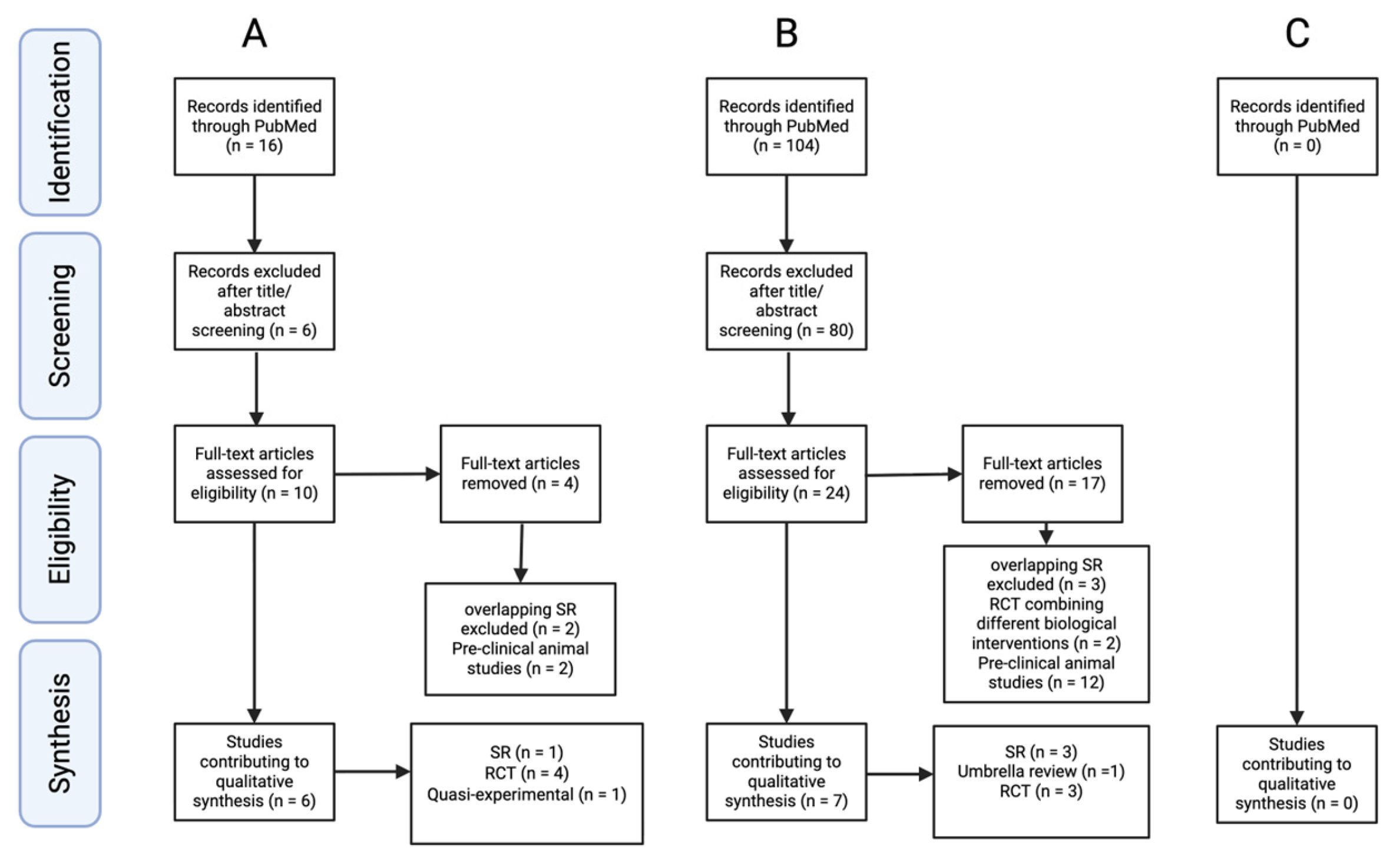
Of 14 studies identified regarding ESWT or RPW in humans with ACL injury or ACL reconstruction (ACLR), eight studies were excluded, as the study protocols combined use of other treatments, including platelet-rich plasma injections or exercise-induced hamstring fatigue (Figure 1A). The studies on ACL reconstruction either used RPW (three studies) or ESWT (two studies), and no studies combined ESWT with RPW. All the studies were conducted in post-operative settings. Four randomized controlled trials were identified, with inconclusive but predominantly positive clinical outcomes. Quasi-experimental research by Rahim et al. provided inconclusive outcomes, suggesting improved graft incorporation, while showing no effects on knee function [22]. These findings remain mainly subjective and have a high risk of bias, as concluded by Shin et al. [21]. This systematic review with meta-analysis included all five studies above and suggested possible improved clinical outcomes following RPW and ESWT after ACLR (Table 1).
B.
Search results for KOA and ESWT or RPW:
A systematic review by Liao et al. demonstrated that higher energy flux density (EFD), up to 0.24 mJ/mm2, resulted in greater improvement in outcomes when treating early KOA, compared to lower EFD [25]. A higher number of impulses (2000–4000) additionally showed better clinical outcomes than a lower number of impulses [25]. An umbrella review by Tang et al. concluded that ESWT is an effective therapy for improving function and pain in patients with KOA [26].
Ko et al. suggest that both ESWT and RPW have significant clinical outcomes in patients with KOA [27]. These findings may suggest that a combination of ESWT and RPW may be effective; using ESWT at a high number of impulses (>2000) and an EFD of 0.24 mJ/mm2 may provide increased improvements in outcomes for individuals with KOA. Due to the limited number of studies included, it is not possible to draw clinically meaningful conclusions regarding separate effects of different energy levels of ESWT and RPWs [26].
C.
Search results for PTOA and ESWT or RPW:
No single study evaluated either ESWT or RPW in the management of PTOA of the knee in humans (Figure 1C). This absence of specific studies evaluating PTOA after ACL injury limited the ability to perform a systematic review. As a result, studies identified through the described methodology were used to inform a perspective on a theoretical framework of how ESWT and RPW may be applied in clinical settings and research to guide treatment of ACL-injured knees, and their potential role in management of PTOA.

4. What Are Extracorporeal Shockwave Therapy (ESWT) and Radial Pressure Waves (RPW)?

It should be noted that the two most common clinical applications of extracorporeal shockwave therapy (ESWT) and radial pressure waves (RPW), although often concomitant, have separate mechanisms of action, and should not be used interchangeably. ESWT can penetrate deeper structures through high-energy sound waves that deliver high energy to more localized targets and, therefore, are recommended for use of osseous applications, such as treatment of non-unions and fractures [16]. In contrast, pressure waves generated through RPW are capable of creating smaller amplitudes of energy, with highest energy generated at the surface of the applicator head, and may have more effective use in soft tissue injuries, such as muscle- or tendon-related pathology [16].
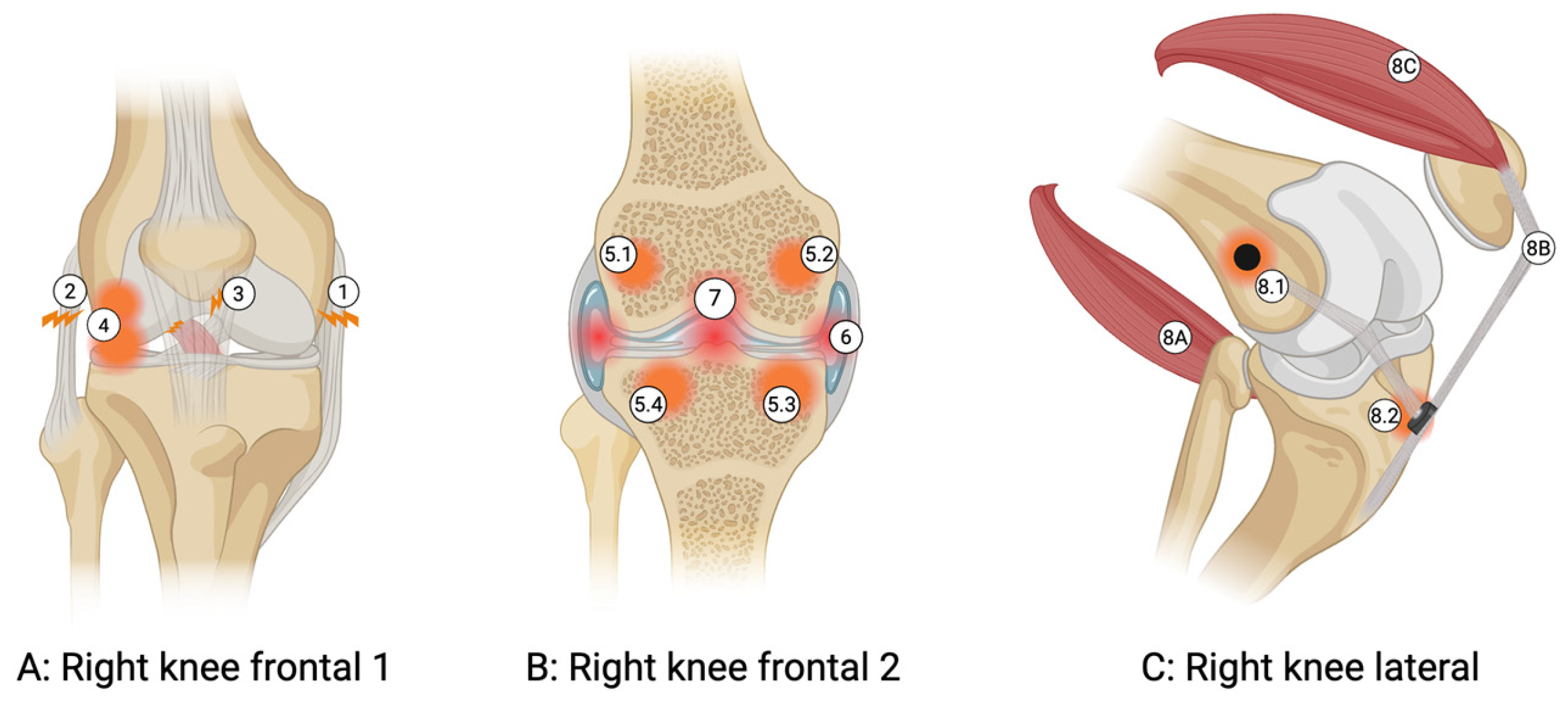
The framework to treatment of the knee is based on considering pain from all intra-articular structures within the joint space (cartilage, meniscus, and synovium); adjacent subchondral bone; and surrounding soft tissues, including tendons, ligaments, and muscles (Figure 2). This concept has been previously proposed, of the knee as an organ system [28]. During surgical repair, additional tissue trauma is generated through harvesting autografts used to perform ACL reconstruction surgery, including the hamstring tendon, bone–patellar tendon–bone, and the quadriceps tendon [29], as well as anchoring the ACL graft within bone. Changes in cartilage, joint fluid, and bone marrow lesions [30], as well as ligament ruptures, may be concomitant injuries sustained during ACL injury [31] (Figure 2). Pre-clinical evidence suggests that ESWT and RPW can result in cellular changes to each of these structures that can be used to target the joint using a systematic approach (Table 2).
An in vitro study of human tenocytes displayed an increase in collagen synthesis and cell growth after treatment with ESWT [32]. In a rodent model, ESWT induced tenocyte proliferation, progressive tendon tissue regeneration, and neovascularization via increasing expression of proliferating cell nuclear antigen (PCNA), transforming growth factor ß1 (TGF-ß1) and insulin-like growth factor 1 (IGF-1) [33].
Humans sustaining acute hamstring muscle injuries have an earlier timeline for return to sport and improved functional recovery following RPW therapy, attributed to reducing strength deficits in the injured leg [34]. RPW therapy was shown as effective in reducing pain and shear modulus, as well as improving function in patients after treatment of myofascial trigger points in the upper trapezius muscle [35]. In the time post-cardiotoxin-induced quadriceps femoris muscle injury in rats, ESWT was correlated with increased mitotic activity (H3P+ cells + cells expressing myoD and myogenin as myogenic regulatory factors) and regeneration in muscle fibers, suggesting an acceleration of the muscle repair process [36].
Pre-clinical models suggest that ESWT may have chondroprotective effects. Both rat and rabbit models of ACL rupture demonstrate reductions in chondrocyte apoptosis and cartilage degradation, as well as improvements in bone remodeling and anti-inflammatory effects [37,38]. ESWT has been shown to have a positive effect on human chondrocytes in vitro by preventing progressive dedifferentiation through an improvement in the Ki67 proliferation index and decreases in p16INK4a/Cdkn2a as markers of chondrosenescence [39]. ESWT may result in an anti-inflammatory and migratory effect on the cytokine-rich environment characterizing OA [39]. ESWT induces overexpression of CD44 receptors on chondrocytes in vitro and may increase interaction with hyaluronic acid (HA), resulting in an enhancement in cartilage health [39].
An additional mechanism for ESWT is promoting the health of subchondral bone. Studies in rabbits show that ESWT accelerates bone healing through increasing vascular endothelial growth factor (VEGF) expression and other angiogenic and osteogenic growth factors [40]. While not documented in humans, prior work in rat models suggested regression of osteoarthritis in the knees after ESWT [41]. Earlier studies in rats demonstrated that ESWT can increase mesenchymal stem cells and expression levels of transforming growth factor (TGF-ß1) and VEGF-A in femoral bone [42,43], leading to increases in angiogenesis and osteogenesis [42,43]. Moreover, an in vivo human study concluded earlier tibial graft maturation (6 months post-operatively) and better femoral/intra-articular maturation (24 months post-operatively) in the RPW group after ACL reconstruction (ACLR) [24] (concept of osteointegration at site of bone tunnel for graft placement illustrated in Figure 2). Notably, prior work suggested that improvements in tibial graft maturation were limited, in one study of human participants receiving six sessions of ESWT, further suggesting the need for standardized, evidence-based treatment protocols [22].
Importantly, the side effects described following ESWT and RPW have generally been minimal and self-limiting, with limited evidence for advanced structural injuries following use. A systematic review by Schmitz et al. indicated ESWT and RPW to be safe for possible treatment of the musculoskeletal system [44]. Most common side effects, such as pain during treatment, post-treatment erythema, skin bruising, or superficial oedema, are often transient [16,45,46]. As a result of their healing effects on soft tissue, ESWT and RPW could hypothetically enhance surgical recovery in peri-operative application after ACL injuries, as unresolved inflammation after surgery and struggles with soft tissue healing remain crucial problems [47,48].
Wess suggests that chronic pain can be seen as a pathological memory trace that enables conditioned reflexes to support a self-maintaining pain spiral, independent of structural damage [49]. These pathological patterns may be disrupted by ESWT and RPW, resulting in durable pain relief [49]. Pain relief may be distributed through the neurological influences of ESWT and RPW, whereas tissue repair via ESWT and RPT primarily derives from local mechanobiological responses (Table 1) [39,40]. Several mechanisms, such as hyperstimulation analgesia and the gate control concept, have been proposed, whereby ESWT and RPW diminish the activity of peripheral nerve endings via overstimulation and altering transmission of pain-related neuropeptides, as well as reduce nociceptive transmission via descending inhibition [50]. In conditions of chronic pain, such as chronic pelvic pain syndrome (CPPS), ESWT and RPW have shown to significantly reduce the level of pain on a visual analog pain scale (VAS) and improve function following treatment [51,52]. In application for conditions of calcifying tendonitis of the rotator cuff, ESWT has also shown non-operative impacts on clinical outcomes, where not only the function of the shoulder joint, but also the pain, improved significantly [53]. In the treatment of patients with OA, ESWT and RPW have been shown to significantly improve pain relief on the Western Ontario and McMaster Universities Osteoarthritis Index (WOMAC), as well as VAS, in a current systematic review and meta-analysis by Wang et al. [54].

5. Existing Evidence for ESWT and RPW in ACL Injury

Five studies evaluated the use of either RPW or ESWT in peri-operative and post-operative timeframes, regarding clinical outcomes. The limited data, as well as the lack of consensus, present a high risk of bias and a gap in the research, yet there are randomized controlled trials suggesting that shockwave could be effective after ACL injuries. The application of ESWT at weeks 4, 5, and 6 post ACLR surgery has provided notable improvements, such as ‘return-to-pivoting-sports’ being faster in patients who received ESWT (27.92 ± 2.99 weeks) compared to control (42.64 ± 5.18 weeks), and a larger portion of patients reached pre-injury activity levels at 12 months with ESWT compared to controls (31 of 37 vs. 6 of 28, respectively) [23]. Wang et al. observed significant improvements in patient-related outcomes (Lysholm score 1 y and 2 y post-op) following ESWT performed immediately after surgery, supporting value of treatment around the time of surgery [17]. A separate human RCT on RPW after ACLR concluded a significantly higher Lysholm score, Tegner score, and enhanced graft maturation compared with controls at 24 months [24]. Additionally, improved graft incorporation after six sessions of RPW treatment was demonstrated in an RCT by Rahim et al. [22]. In contrast, Song et al. reported no significant differences in Lysholm score, range of motion (ROM), IKDC score, or VAS after RPW treatment following ACLR at 24 weeks [19].
A systematic review with meta-analysis, performed in 2025, did not observe differences in functional outcomes exceeding the minimal clinically important difference (MCID) in those receiving ESWT after ACLR [21]. The review highlighted issues with variability in treatment protocols, use of ESWT and RPW, and lack of standardization in clinical outcomes, including Lysholm score, IKDC score, VAS, and ROM. Standard procedural recommendations in using ESWT and RPW in management of sports injuries has been proposed and should be applied to future research [16].
Reviewing the literature, the most common target sites for ESWT after ACL injury include the central intra-articular graft, as well as the femoral and tibial tunnels [17,23]. The aim in many of these studies was to modulate graft ligamentization and bone–tendon interface healing. The typical target for patients with bone marrow edema (BME) of the knee is the radiographic-defined BME lesion, which can occur in the medial femoral condyle [55]. For ACL injuries, specifically, the lateral tibial and femoral condyles are the most common localizations of BME, due to the lateral bone contusions after ACL tear; outcomes targeting these potential pain generators have been limited [30].

6. Application of ESWT and RPW in ACL Rehabilitation and Future Directions

ESWT and RPW may have multiple uses in advancing recovery from ACL injuries by targeting associated sites of knee injury that are often not directly addressed during surgery. ESWT has been shown to significantly improve clinical outcomes (WOMAC Osteoarthritis Index and SF-36 scores) and pain relief (VAS) in early treatment (1 to 6 months) of primary BME [55]. The BME regression seen on MRI was significantly higher (95% vs. 65% control) at 6 months, with 100% complete regression by the 1-year timepoint in patients who received ESWT compared to controls [55]. Similarly, better patient-reported outcomes in function and pain for patients being treated with ESWT after BME have been observed [56]. In a network meta-analysis from 2025, ESWT and RPW were ranked among the leading nonsurgical modalities for alleviating functional impairment, stiffness, and pain in patients with knee OA [57].
ESWT and RPW have demonstrated chondroprotective and osteoprotective effects, as well as positive effects on tenocytes after tendon injuries and on graft healing and maturation after ACLR surgery [24,32,33,37,38]. These effects occurred while not showing any significant harm due to ESWT and RPW treatment [44]. In addition to the effects of ESWT and RPW on RTS and knee OA outcome measures, different animal models (trials with rats and rabbits) demonstrated significant decreases in progression of PTOA through the use of ESWT after induced ACL injury [37,38,41]. However, work to characterize outcomes in management of knee PTOA after ACL injuries with ESWT and RPW have not been described and are currently being explored in our lab (A.S.T.).
We propose that ESWT and RPW may be reasonable to apply during pre-operative management of ACL injuries or following post-operative management. A recent case–control series observed that ESWT and RPW around the time of insertional Achilles tendon repair resulted in an earlier return to activity following surgery, by 1.3 months [58]. Additional work by this author group includes earlier evidence of bone healing at 4 weeks in those treated with ESWT combined with electromagnetic transduction therapy [59]. These results are promising and suggest value in future research on treatment in the pre-, peri-, and post-operative management of ACL injuries, taking a more global approach to recognized sites of injury, including BME and graft harvest locations, in addition to promotion of surgical site osteointegration. From these studies, it can be concluded that applying focused ESWT to the osseous structures may potentially help to stimulate bone healing and osteointegration at the sites of ACL repair, and should target subchondral bone edema, particularly at sites of identified injury and, ideally, using high energy settings. RPW may hypothetically be applied generally to the soft tissue surrounding structures, using moderate energy settings to treat tendons and muscles. Based on prior work on treatment of tendon and bone pathology, this would include 3–4 separate sessions, every 1–2 weeks, with effects of treatment expected at 2–3 months following application [16]. On review of the limited available studies on ESWT and RPW use to treat peri- and post-operative ACL injury, we propose that the use of both treatments may require more detailed treatment of both intra- and extra-articular structures with appropriate energy settings. Extrapolating from the literature on ESWT or RPW after ACL repair, and findings in general knee OA, we propose a goal to reach moderate energy settings and 2000–4000 shocks with ESWT. This is supported by findings that higher EFD (up to 0.24 mJ/mm2) and a higher number of impulses (2000–4000) have been shown to improve outcomes in individuals with KOA [25,26]. We propose that ESWT be combined with RPW, with RPW, delivered for 2000–3000 counts, over tendons and soft tissue areas of injury, given the different methods, as pressure waves transmit energy more diffusely.
The absence of disease-modifying clinical therapies for PTOA remains a challenge [9]. Based on the findings in this perspective, we hypothesize that peri-operative treatment with ESWT and RPW may have the potential to improve clinical outcomes after ACL injuries. While this perspective aims to synthesize existing data on use of ESWT and RPW following ACL injury and in KOA, there are limitations to this work. These include the absence of human subject research to test this combined approach in individuals with PTOA of the knee following ACL repair; furthermore, a high degree of variability in use of ESWT and RPW was observed across studies in number of sessions, energy settings, and anatomical sites targeted, which all limit our understanding of how these treatments may affect the long-term outcomes for patients with KOA or after ACL injury. These limitations need to be acknowledged in clinical approaches to management of individuals following ACL injury and those who develop PTOA.

7. Conclusions

In conclusion, ESWT and RPW have been shown to have clinical impacts in the treatment of many musculoskeletal injuries and, more recently, in pre-surgical application. There is a gap in the research for the use of ESWT and RPW in ACL surgery patients, both for the improvement of surgical outcomes and RTS, as well as for PTOA prevention. The ongoing interest in optimizing the management of ACL injuries requires ongoing work to optimize surgical care, rehabilitation, and other novel interventions, including the potential use of ESWT and RPW.

Author Contributions

Conceptualization, F.J.Z., L.W.G., and A.S.T.; writing—original draft preparation, F.J.Z., L.W.G., M.B., and A.S.T.; writing—review and editing, F.J.Z., L.W.G., M.B., J.S., L.G., and A.S.T.; visualization, F.J.Z.; supervision, A.S.T. All authors have read and agreed to the published version of the manuscript.

Funding

This research received no external funding.

Conflicts of Interest

The authors declare no conflicts of interest, and no funding was received to support this manuscript. Ludger Gerdesmeyer received equipment, honoraria, research, and travel support from Storz Medical AG and Curamedix. Adam Tenforde serves as Senior editor for PM&R Journal. He gives professional talks, such as grand rounds and medical conference plenary lectures, and receives honoraria from conference organizers. He has participated in research funded by the Arnold P. Gold Foundation (physician and patient care disparities), Football Player Health Study at Harvard (health in American-Style Football players), American Medical Society for Sports Medicine (bone density research), Uniform Health Service and Enovis (Achilles tendinopathy), and MTEC/Department of Defense (bone stress injuries with shockwave). He is a paid consultant for State Farm Insurance and Strava. He receives industry support from Enovis, Sanuwave, and Storz for equipment use for research studies on treatment of tendinopathy, knee osteoarthritis, and bone stress injuries.

References

  1. Kaeding, C.C.; Léger-St-Jean, B.; Magnussen, R.A. Epidemiology and Diagnosis of Anterior Cruciate Ligament Injuries. Clin. Sports Med. 2017, 36, 1–8. [Google Scholar] [CrossRef]
  2. Ardern, C.L.; Taylor, N.F.; Feller, J.A.; Webster, K.E. Fifty-Five per Cent Return to Competitive Sport Following Anterior Cruciate Ligament Reconstruction Surgery: An Updated Systematic Review and Meta-Analysis Including Aspects of Physical Functioning and Contextual Factors. Br. J. Sports Med. 2014, 48, 1543–1552. [Google Scholar] [CrossRef] [PubMed]
  3. Welling, W.; Benjaminse, A.; Seil, R.; Lemmink, K.; Zaffagnini, S.; Gokeler, A. Low Rates of Patients Meeting Return to Sport Criteria 9 Months after Anterior Cruciate Ligament Reconstruction: A Prospective Longitudinal Study. Knee Surg. Sports Traumatol. Arthrosc. 2018, 26, 3636–3644. [Google Scholar] [CrossRef] [PubMed]
  4. Webster, K.E.; Hewett, T.E. Anterior Cruciate Ligament Injury and Knee Osteoarthritis: An Umbrella Systematic Review and Meta-Analysis. Clin. J. Sport Med. 2022, 32, 145–152. [Google Scholar] [CrossRef] [PubMed]
  5. Segawa, H.; Omori, G.; Koga, Y. Long-Term Results of Non-Operative Treatment of Anterior Cruciate Ligament Injury. Knee 2001, 8, 5–11. [Google Scholar] [CrossRef]
  6. Davi, S.M.; Ahn, A.; White, M.S.; Butterfield, T.A.; Kosmac, K.; Kwon, O.S.; Lepley, L.K. Long-Lasting Impairments in Quadriceps Mitochondrial Health, Muscle Size, and Phenotypic Composition Are Present After Non-Invasive Anterior Cruciate Ligament Injury. Front. Physiol. 2022, 13, 805213. [Google Scholar] [CrossRef]
  7. Chinzei, N.; Brophy, R.H.; Duan, X.; Cai, L.; Nunley, R.M.; Sandell, L.J.; Rai, M.F. Molecular Influence of Anterior Cruciate Ligament Tear Remnants on Chondrocytes: A Biologic Connection between Injury and Osteoarthritis. Osteoarthr. Cartil. 2018, 26, 588–599. [Google Scholar] [CrossRef]
  8. Luc, B.; Gribble, P.A.; Pietrosimone, B.G. Osteoarthritis Prevalence Following Anterior Cruciate Ligament Reconstruction: A Systematic Review and Numbers-Needed-to-Treat Analysis. J. Athl. Train. 2014, 49, 806–819. [Google Scholar] [CrossRef]
  9. Dilley, J.E.; Bello, M.A.; Roman, N.; McKinley, T.; Sankar, U. Post-Traumatic Osteoarthritis: A Review of Pathogenic Mechanisms and Novel Targets for Mitigation. Bone Rep. 2023, 18, 101658. [Google Scholar] [CrossRef]
  10. Dye, S.F. The Knee as a Biologic Transmission With an Envelope of Function: A Theory. Clin. Orthop. Relat. Res. 1996, 325, 10–18. [Google Scholar] [CrossRef]
  11. Wick, E.C.; Grant, M.C.; Wu, C.L. Postoperative Multimodal Analgesia Pain Management With Nonopioid Analgesics and Techniques: A Review. JAMA Surg. 2017, 152, 691. [Google Scholar] [CrossRef] [PubMed]
  12. Curtis, E.; Fuggle, N.; Shaw, S.; Spooner, L.; Ntani, G.; Parsons, C.; Corp, N.; Honvo, G.; Baird, J.; Maggi, S.; et al. Safety of Cyclooxygenase-2 Inhibitors in Osteoarthritis: Outcomes of a Systematic Review and Meta-Analysis. Drugs Aging 2019, 36, 25–44. [Google Scholar] [CrossRef] [PubMed]
  13. Bhala, N.; Emberson, J.; Merhi, A.; Abramson, S.; Arber, A.; Baron, J.; Bombardier, C.; Cannon, C.; E Farkouh, M. Vascular and Upper Gastrointestinal Effects of Non-Steroidal Anti-Inflammatory Drugs: Meta-Analyses of Individual Participant Data from Randomised Trials. Lancet 2013, 382, 769–779. [Google Scholar] [CrossRef] [PubMed]
  14. Ussai, S.; Miceli, L.; Pisa, F.; Bednarova, R.; Petelin, R.; Della Rocca, G.; Giordano, A. Impact of Potential Inappropriate NSAIDs UseInChronic Pain. DDDT 2015, 2015, 2073–2077. [Google Scholar] [CrossRef]
  15. Parisien, M.; Lima, L.V.; Dagostino, C.; El-Hachem, N.; Drury, G.L.; Grant, A.V.; Huising, J.; Verma, V.; Meloto, C.B.; Silva, J.R.; et al. Acute Inflammatory Response via Neutrophil Activation Protects against the Development of Chronic Pain. Sci. Transl. Med. 2022, 14, eabj9954. [Google Scholar] [CrossRef]
  16. Rhim, H.C.; Singh, M.; Maffulli, N.; Saxena, A.; Leal, C.; Gerdesmeyer, L.; Quirolgico, K.; Furia, J.P.; Zwerver, J.; Liao, C.-D.; et al. Recommendations for Use of Extracorporeal Shockwave Therapy in Sports Medicine: An International Modified Delphi Study. Br. J. Sports Med. 2025, 59, 1287–1301. [Google Scholar] [CrossRef]
  17. Wang, C.-J.; Ko, J.-Y.; Chou, W.-Y.; Hsu, S.-L.; Ko, S.-F.; Huang, C.-C.; Chang, H.-W. Shockwave Therapy Improves Anterior Cruciate Ligament Reconstruction. J. Surg. Res. 2014, 188, 110–118. [Google Scholar] [CrossRef]
  18. Cacchio, A.; Giordano, L.; Colafarina, O.; Rompe, J.D.; Tavernese, E.; Ioppolo, F.; Flamini, S.; Spacca, G.; Santilli, V. Extracorporeal Shock-Wave Therapy Compared with Surgery for Hypertrophic Long-Bone Nonunions. J. Bone Jt. Surg. 2009, 91, 2589–2597. [Google Scholar] [CrossRef]
  19. Song, Y.; Che, X.; Wang, Z.; Li, M.; Zhang, R.; Wang, D.; Shi, Q. A Randomized Trial of Treatment for Anterior Cruciate Ligament Reconstruction by Radial Extracorporeal Shock Wave Therapy. BMC Musculoskelet. Disord. 2024, 25, 57. [Google Scholar] [CrossRef]
  20. Rhim, H.C.; Shin, J.; Kang, J.; Dyrek, P.; Crockett, Z.; Galido, P.; Wade, C.; Hollander, K.; Borg-Stein, J.; Sampson, S.; et al. Use of Extracorporeal Shockwave Therapies for Athletes and Physically Active Individuals: A Systematic Review. Br. J. Sports Med. 2024, 58, 154–163. [Google Scholar] [CrossRef]
  21. Shin, J.; Rhim, H.C.; Kim, J.; Guo, R.; Elshafey, R.; Jang, K.-M. Use of Extracorporeal Shockwave Therapy Combined with Standard Rehabilitation Following Anterior Cruciate Ligament Reconstruction: A Systematic Review with Meta-Analysis. BMC Musculoskelet. Disord. 2025, 26, 79. [Google Scholar] [CrossRef] [PubMed]
  22. Rahim, N.; Ooi, F.; Mt, M.; Chen, C.; Musa, A. The Effects of Three and Six Sessions of Low Energy Extracorporeal Shockwave Therapy on Graft Incorporation and Knee Functions Post Anterior Cruciate Ligament Reconstruction. Malays. Orthop. J. 2022, 16, 28–39. [Google Scholar] [CrossRef] [PubMed]
  23. Weninger, P.; Thallinger, C.; Chytilek, M.; Hanel, Y.; Steffel, C.; Karimi, R.; Feichtinger, X. Extracorporeal Shockwave Therapy Improves Outcome after Primary Anterior Cruciate Ligament Reconstruction with Hamstring Tendons. JCM 2023, 12, 3350. [Google Scholar] [CrossRef] [PubMed]
  24. Zhang, S.; Wen, A.; Li, S.; Yao, W.; Liu, C.; Lin, Z.; Jin, Z.; Chen, J.; Hua, Y.; Chen, S.; et al. Radial Extracorporeal Shock Wave Therapy Enhances Graft Maturation at 2-Year Follow-up After ACL Reconstruction: A Randomized Controlled Trial. Orthop. J. Sports Med. 2022, 10, 23259671221116340. [Google Scholar] [CrossRef]
  25. Liao, P.-C.; Chou, S.-H.; Shih, C.-L. A Systematic Review of the Use of Shockwave Therapy for Knee Osteoarthritis. J. Orthop. 2024, 56, 18–25. [Google Scholar] [CrossRef]
  26. Tang, P.; Wen, T.; Lu, W.; Jin, H.; Pan, L.; Li, H.; Zeng, B.; Zhou, Y.; Xiao, W.; Li, Y. The Efficacy of Extracorporeal Shock Wave Therapy for Knee Osteoarthritis: An Umbrella Review. Int. J. Surg. 2024, 110, 2389–2395. [Google Scholar] [CrossRef]
  27. Ko, N.-Y.; Chang, C.-N.; Cheng, C.-H.; Yu, H.-K.; Hu, G.-C. Comparative Effectiveness of Focused Extracorporeal versus Radial Extracorporeal Shockwave Therapy for Knee Osteoarthritis—Randomized Controlled Study. IJERPH 2022, 19, 9001. [Google Scholar] [CrossRef]
  28. Loeser, R.F.; Goldring, S.R.; Scanzello, C.R.; Goldring, M.B. Osteoarthritis: A Disease of the Joint as an Organ. Arthritis Rheum. 2012, 64, 1697–1707. [Google Scholar] [CrossRef]
  29. Runer, A. Current Trends in Graft Choice for Anterior Cruciate Ligament Reconstruction—Part I: Anatomy, Biomechanics, Graft Incorporation and Fixation. J. Exp. Orthop. 2023, 10, 37. [Google Scholar] [CrossRef]
  30. Frobell, R.B.; Le Graverand, M.-P.; Buck, R.; Roos, E.M.; Roos, H.P.; Tamez-Pena, J.; Totterman, S.; Lohmander, L.S. The Acutely ACL Injured Knee Assessed by MRI: Changes in Joint Fluid, Bone Marrow Lesions, and Cartilage during the First Year. Osteoarthr. Cartil. 2009, 17, 161–167. [Google Scholar] [CrossRef]
  31. The Swdish National Knee Ligament Registry. The Swedish Knee Ligament Registry. Annual Report 2020. Available online: https://www.aclregister.nu/media/uploads/Annual%20reports/rapport2020en.pdf (accessed on 3 October 2025).
  32. Vetrano, M.; d’Alessandro, F.; Torrisi, M.R.; Ferretti, A.; Vulpiani, M.C.; Visco, V. Extracorporeal Shock Wave Therapy Promotes Cell Proliferation and Collagen Synthesis of Primary Cultured Human Tenocytes. Knee Surg. Sports Traumatol. Arthrosc. 2011, 19, 2159–2168. [Google Scholar] [CrossRef] [PubMed]
  33. Chen, Y.; Wang, C.; Yang, K.D.; Kuo, Y.; Huang, H.; Huang, Y.; Sun, Y.; Wang, F. Extracorporeal Shock Waves Promote Healing of Collagenase-induced Achilles Tendinitis and Increase TGF-β1 and IGF-I Expression. J. Orthop. Res. 2004, 22, 854–861. [Google Scholar] [CrossRef] [PubMed]
  34. Crupnik, J.; Silveti, S.; Wajnstein, N.; Rolon, A.; Wuerfel, T.; Stiller, P.; Morral, A.; Furia, J.P.; Maffulli, N.; Schmitz, C. Radial ESWT Combined with a Specific Rehabilitation Program (rESWT + RP) Is More Effective than Sham rESWT + RP for Acute Hamstring Muscle Complex Injury Type 3b: A Randomized, Controlled Trial. Br. Med. Bull. 2025, 155. [Google Scholar] [CrossRef] [PubMed]
  35. Luan, S.; Zhu, Z.; Ruan, J.; Lin, C.; Ke, S.; Xin, W.; Liu, C.; Wu, S.; Ma, C. Randomized Trial on Comparison of the Efficacy of Extracorporeal Shock Wave Therapy and Dry Needling in Myofascial Trigger Points. Am. J. Phys. Med. Rehabil. 2019, 98, 677–684. [Google Scholar] [CrossRef]
  36. Zissler, A.; Steinbacher, P.; Zimmermann, R.; Pittner, S.; Stoiber, W.; Bathke, A.C.; Sänger, A.M. Extracorporeal Shock Wave Therapy Accelerates Regeneration After Acute Skeletal Muscle Injury. Am. J. Sports Med. 2017, 45, 676–684. [Google Scholar] [CrossRef]
  37. Zhao, Z.; Ji, H.; Jing, R.; Liu, C.; Wang, M.; Zhai, L.; Bai, X.; Xing, G. Extracorporeal Shock-Wave Therapy Reduces Progression of Knee Osteoarthritis in Rabbits by Reducing Nitric Oxide Level and Chondrocyte Apoptosis. Arch. Orthop. Trauma Surg. 2012, 132, 1547–1553. [Google Scholar] [CrossRef]
  38. Wang, C.-J.; Weng, L.-H.; Ko, J.-Y.; Sun, Y.-C.; Yang, Y.-J.; Wang, F.-S. Extracorporeal Shockwave Therapy Shows Chondroprotective Effects in Osteoarthritic Rat Knee. Arch. Orthop. Trauma Surg. 2011, 131, 1153–1158. [Google Scholar] [CrossRef]
  39. Vetrano, M.; Ranieri, D.; Nanni, M.; Pavan, A.; Malisan, F.; Vulpiani, M.C.; Visco, V. Hyaluronic Acid (HA), Platelet-Rich Plasm and Extracorporeal Shock Wave Therapy (ESWT) Promote Human Chondrocyte Regeneration in Vitro and ESWT-Mediated Increase of CD44 Expression Enhances Their Susceptibility to HA Treatment. PLoS ONE 2019, 14, e0218740. [Google Scholar] [CrossRef]
  40. Wang, C.-J.; Huang, K.-E.; Sun, Y.-C.; Yang, Y.-J.; Ko, J.-Y.; Weng, L.-H.; Wang, F.-S. VEGF Modulates Angiogenesis and Osteogenesis in Shockwave-Promoted Fracture Healing in Rabbits. J. Surg. Res. 2011, 171, 114–119. [Google Scholar] [CrossRef]
  41. Wang, C.-J.; Weng, L.-H.; Ko, J.-Y.; Wang, J.-W.; Chen, J.-M.; Sun, Y.-C.; Yang, Y.-J. Extracorporeal Shockwave Shows Regression of Osteoarthritis of the Knee in Rats. J. Surg. Res. 2011, 171, 601–608. [Google Scholar] [CrossRef]
  42. Wang, F.S.; Yang, K.D.; Chen, R.F.; Wang, C.J.; Sheen-Chen, S.M. Extracorporeal Shock Wave Promotes Growth and Differentiation of Bone-Marrow Stromal Cells towards Osteoprogenitors Associated with Induction of TGF-β1. J. Bone Joint Surg. 2002, 84, 457–461. [Google Scholar] [CrossRef]
  43. Chen, Y.-J.; Kuo, Y.-R.; Yang, K.D.; Wang, C.-J.; Sheen Chen, S.-M.; Huang, H.-C.; Yang, Y.-J.; Yi-Chih, S.; Wang, F.-S. Activation of Extracellular Signal-Regulated Kinase (ERK) and P38 Kinase in Shock Wave-Promoted Bone Formation of Segmental Defect in Rats. Bone 2004, 34, 466–477. [Google Scholar] [CrossRef] [PubMed]
  44. Schmitz, C.; Császár, N.B.M.; Milz, S.; Schieker, M.; Maffulli, N.; Rompe, J.-D.; Furia, J.P. Efficacy and Safety of Extracorporeal Shock Wave Therapy for Orthopedic Conditions: A Systematic Review on Studies Listed in the PEDro Database. Br. Med. Bull. 2015, ldv047. [Google Scholar] [CrossRef] [PubMed]
  45. Roerdink, R.L.; Dietvorst, M.; Van Der Zwaard, B.; Van Der Worp, H.; Zwerver, J. Complications of Extracorporeal Shockwave Therapy in Plantar Fasciitis: Systematic Review. Int. J. Surg. 2017, 46, 133–145. [Google Scholar] [CrossRef] [PubMed]
  46. Tenforde, A.S.; Borgstrom, H.E.; DeLuca, S.; McCormack, M.; Singh, M.; Hoo, J.S.; Yun, P.H. Best Practices for Extracorporeal Shockwave Therapy in Musculoskeletal Medicine: Clinical Application and Training Consideration. PM&R 2022, 14, 611–619. [Google Scholar] [CrossRef]
  47. Larsson, S.; Struglics, A.; Lohmander, L.S.; Frobell, R. Surgical Reconstruction of Ruptured Anterior Cruciate Ligament Prolongs Trauma-Induced Increase of Inflammatory Cytokines in Synovial Fluid: An Exploratory Analysis in the KANON Trial. Osteoarthr. Cartil. 2017, 25, 1443–1451. [Google Scholar] [CrossRef]
  48. Claes, S.; Verdonk, P.; Forsyth, R.; Bellemans, J. The “Ligamentization” Process in Anterior Cruciate Ligament Reconstruction: What Happens to the Human Graft? A Systematic Review of the Literature. Am. J. Sports Med. 2011, 39, 2476–2483. [Google Scholar] [CrossRef]
  49. Wess, O.J. A Neural Model for Chronic Pain and Pain Relief by Extracorporeal Shock Wave Treatment. Urol. Res. 2008, 36, 327–334. [Google Scholar] [CrossRef]
  50. Ryskalin, L.; Morucci, G.; Natale, G.; Soldani, P.; Gesi, M. Molecular Mechanisms Underlying the Pain-Relieving Effects of Extracorporeal Shock Wave Therapy: A Focus on Fascia Nociceptors. Life 2022, 12, 743. [Google Scholar] [CrossRef]
  51. Vahdatpour, B.; Alizadeh, F.; Moayednia, A.; Emadi, M.; Khorami, M.H.; Haghdani, S. Efficacy of Extracorporeal Shock Wave Therapy for the Treatment of Chronic Pelvic Pain Syndrome: A Randomized, Controlled Trial. ISRN Urol. 2013, 2013, 1–6. [Google Scholar] [CrossRef]
  52. Zimmermann, R.; Cumpanas, A.; Miclea, F.; Janetschek, G. Extracorporeal Shock Wave Therapy for the Treatment of Chronic Pelvic Pain Syndrome in Males: A Randomised, Double-Blind, Placebo-Controlled Study. Eur. Urol. 2009, 56, 418–424. [Google Scholar] [CrossRef]
  53. Gerdesmeyer, L.; Wagenpfeil, S.; Haake, M.; Maier, M.; Loew, M.; Wörtler, K.; Lampe, R.; Seil, R.; Handle, G.; Gassel, S.; et al. Extracorporeal Shock Wave Therapy for the Treatment of Chronic Calcifying Tendonitis of the Rotator Cuff: A Randomized Controlled Trial. JAMA 2003, 290, 2573. [Google Scholar] [CrossRef] [PubMed]
  54. Wang, Y.-C.; Huang, H.-T.; Huang, P.-J.; Liu, Z.-M.; Shih, C.-L. Efficacy and Safety of Extracorporeal Shockwave Therapy for Treatment of Knee Osteoarthritis: A Systematic Review and Meta-Analysis. Pain Med. 2020, 21, 822–835. [Google Scholar] [CrossRef] [PubMed]
  55. Gao, F.; Sun, W.; Li, Z.; Guo, W.; Wang, W.; Cheng, L.; Yue, D.; Zhang, N.; Savarin, A. Extracorporeal Shock Wave Therapy in the Treatment of Primary Bone Marrow Edema Syndrome of the Knee: A Prospective Randomised Controlled Study. BMC Musculoskelet. Disord. 2015, 16, 379. [Google Scholar] [CrossRef] [PubMed]
  56. Häußer, J.; Wieber, J.; Catalá-Lehnen, P. The Use of Extracorporeal Shock Wave Therapy for the Treatment of Bone Marrow Oedema—A Systematic Review and Meta-Analysis. J. Orthop. Surg. Res. 2021, 16, 369. [Google Scholar] [CrossRef]
  57. Lan, X.; Li, L.; Jia, Q.; He, F.; Kuang, G.; Zeng, W.; Chen, M.; Guo, C.; Wen, Z.; Chen, Q. Physical Modalities for the Treatment of Knee Osteoarthritis: A Network Meta-Analysis. Aging Clin. Exp. Res. 2025, 37, 121. [Google Scholar] [CrossRef]
  58. Saxena, A.; Bondi, E.; Gerdesmeyer, L.; Tenforde, A.S. Extracorporeal Shock Wave Therapy (ESWT) and Radial Pressure Waves (RPW) May Improve Post-Operative Recovery Following Insertional Achilles Tendon Surgeries: A Case-Control Pilot Study. J. Orthop. Surg. Res. 2025, 20, 751. [Google Scholar] [CrossRef]
  59. Saxena, A.; Bondi, E.; Gerdesmeyer, L.; Leal, C.; Knobloch, K.; Maffulli, N.; Gerdesmeyer, L.; Tenforde, A.S. Focused Shock Wave Therapy (ESWT) and Electromagnetic Transduction Therapy (EMTT) May Improve Post-Operative Bone Healing in Selective Foot and Ankle Surgeries: A Case-Control Pilot Study. J. Orthop. Surg. Res. 2025, 20, 1018. [Google Scholar] [CrossRef]
Figure 1. Flowchart showing synthesis of articles associated with each set of search terms: (A)—ACL injury/reconstruction and ESWT or RPW, (B)—knee osteoarthritis and ESWT or RPW, (C)—PTOA and ESWT or RPW. SR = systematic review, RCT = randomized controlled trial.
Applsci 16 01344 g001
Figure 2. The knee as an organ system—concomitant injuries after ACL tear. Important locations of concomitant injuries associated with ACL injuries. (A) 1: medial collateral ligament sprain; 2: lateral collateral ligament sprain; 3: posterior cruciate ligament sprain; 4: cortical depression fractures in the lateral femoral condyle and posterior lateral tibial condyle; (B) 5.1: bone marrow lesions in the lateral femoral condyle; 5.2: bone marrow lesions in the medial femoral condyle; 5.3: bone marrow lesions in the medial tibial plateau; 5.4: bone marrow lesions in the lateral tibial plateau; 6: increase in joint fluid volume/effusion; 7: reductions in cartilage surface area, cartilage volume, and cartilage thickness in the trochlea femur and the central medial femur; (C) Anterior Cruciate Ligament reconstruction autograft (hamstring tendon (8A), bone–patellar–bone (8B) or quadriceps tendon (8C)) requires fixation and osteointegration in the femoral (8.1) and tibial bone tunnels (8.2) and may cause bone marrow edema at surgical drilling site.
Applsci 16 01344 g002
Table 1. Effects of ESWT and RPW after ACL injury.
Author/YearStudy DesignTreatment ProtocolDevice UsedEnergy SettingsLimitationsOutcomes
Song 2024 [19]RCT, Level of evidence 1RPW once a week for 6 weeks, starting on the second day after ACLRRadial pressure wave (RPW: Swiss Dolor-Clast, EMS Nyon, Switzerland)2500 Impulses at 6–8 Hz (0.08–0.298 mJ/mm2 EFD)
-
Clinical outcome measures only
-
Moderate follow-up duration (24 weeks)
Inconclusive Outcomes:
-
Improved clinical scores (Lysholm, IKDC, VAS, ROM) at 4 and 6 weeks after treatment
-
No significant differences at 24 weeks after treatment
Weninger 2023 [23] RCT, Level of evidence 1ESWT at 4, 5, and 6 weeks after hamstring autograft ACLRFocused ESWT (Duolith SD1 «ultra», Storz Medical AG, Tägerwilen, Switzerland)1500 Impulses at 5 Hz (0.25 mJ/mm2 EFD)
-
Absence of placebo group as control group
-
Inter-observer reliability as a confounder
-
Only a single MRI investigation per patient, 12 months post-operation
Positive Outcomes:
-
Increased graft maturation
-
Improved clinical scores (Lysholm, IKDC, VAS)
-
Shortened time to ‘return-to-pivoting-sports’
Zhang 2022 [24] RCT, Level of evidence 1RPW once a week for 5 weeks, combined with a 5-week rehabilitation training program, starting at 3 months after ACLRRadial pressure wave (RPW: EMS Swiss Dolor-Clast, EMS Nyon, Switzerland)2000 Impulses at 6–8 Hz (0.05–0.11 mJ/mm2 EFD)
-
Male patients only
-
Selected timing of RPW based on clinical experience
-
No established causality between graft maturation and clinical outcomes
Positive Outcomes:
-
Enhanced graft maturation at 24-month follow-up
-
Improved clinical scores (Lysholm, Tegner)
Rahim 2022 [22]Quasi-experimental, Level of evidence 2RPW once per week for either 3 or 6 weeks, starting at week 7 after ACLRRadial pressure wave (ShockMaster 300, GymnaUniphy NV, Bilzen-Hoeselt, Belgium)500 Impulses at 1.5 bar (0.09/0.18 mJ/mm2 EFD)
-
No random selection and allocation of participants
-
Small sample size
-
Moderate follow-up duration (6 months)
Inconclusive Outcomes:
-
Improved graft incorporation after 6 sessions of RPW
-
No effects on knee function score at 6 months after ACLR
Wang 2014 [17]RCT, Level of evidence 1Single session of ESWT to the middle third of the tibial tunnel, immediately after hamstring autograft ACLRFocused ESWT (OssaTron High-medical Technology, Lengwil, Switzerland)1500 Impulses of ESWT at 20 kV (0.298 mJ/mm2 EFD)
-
Short follow-up duration
-
No biopsy to confirm tendon–bone healing
-
ESWT dosage chosen based on clinical experience only
-
Concomitant surgery for meniscal tear or chondral lesion
Positive Outcomes:
Improvement in Lysholm score
Decreased middle 1/3 tibia tunnel enlargement
Shin 2025 [21]Systematic Review with meta-analysis, Level of evidence 1/ESWT and RPW, depending on the study500–2500 Impulses (0.08–0.298 mJ/mm2 EFD)
-
Only 5 studies included
-
Risks of bias for all RCTs were ‘high’ and ‘serious’
Positive Outcomes:
-
ESWT and RPW with standard rehabilitation may lead to better clinical outcomes
-
Cautious interpretation is needed
Relevant studies examining effects of ESWT and RPWs after ACLR. RCT = randomized controlled trial, ACLR = Anterior Cruciate Ligament Reconstruction, EFD = energy flux density, ESWT = extracorporeal shockwave therapy, IKDC-Score = International Knee Documentation Committee Score, VAS = visual analog pain scale, ROM = range of motion, RPW = radial pressure wave.
Table 2. Additional effects of ESWT and RPW on the structures of the knee.
Author/YearStudy DesignTarget Tissue/MechanismProposed ActionSupporting Evidence
Vetrano 2011 [32]In vitro primary cultured human tenocytes treated with ESWTTendon/TenocytesESWT stimulates tenocyte proliferation, impairs dedifferentiation, increases collagen synthesis
-
Increased Ki67 expression and cell viability (in proliferation assay)
-
Increased collagen type 1 synthesis
Chen 2004/A [33]In vivo animal study on rats with ESWT after Achilles tendinitisTendon/TenocytesESWT stimulates tenocyte proliferation, resolves inflammation, restores tendon structure
-
Increased PCNA (1–6 wk), TGF-ß1 (<4 wk), IGF-1 (1–12 wk)
-
200 impulses restored stiffness and load-to-failure, and resolved edema
Crupnik 2025 [34]In vivo human RCT on RPW for hamstring muscle injuriesSkeletal muscleRPW improves functional recovery by avoiding strength deficits, and reduces RTS timeline
-
Strength: no strength deficit in RPW group in post hoc testing
-
RTS: 25.4 ± 3.5 days (RPW) vs. 28.3 ± 4.5 days (sham)
Luan 2019 [35]In vivo human RCT on RPW for active myofascial trigger points in the upper trapezius muscle
Skeletal muscle/myofascial tissueRPW reduces pain and shear modulus, and improves function
-
Improvements in visual analog scale, pressure–pain threshold, and neck disability index
-
Reduction in shear modulus measured via shear wave elastography
Zissler 2017 [36]In vivo animal study on rats with ESWT after cardiotoxin-induced acute quadriceps muscle injury Skeletal muscleESWT accelerates muscle repair process
-
Increased mitotic activity (H3P+ cells, cells expressing myoD and myogenin as myogenic regulatory factors)
-
Significantly larger regeneration fiber size vs. non-treated injured muscles
Zhao 2012 [37]In vivo animal study on rabbits with ESWT after OA induced by ACL transection (ACLT)Cartilage/chondrocytesESWT slows OA progression by protecting cartilage matrix
-
Reduction in chondrocyte apoptosis
-
Decreased nitric oxide levels
-
Less degeneration of cartilage surface histologically vs. untreated OA group
Wang 2011/A [38]In vivo animal study on rats with ESWT after OA induced by ACLTCartilage/Chondrocytes and Subchondral BoneESWT slows OA progression by protecting cartilage matrix and improvement in subchondral bone remodeling
-
Increased chondrocyte activity
-
Slower radiographical and histomorphological OA progression
-
Improved bone mineral density, serum biomarkers (osteocalcin, cartilage oligomeric protein), and urinary concentration of CTX-2
Vetrano 2019 [39]In vitro primary cultured human chondrocytes treated with ESWT, HA, PRPCartilage/ChondrocytesESWT enhances chondrocyte regeneration, anti-inflammation, migration, and proliferation, and improves the effects of HA
-
Increased COL2/COL1 ratio
-
Improved Ki67 proliferation index
-
Decreases in senescence markers p16INK4a/Cdkn2a
-
Increased anti-inflammatory IL-10 and decreases in pro-inflammatory cytokines
-
Upregulation of CD44 receptor for improved HA effect
Wang 2011/B [40]In vivo animal study on rabbits with ESWT after tibia fracturesBoneESWT accelerates bone healing
-
Increased VEGF, vWF, PCNA, BMP-2 and osteocalcin expression
-
Better remodeling and bone tissue formation in callus histomorphology
Wang 2011/C [41]In vivo animal study on rats with ESWT after OA induced by ACLTCartilage and Subchondral BoneESWT shows regression of OA
-
Increased subchondral bone remodeling
-
Lower cartilage degradation
-
Higher bone strength and bone mineral density at 24 wks post-ACLT
Wang 2002 [42]In vivo animal study on rats with ESWT to the femur
Bone ESWT improves osteoprogenitor differentiation and production
-
Increased CFU-Osteoprogenitors, alkaline phosphatase activity, and TGF-ß1 secretion, and enhanced bone nodule formation
Chen 2004/B [43]In vivo animal study on rats with ESWT after segmental defect in femoral boneBone/CallusESWT promotes proliferation and differentiation of cells in the bone defect
-
Increased collagen 1 + 2 and osteocalcin synthesis in the callus
-
Histological endochondral and intramembranous ossification in ESW-treated defects
-
Increased p38 activation in cartilage of the callus
-
Enhanced ERK phosphorylation in callus-related cells immediately after ESWT
Zhang 2022 [24]In vivo human RCT on RPW after ACLR surgeryACL Graft/Ligamentous Tissue/Bone TunnelsRPW accelerates graft maturation
-
Improved Lysholm and Tegner scores, and graft maturation 24 months after ACLR vs. control group
Rahim 2022 [22]In vivo human study on RPW after ACLR surgeryACL Graft/Bone TunnelsRPW accelerates graft maturation
-
Improved graft incorporation in the tibial tunnel after six sessions of low-energy RPW
Relevant studies examining effects of ESWT and RPW. Abbreviations: transforming growth factor ß1 (TGF-ß1), insulin-like growth factor 1 (IGF-1), randomized controlled trial (RCT), C-terminal cross-linked telopeptides of type 2 collagen (CTX-2), platelet-rich plasma (PRP), collagen 2/collagen 1 (COL2/COL1 ratio), cyclin-dependent kinase inhibitor 2a (p16INK4A/Cdkn2A), interleukin 10 (IL-10), vascular endothelial growth factor (VEGF), von Willebrand Factor (vWF), extracellular signal-regulated kinase (ERK).
Disclaimer/Publisher’s Note: The statements, opinions and data contained in all publications are solely those of the individual author(s) and contributor(s) and not of MDPI and/or the editor(s). MDPI and/or the editor(s) disclaim responsibility for any injury to people or property resulting from any ideas, methods, instructions or products referred to in the content.