Article

Comparative Evaluation of the Translucency Properties of CAD/CAM Anterior Crowns

by
Hatice Banu Özel
*,
Mine Helvacıoğlu Özkardeş
and
Erkut Kahramanoğlu
Department of Prosthodontics, Faculty of Dentistry, Marmara University, Istanbul 34854, Turkey
*
Author to whom correspondence should be addressed.
Appl. Sci. 2026, 16(2), 663; https://doi.org/10.3390/app16020663
Submission received: 1 December 2025 / Revised: 29 December 2025 / Accepted: 4 January 2026 / Published: 8 January 2026
(This article belongs to the Special Issue Optical Technology in Dentistry)

Abstract

This study aimed to compare the translucency values of anterior crowns fabricated with ultra-high-translucent (UHT) zirconia, low-translucent (LT) lithium disilicate (LDS), and LT zirconia-reinforced lithium silicate (ZLS) glass-ceramics. In total, 48 central incisor crowns (n = 12) were fabricated from IPS e.max CAD LT (IPS), Celtra Duo LT (CD), and GC Initial UHT zirconia (GC, GC1). A standard of 1.5 mm labial thickness of the crowns was determined for three groups (IPS, CD, GC), and 1 mm labial thickness was determined for GC1. The crowns’ translucency values were assessed in terms of the contrast ratio (CR). One-way ANOVA and Tamhane tests were used for analyzing the data. The mean CRs of GC1, GC, IPS, and CD specimens were 0.13 ± 0.01, 0.22 ± 0.01, 0.22 ± 0.04, and 0.29 ± 0.04, respectively. The CD group had significantly higher CR values than the other groups. The difference between the CR values of GC and IPS groups was not statistically significant. The GC1 group’s CR was significantly lower than the GC group. It is critical to select monolithic materials in order to achieve esthetic restorations, particularly for anterior teeth. The translucency of monolithic restorative materials was influenced by the type and the thickness of the material used.

1. Introduction

In the field of dentistry, all-ceramic restorations are usually chosen, particularly when maintaining a natural tooth appearance is required [1]. Monolithic dental restorations manufactured with computer-aided design/computer-aided manufacturing (CAD/CAM) technologies have gained popularity recently due to their superior mechanical qualities as well as pleasing esthetics, which eliminate the need for veneering ceramics and extensive tooth reduction [2,3,4].
New materials with distinct chemical structures for CAD/CAM systems have been introduced including lithium disilicate (LDS), zirconium oxide, and zirconia-reinforced lithium silicate (ZLS) [5,6]. LDS and ZLS are two types of glass ceramics that have been developed to offer adequate mechanical strength without compromising the restoration’s esthetic results [5]. LDS is the most popular of the monolithic CAD/CAM materials for anterior restorations and posterior single crowns that meet the highest esthetic demands [5,7]. The bluish colored LDS is in a partially crystallized state. After milling, restorations are subjected to crystallization firing in order to improve mechanical strength and meet esthetic standards [8]. LDS glass-ceramic materials are remarkably translucent thanks to their low refractive index, despite having a high crystalline content [9,10]. However, because of their brittleness, new monolithic materials have been developed by adding various types of filler particles to their compositions in order to improve the mechanical strengths of glass-ceramics [6,11,12,13].
As a recently produced CAD/CAM material, ZLS is a translucent glass-ceramic that contains a lot of glassy matrix and silicate crystals embedded in it. It is also enhanced with tetragonal zirconia fillers (about 10% by weight) to reinforce the material by providing crack-interruption mechanisms [14,15]. Furthermore, it is proposed that the smaller lithium silicate crystals in the glassy matrix may result in superior translucency than LDS ceramics [13].
Another frequently used CAD/CAM material, monolithic zirconia, has been subjected to several changes, such as the sintering temperature, coloring liquid application, and production techniques, to enhance its esthetic qualities [16]. This newly developed material called ‘translucent zirconia’ is characterized by a content of 30–35% of cubic crystals. In addition to having better optical properties, this allotropic component does not exhibit hydrothermal degradation, or aging, when such a cubic phase is present [17].
Translucency is the ability of a substance to transmit light through it [18], and the ability of a restorative material to replicate the natural appearance of enamel and dentine is considered to be primarily affected by its translucency [7,19]. This optical characteristic, which stands between full opacity and transparency, is affected by the material’s thickness, manufacturing method, illuminants, and shade, in addition to its composition [19,20]. Translucency has a key role in maintaining esthetics, making it an important consideration when choosing restorative materials [16,21]. The evaluation of dental ceramics’ translucency has been addressed by a number of authors, who generally measure the materials’ contrast ratio (CR) or translucency parameter (TP) [19,22,23]. The CR is the ratio of a specimen’s reflectance over a black backing to that over a white backing, whereas the TP is the color difference of a substance with a precisely defined thickness on a black and white background [24,25].
The VITA Easyshade (VITA Zahnfabrik) is a digital dental spectrophotometer that analyzes light reflected from the tooth surface in order to generate objective and accurate color measurements of dental tissues. Equipped with an integrated optical system and a photosensitive sensor, the device records chromatic parameters and presents the results on its built-in display [26].
Since data concerning the translucency of zirconia in comparison to LDS and ZLS are limited, the aim of this in vitro study was to evaluate and compare the translucency of recently introduced translucent zirconia of two different thickness, ZLS and LDS. Our null hypotheses were as follows: (1) There will be no difference in CR values among three different CAD-CAM materials. (2) In the translucent zirconia group, translucency will not change with reductions in the thickness in terms of CR values.
The results are anticipated to provide guidance on which restorative material is more suitable for all ceramic anterior restorations that are esthetically pleasing and require less tooth reduction.

2. Materials and Methods

A total of 48 central incisor crowns (n = 12 per group) were fabricated from low-translucent (LT) LDS (IPS e.max CAD, LT, IPS), LT ZLS (Celtra Duo, LT, CD), and ultra-high-translucent (UHT) zirconia in two different thicknesses (GC Initial UHT zirconia, GC, GC1) (Table 1). All the specimens were selected from A1 shade. The sample size of the study was determined using G* Power 3.1 software (v.3.1; Heinrich-Heine-Universität Düsseldorf, Düsseldorf, Germany), with α = 0.05 and a power of 80%.

2.1. Specimen Preparation

A resin maxillary central incisor was prepared following a standardized preparation protocol for all-ceramic fixed partial dentures with a 1.5 mm axial reduction, a 1.0 mm marginal chamfer, a 2 mm incisal reduction, and 6° convergence angle using parallelometer. In order to standardize crown samples, the impression of the prepared tooth was made using condensation type silicone impression material (Zetaplus, Zhermack SpA, Badia Polesine, Italy), and two epoxy resin replicas (Tekno Marin Era 4000, Tekno Marin, İstanbul, Turkey) were obtained using these indexes by employing the technique reported by Zahran et al. [26]. All restorations were designed using inLab Software 18.1 (Dentsply Sirona, Bensheim, Germany). A standard of 1.5 mm labial thickness was determined for three groups, and 1 mm labial thickness was determined for the translucent zirconia group, according to the manufacturers’ suggested minimum thicknesses. In order to standardize labial thickness, the thickness was measured and adjusted at ten distinct locations on the labial surface (Figure 1).
Restorations were milled with inLab MC X5 (Dentsply Sirona, Bensheim, Germany). After crystallization of the IPS and CD crowns and the sintering of GC and GC1 crowns, all specimens were glazed according to the manufacturers’ instructions.

2.2. Evaluation of the CR of the Groups

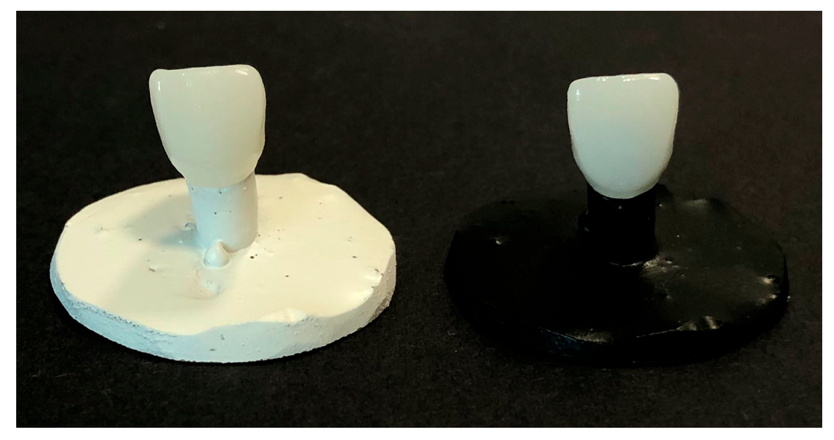
The restorations underwent a 10-min ultrasonic cleaning in distilled water before the measuring process. Two epoxy dies were painted with white (RAL 9010) or black (RAL 9005) acrylic spray varnish to simulate black and white backgrounds. To perform optical measurements, the crowns were placed on the white and black epoxy replicas in order (Figure 2).
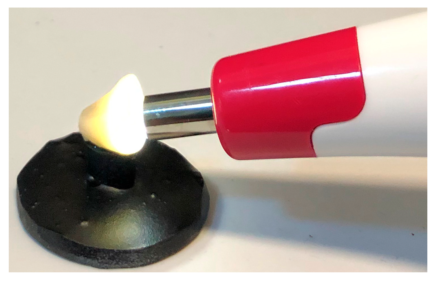
The measurements were performed with a spectrophotometer (Vita Easyshade V Spectrophotometer, Vita, Bäd Sackingen, Germany). The probe was positioned at a 90° angle to the center of the crown’s labial surface (Figure 3).
Three repeated measurements on each crown were made by one investigator in the same location and under D65 standard illumination. Translucency was quantitatively measured by comparing the reflectance of light (Y) through the test specimen over a high-reflectance backing (white backing, Yw) with that of a high-absorbance backing (black backing, Yb). Data were recorded in CIEXYZ colorimetric systems. The instrument output recorded (Yb/w) for each specimen evaluation over the black and white backings was a single number that represented the average of ten successive, automatic measurements. The following formula was used to calculate the contrast ratio [27]:
CR = Yb/Yw, where Y = [(L + 16)/116]3 × 100.
According to this calculation, CR = 0 is transparent, while CR = 1 is completely opaque.

2.3. Statistical Analysis

After measuring the samples’ translucency, comparisons between groups were made using a statistical software program (IBM SPSS Statistics, v24; IBM Corp., Armonk, NY, USA). The distribution of data was evaluated using the Kolmogorov–Smirnov test. For normally distributed data, groups were compared using one-way ANOVA. To perform binary comparisons of the groups, post hoc Tamhane tests were used. Analysis results were presented as means and standard deviation, and a p value < 0.05 was considered significant.

3. Results

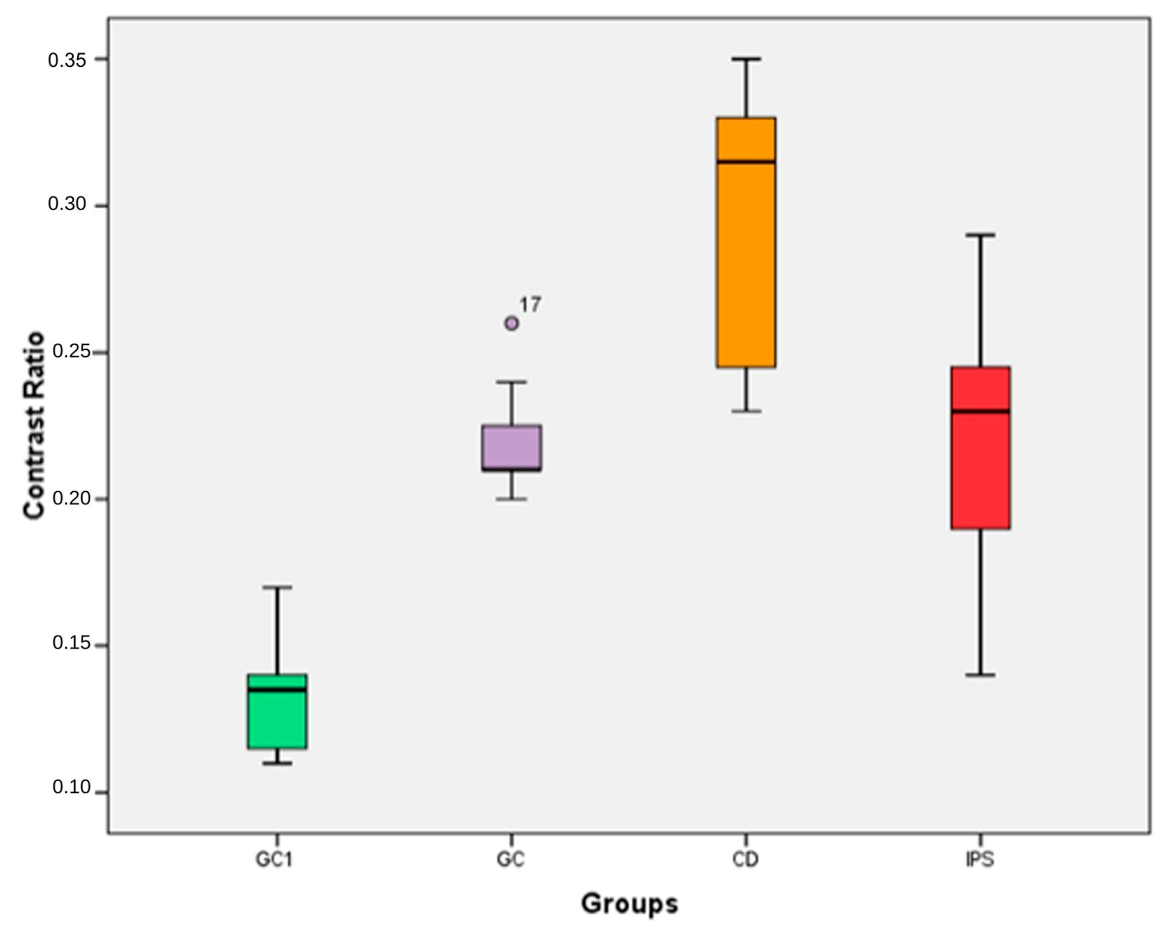
The results showed that GC1 had the lowest CR value (0.13 ± 0.01), the GC and IPS groups had equal values (0.22 ± 0.01, 0.22 ± 0.04 respectively), and the CD group showed the highest CR value (0.29 ± 0.04) (Table 2) (Figure 4).
The CD group had a significantly higher CR value than the other groups (p < 0.01) (Table 3) (Figure 4). The difference between the CR values of GC and IPS groups was not statistically significant. The GC1 group’s CR was significantly lower than the GC group (p < 0.01) (Table 3) (Figure 4).

4. Discussion

This in vitro study compared the translucency of three different CAD/CAM materials. Based on the statistical analysis of the results, the first null hypothesis indicating that there will be no difference of CR values among three different CAD/CAM materials was partially accepted. The second null hypothesis indicating that translucency of translucent monolithic zirconia will not change with reduction of the thickness was rejected. Differences were found in the CR based on type of ceramic material or thickness.
Translucency, masking ability, and lightness (L*) are interdependent optical parameters that play a critical role in the esthetic outcome of anterior restorations. Increased translucency enhances natural light transmission but may compromise masking ability, particularly over darker substrates. In addition, the lightness of the underlining background has probably the strongest effect on the final color of the ceramics [28,29,30]. Therefore, an optimal balance among these parameters is essential when selecting ceramic materials for the anterior region.
Translucency is a significant optical property and an important criteria for choosing materials, particularly for esthetic zone restorations. Natural-looking restorations require optimal translucency with satisfactory durability. Chemical composition, microstructure, particle shape and size, distribution and refractive index of crystalline phase, fabrication processes, and porosity all affect the optical and translucency characteristics of dental ceramics [31,32]. Given that translucency varies with material thickness and an ISO guideline for translucency assessment in dentistry is not yet established [33], the present study adopted a labial thickness of 1.5 mm for the three material groups, while a 1.0 mm labial thickness was used for the translucent zirconia group based on the minimum thickness recommendations provided by the manufacturers.
Regarding the various techniques used for evaluating translucency, there is little consensus in dental research [34]. Translucency is often assessed using either CR or TP [31]. While TP uses color differences, CR is a reflectance value ratio [35]. Evaluation of TP is one of the most widely used methods for assessing how light interacts with dental restorative materials. Although the translucency parameter provides the benefit of a direct visual evaluation of translucency, dental crowns cannot be assessed using this parameter due to their curved surfaces; for this reason, CR was employed in the present study [27]. The CR evaluation was performed on the labial surface of the crowns, and the spectrophotometer recorded changes in reflectance when the background switched from white to black, which were then translated into a translucency value [36].
In restorative dentistry, translucency plays a critical role as it replicates the optical behavior of natural teeth. Materials with a high degree of translucency are necessary for CAD/CAM systems to produce restorations that seem natural [33]. According to Dietschi et al., the mean CR was found to be 0.45 for enamel and 0.65 for dentin [37].
In this study, the mean CR values for GC1, GC, IPS, and CD were 0.13 and 0.22, 0.22 and 0.29, respectively. None of the evaluated materials exhibited translucency levels comparable to those of human enamel (all CR values < 0.45). Although translucency is essential for achieving a natural-looking restoration, excessive translucency may lead to a reduction in lightness, resulting in a grayish appearance [31]. In certain clinical scenarios—such as cases involving a discolored tooth or a cast metal post and core in the anterior esthetic region—translucency may be undesirable, and the use of a prosthetic material with enhanced masking properties and lower translucency becomes necessary [38]. The final color of the restoration is affected by ceramic translucency, restoration thickness, background color, and cement shade [39]. Therefore, in cases where masking of the underlying tooth structure is required, the material thickness should be carefully determined, and both the translucency of the material and the type of luting cement should be appropriately selected.
The translucency threshold for perception evaluated using the CR has been determined in a study by Liu et al. [36]. They stated that half of the participants in the study perceived a translucency difference of 0.06 and for all subjects, and the mean translucency perception threshold was found to be 0.07. A CR difference between 0.06 and 0.07 is the perceptual threshold that is detectable by the human eye [36]. According to the CR values of different brands at the same thickness, the difference between the CR values of IPS and CD as well as the CR difference between GC and CD were both 0.07. The difference in translucency was perceptible according to Liu et al. They also stated that, in order to achieve a translucency difference of 0.06 between two thicknesses of the same brand, there need to be a minimum 0.6 mm increase in thickness between the two [36]. In the current study, even though the thickness difference was 0.5 mm, the difference in CR values between GC1 and GC was 0.09. According to these findings, all the CR differences obtained in this study might be clinically detectable.
In the study of Church et al., the results showed that high-translucent (HT) LDS had significantly greater translucency than the HT zirconia materials at each thickness [40]. In the present study, the translucency levels of GC and IPS were similar. The reason for the difference in our study may be that they evaluated LDS ceramics with high translucency. In contrast with our study, Harada et al. measured the translucency of recently developed translucent zirconia materials and compared them with LDS by producing disk shaped specimens. They concluded that at thicknesses of both 0.5 mm and 1.0 mm, ultra-translucent (UT) zirconia was significantly more translucent than all other zirconia materials, and LT LDS was significantly more translucent than all zirconia materials [41]. Similar to Harada et al., Nassary Zadeh et al. also reported that when compared to zirconia materials, LT LDS samples with a thickness of 1 mm displayed the maximum translucency [42]. The difference in results may be due to comparing different brands of cubic zirconia and ceramic sample forms. In the present study, samples were compared in the form of anterior crowns. Studies using flat discs or tablet-shaped specimens do not represent for optical effects associated to surface forms; hence, the translucency findings collected here may be more in line with clinical observations.
There are limited studies in the literature that evaluated the translucency of ZLS. Similar to our data, Alp et al. reported that LDS presented higher translucency than ZLS before and after coffee thermocycling [43]. According to Porojan et al., who evaluated the TP of 1.5 mm MT ZLS and LDS discs in their study, LDS samples had higher translucency compared to ZLS, but the difference was insignificant [44]. In another study, Sen and Us evaluated the translucency of HT ZLS and LDS discs and stated that ZLS samples were more translucent than the LDS samples. Unlike their findings, in the present study, the translucency of LDS crowns was significantly higher than ZLS samples [5]. The difference of the results may be due to the fact that LT ZLS were used in the current study.
According to the manufacturer, newly produced UHT zirconia with reduced alumina (trace) and enhanced yttria (5.5 mol%; 9 wt%) stimulated the formation of certain cubic and tetragonal zirconia grains, which results in greater translucency. The cubic grains have an isotropic refractive index, which lowers the strong scattering at the grain borders. When isotropic cubic grains are used to replace the majority of tetragonal grains, the translucency of the ceramic is constantly increased [45]. The finding obtained from this research indicated that the translucency of GC1 was noticeably higher than that of GC since translucency decreases with increasing ceramic thickness [46,47]. This result was consistent with the part of the findings of Baldissara et al., who evaluated the CR values of 1.0 and1.5 mm UT monolithic zirconia crowns and founded that 1.0 mm UT monolithic zirconia crowns were more translucent than 1.5 mm UT monolithic zirconia crowns [27].
In addition to having the advantage of a more conservative preparation, a thinner and more translucent crown allows a greater amount of light to pass through the ceramic and positively affects polymerization during cementation with resin-based materials [35]. Therefore, a ceramic that achieves the desired degree of translucency at the minimum restoration thickness should be selected. The minimal thickness recommended by the manufacturers is 1.5 mm for IPS and CD and 1 mm for GC. Here, 1.5 mm GC was included in the present study for comparison with IPS and CD.
According to the current study, GC had comparable translucency to IPS, while GC1 showed better translucency than IPS. The results of the current study were similar with the results of Church et al., who stated that 0.5 mm HT monolithic zirconia samples were more translucent than the 1.0 mm HT LDS samples (IPS e.max CAD HT) [40]. Our findings were also consistent with the study by Baldissara et al. [27]. They stated that 1.0 mm UT monolithic zirconia crowns were more translucent than 1.5 mm LT LDS crowns [27].
One of the strongest aspects of this study is that in vitro studies enable parameters to be evaluated separately under standard conditions. In addition, the fact that the samples were prepared in the form of crowns allowed the effect of surface shapes on the optical properties to be evaluated, so the results obtained better reflect the clinical observations. However, the inclusion of a single ceramic material per manufacturer restricts the validity of the findings to the evaluated commercial systems. Additionally, SEM or XRD analyses were not performed in this study; therefore, the microstructural descriptions are based on previously published literature and manufacturers’ technical documentation [48,49]. Furthermore, the exclusion of the luting cement layer and substrate color constitutes a limitation of this study, as both factors are known to affect the optical properties of translucent restorative materials [39]. Finally, the oral environmental conditions such as the application of restorations to the natural teeth and presence of saliva could not be assessed. These factors represent the limitations of this study.
Further research that includes a wider range of ceramic materials, incorporates microstructural analyses, and better simulates clinical conditions is needed.

5. Conclusions

Within the limitations of this current study, the following conclusions can be drawn:
  • The translucency was affected by the type of CAD/CAM restorative material.
  • LDS and UHT monolithic zirconia presented higher translucency than ZLS.
  • The translucency of UHT monolithic zirconia is highly influenced by the thickness of the material.
  • Owing to its higher translucency, UHT monolithic zirconia appears to be a promising material for anterior esthetic restorations.

Author Contributions

Conceptualization, E.K.; methodology, E.K., H.B.Ö. and M.H.Ö.; validation, H.B.Ö.; formal analysis, H.B.Ö.; investigation, M.H.Ö.; data curation, M.H.Ö.; writing—original draft preparation, H.B.Ö.; writing—review and editing, E.K. and H.B.Ö.; supervision, E.K. All authors have read and agreed to the published version of the manuscript.

Funding

This research received no external funding.

Institutional Review Board Statement

Not applicable.

Informed Consent Statement

Not applicable.

Data Availability Statement

The data that support the findings of this study are available from the corresponding author upon reasonable request.

Acknowledgments

The authors would like to thank Ivoclar Vivadent, Dentsply Sirona, and GC for generously supplying the materials.

Conflicts of Interest

The authors declare no conflicts of interest.

Abbreviations

The following abbreviations are used in this manuscript:
CAD/CAMComputer-aided design/computer-aided manufacturing
LDSLithium disilicate
ZLSZirconia reinforced lithium silicate
UHTUltra-high translucent
LTLow translucent
HTHigh translucent
CRContrast ratio
TPTranslucency parameter

References

  1. Kelly, J.R.; Nishimura, I.; Campbell, S.D. Ceramics in dentistry: Historical roots and current perspectives. J. Prosthet. Dent. 1996, 75, 18–32. [Google Scholar] [CrossRef] [PubMed]
  2. Kim, H.K.; Kim, S.H. Effect of the number of coloring liquid applications on the optical properties of monolithic zirconia. Dent. Mater. 2014, 30, 229–237. [Google Scholar] [CrossRef] [PubMed]
  3. Culp, L.; McLaren, E.A. Lithium disilicate: The restorative material of multiple options. Compend. Contin. Educ. Dent. 2010, 31, 716–720. [Google Scholar] [PubMed]
  4. Basegio, M.M.; Pecho, O.E.; Ghinea, R.; Perez, M.M.; Della Bona, A. Masking ability of indirect restorative systems on tooth-colored resin substrates. Dent. Mater. 2019, 35, 122–130. [Google Scholar] [CrossRef]
  5. Sen, N.; Us, Y.O. Mechanical and optical properties of monolithic CAD-CAM restorative Materials. J. Prosthet. Dent. 2018, 119, 593–599. [Google Scholar] [CrossRef]
  6. Denry, I.; Kelly, J.R. Emerging ceramic-based materials for dentistry. J. Dent. Res. 2014, 93, 1235–1242. [Google Scholar] [CrossRef]
  7. Kelly, J.R.; Benetti, P. Ceramic materials in dentistry: Historical evolution and current practice. Aust. Dent. J. 2011, 56, 84–96. [Google Scholar] [CrossRef]
  8. Van Noort, R. Introduction to Dental Materials, 4th ed.; Mosby/Elsevier: Edinburgh, UK, 2013; pp. 231–245. [Google Scholar]
  9. Giordano, R.; McLaren, E.A. Ceramics overview: Classification by microstructure and processing methods. Compend. Contin. Educ. Dent. 2010, 31, 682–684. [Google Scholar]
  10. Chaiyabutr, Y.; Kois, J.C.; Lebeau, D.; Nunokawa, G. Effect of abutment tooth color, cement color, and ceramic thickness on the resulting optical color of a CAD/CAM glass-ceramic lithium disilicate reinforced crown. J. Prosthet. Dent. 2011, 105, 83–90. [Google Scholar] [CrossRef]
  11. Reich, S. Tooth-colored CAD/CAM monolithic restorations. Int. J. Comput. Dent. 2015, 18, 131–146. [Google Scholar]
  12. Datla, S.R.; Alla, R.K.; Alluri, V.R.; Babu, J.P.; Konakanchi, A. Dental ceramics: Part II—Recent advances in dental ceramics. Am. J. Mater. Eng. Technol. 2015, 3, 19–26. [Google Scholar] [CrossRef]
  13. Awad, D.; Stawarczyk, B.; Liebermann, A.; Ilie, N. Translucency of esthetic dental restorative CAD-CAM materials and composite resins with respect to thickness and surface roughness. J. Prosthet. Dent. 2015, 113, 534–540. [Google Scholar] [CrossRef] [PubMed]
  14. Elsaka, S.E.; Elnaghy, A.M. Mechanical properties of zirconia reinforced lithium silicate glass-ceramic. Dent. Mater. 2016, 32, 908–914. [Google Scholar] [CrossRef] [PubMed]
  15. Celtra® Duo. Zirconia-Reinforced Lithium Silicate (ZLS) Block; Technical Monograph; Dentsply Sirona Inc., DeguDent GmbH: Milford, DE, USA, 2016. Available online: https://assets.dentsplysirona.com/dentsply/microsites/celtra/celtraduo-tech-monograph.pdf (accessed on 20 February 2025).
  16. Vichi, A.; Sedda, M.; Fabian Fonzar, R.; Carrabba, M.; Ferrari, M. Comparison of Contrast Ratio, Translucency Parameter, and Flexural Strength of Traditional and “Augmented Translucency” Zirconia for CEREC CAD/CAM System. J. Esthet. Restor. Dent. 2016, 28, 32–39. [Google Scholar] [CrossRef]
  17. Camposilvan, E.; Leone, R.; Gremillard, L.; Sorrentino, R.; Zarone, F.; Ferrari, M.; Chevalier, J. Aging resistance, mechanical properties and translucency of different yttria- stabilized zirconia ceramics for monolithic dental crown applications. Dent. Mater. 2018, 34, 879–890. [Google Scholar] [CrossRef]
  18. Bona, A.D.; Nogueira, A.D.; Pecho, O.E. Optical properties of CAD/CAM ceramic systems. J. Dent. 2014, 42, 1202–1209. [Google Scholar] [CrossRef]
  19. Yu, B.; Ahn, J.S.; Lee, Y.K. Measurement of translucency of tooth enamel and dentin. Acta Odontol. Scand. 2009, 67, 57–64. [Google Scholar] [CrossRef]
  20. Yu, B.; Lee, Y.K. Color difference of all-ceramic materials by the charge of illuminants. Am. J. Dent. 2009, 22, 73–78. [Google Scholar]
  21. Tuncel, I.; Turp, I.; Üşümez, A. Evaluation of translucency of monolithic zirconia and framework zirconia materials. J. Adv. Prosthodont. 2016, 8, 181–186. [Google Scholar] [CrossRef]
  22. Pop-Ciutrila, I.S.; Dudea, D.; Eugenia Badea, M.; Moldovan, M.; Cimpean, S.I.; Ghinea, R. Shade correspondence, color, and translucency differences between human dentine and a CAD/CAM hybrid ceramic system. J. Esthet. Restor. Dent. 2016, 28, 46–55. [Google Scholar] [CrossRef]
  23. Espinar, C.; Bona, A.D.; Pérez, M.M.; Tejada-Casado, M.; Pulgar, R. The influence of printing angle on color and translucency of 3D printed resins for dental restorations. Dent. Mater. Off. Publ. Acad. Dent. Mater. 2023, 39, 410–417. [Google Scholar] [CrossRef] [PubMed]
  24. Johnston, W.M.; Ma, T.; Kienle, B.H. Translucency parameter of colorants for maxillofacial prostheses. Int. J. Prosthodont. 1995, 8, 79–86. [Google Scholar] [PubMed]
  25. Miyagawa, Y.; Powers, J.M.; O’Brien, W.J. Optical properties of direct restorative materials. J. Dent. Res. 1981, 60, 890–894. [Google Scholar] [CrossRef] [PubMed]
  26. Zahran, M.; El-Mowafy, O.; Tam, L.; Watson, P.A.; Finer, Y. Fracture Strength and Fatigue Resistance of All-Ceramic Molar Crowns Manufactured with CAD/CAM Technology. J. Prosthodont. 2008, 17, 370–377. [Google Scholar] [CrossRef]
  27. Baldissara, P.; Wandscher, V.F.; Marchionatti, A.M.E.; Parisi, C.; Monaco, C.; Ciocca, L. Translucency of IPS e.max and cubic zirconia monolithic crowns. J. Prosthet. Dent. 2018, 120, 269–275. [Google Scholar] [CrossRef]
  28. Skyllouriotis, A.L.; Yamamoto, H.L.; Nathanson, D. Masking properties of ceramics for veneer restorations. J. Prosthet. Dent. 2017, 118, 517–523. [Google Scholar] [CrossRef]
  29. Ayata, M.; Kilic, K.; Al-Haj Husain, N.; Ozcan, M. Effect of thickness and translucency on color change and masking ability of ceramic materials used for laminate veneers. Eur. J. Prosthodont. Restor. Dent. 2023, 31, 383–390. [Google Scholar] [CrossRef]
  30. Pala, K.; Reinshagen, E.M.; Attin, T.; Hüsler, J.; Jung, R.E.; Ioannidis, A. Masking capacity of minimally invasive lithium disilicate restorations on discolored teeth-The impact of ceramic thickness, the material’s translucency, and the cement color. J. Esthet. Restor. Dent. 2024, 36, 107–115. [Google Scholar] [CrossRef]
  31. Vichi, A.; Carrabba, M.; Paravina, R.; Ferrari, M. Translucency of ceramic materials for CEREC CAD-CAM system. J. Esthet. Restor. Dent. 2014, 26, 224–231. [Google Scholar] [CrossRef]
  32. Nejatidanesh, F.; Azadbakht, K.; Savabi, O.; Sharifi, M.; Shirani, M. Effect of repeated firing on the translucency of CAD-CAM monolithic glass-ceramics. J. Prosthet. Dent. 2020, 123, 530.e1–530.e6. [Google Scholar] [CrossRef]
  33. Vichi, A.; Balestra, D.; Scotti, N.; Louca, C.; Paolone, G. Translucency of CAD/CAM and 3D printable composite materials for permanent dental restorations. Polymers 2023, 15, 1443. [Google Scholar] [CrossRef]
  34. Nogueira, A.D.; Della Bona, A. The effect of a coupling medium on color and translucency of CAD-CAM ceramics. J. Dent. 2013, 41, 18–23. [Google Scholar] [CrossRef] [PubMed]
  35. Sulaiman, T.A.; Abdulmajeed, A.A.; Donovan, T.E.; Ritter, A.V.; Vallittu, P.K.; Närhi, T.O.; Lassila, L.V. Optical properties and light irradiance of monolithic zirconia at variable thicknesses. Dent. Mater. 2015, 31, 1180–1187. [Google Scholar] [CrossRef] [PubMed]
  36. Liu, M.C.; Aquilino, S.A.; Lund, P.S.; Vargas, M.A.; Diaz-Arnold, A.M.; Gratton, D.G.; Qian, F. Human perception of dental porcelain translucency correlated to spectrophotometric measurements. J. Prosthodont. 2010, 19, 187–193. [Google Scholar] [CrossRef] [PubMed]
  37. Dietschi, D.; Ardu, S.; Krejci, I. A new shading concept based on natural tooth color applied to direct composite restorations. Quintessence Int. 2006, 37, 91–102. [Google Scholar]
  38. Alfouzan, A.F.; Al-Otaibi, H.N.; Labban, N.; Al Taweel, S.M.; Al-Tuwaijri, S.; Algazlan, A.S.; Tashkandi, E.A. Effects of thickness and background color on the translucency of CAD/CAM ceramic materials. Ceram. Silikáty 2020, 64, 418–422. [Google Scholar]
  39. Zhu, J.; Xia, Y.; Lui, S.; Wang, W.; Liang, S.; Huang, C. Masking ability of CAD-CAM resin-matrix ceramics with different translucencies and thicknesses combined with four cement shades against varying background colors when facing veneer restorations. BMC Oral Health 2024, 24, 1198. [Google Scholar] [CrossRef]
  40. Church, T.D.; Jessup, J.P.; Guillory, V.L.; Vandewalle, K.S. Translucency and strength of high-translucency monolithic zirconium oxide materials. Gen. Dent. 2017, 65, 48–52. [Google Scholar]
  41. Harada, K.; Raigrodski, A.J.; Chung, K.H.; Flinn, B.D.; Dogan, S.; Mancl, L.A. A comparative evaluation ofthe translucency of zirconias and lithium disilicate for monolithic restorations. J. Prosthet. Dent. 2016, 116, 257–263. [Google Scholar] [CrossRef]
  42. Nassary Zadeh, P.; Lümkemann, N.; Sener, B.; Eichberger, M.; Stawarczyk, B. Flexural strength, fracture toughness, and translucency of cubic/tetragonal zirconia materials. J. Prosthet. Dent. 2018, 120, 948–954. [Google Scholar] [CrossRef]
  43. Alp, G.; Subasi, M.G.; Johnston, W.M.; Yilmaz, B. Effect of surface treatments and coffee thermocycling on the color and translucency of CAD-CAM monolithic glass-ceramic. J. Prosthet. Dent. 2018, 120, 263–268. [Google Scholar] [CrossRef]
  44. Porojan, L.; Vasiliu, R.D.; Bîrdeanu, M.I.; Porojan, S.D. Surface characterization and optical properties of reinforced dental glass-ceramics related to artificial aging. Molecules 2020, 25, 3407. [Google Scholar] [CrossRef]
  45. Zhang, Y. Making yttria-stabilized tetragonal zirconia translucent. Dent. Mater. 2014, 30, 1195–1203. [Google Scholar] [CrossRef]
  46. Ilie, N.; Stawarczyk, B. Quantification of the amount of blue light passing through monolithic zirconia with respect to thickness and polymerization conditions. J. Prosthet. Dent. 2015, 113, 114–121. [Google Scholar] [CrossRef]
  47. Wang, F.; Takahashi, H.; Iwasaki, N. Translucency of dental ceramics with different thicknesses. J. Prosthet. Dent. 2013, 110, 14–20. [Google Scholar] [CrossRef]
  48. Romanyk, D.L.; Guo, Y.; Rae, N.; Veldhuis, S.; Sirovica, S.; Fleming, G.J.; Addison, O. Strength-limiting damage and its mitigation in CAD-CAM zirconia-reinforced lithium-silicate ceramics machined in a fully crystallized state. Dent. Mater. Off. Publ. Acad. Dent. Mater. 2020, 36, 1557–1565. [Google Scholar] [CrossRef]
  49. Čokić, S.M.; Cóndor, M.; Vleugels, J.; Meerbeek, B.V.; Oosterwyck, H.V.; Inokoshi, M.; Zhang, F. Mechanical properties-translucency-microstructure relationships in commercial monolayer and multilayer monolithic zirconia ceramics. Dent. Mater. Off. Publ. Acad. Dent. Mater. 2022, 38, 797–810. [Google Scholar] [CrossRef]
Figure 1. Design of anterior crowns.
Applsci 16 00663 g001
Figure 2. Epoxy resin dies simulating black and white backgrounds.
Applsci 16 00663 g002
Figure 3. Measuring the reflectance of the crowns.
Applsci 16 00663 g003
Figure 4. CR values of the groups. GC1: GC Initial UHT Zr 1 mm, GC: Initial UHT Zr 1.5 mm, CD: Celtra Duo LT 1.5 mm, IPS: IPS e.max CAD LT 1.5 mm.
Applsci 16 00663 g004
Table 1. Study groups, materials used in the study, contents, and manufacturers.
CAD/CAM Materials and GroupsContentManufacturer
IPS e.max CAD, LT
IPS: 1.5 mm Lithium Disilicate
SiO257–80%Ivoclar Vivadent, Schaan, Liechtenstein
Li2O11–19%
ZrO20.8%
and others
Celtra Duo, LT
CD: 1.5 mm Zirconia-Reinforced Lithium Silicate
SiO256–64%Sirona, Dentsply, Bensheim, Germany
Li2O15–21%
ZrO210%
and others
GC Initial UHT
GC: 1.5 mm Monolithic Zirconia
GC1: 1.0 mm Monolithic Zirconia
ZrO290%GC, GC Europe N.V, Leuven, Belgium
Y2O35.5%
Al2O3trace
and others
Table 2. Mean ± SD and medians obtained with CR analysis a.
GroupMean ± SD bMedian
GC10.13 ± 0.01 A0.13
GC0.22 ± 0.01 B0.21
IPS0.22 ± 0.04 B0.23
CD0.29 ± 0.04 C0.31
GC1: GC Initial UHT Zr 1 mm, GC: Initial UHT Zr 1.5 mm, CD: Celtra Duo LT 1.5 mm, IPS: IPS e.max CAD LT 1.5 mm. a A value of 0 means most translucent; a value of 1 indicates most opaque. b Different superscript uppercase letters indicate statistical differences of materials according to one-way ANOVA with Tamhane post hoc test. (p < 0.05).
Table 3. Comparison of samples’ CR values.
GROUPp95% Confidence Interval
Lower BoundUpper Bound
GC1GC<0.001 *−0.1083−0.0667
CD<0.001 *−0.2066−0.1201
IPS<0.001 *−0.1305−0.0445
GCGC1<0.001 *−0.06670.1083
CD0.001 *−0.1191−0.0326
IPS1.000−0.04290.0429
CDGC1<0.001 *−0.12010.2066
GC0.001 *−0.03260.1191
IPS0.003 *−0.02200.1297
IPSGC1<0.001 *−0.04450.1305
GC1.000−0.04290.0429
CD0.003 *−0.1297−0.0220
GC1: GC Initial UHT Zr 1 mm, GC: Initial UHT Zr 1.5 mm, CD: Celtra Duo LT 1.5 mm, IPS: IPS e.max CAD LT 1.5 mm. * Statistically significant difference (p < 0.05) with the post hoc Tamhane test.
Disclaimer/Publisher’s Note: The statements, opinions and data contained in all publications are solely those of the individual author(s) and contributor(s) and not of MDPI and/or the editor(s). MDPI and/or the editor(s) disclaim responsibility for any injury to people or property resulting from any ideas, methods, instructions or products referred to in the content.