Article

Phytochemical Characterization of Schinus terebinthifolia Leaf Phytobiotics and Their Potential as Natural Antibiotic Adjuvants Against Carbapenem-Resistant Bacteria

by
Fatima Mourabiti
1,*,
Fatimazahra Jouga
1,
José Luis Rodríguez Rama
2,
Abdelhakim Elmakssoudi
3,
Souraya Sakoui
1,
Lorena G. Calvo
2,
Abdelaziz Soukri
1,
Trinidad de Miguel
2 and
Bouchra El Khalfi
1,*
1
Laboratory of Health, Environment and Biotechnology, Team of Physiopathology, Molecular Genetics and Biotechnology, Faculty of Sciences Ain Chock, Hassan II University of Casablanca, Casablanca 20100, Morocco
2
Department of Microbiology and Parasitology, Universidade de Santiago de Compostela, E-15782 Santiago de Compostela, Spain
3
Laboratory of Molecular Synthesis, Extraction Valorization, Faculty of Sciences Ain Chock, Hassan II University of Casablanca, Casablanca 20100, Morocco
*
Authors to whom correspondence should be addressed.
Appl. Sci. 2026, 16(18), 9212; https://doi.org/10.3390/app16189212
Submission received: 10 August 2026 / Revised: 10 September 2026 / Accepted: 11 September 2026 / Published: 17 September 2026
(This article belongs to the Special Issue Synthesis and Biological Evaluation of New Compounds)

Abstract

The emergence of carbapenem-resistant Gram-negative bacteria has intensified the search for natural compounds capable of restoring antibiotic efficacy by targeting β-lactamase-mediated resistance. This study investigated the antibacterial, β-lactamase inhibitory, and in silico potential of Schinus terebinthifolia leaf phytobiotics. Soxhlet (STS), maceration (STM), and essential oil (STEOL) extracts were evaluated against carbapenem-resistant Escherichia coli, Klebsiella pneumoniae, and Pseudomonas aeruginosa using agar diffusion, broth microdilution, checkerboard, and colorimetric β-lactamase inhibition assays. In parallel, the phytochemicals identified by GC–MS and LC–MS were virtually screened against the VIM and OXA-48-like β-lactamases using PyRx, followed by drug-likeness prediction using SwissADME. All phytobiotics exhibited significant antibacterial activity and synergistically enhanced the efficacy of cefotaxime against the majority of the tested bacterial strains (ΣFICI ≤ 0.5). Moreover, they markedly inhibited β-lactamase activity, with complete inhibition achieved at MIC/2. Among the tested phytobiotics, STS exhibited the greatest β-lactamase inhibitory potency, with an IC50 of 49.8 μg/mL. Virtual screening identified diosmetin, catechin, quercetin, ellagic acid, and epicatechin gallate as the most promising candidate β-lactamase inhibitors, exhibiting strong binding affinities toward VIM and OXA-48-like β-lactamases. SwissADME analysis further indicated favorable drug-like properties for the top-ranked compounds. Collectively, these findings suggest that S. terebinthifolia phytobiotics have promising antibacterial and β-lactamase-inhibitory potential and may represent a source of candidate compounds for further investigation as antibiotic adjuvants against carbapenem-resistant Gram-negative pathogens.

1. Introduction

β-Lactams are among the most widely used and diverse classes of antibiotics. Their mode of action relies on interacting with and blocking penicillin-binding proteins (PBPs), which are implicated in the final steps of cell wall formation [1]. Carbapenems are a critical class of β-lactams, often employed as the ultimate therapeutic option against multidrug-resistant bacteria [2]. Carbapenems are structurally different from other β-lactams and have a broad range of antimicrobial activity and high strength against β-lactamases [2]. However, since the 2000s, the global rise in carbapenem-resistant microorganisms has become a major public health concern [3]. Resistance is particularly mediated by the production of β-lactamases, reduced membrane permeability, and efflux pumps, all of which significantly compromise therapeutic efficacy [4]. Therefore, the World Health Organization (WHO) recently identified ESKAPE bacteria (Enterococcus faecium, Staphylococcus aureus, Klebsiella pneumoniae, Acinetobacter baumannii, Pseudomonas aeruginosa, and Enterobacter species) as being in the critical group as one of the most significant pathogens driving the need for research, development, and strategic interventions to combat antimicrobial resistance [5,6]. In 2022, antimicrobial resistance led to an estimated five million deaths across the world, highlighting the grave impact of infections caused by Enterobacterales [7]. This growing concern highlights that carbapenem resistance constitutes a critical global health challenge, with particularly alarming implications in tropical regions [8].
In response to the rising threat of carbapenem resistance, it has become increasingly urgent to explore new antibiotics. Researchers have now shifted their focus from traditional microbial sources to alternative ones, particularly plants. Natural extracts have emerged as promising alternatives to conventional antimicrobials due to their low toxicity and multitargeting activity [9,10]. Phytobiotics are bioactive plant-derived substances, including plant extracts, essential oils, and phytochemicals such as flavonoids, phenolic acids, alkaloids, and terpenoids, that exhibit diverse biological activities, particularly antimicrobial effects. Their ability to inhibit microbial growth and potentially enhance antibiotic activity has stimulated interest in their investigation as natural antibiotic adjuvants in the context of antimicrobial resistance [11]. Their broad-spectrum activity stems from their wide diversity of secondary metabolites [12].
Hence, research on natural products is increasing, particularly in the identification and characterization of phytochemicals and plant species with antimicrobial activity, primarily against carbapenem-resistant bacteria. The flora of Brazil is particularly rich, with many species belonging to families such as Anacardiaceae, Piperaceae, Meliaceae, Euphorbiaceae, Fabaceae, Amaranthaceae, Rubiaceae, Malvaceae, Malpighiaceae, Annonaceae, Sapindaceae, and Myrtaceae. These botanical groups are known to be valuable sources of diverse phytochemicals with promising antibacterial properties [13]. However, it is essential to identify and characterize the species with antimicrobial efficacy within these families [13]. Schinus terebinthifolia Raddi is a Brazilian medicinal plant known for its diverse phytochemicals and antimicrobial properties [13]. Several studies have demonstrated the antimicrobial potential of S. terebinthifolia, supporting further investigation of this species as a source of bioactive agents [14]. Mu et al. reported that extracts from Schinus terebinthifolia exhibited strong activity against Gram-negative bacteria and inhibited the growth of Gram-positive food-associated microorganisms [15]. Similarly, the leaf essential oil from Zimbabwe showed inhibitory activity against a broad range of bacteria, including Pseudomonas aeruginosa, Yersinia enterocolitica, Escherichia coli, Acinetobacter calcoaceticus, Klebsiella pneumoniae, and Bacillus subtilis. This activity has been associated, at least in part, with monoterpenes, which can disrupt bacterial membrane integrity by increasing membrane permeability and fluidity, altering membrane-associated proteins, and disturbing cellular respiration and ion homeostasis [16].
Therefore, the objective of our research was to investigate the antimicrobial effects of the essential oil and two leaf extracts of S. terebinthifolia against reference and carbapenem-resistant Gram-negative bacteria, including Klebsiella pneumoniae, Escherichia coli, and Pseudomonas aeruginosa, particularly carbapenemase-producing isolates associated with human nosocomial infections. The synergistic effects of cefotaxime combined with S. terebinthifolia phytobiotics, together with their β-lactamase inhibitory activity, were evaluated to further investigate their potential as antibiotic adjuvants. Moreover, in silico analyses were performed to explore the potential interactions of phytochemical constituents with clinically relevant β-lactamases and provide mechanistic insights into the observed inhibitory activity. Although S. terebinthifolia has been traditionally used for medicinal purposes in several countries, in Morocco the plant is primarily encountered as an ornamental species, and its potential antimicrobial benefits remain largely unexplored. We therefore sought to investigate its potential as a source of natural antibiotic adjuvants against multidrug-resistant Gram-negative bacteria. Importantly, the novelty of this study does not lie in the identification of new phytochemical compounds but rather in providing integrated evidence linking the phytochemical profile of S. terebinthifolia with antibacterial activity, antibiotic potentiation, and β-lactamase inhibition against clinically relevant carbapenem-resistant pathogens. To our knowledge, this is the first study to comprehensively evaluate the potential of S. terebinthifolia leaf phytobiotics against carbapenemase-producing Gram-negative bacteria using a combined in vitro and in silico approach. This work provides new insights into the potential application of this traditionally underexplored Moroccan ornamental plant as a source of natural antibiotic adjuvants and contributes to the search for alternative therapeutic strategies in regions heavily affected by multidrug-resistant Gram-negative infections.

2. Materials and Methods

2.1. Botanical Material

The leaves of S. terebinthifolia Raddi were collected in November 2022 from the Faculty of Science Ain Chock, Hassan II University, Casablanca, Morocco (geographic coordinates: 33.5454° N, 7.6565° W). The plant samples were botanically identified based on similarity to the Herbarium of USAMV Cluj-Napoca CLA under sample code No. 30358. We rapidly washed the Brazilian pepper leaves with tap water and dried them on a paper towel. After air-drying in a dark environment for two weeks, the plant samples were finely ground with an electric blender. After grinding, the powdered leaves were stored in airtight containers at room temperature and protected from light until extraction.

2.2. Extraction of Leaves of Schinus terebinthifolia

Plant material was extracted using two methods: Soxhlet extraction and maceration. Approximately 62 g of the powder was weighed and extracted with ethanol for 6 h in Soxhlet equipment, and another roughly 62 g of leaf powder was extracted using maceration at room temperature on a magnetic stirrer for 72 h. Extracts were filtered through Whatman No.1 filter paper; the filtrate was concentrated, the solvent was removed, and it was then concentrated on a rotovap under reduced pressure at 79 °C. The extraction yield was expressed as the fraction of the extract mass over the dry mass of the plant material for the two ethanolic extracts. Yields were approximately 20.22% for the extract obtained by maceration (STM) and 26.82% for the one obtained by Soxhlet extraction (STS). The concentrated extract was stored at 4 °C until further analysis.
Essential oil (EO) was obtained from 200 g of S. terebinthifolia leaves by hydrodistillation using a Clevenger-type apparatus. The plant material was distilled with 1.5 L of water for 3 h. The EO yield was calculated on a dry-weight basis by dividing the mass of recovered EO by the dry mass of plant material subjected to extraction. Following extraction, the EO was stored at 4 °C until chemical and biological analyses.

2.3. Chemical Composition of Schinus terebinthifolia Photobiotics

2.3.1. Total Phenolic Content

Total phenolic content (TPC) was assessed in the two ethanolic extracts using the Folin–Ciocalteu assay, following the procedure reported by Dirar et al. [17], with minor modifications. Briefly, each sample was reconstituted in 3 mL of methanol/water (50:50, v/v). The resulting suspensions were subjected to ultrasound-assisted extraction at 25 °C for 10 min at a frequency of 50 kHz. After obtaining homogeneous extracts, the supernatants were passed through 0.22 µm PTFE (polytetrafluoroethylene) membrane filters. A gallic acid (GA) calibration curve was used for quantification. TPC values were calculated from the corresponding GA calibration equation and reported as milligrams of gallic acid equivalents per gram of extract (mg GAE/g extract).

2.3.2. Liquid Chromatography–Tandem Mass Spectrometry Analysis

Polyphenolic compounds present in the S. terebinthifolia extracts were identified and quantified by liquid chromatography–tandem mass spectrometry (LC-MS/MS), according to the method described previously [18]. Analyses were conducted using a Thermo Scientific system (San José, CA, USA) consisting of a TSQ Quantum Ultra™ triple-quadrupole mass spectrometer fitted with a heated electrospray ionization source (HESI-II) and an Accela Open autosampler equipped with a 20 µL injection loop. Chromatographic separation was performed on a Kinetex C18 column (100 × 2.1 mm, 2.6 µm, 100 Å; Phenomenex) maintained at 50 °C. The mobile phase comprised water (solvent A) and methanol (solvent B), both containing 0.1% formic acid. The gradient program began at 5% B, followed by an increase to 90% B over 11 min. This composition was maintained for an additional 3 min before returning to the initial conditions over 9 min. The flow rate was 0.2 mL min−1, and 10 µL of sample was injected for each analysis, giving a total chromatographic run time of 25 min.
The mass spectrometer was operated in both positive- and negative-ionization modes, with two or three MS/MS transitions monitored for each target compound. External calibration curves were prepared in water over the concentration range of 0.1–10 mg L−1 for quantitative determination. The calibration models showed good linearity, with coefficients of determination (R2) greater than 0.9952 for all analyzed compounds.

2.3.3. Gas Chromatography/Mass Spectrometry

The volatile constituents of the EO were characterized by gas chromatography–mass spectrometry (GC-MS). The analyses were performed on a Shimadzu GC-2010 Plus gas chromatograph equipped with a flame ionization detector (FID) and a BP-5 capillary column (30 m × 0.25 mm i.d.; film thickness, 0.25 µm; SGE Ltd, Melbourne, Australia), coupled to a Shimadzu QP2010 Plus mass spectrometer and operated using software version 2.50 SU1.
The chromatographic conditions were as follows: transfer-line temperature, 300 °C; ion-source temperature, 200 °C; helium as the carrier gas at a linear velocity of 36.5 cm s−1; split ratio, 1:40; electron ionization energy, 70 eV; mass range, 40–400 u; and scan time, 1 s. Compound assignment was based primarily on retention indices calculated relative to C9–C20 n-alkanes analyzed under the same chromatographic conditions. Identification was further supported by matching the obtained mass spectra against the Shimadzu and NIST05 spectral databases, as well as an in-house library. The latter was established using reference essential oils, laboratory-synthesized compounds, commercially available standards, and previously reported spectral and chromatographic data [19].

2.4. Characterization of the Antibacterial Activity of S. terebinthifolia Phytobiotics

2.4.1. Bacterial Strains and Culture Media

Six bacterial isolates were employed in the biological effect assays: three ATCC bacteria (Pseudomonas aeruginosa ATCC27853 (P. aeruginosa), Klebsiella pneumoniae ATCC13883 (K. pneumoniae), and E. coli ATCC25922) and three clinical isolates resistant to carbapenems (P. aeruginosa, K. pneumoniae, and E. coli). They were obtained from urinary tract infections, characterized as carbapenemase-producing Gram-negative bacteria, preserved in glycerol, and stored at −20 °C. All strains were routinely subcultured on various culture media, such as Cetrimide agar, MacConkey agar plates, and Luria–Bertani broth, and isolate identification was reconfirmed. Culture media were purchased from Oxoid (Basingstoke, UK).

2.4.2. Well Diffusion Method

The antibacterial effect of the three leaf phytobiotics (STEOL, STS and STM) against the six pathogenic bacteria, including the reference and the clinical strains, was determined using the agar diffusion test as outlined in previous studies [20]. Briefly, Petri plates containing Mueller–Hinton agar (MHA) were inoculated with a 106 CFU/mL fresh bacterial suspension. After the agar had solidified, 5 mm diameter wells were carefully formed. Then, 70 μL of each phytobiotic preparation was added to each well (10,000 µg/mL for STEOL, 10,000 µg/mL for STM, and 15,000 µg/mL for STS). The STS and STM extracts were diluted in ethanol, whereas the essential oil (STEOL) was dissolved and diluted in DMSO to obtain the required concentrations. The corresponding solvent controls were included in the assay, and meropenem was included as the reference antibiotic for comparison. All plates were maintained at 4 °C for 4 h and then incubated at 37 °C for 24 h. The inhibition zone diameter (IZD) of bacterial growth around the well was measured at three different points in the circular zones. The experiments were repeated three times.

2.4.3. Assessment of Antibacterial Activity Using the AlamarBlue Assay

The broth microdilution method was used to determine the minimum inhibitory concentration (MIC) of the S. terebinthifolia phytobiotics against the six bacterial strains included in this study, according to the guidelines of the Clinical and Laboratory Standards Institute [21]. The assays were performed in sterile 96-well microtiter plates using two-fold serial dilutions of the phytobiotics at concentrations of 10, 5, 2.5, 1.25, 0.6, 0.3, 0.15, and 0.07 mg/mL. DMSO and ethanol were used as solvents, and the corresponding solvent controls were included to verify that the solvents themselves did not affect bacterial growth. A growth control containing the bacterial inoculum without phytobiotics was also included. In addition, an Alamar Blue (resazurin) assay was performed to assess the effect of S. terebinthifolia phytobiotics on the metabolic activity of the studied strains, based on a modification of a previously described method [22]. The MIC was considered the lowest phytobiotic concentration at which no visible bacterial growth was detected. Bacterial metabolic activity was monitored by measuring the fluorescence generated by the reduction of resazurin to resorufin. Fluorescence was recorded with a FLUOstar Omega microplate reader (BMG Labtech, Ortenberg, Germany) at 544 nm excitation and 590 nm emission wavelengths. MIC values were reported in µg/mL, taking. The concentration producing 50% inhibition of bacterial metabolic activity (IC50) was estimated using GraphPad Prism version 8.0.2. Each experiment was conducted in triplicate.

2.4.4. Minimum Bactericidal Concentration Determination

The minimum bactericidal concentration (MBC) was determined by subculturing samples from MIC wells in which no visible bacterial growth was detected. Aliquots were transferred onto Mueller–Hinton agar (MHA) plates and incubated at 37 °C for 24 h. The MBC was recorded as the lowest extract concentration that resulted in a complete absence of visible colony growth following incubation [22].

2.5. Evaluation of the Interaction Between S. terebinthifolia Phytobiotics and Cefotaxime

The interaction between the S. terebinthifolia phytobiotics and cefotaxime (CTX) was investigated by the checkerboard microdilution method in 96-well plates, based on a previously described procedure [23] with minor modifications. Cefotaxime was selected as the β-lactam partner because the tested isolates exhibited resistance to this antibiotic, allowing us to assess whether the extracts and essential oil could enhance its antibacterial activity. We tested S. terebinthifolia extract and essential oil concentrations (2× MIC, MIC, MIC/2, MIC/4, MIC/8, and MIC/16) in combination with six CTX concentrations ranging from 0.003 to 0.1 mg/mL. The bacterial inoculum was adjusted to 1.5 × 10⁶ CFU/mL, and the plates were incubated at 37 °C for 24 h. Following incubation, bacterial viability was assessed using resazurin. Optical density and fluorescence measurements were obtained at 544/590 nm with a FLUOstar Omega microplate reader (BMG Labtech). Three independent experiments were performed.
The fractional inhibitory concentration index (FICI) was determined according to
ΣFICI = FIC(A) + FIC(B)
where FIC(A) corresponds to the MIC of the S. terebinthifolia phytobiotic in combination divided by its MIC when tested alone, while FIC(B) represents the MIC of CTX in combination divided by the MIC of CTX alone. The interactions were interpreted as synergistic when ΣFICI ≤ 0.5, additive when 0.5 < ΣFICI < 1.0, non-interactive when 1 ≤ ΣFICI < 4, and antagonistic when ΣFICI ≥ 4.0 [23].

2.6. Evaluation of the β-Lactamase Inhibitory Effect of S. terebinthifolia Phytobiotics

The ability of the S. terebinthifolia extracts to inhibit β-lactamase-mediated hydrolysis was evaluated using the Colorimetric β-Lactamase Activity Assay Kit (AAT Bioquest, Pleasanton, CA, USA), according to the manufacturer’s protocol. The standardized enzyme provided with the kit was used as the β-lactamase source. Reactions were conducted in 96-well microplates and incubated at 37 °C, followed by measurement with a microplate reader.
The extent of β-lactam hydrolysis and enzyme inhibition was calculated from the optical density measurements using the following equations:
% inhibition = [(ODcontrol − ODtreated)/ODcontrol] × 100
% hydrolysis = 100 − % inhibition

2.7. Molecular Docking Study

2.7.1. Protein Preparation

Crystal structures of the β-lactamase targets were retrieved from the RCSB Protein Data Bank in PDB format. VIM and OXA-48-like β-lactamases were selected as representative targets because of their importance in carbapenem resistance among Gram-negative bacteria and their relevance to the resistant species examined in this study (K. pneumoniae, E. coli, and P. aeruginosa) [24]. The corresponding PDB structures used for docking were VIM (PDB ID: 8HXO) and OXA-48-like (PDB ID: 7AW5).
Before molecular docking, the protein structures were prepared by removing crystallographic water molecules, co-crystallized ligands, and other non-protein heteroatoms. Polar hydrogen atoms and Kollman charges were added, and the receptors were converted into PDBQT format using PyRx. The active binding pockets and interacting amino acid residues were identified using the DoGSiteScorer web server [25], and these residues were used to define the docking grid.

2.7.2. Ligand Preparation

Of the 39 phytochemicals identified in the ethanolic extracts (Soxhlet and maceration) and essential oil from S. terebinthifolia leaves, 38 were subjected to virtual screening to evaluate their potential as β-lactamase inhibitors. The three-dimensional chemical structures of the selected compounds were downloaded from the PubChem database (https://pubchem.ncbi.nlm.nih.gov/) in SDF format. The ligands were imported into PyRx 0.8, where their geometries were energy-minimized using the Open Babel module. The optimized structures were subsequently converted into PDBQT format for molecular docking.

2.7.3. Virtual Screening and Molecular Docking Simulations

Virtual screening and molecular docking calculations were carried out in PyRx v0.8 using the AutoDock Vina 1.1.2 engine. The prepared ligands were individually evaluated against the VIM and OXA-48-like β-lactamase structures. For each target, the docking region was defined according to the experimentally characterized binding site of the co-crystallized reference ligand or inhibitor. The catalytic pocket was further localized using DoGSiteScorer, and the resulting coordinates were used to establish the docking grid.
For VIM, the grid was centered at X = 11.7385, Y = 1.7180, and Z = 32.3516, with dimensions of 23.8230 × 25.3451 × 22.8063 Å.
For OXA-48-like β-lactamase, the grid center was positioned at X = 1.2402, Y = −5.7259, and Z = 58.3599 Å, with dimensions of 22.3426 × 27.1057 × 29.0488 Å.
Docking calculations were conducted using the standard AutoDock Vina settings available in PyRx. For each ligand, the resulting docking score was reported as the predicted binding affinity in kcal/mol. The highest-ranked pose was retained for subsequent interaction analysis. Ligand–protein interactions were examined with BIOVIA Discovery Studio Visualizer 2021, including hydrogen bonding, hydrophobic contacts, π-interactions, and the corresponding amino acid residues. The same software was used to generate the two-dimensional interaction and three-dimensional representations of the docked complexes.

2.7.4. Reference/Co-Crystallized Inhibitors and Validation of the Docking Protocol

The co-crystallized β-lactamase inhibitors, namely 2-amino-5-isobutylthiazole-4-carboxylic acid and 4-[(E)-[3-(4-chlorophenyl)-5-sulfanylidene-1H-1,2,4-triazol-4-yl]iminomethyl]benzoic acid, were used as reference ligands to benchmark the docking results. These compounds were used as structural benchmarks because they were co-crystallized with the corresponding β-lactamases in experimentally determined crystal structures.
The reliability of the docking protocol was validated by redocking the co-crystallized ligand into the active site of each enzyme. The predicted binding pose was compared with the corresponding crystallographic conformation, resulting in RMSD values of 0.703 Å for OXA-48-like β-lactamases and 1.013 Å for VIM. Both values were below the commonly accepted threshold of 2 Å, confirming the reliability and accuracy of the docking protocol.

2.8. Prediction of Drug-Likeness Properties

The physicochemical and drug-likeness characteristics of the compounds selected from the docking analysis were predicted using the SwissADME web server (http://www.swissadme.ch/) [26]. Canonical SMILES representations of the selected compounds were entered into the platform for computational assessment.
Drug-likeness was primarily assessed according to Lipinski’s Rule of Five, considering molecular weight, lipophilicity (LogP), hydrogen-bond donor count, and hydrogen-bond acceptor count. The commonly applied thresholds were a molecular weight ≤ 500 Da, LogP ≤ 5, no more than five hydrogen-bond donors, and no more than ten hydrogen-bond acceptors.
The Brain Or IntestinaL EstimateD permeation (BOILED-Egg) model available in SwissADME was additionally applied to estimate gastrointestinal absorption and blood–brain barrier permeability from the compounds’ physicochemical characteristics, particularly lipophilicity and topological polar surface area (TPSA). The analysis also provided predictions regarding interaction with P-glycoprotein (P-gp), an efflux transporter that can influence the absorption and distribution of xenobiotic compounds.

2.9. Data Analysis

An Excel-based statistical tool was used to enter the data. Statistical analysis and visualization were performed using GraphPad Prism version 8.0.2 software. The outcomes were expressed as the average, and the measure of variability was the standard deviation derived from three independent experiments (means ± standard deviations). Data were analyzed using one-way analysis of variance (ANOVA) followed by Šídák’s multiple-comparison test for MIC and MBC determinations and Tukey’s multiple-comparison test for the β-lactamase inhibition assay. All experiments were performed in triplicate. Descriptive analyses, such as counts, frequencies, and percentages, were computed. p-values less than or equal to 0.05 were considered statistically significant.

3. Results

3.1. Characterization of Chemical Compounds of the Phytobiotics

3.1.1. Characterization of the Polyphenolic Compounds

The total phenolic content index (TPI) was analyzed using the Folin–Ciocalteu method. Results revealed that the extract obtained by Soxhlet had a value of 18,882 ± 524 mg GAE/L, while the extract obtained by maceration had a value of 14,803 ± 496 mg GAE/L. The LC-MS/MS analysis provided the characterization of polyphenolic profiles in the ST extracts (STS and STM), as shown in Table 1. A total of 15 compounds in the STM and 16 in the STS were reported in Table 1. Among them were flavonol constituents, mainly consisting of quercetin derivatives; the rest were represented by hydroxybenzoic acids, ellagitannins, and hydroxycinnamic acids. The major compounds were gallic acid (1142 ± 222 mg L−1), followed by quercetin-3-glucoside (812 ± 51 mg L−1) and quercetin (483 ± 21 mg L−1) in the STS, whereas the STM contained as the main compounds quercetin (430 ± 21 mg L−1), followed by quercetin-3-glucoside (422 ± 21 mg L−1) and gallic acid (207 ± 53 mg L−1).

3.1.2. GC-MS Analysis of the Essential Oil of S. terebinthifolia

GC–MS analysis of the leaf essential oil of S. terebinthifolia (STEOL) led to the identification of 23 compounds. The major constituents were (+)-4-carene (64.88%), p-cymene (11.74%), and trans-2-carene-4-ol (6.75%). The obtained chromatograms clearly illustrated the distribution of the identified compounds and revealed a marked predominance of monoterpenes in the essential oil (Table 2).

3.2. Characterization of the Antibacterial Activity

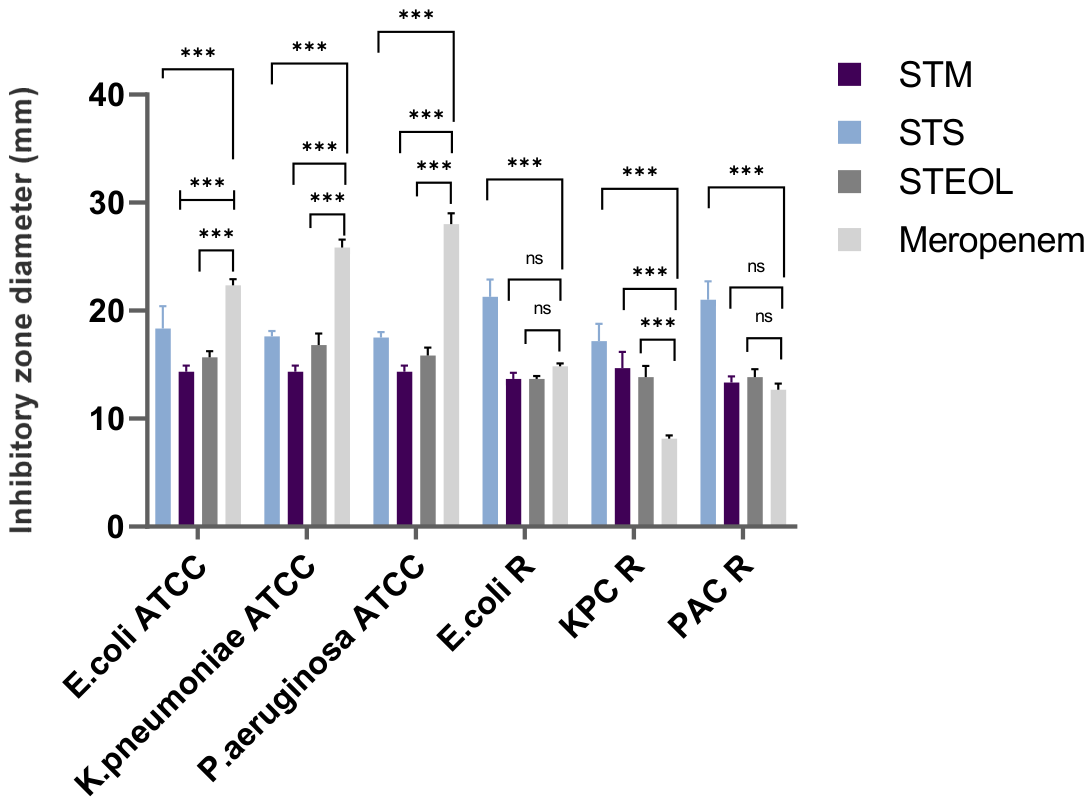
Antibacterial activity assays using the well diffusion and microdilution methods showed that the three phytobiotics were active against both the reference and the clinical isolates. The well diffusion method was used to qualitatively assess the antibacterial effect of the ST extracts by measuring the zone of inhibition (mm). The three phytobiotics, STEOL, STS, and STM, inhibited all studied strains, particularly the STS extract, which showed strong potential with inhibition zone diameters ranging from 16.5 to 23 mm, whereas STM produced inhibition zones between 13.5 and 15 mm. The results are reported in Figure 1. Regarding the leaf essential oil (STEOL), it exhibited inhibition zones ranging from 13.5 to 16.5 mm against the tested bacterial strains.
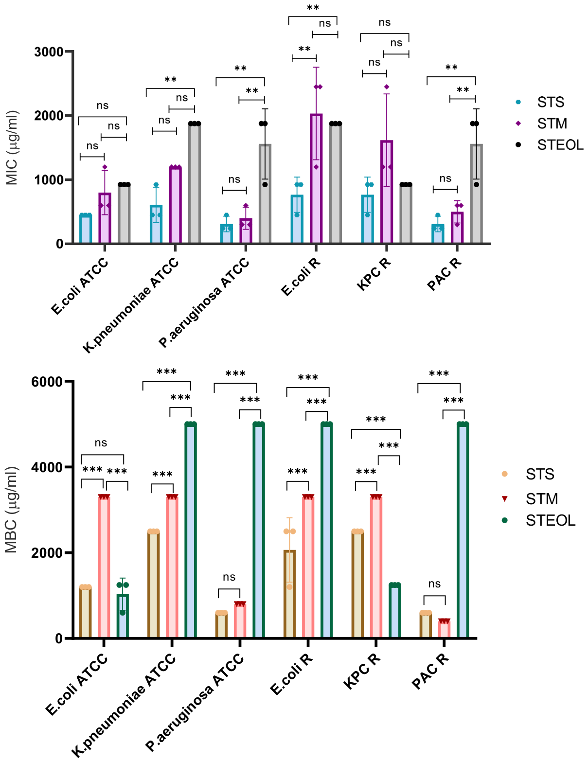
The quantification assay of the antibacterial effect was conducted using the microdilution method. To confirm the impact of ST extracts on bacterial viability, a cell viability assay with Alamar Blue was performed. The results displayed in Figure 2 indicate that STS, STM, and STEOL produced a significant reduction in bacterial viability, especially at higher concentrations. Before outlining MIC and MBC values for each extract on each isolate, an interesting antibacterial effect was noted for the STS extract, as already noted with the agar well diffusion test, which demonstrated the highest activity against the bacterial growth of the studied isolates. For STS, the MIC ranged from 310 to 766 µg/mL, and the MBC ranged from 600 to 2500 µg/mL. The MIC and MBC values of the STM extract ranged from 400 to 2000 μg/mL and from 400 to 3300 μg/mL, respectively. In contrast, the leaf essential oil (STEOL) exhibited MIC values ranging from 925 to 1875 μg/mL and MBC values ranging from 1250 to 5000 μg/mL.
The MBC/MIC ratio was used to determine the antibacterial mode of action of the tested phytobiotics (Supplementary Materials). The STS extract exhibited a bactericidal effect (MBC/MIC ≤ 4) against all tested strains, except for carbapenem-resistant E. coli, against which it showed a bacteriostatic effect. Similarly, STM exhibited bactericidal activity against all tested strains, except for E. coli ATCC, for which a bacteriostatic effect was observed. In contrast, the leaf essential oil (STEOL) exhibited bactericidal activity against all tested strains.
Moreover, the Alamar Blue assay showed that the three S. terebinthifolia phytobiotics reduced bacterial viability, with STS generally exhibiting the strongest effect, as indicated by its lower IC50 values (Table 3). The lowest IC50 value was observed for STS against P. aeruginosa CR (4.74 μg/mL ± 1.32), followed by P. aeruginosa ATCC (18.81 μg/mL ± 0.20) and E. coli ATCC (24.66 μg/mL ± 2.20). In contrast, the highest values were observed mainly with STEOL, particularly against K. pneumoniae CR (127.23 μg/mL ± 0.013) and P. aeruginosa CR (114.2 μg/mL ± 0.008). Overall, the results indicate strain- and phytobiotic-dependent variation in antibacterial activity.

3.3. Interaction Between S. terebinthifolia Phytobiotics and Cefotaxime

In our study, after confirming the growth inhibition activity exhibited by S. terebinthifolia phytobiotics toward the carbapenem-resistant strains, we explored the synergistic effect generated when combining the three phytobiotics with cefotaxime. Using the checkerboard method, we assessed the synergistic effect generated by the three extracts tested, which significantly decreased the MIC value of the antibiotic compared to cefotaxime alone (FIC index value was ≤0.5). The highest reduction in the MIC of CTX was obtained when it was combined with S. terebinthifolia phytobiotics against carbapenem-resistant E. coli and K. pneumoniae isolates, restoring the effect of CTX, which was ineffective alone against the two resistant bacteria. Also, the combination of STS with CTX modulated the action of CTX against carbapenem-resistant K. pneumoniae, carbapenem-resistant P. aeruginosa, and carbapenem-resistant E. coli strains, reducing its MIC values from 528 µg/mL to 50 µg/mL, 278 µg/mL to 1.5 µg/mL, and 235 µg/mL to 3.12 µg/mL, respectively. The checkerboard method results are summarized in Table 4 and Table 5.

3.4. β-Lactamase Inhibitory Effect of S. terebinthifolia Phytobiotics

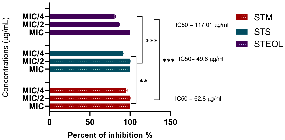
The β-lactamase inhibitory activity of the three S. terebinthifolia leaf phytobiotics was evaluated. The inhibitory effects of the ethanolic extracts and the essential oil are presented in Figure 3. Remarkably, all three phytobiotics (STEOL, STS, and STM) exhibited strong β-lactamase inhibitory activity, achieving 100% inhibition at their MIC/2. Among them, the Soxhlet leaf extract (STS) demonstrated the highest inhibitory potency, with an IC50 value of 49.8 μg/mL, indicating superior β-lactamase inhibition compared with the ethanolic extracts.

3.5. In Silico Study

3.5.1. Molecular Docking Simulations

In this study, a total of 38 chemical compounds identified in the three S. terebinthifolia leaf phytobiotics were subjected to virtual screening against two clinically relevant β-lactamases, VIM and OXA-48-like, using PyRx software. The docking results revealed that the majority of the identified compounds exhibited higher binding affinities (more negative binding energies) than the reference antibiotic (Table 6 and Table 7), suggesting a stronger interaction with the active sites of the target enzymes. Among the screened compounds, diosmetin, catechin, and quercetin exhibited the highest binding affinities for VIM, with docking scores of −9.3, −8.9, and −8.9 kcal/mol, respectively. Against OXA-48-like, ellagic acid, catechin, and epicatechin gallate were identified as the top-ranked compounds, with binding energies of −9.2, −8.9, and −8.7 kcal/mol, respectively. These findings suggest that the phytochemicals present in S. terebinthifolia may exhibit favorable predicted interactions within the catalytic pockets of both β-lactamases, highlighting these compounds as potential candidate inhibitors of VIM and OXA-48-like enzymes.
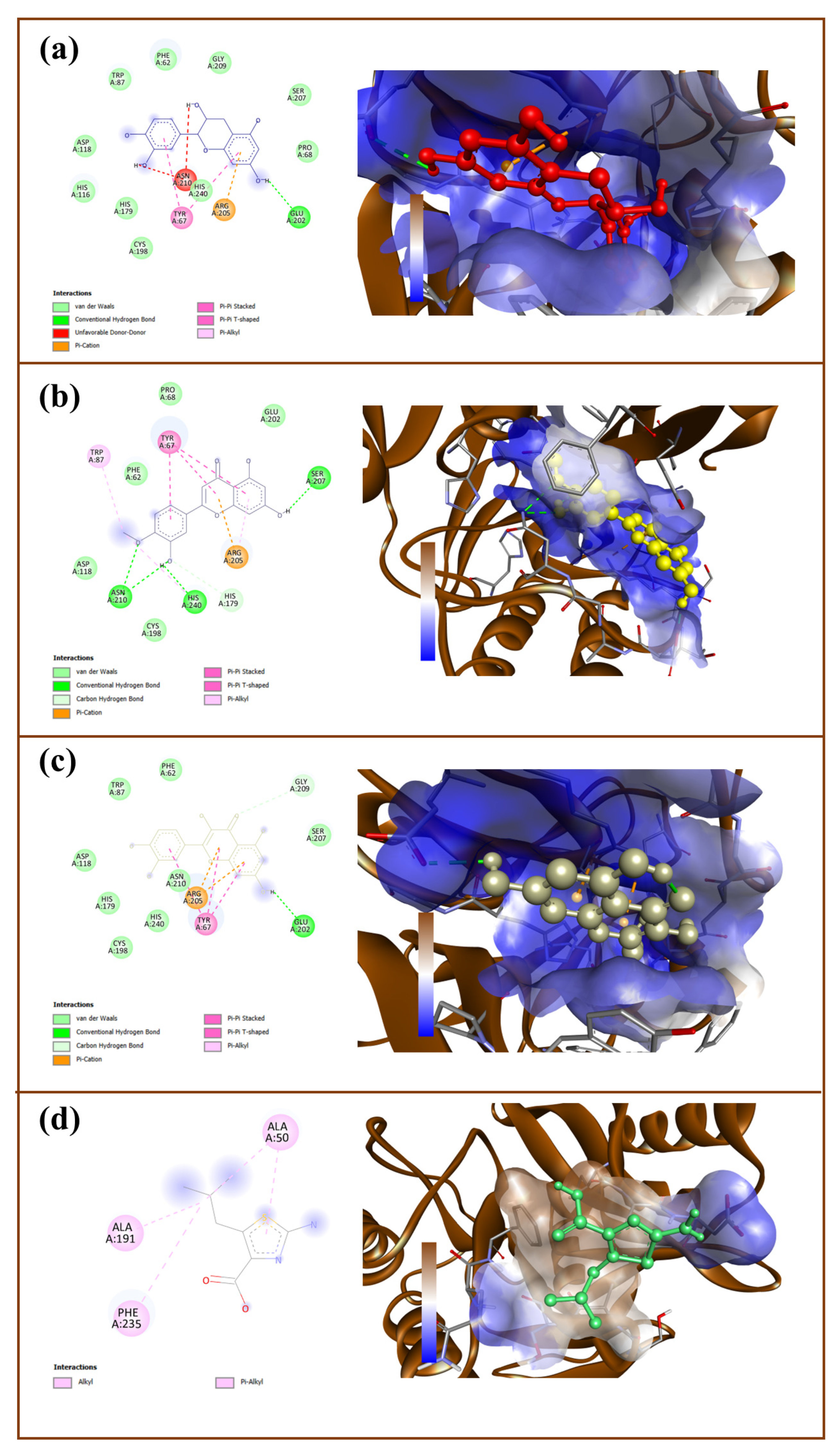
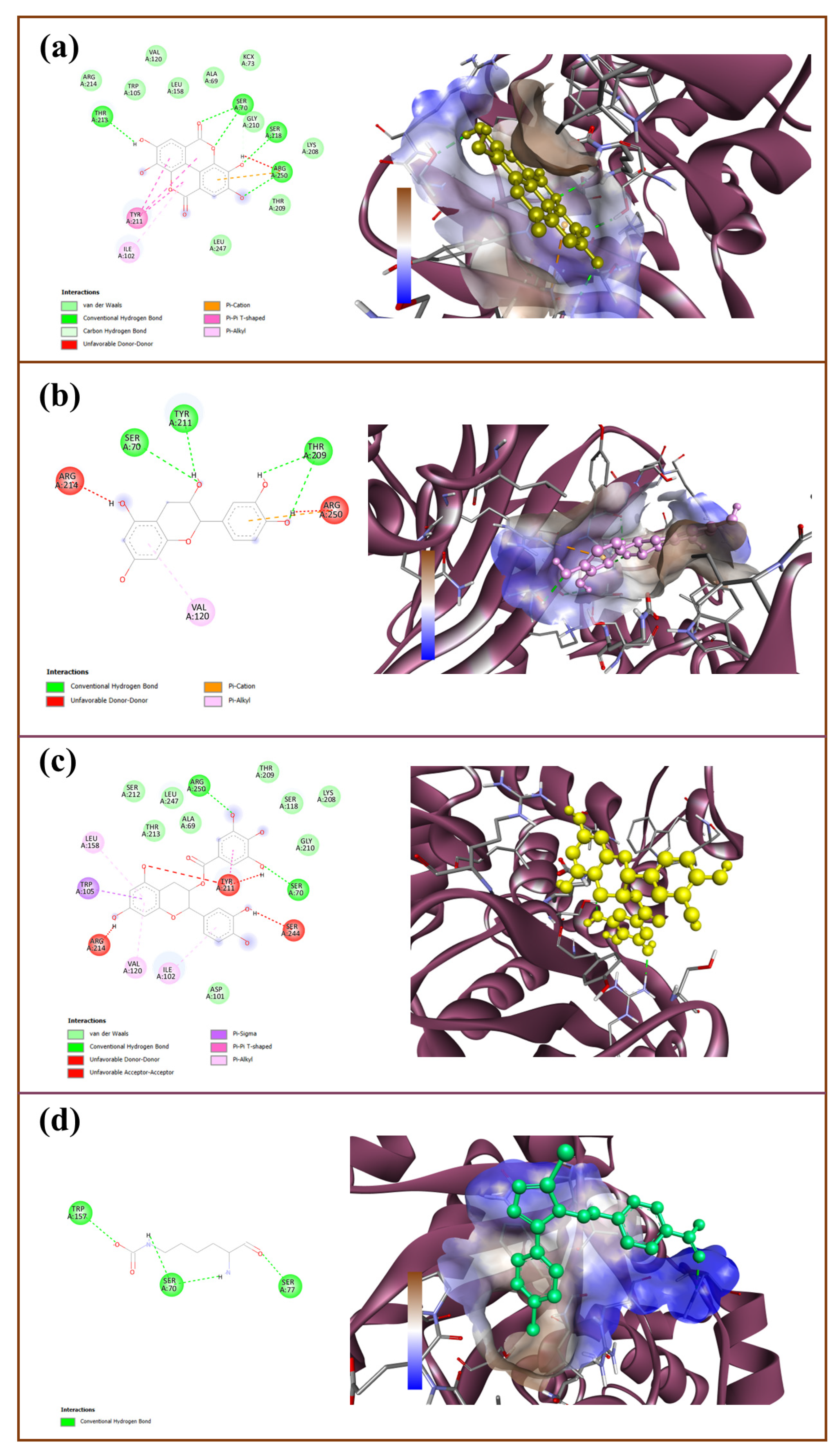
Furthermore, to gain deeper insight into the binding mechanisms of the docked complexes, both 3D and 2D interaction analyses were performed using Discovery Studio Visualizer, as presented in Figure 4 and Figure 5. The results showed that various intermolecular interactions mediated the binding between the tested compounds and the three proteins, highlighting their potential to interact with key amino acid residues in the binding pocket. For example, epicatechin gallate interacts with Oxa-48 via various residues through significant interactions, including van der Waals forces with Asp101, Ser212, Thr213, Leu247, Ala69, Thr209, Ser118, Lys208, and Gly210; a Pi–Sigma interaction with Trp105; conventional hydrogen bonds (H-bonds) with Arg250 and Ser70; and Pi–Pi T-shaped interactions with Ile102, Val129, and Leu158.

3.5.2. Drug-Likeness Properties Prediction

To identify potential lead compounds with favorable drug-like properties, the top-ranked phytochemicals were evaluated using the SwissADME web server. Drug-likeness was assessed according to Lipinski’s Rule of Five, the Ghose filter, and the Veber rule, based on key physicochemical descriptors including molecular weight, lipophilicity (LogP), hydrogen-bond donors, hydrogen-bond acceptors, topological polar surface area (TPSA), and the number of rotatable bonds (Table 8). Overall, all selected compounds complied with Lipinski’s Rule of Five and fulfilled the Ghose criteria, indicating favorable drug-like characteristics. Furthermore, all compounds satisfied the Veber rule, except for epicatechin gallate, which exceeded the recommended TPSA threshold of 140 Å2. Diosmetin and quercetin exhibited TPSA values below 130 Å2, suggesting good membrane permeability.
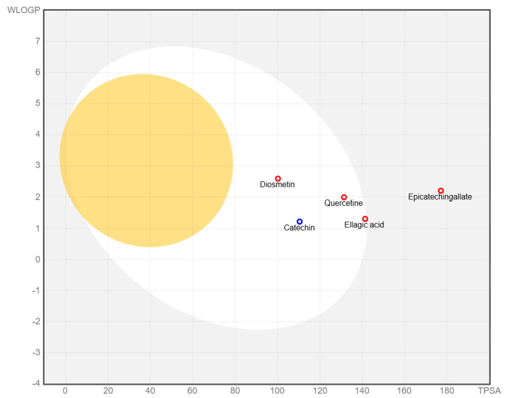
The BOILED-Egg model was further used to predict gastrointestinal (GI) absorption, blood–brain barrier (BBB) permeability, and P-glycoprotein (P-gp) substrate status (Figure 6). The analysis indicated that all compounds, except epicatechin gallate, were located within the white region, suggesting a high probability of passive gastrointestinal absorption. Epicatechin gallate was positioned outside the BOILED-Egg predictive regions, indicating poor passive absorption. Regarding P-gp substrate prediction, catechin was classified as a P-gp substrate (blue), whereas the remaining compounds were predicted to be non-substrates (red). None of the evaluated compounds were located in the yellow region of the BOILED-Egg plot, indicating a low probability of blood–brain barrier permeation.

4. Discussion

The current study explored the potential antibacterial properties of extracts and essential oil from S. terebinthifolia leaves in fighting carbapenemase-producing Gram-negative bacteria. Results indicated that Soxhlet extraction was more efficient in extracting phenolic compounds than the maceration method. The difference in total phenolic content determined is linked to the extraction methods. The content of total phenolics detected in the aroeira leaves included in the current investigation was markedly superior to that presented in the literature for the same species of leaf extract: 384.64 mg GAE/g [27]. Soxhlet extraction varied between 5.44 and 309.03 mg GAE/g of Soxhlet extract, while extracts obtained by maceration ranged between 73.90 and 228.51 mg GAE/g [28]. A previous report showed that in the Moroccan S. terebinthifolia Raddi, the major compounds are represented by phenolic compounds, principally including galloyl derivatives; flavonoids represented the rest, particularly flavonols, namely myricetin, quercetin, and kaempferol derivatives [29]. Another study showed the presence of catechin, ellagic acid, gallic acid, and epicatechin in S. terebinthifolia extract [30]. Sereniki et al. (2016) confirmed that the S. terebinthifolia sample demonstrated the existence of the prevailing compounds, namely phenolic acids and flavonoids [31]. It exhibited a phenolic profile characteristic of the Schinus species, and phenolic acids and flavonoids were identified [32].
The volatile composition analysis of the leaf essential oil of S. terebinthifolia revealed a predominance of monoterpenes, with (+)-4-carene, p-cymene, and trans-2-carene-4-ol identified as the major constituents. Similar monoterpene-rich profiles have been reported for S. terebinthifolia essential oils from different geographical regions. For example, in Tunisia, the essential oil of S. terebinthifolia was also characterized by a predominance of monoterpenes, particularly α-pinene (14.85–15.18%) and limonene (6.62–8.79%) [33]. However, other studies have reported different chemical profiles. For example, Belhoussaine et al. (2022) found that the leaf essential oil of S. terebinthifolia collected in Rabat, Morocco, was dominated by globulol, γ-elemene, and β-elemene [29]. Such variations in the chemical composition of essential oils may be attributed to several factors, including geographical origin, climatic and environmental conditions, soil characteristics, plant genotype, developmental stage, harvesting season, and extraction method.
The in vitro biological investigations conducted on S. terebinthifolia extracts displayed their biological capabilities, including their antibacterial properties, for potential use against urinary and respiratory tract infections [34]. Our results demonstrated that P. aeruginosa was the most susceptible bacterial species to the S. terebinthifolia phytobiotics. This was evidenced by the lowest MIC values, particularly for the Soxhlet leaf extract (STS), which exhibited an MIC of 310 μg/mL. In contrast, K. pneumoniae was the least susceptible species, displaying the highest MIC values among the tested bacteria (Figure 2). Our findings agree with those previously published that describe the antimicrobial potential of S. terebinthifolia leaf phytobiotics against Gram-negative bacteria, including P. aeruginosa and E. coli, with an MIC value of 500 μg/mL [35]. Additionally, prior research revealed that the leaves of the Anacardiaceae family plant had antibacterial activities against carbapenem-resistant Enterobacteriaceae, including K. pneumoniae, with MICs ranging from 512 to 1024 µg/mL [36]. Similarly, antibacterial activities of S. terebinthifolia extracts reported that the IZD values were about 15.3 mm and 13 mm at concentrations of 2000 and 1000 µg/mL, respectively, against E. coli, and 18.3 mm at 2000 µg/mL for P. aeruginosa [37]. These studies obtained MIC values close to those found in the present study.
In contrast to our findings, Giordani et al. (2021) suggested that S. terebinthifolia phytobiotics were active against Gram-negative bacteria at an MIC value of 3750 μg/mL [38], an MIC value higher than what we found in our research. Another study reported that ST extracts exhibit their effect against E. coli with an MIC of 78 μg mL−1 [39]. This greater activity could be attributed to the richness of phenolic constituents because compounds of this class have demonstrated interesting antibacterial activity [40]. The variations in results may be linked to the variation in the chemical profile of each extract.
New antibacterial agents are being investigated to be used alone and in association with drugs to surmount antibiotic resistance. Plant metabolites have been used as a promising alternative because they can act via different mechanisms and have fewer side effects [41]. Natural products can induce an alteration in the effect of antibiotics, either by antagonizing or increasing the antibiotic effect [42]. The study of the application of natural compounds in association with antibiotics could be useful to treat infectious diseases caused by ESBL-producing Enterobacteriaceae [43]. In particular, S. terebinthifolia phytobiotics could be used as an excipient for β-lactam antibiotics, decreasing their dose. This alternative seems promising [44]. Based on a more in-depth analysis of activity, it is possible to observe that the combination with the three phytobiotics potentiated the action of cefotaxime against all bacteria included in this study by increasing the sensitivity of the tested strains toward cefotaxime (Table 4). In this context, similar to our findings, numerous studies have highlighted the synergistic effect of natural extracts with β-lactam antibiotics, which is a potential approach to combat β-lactamase-producing isolates. The S. terebinthifolia extracts potentiated the action of two conventional β-lactam antibiotics; exploring the association of S. terebinthifolia extracts with β-lactam antibiotics such as gentamicin induced a reduction of 50 to 96.87% in the MIC of antibiotics, indicating the capacity to enhance the antibiotic effect against MDR strains, with a significant effect against E. coli, where the MIC was decreased from 64 µg/mL to 2 µg/mL [45]. Furthermore, the lectin obtained from the leaf of S. terebinthifolia improved the action of ampicillin and presented a synergistic effect against MDR strains [46].
Indeed, it was shown that combining a natural compound with cefotaxime against metallo-β-lactamase-producing isolates presented an excellent synergistic effect [42]. Thus, Zhou et al. (2013) indicated that the plant extract had a synergistic impact on cefotaxime against ESBL-producing isolates, and they demonstrated that it exhibited enzymatic inhibition activity of β-lactamase against these isolates without any mutation in the ESBL gene [43]. However, other results evaluating the effects of the association of different antibiotics with different essential oils showed good activity against MDR strains producing extended-spectrum β-lactamase, such as K. pneumoniae and E. coli [47]. Additionally, it was confirmed that natural extracts against oxacillin-resistant isolates demonstrated synergistic action with cefotaxime against ESBL-producing E. coli strains [44]. In this case, the resistance mechanism involves the production of β-lactamase that stimulates the hydrolysis of the β-lactam ring, inducing its neutralization [48]. Interestingly, botanicals ameliorated the actions of several antibiotics and potentiated the effect of antibiotics against K. pneumoniae [37]. Thereby, the combination of an Anacardiaceae plant with imipenem decreased the MIC value of the antibiotic against MDR P. aeruginosa [43]. Another study by [45] indicated that some phytocompounds exhibited a synergistic activity when combined with imipenem. However, mechanisms of synergy are still poorly understood; nevertheless, various researchers propose that molecules disrupt the cell wall or increase the permeability of the cytoplasmic membrane and thereby produce efflux pump inhibitors, inhibit penicillin-binding proteins, or promote the influx of antibiotics [45].
Limited studies have been conducted to examine the impact of terebinthifolia extracts on inhibiting β-lactams hydrolysis. This is the first study reporting the inhibitory activity of β-lactamase using S. terebinthifolia extracts. Costa da Silva et al. (2022) attributed the effect of S. terebinthifolia essential oil on strains resistant to a β-lactam antibiotic to the inhibition of the β-lactam ring by the essential oil [46]. On the other hand, Winsou et al. (2022) observed strong β-lactamase inhibition activity with Terminalia superba and Annona senegalensis [49]. Indeed, a hexane extract from Anacardium occidentale exhibited β-lactamase inhibitory activity on two strains resistant to penicillin by the synthesis of oxacillinase [50]. Moreover, [45] have reported that there is a relationship between the chemical compounds and the antibacterial effect of the plants [51].
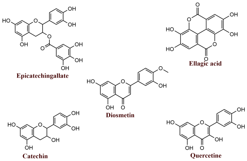
In the present study, the extracts from S. terebinthifola had potent antibacterial activity against carbapenemase-producing strains, targeting the hydrolysis of β-lactam antibiotics by the β-lactamase mechanism. Several natural constituents present in the plant employed in the current research could have contributed to the inhibition of β-lactamase in the included isolates. Furthermore, in silico analyses, including molecular docking, confirmed that phytochemicals of S. terebinthifolia exhibit a strong binding affinity within the pocket of carbapenemase enzymes, suggesting their effectiveness as potential inhibitors (Figure 7). Additionally, Spyrakis et al. [52] have suggested the β-lactamase inhibitory activity of natural compounds. The presence of the 4-oxo function in flavonoids, comparable to that in clavulanic acid and penicillin G, underlies this property [52]. Several in silico findings have highlighted that flavonoids can inhibit NDM-1, which is responsible for inducing resistance to standard antibiotics [53,54]. Moreover, Zhang et al. (2022) identified kaempferol, apigenin, fisetin, taxifolin, quercetin, and luteolin as effective inhibitors of OXA-48 [55].
Although the present study provides evidence for the antibacterial and β-lactamase inhibitory potential of S. terebinthifolia leaf extracts and essential oil, the observed antibacterial effects may involve multiple complementary mechanisms. In addition to β-lactamase inhibition, the antimicrobial effects of plant-derived preparations may result from interactions of their diverse constituents with bacterial cellular structures and physiological processes. The observed enhancement of cefotaxime activity, together with the β-lactamase inhibition results, suggests that interference with β-lactam resistance may contribute to the overall antibacterial effect. The molecular docking results further support the potential interaction of selected phytochemicals with β-lactamases.


5. Conclusions

In conclusion, S. terebinthifolia leaf extracts and essential oil exhibited antibacterial activity against the tested carbapenem-resistant Gram-negative isolates and enhanced the activity of cefotaxime in combination assays. The phytobiotics also showed β-lactamase inhibitory activity, supporting their potential contribution to the observed antibiotic-potentiating effect. Molecular docking analyses further identified diosmetin, catechin, quercetin, ellagic acid, and epicatechin gallate as candidate β-lactamase-interacting compounds based on their predicted binding profiles. Overall, the findings support the potential of S. terebinthifolia phytobiotics as sources of natural antibiotic-adjuvant candidates. Further biochemical, toxicity, and in vivo studies are required to confirm the underlying mechanisms and assess their therapeutic relevance.

Supplementary Materials

The following supporting information can be downloaded at https://www.mdpi.com/article/10.3390/app16189212/s1, Table S1. The MIC, MBC, and MBC/MIC (μg/ml) values of Soxhlet Leaf Extract (STS) against E. coli, K. pneumoniae, and P. aeruginosa. Table S2. The MIC, MBC, and MBC/MIC (μg/ml) values of Leaf Maceration Extract (STM) against E. coli, K. pneumoniae, and P. aeruginosa. Table S3. The MIC, MBC, and MBC/MIC (μg/ml) values of Leaf Essential Oil (STEOL) against E. coli, K. pneumoniae, and P. aeruginosa.

Author Contributions

Conceptualization, F.M.; methodology, F.M. and J.L.R.R.; software, F.M.; validation, B.E.K. and T.d.M.; formal analysis, F.M.; investigation, F.M.; resources, F.M.; data curation, F.M.; writing—original draft preparation, F.M.; writing—review and editing, F.J., J.L.R.R., L.G.C., S.S., A.S., T.d.M., B.E.K. and A.E.; visualization, B.E.K. and T.d.M.; supervision, B.E.K. and A.S. All authors have read and agreed to the published version of the manuscript.

Funding

This research received no external funding.

Institutional Review Board Statement

Not applicable.

Informed Consent Statement

Not applicable.

Data Availability Statement

The data sets generated during and/or analyzed during the current study are available from the corresponding author on reasonable request.

Acknowledgments

This research was supported by the Erasmus+ program, which provided funding for Fatima Mourabiti’s predoctoral mobility from Hassan II University (Morocco) to the University of Santiago de Compostela (Spain), within the framework of the cooperation agreement established between the two institutions. The authors also gratefully acknowledge the Pan-African PGS Education and Research Initiative (PAPERI) and the Sustainable Sciences Institute (SSI) for their valuable training and guidance provided through the Scientific Writing Workshop.

Conflicts of Interest

The authors declare no conflicts of interest.

Abbreviations

The following abbreviations are used in this manuscript:
BBBblood–brain barrier
BOILED-EggBrain Or IntestinaL EstimateD Permeation method
CTXcefotaxime
E. coli Rcarbapenem-resistant E. coli
EOessential oil
ESKAPEEnterococcus faecium, Staphylococcus aureus, Klebsiella pneumoniae, Acinetobacter baumannii, Pseudomonas aeruginosa, and Enterobacter species
FICIfractional inhibitory concentration index
GAgallic acid
GC/MSgas chromatography–mass spectrometry
GIgastrointestinal
H-bondhydrogen bond
IZDinhibition zone diameter
KPC Rcarbapenem-resistant K. pneumoniae
LC-MS/MSliquid chromatography–tandem mass spectrometry
MBCminimum bactericidal concentration
MDRmultidrug resistance
MHAMueller–Hinton agar
MICminimum inhibitory concentration
MWmolecular weight
ODoptical density
PAC Rcarbapenem-resistant P. aeruginosa
PBPspenicillin-binding proteins
P-gpP-glycoprotein
S. terebinthifoliaSchinus terebinthifolia Raddi
SMILESSimplified Molecular Input Line Entry System
STEOLleaf essential oil of S. terebinthifolia
STMextract obtained by maceration
STSextract obtained by Soxhlet extraction
TPCtotal phenolic content
TPSAtopological polar surface area
WHOWorld Health Organization

References

  1. Tooke, C.L.; Hinchliffe, P.; Bragginton, E.C.; Colenso, C.K.; Hirvonen, V.H.A.; Takebayashi, Y.; Spencer, J. β-Lactamases and β-Lactamase Inhibitors in the 21st Century. J. Mol. Biol. 2019, 431, 3472–3500. [Google Scholar] [CrossRef] [Scilit] [PubMed]
  2. El-Gamal, M.I.; Brahim, I.; Hisham, N.; Aladdin, R.; Mohammed, H.; Bahaaeldin, A. Recent Updates of Carbapenem Antibiotics. Eur. J. Med. Chem. 2017, 131, 185–195. [Google Scholar] [CrossRef] [Scilit] [PubMed]
  3. Vázquez-Ucha, J.C.; Arca-Suárez, J.; Bou, G.; Beceiro, A. New Carbapenemase Inhibitors: Clearing the Way for the β-Lactams. Int. J. Mol. Sci. 2020, 21, 9308. [Google Scholar] [CrossRef] [Scilit] [PubMed]
  4. Rahdar, H.A.; Malekabad, E.S.; Dadashi, A.R.; Takei, E.; Keikha, M.; Kazemian, H.; Karami-Zarandi, M. Correlation between Biofilm Formation and Carbapenem Resistance among Clinical Isolates of Klebsiella pneumoniae. Ethiop. J. Health Sci. 2019, 29, 745–750. [Google Scholar] [CrossRef] [Scilit] [PubMed]
  5. Nordmann, P.; Poirel, L. Epidemiology and Diagnostics of Carbapenem Resistance in Gram-Negative Bacteria. Clin. Infect. Dis. 2019, 69, S521–S528. [Google Scholar] [CrossRef] [Scilit] [PubMed]
  6. Cusumano, J.A.; Caffrey, A.R.; Daffinee, K.E.; Luther, M.K.; Lopes, V.; LaPlante, K.L. Weak Biofilm Formation among Carbapenem-Resistant Klebsiella pneumoniae. Diagn. Microbiol. Infect. Dis. 2019, 95, 114877. [Google Scholar] [CrossRef] [Scilit] [PubMed]
  7. Bhat, D.; Rajan, A.K.; Aditya, V.; Eshwara, V.K.; Varma, M.; Umakanth, S.; Thunga, G.; Shanbhag, V. Does Carbapenem Resistance Influence Clinical Outcomes in ICU Patients with Klebsiella pneumoniae Bacteraemia? A Comparative Analysis of Carbapenem-Resistant and Susceptible Isolates. Acta Trop. 2025, 269, 107744. [Google Scholar] [CrossRef] [Scilit] [PubMed]
  8. Somda, N.S.; Nyarkoh, R.; Kotey, F.C.N.; Tetteh-Quarcoo, P.B.; Donkor, E.S. A Systematic Review and Meta-Analysis of Carbapenem-Resistant Enterobacteriaceae in West Africa. BMC Med. Genom. 2024, 17, 267. [Google Scholar] [CrossRef] [Scilit] [PubMed]
  9. Moo, C.L.; Osman, M.A.; Yang, S.K.; Yap, W.S.; Ismail, S.; Lim, S.H.E.; Chong, C.M.; Lai, K.S. Antimicrobial Activity and Mode of Action of 1,8-Cineol against Carbapenemase-Producing Klebsiella pneumoniae. Sci. Rep. 2021, 11, 20824. [Google Scholar] [CrossRef] [Scilit] [PubMed]
  10. Iseppi, R.; Di Cerbo, A.; Aloisi, P.; Manelli, M.; Pellesi, V.; Provenzano, C.; Camellini, S.; Messi, P.; Sabia, C. In Vitro Activity of Essential Oils against Planktonic and Biofilm Cells of Extended-Spectrum β-Lactamase (ESBL)/Carbapenamase-Producing Gram-Negative Bacteria Involved in Human Nosocomial Infections. Antibiotics 2020, 9, 272. [Google Scholar] [CrossRef] [Scilit] [PubMed]
  11. Rachwał, K.; Gustaw, K. Plant-Derived Phytobiotics as Emerging Alternatives to Antibiotics Against Foodborne Pathogens. Appl. Sci. 2025, 15, 6774. [Google Scholar] [CrossRef] [Scilit]
  12. Yang, S.K.; Yusoff, K.; Ajat, M.; Yap, W.S.; Lim, S.H.E.; Lai, K.S. Antimicrobial Activity and Mode of Action of Terpene Linalyl Anthranilate against Carbapenemase-Producing Klebsiella pneumoniae. J. Pharm. Anal. 2021, 11, 210–219. [Google Scholar] [CrossRef] [Scilit] [PubMed]
  13. Vasconcelos, N.G.; Vaz, M.S.M.; Radai, J.A.S.; Kassuya, C.A.L.; Formagio, A.S.N.; Graciani, F.S.; Leal, M.L.; Oliveira, R.J.; da Silva, K.E.; Croda, J.; et al. Antimicrobial Activity of Plant Extracts against Carbapenem-Producing Klebsiella pneumoniae and in Vivo Toxicological Assessment. J. Toxicol. Environ. Health-Part A Curr. Issues 2020, 83, 719–729. [Google Scholar] [CrossRef] [Scilit] [PubMed]
  14. Dannenberg, G.d.S.; Funck, G.D.; da Silva, W.P.; Fiorentini, Â.M. Essential Oil from Pink Pepper (Schinus terebinthifolius Raddi): Chemical Composition, Antibacterial Activity and Mechanism of Action. Food Control 2019, 95, 115–120. [Google Scholar] [CrossRef] [Scilit]
  15. Mu, Q.; Chen, L.; Gao, X.; Shen, S.; Sheng, W.; Min, J.; Wang, F. The Role of Iron and Siderophores in Infection, and the Development of Siderophore Antibiotics. Clin. Infect. Dis. 2019, 69, S529–S537. [Google Scholar] [CrossRef] [Scilit] [PubMed]
  16. El-Nashar, H.A.S.; Mostafa, N.M.; Abd El-Ghffar, E.A.; Eldahshan, O.A.; Singab, A.N.B. The Genus Schinus (Anacardi-aceae): A Review on Phytochemicals and Biological Aspects. Nat. Prod. Res. 2022, 36, 4839–4857. [Google Scholar] [CrossRef] [Scilit] [PubMed]
  17. Dirar, A.I.; Alsaadi, D.H.M.; Wada, M.; Mohamed, M.A.; Watanabe, T.; Devkota, H.P. Effects of Extraction Solvents on Total Phenolic and Flavonoid Contents and Biological Activities of Extracts from Sudanese Medicinal Plants. S. Afr. J. Bot. 2019, 120, 261–267. [Google Scholar] [CrossRef] [Scilit]
  18. Celeiro, M.; Lamas, J.P.; Arcas, R.; Lores, M. Antioxidants Profiling of By-Products from Eucalyptus Greenboards Manufacture. Antioxidants 2019, 8, 263. [Google Scholar] [CrossRef] [Scilit] [PubMed]
  19. Essaqui, A.; Elamrani, A.; Cayuela, J.A.; Benaissa, M. Chemical Composition of the Essential Oil of Warionia Saharae from Morocco. J. Essent. Oil-Bear. Plants 2007, 10, 241–246. [Google Scholar] [CrossRef] [Scilit]
  20. Derdak, R.; Quinteiro, J.; Sakoui, S.; Addoum, B.; Rodríguez Castro, J.; Rey Méndez, M.; Soukri, A.; El Khalfi, B. Isolation and Identification of Dominant Bacteria from Raw Donkey Milk Produced in a Region of Morocco by QIIME 2 and Evaluation of Their Antibacterial Activity. Sci. World J. 2021, 2021, 6664636. [Google Scholar] [CrossRef] [Scilit] [PubMed]
  21. Weinstein, M.P. Performance Standards for Antimicrobial Susceptibility Testing, 28th ed.; Clinical and Laboratory Standards Institute: Wayne, PA, USA, 2019; Volume 38. [Google Scholar]
  22. Mourabiti, F.; Jouga, F.; Calvo, L.G.; Villarino, R.A.; Zouheir, Y.; Soukri, A.; de Miguel, T.; El Khalfi, B. Polyphenols as Potential β-Lactamase Inhibitors: An Integrated Computational and Experimental Study. Molecules 2025, 30, 4416. [Google Scholar] [CrossRef] [Scilit] [PubMed]
  23. Pimentel Arantes, J.; Dillis Faccin, I.; Coutinho, E.J.; Lima Cardoso, C.A.; Lopes Fernandes, S.S.; Rossato, L.; Simionatto, E.; Simionatto, S. An Approach to Combat Multidrug-Resistant K. pneumoniae Strain Using Synergistic Effects of Ocotea diospyrifolia Essential Oil in Combination with Amikacin. Microb. Pathog. 2024, 193, 106782. [Google Scholar] [CrossRef] [Scilit] [PubMed]
  24. Mourabiti, F.; Jouga, F.Z.; Mouzoun, Y.; Arslan, S.; Schena, R.; Zouheir, Y.; Soukri, A.; De Martino, L.; Nocera, F.P.; El Khalfi, B. Phenotypic and Genotypic Characterization of Carbapenem Encoding Genes among Carbapenem-Resistant Gram-Negative Bacteria Isolated from North Casablanca, Morocco. Comp. Immunol. Microbiol. Infect. Dis. 2025, 123, 102399. [Google Scholar] [CrossRef] [Scilit] [PubMed]
  25. Ehrt, C.; Schulze, T.; Graef, J.; Diedrich, K.; Pletzer-Zelgert, J.; Rarey, M. ProteinsPlus: A Publicly Available Resource for Protein Structure Mining. Nucleic Acids Res. 2025, 53, W478–W484. [Google Scholar] [CrossRef] [Scilit] [PubMed]
  26. Lipinski, C.A.; Lombardo, F.; Dominy, B.W.; Feeney, P.J. Experimental and Computational Approaches to Estimate Solubility and Permeability in Drug Discovery and Development q Settings. Adv. Drug Deliv. Rev. 2001, 46, 3–26. [Google Scholar] [CrossRef] [Scilit] [PubMed]
  27. Bulla, M.K.; Hernandes, L.; Baesso, M.L.; Nogueira, A.C.; Bento, A.C.; Bortoluzzi, B.B.; Serra, L.Z.; Cortez, D.A.G. Evaluation of Photoprotective Potential and Percutaneous Penetration by Photoacoustic Spectroscopy of the Schinus terebinthifolius Raddi Extract. Photochem. Photobiol. 2015, 91, 558–566. [Google Scholar] [CrossRef] [Scilit] [PubMed]
  28. Candice Costa Silva, J.; Medeiros Santos, N.; de Sousa Silva, N.; Cristina Silveira Martins, A.; Maria Gomes Dutra, L.; Eduardo Alves Dantas, C.; dos Santos Lima, M.; Fechine Tavares, J.; Sobral da Silva, M.; Mangueira do Nascimento, Y. Characterization of Flours from the Aroeira Leaf (Schinus terebinthifolius Raddi), Obtained by Different Drying Methods. J. Chromatogr. B Anal. Technol. Biomed. Life Sci. 2024, 1239, 124126. [Google Scholar] [CrossRef] [Scilit] [PubMed]
  29. Belhoussaine, O.; El Kourchi, C.; Harhar, H.; Bouyahya, A.; El Yadini, A.; Fozia, F.; Alotaibi, A.; Ullah, R.; Tabyaoui, M. Chemical Composition, Antioxidant, Insecticidal Activity, and Comparative Analysis of Essential Oils of Leaves and Fruits of Schinus molle and Schinus terebinthifolius. Evid.-Based Complement. Altern. Med. 2022, 2022, 4288890. [Google Scholar] [CrossRef] [Scilit] [PubMed]
  30. da Silva, M.M.; Iriguchi, E.K.K.; Kassuya, C.A.L.; Vieira, M.d.C.; Foglio, M.A.; de Carvalho, J.E.; Ruiz, A.L.T.G.; Souza, K.d.P.; Formagio, A.S.N. Schinus terebinthifolius: Phenolic Constituents and in Vitro Antioxidant, Antiproliferative and in Vivo Anti-Inflammatory Activities. Rev. Bras. Farmacogn. 2017, 27, 445–452. [Google Scholar] [CrossRef] [Scilit]
  31. Sereniki, A.; Linard-Medeiros, C.F.B.; Silva, S.N.; Silva, J.B.R.; Peixoto Sobrinho, T.J.S.; da Silva, J.R.; Alvesa, L.D.S.; Smailic, S.S.; Wanderley, A.G.; Lafayette, S.S.L. Schinus terebinthifolius Administration Prevented Behavioral and Biochemical Alterations in a Rotenone Model of Parkinson’s Disease. Rev. Bras. Farmacogn. 2016, 26, 240–245. [Google Scholar] [CrossRef] [Scilit]
  32. Feuereisen, M.M.; Hoppe, J.; Zimmermann, B.F.; Weber, F.; Schulze-Kaysers, N.; Schieber, A. Characterization of Phenolic Compounds in Brazilian Pepper (Schinus terebinthifolius Raddi) Exocarp. J. Agric. Food Chem. 2014, 62, 6219–6226. [Google Scholar] [CrossRef] [Scilit] [PubMed]
  33. Ennigrou, A.; Casabianca, H.; Laarif, A.; Hanchi, B.; Hosni, K. Maturation-Related Changes in Phytochemicals and Biological Activities of the Brazilian Pepper Tree (Schinus terebinthifolius Raddi) Fruits. S. Afr. J. Bot. 2017, 108, 407–415. [Google Scholar] [CrossRef] [Scilit]
  34. Tlili, N.; Yahia, Y.; Feriani, A.; Labidi, A.; Ghazouani, L.; Nasri, N.; Saadaoui, E.; Khaldi, A. Schinus terebinthifolius vs Schinus Molle: A Comparative Study of the Effect of Species and Location on the Phytochemical Content of Fruits. Ind. Crops Prod. 2018, 122, 559–565. [Google Scholar] [CrossRef] [Scilit]
  35. Shehata, M.E.; El-Sherbiny, G.M.; Sharaf, M.H.; Kalaba, M.H.; Shaban, A.S. Phytochemical Analysis, Antimicrobial, Antioxidant, and Cytotoxicity Activities of Schinus molle (L.) Extracts. Biomass Convers. Biorefin. 2024, 15, 3753–3770. [Google Scholar] [CrossRef] [Scilit]
  36. Matieta, V.Y.; Kuete, V.; Mbaveng, A.T. Anti-Klebsiella and Antibiotic-Potentiation Activities of the Methanol Extracts of Seven Cameroonian Dietary Plants against Multidrug-Resistant Phenotypes over-Expressing AcrAB-TolC Efflux Pumps. Investig. Med. Chem. Pharmacol. 2023, 6, 73. [Google Scholar] [CrossRef] [Scilit]
  37. Salem, M.Z.M.; El-Hefny, M.; Ali, H.M.; Elansary, H.O.; Nasser, R.A.; El-Settawy, A.A.A.; El Shanhorey, N.; Ashmawy, N.A.; Salem, A.Z.M. Antibacterial Activity of Extracted Bioactive Molecules of Schinus terebinthifolius Ripened Fruits against Some Pathogenic Bacteria. Microb. Pathog. 2018, 120, 119–127. [Google Scholar] [CrossRef] [Scilit] [PubMed]
  38. Giordani, C.; Waller, S.B.; Guterres, K.A.; da Silva, C.C.; Lobo, R.R.; Hoffmann, J.F.; Perera, S.C.; Chaves, F.C.; Ladeira, S.R.L.; Sallis, E.S.V.; et al. Multiresistant Bacteria Isolated from Domestic and Wild Animals with Skin Lesions Were Susceptible to Native Plants from Southern Brazil. Nat. Prod. Res. 2022, 36, 2927–2931. [Google Scholar] [CrossRef] [Scilit] [PubMed]
  39. da Silva, J.H.S.; Simas, N.K.; Alviano, C.S.; Alviano, D.S.; Ventura, J.A.; de Lima, E.J.; Seabra, S.H.; Kuster, R.M. Anti-Escherichia coli Activity of Extracts from Schinus terebinthifolius Fruits and Leaves. Nat. Prod. Res. 2018, 32, 1365–1368. [Google Scholar] [CrossRef] [Scilit] [PubMed]
  40. Gomes, R.B.d.A.; de Souza, E.S.; Gerhardt Barraqui, N.S.; Tosta, C.L.; Nunes, A.P.F.; Schuenck, R.P.; Ruas, F.G.; Ventura, J.A.; Filgueiras, P.R.; Kuster, R.M. Residues from the Brazilian Pepper Tree (Schinus terebinthifolia Raddi) Processing Industry: Chemical Profile and Antimicrobial Activity of Extracts against Hospital Bacteria. Ind. Crops Prod. 2020, 143, 111430. [Google Scholar] [CrossRef] [Scilit]
  41. da Silva, P.M.; da Silva, B.R.; de Oliveira Silva, J.N.; de Moura, M.C.; Soares, T.; Feitosa, A.P.S.; Brayner, F.A.; Alves, L.C.; Paiva, P.M.G.; Damborg, P.; et al. Punica granatum Sarcotesta Lectin (PgTeL) Has Antibacterial Activity and Synergistic Effects with Antibiotics against β-Lactamase-Producing Escherichia coli. Int. J. Biol. Macromol. 2019, 135, 931–939. [Google Scholar] [CrossRef] [Scilit] [PubMed]
  42. Barbosa-Filho, V.M.; Waczuk, E.P.; Leite, N.F.; Menezes, I.R.A.; da Costa, J.G.M.; Lacerda, S.R.; Adedara, I.A.; Coutinho, H.D.M.; Posser, T.; Kamdem, J.P. Phytocompounds and Modulatory Effects of Anacardium microcarpum (Cajui) on Antibiotic Drugs Used in Clinical Infections. Drug Des. Devel. Ther. 2015, 9, 5965–5972. [Google Scholar] [CrossRef] [Scilit] [PubMed][Green Version]
  43. Zhou, X.Z.; Jia, F.; Liu, X.M.; Yang, C.; Zhao, L.; Wang, Y.J. Total Alkaloids from Sophora alopecuroides L. Increase Susceptibility of Extended-Spectrum β-Lactamases Producing Escherichia coli Isolates to Cefotaxime and Ceftazidime. Chin. J. Integr. Med. 2013, 19, 945–952. [Google Scholar] [CrossRef] [Scilit] [PubMed]
  44. Vasconcelos, N.G.; Mallmann, V.; Costa, É.R.; Simionatto, E.; Coutinho, E.J.; De Lara Da Silva, R.C.; Ribeiro, S.M.; Franco, O.L.; Migliolo, L.; Croda, J.; et al. Antibacterial Activity and Synergism of the Essential Oil of Nectandra megapotamica (L.) Flowers against OXA-23-Producing Acinetobacter baumannii. J. Essent. Oil Res. 2020, 32, 260–268. [Google Scholar] [CrossRef] [Scilit]
  45. Costa da Silva, M.M.; Bezerra de Araújo Neto, J.; Lucas dos Santos, A.T.; de Morais Oliveira-Tintino, C.D.; de Araújo, A.C.J.; Freitas, P.R.; da Silva, L.E.; do Amaral, W.; Deschamps, C.; de Azevedo, F.R.; et al. Antibiotic-Potentiating Activity of the Schinus terebinthifolius Raddi Essential Oil against MDR Bacterial Strains. Plants 2023, 12, 1587. [Google Scholar] [CrossRef] [Scilit] [PubMed]
  46. de Souza Feitosa Lima, I.M.; Zagmignan, A.; Santos, D.M.; Maia, H.S.; dos Santos Silva, L.; da Silva Cutrim, B.; Vieira, S.L.; Bezerra Filho, C.M.; de Sousa, E.M.; Napoleão, T.H.; et al. Schinus terebinthifolia Leaf Lectin (SteLL) Has Anti-Infective Action and Modulates the Response of Staphylococcus aureus-Infected Macrophages. Sci. Rep. 2019, 9, 18159. [Google Scholar] [CrossRef] [Scilit] [PubMed]
  47. Mahmoud, A.M.; Mahmoud, R.; El-Baky, A.; Bakr, A.; Ahmed, F.; Fadl, G.; Gad, M. Antibacterial Activity of Essential Oils and in Combination with Some Standard Antimicrobials against Different Pathogens Isolated from Some Clinical Specimens. Am. J. Microbiol. Res. 2016, 4, 16–25. [Google Scholar]
  48. Guo, Y.; Song, G.; Sun, M.; Wang, J.; Wang, Y. Prevalence and Therapies of Antibiotic-Resistance in Staphylococcus aureus. Front. Cell. Infect. Microbiol. 2020, 10, 107. [Google Scholar] [CrossRef] [Scilit] [PubMed]
  49. Winsou, I.; Eugenie, A.; Victorien, D.; Adole, A.; Brice, F.; Eustache, A. Antibacterial Plant Extracts Inhibit the Beta-Lactamase of Escherichia coli Clinical Isolates. J. Med. Plants Stud. 2023, 11, 49–54. [Google Scholar] [CrossRef] [Scilit]
  50. Bouttier, S.; Fourniat, J.; Garofalo, C.; Gleye, C.; Laurens, A.; Hocquemiller, R. β-Lactamase Inhibitors from Anacardium Occidentale. Pharm. Biol. 2002, 40, 231–234. [Google Scholar] [CrossRef] [Scilit]
  51. Saeidi, S.; Boroujeni, N.A.; Ahmadi, H.; Hassanshahian, M. Antibacterial Activity of Some Plant Extracts against Extended- Spectrum Beta-Lactamase Producing Escherichia coli Isolates. Jundishapur J. Microbiol. 2015, 8, e15434. [Google Scholar] [CrossRef] [Scilit] [PubMed]
  52. Spyrakis, F.; Bellio, P.; Quotadamo, A.; Linciano, P.; Benedetti, P.; D’Arrigo, G.; Baroni, M.; Cendron, L.; Celenza, G.; Tondi, D. First Virtual Screening and Experimental Validation of Inhibitors Targeting GES-5 Carbapenemase. J. Comput. Aided. Mol. Des. 2019, 33, 295–305. [Google Scholar] [CrossRef] [Scilit] [PubMed]
  53. Guo, Y.; Yang, Y.; Xu, X.; Li, L.; Zhou, Y.; Jia, G.; Wei, L.; Yu, Q.; Wang, J. Metallo-β-Lactamases Inhibitor Fisetin Attenuates Meropenem Resistance in NDM-1-Producing Escherichia coli. Eur. J. Med. Chem. 2022, 231, 114108. [Google Scholar] [CrossRef] [Scilit] [PubMed]
  54. Biharee, A.; Sharma, A.; Kumar, A.; Jaitak, V. Antimicrobial Flavonoids as a Potential Substitute for Overcoming Antimicrobial Resistance. Fitoterapia 2020, 146, 104720. [Google Scholar] [CrossRef] [Scilit] [PubMed]
  55. Zhang, Y.; Chen, C.; Cheng, B.; Gao, L.; Qin, C.; Zhang, L.; Zhang, X.; Wang, J.; Wan, Y. Discovery of Quercetin and Its Analogs as Potent OXA-48 Beta-Lactamase Inhibitors. Front. Pharmacol. 2022, 13, 926104. [Google Scholar] [CrossRef] [Scilit] [PubMed]
Figure 1. The antibacterial activity of S. terebinthifolia phytobiotics against E. coli, K. pneumoniae, and P. aeruginosa was determined by the well diffusion assay. E. coli R: carbapenem-resistant E. coli, KPC R: carbapenem-resistant K. pneumoniae, PAC R: carbapenem-resistant P. aeruginosa. STS: Soxhlet leaf extract, STM: leaf maceration extract, STEOL: leaf essential oil. *** indicates p < 0.001, while ns indicates p > 0.05.
Applsci 16 09212 g001
Figure 2. Minimum inhibitory concentration (MIC) and minimum bactericidal concentration (MBC) of S. terebinthifolia phytobiotics against E. coli, K. pneumoniae, and P. aeruginosa strains. E. coli R: carbapenem-resistant E. coli, KPC R: carbapenem-resistant K. pneumoniae, PAC R: carbapenem-resistant P. aeruginosa. STS: Soxhlet leaf extract, STM: leaf maceration extract, STEOL: leaf essential oil. ** indicates p < 0.01, *** indicates p < 0.001, while ns indicates p > 0.05.
Applsci 16 09212 g002
Figure 3. Percentage of β-lactamase activities inhibited by S. terebinthifolia leaf phytobiotics. ** indicates p < 0.01; *** p < 0.001.
Applsci 16 09212 g003
Figure 4. 2D and 3D visualization of the intermolecular interactions between VIM and the top-ranked phytochemicals (catechin (a), diosmetin (b), quercetine (c), and the reference inhibitor (d)).
Applsci 16 09212 g004
Figure 5. 2D and 3D visualization of the intermolecular interactions between OXA-48-like β-lactamases and the top-ranked phytochemicals (ellagic acid (a), catechin (b), epicatechingallate (c), and the reference inhibitor (d)).
Applsci 16 09212 g005
Figure 6. BOILED−Egg plot analysis of the top−ranked phytochemicals.
Applsci 16 09212 g006
Figure 7. Chemical structures of the most promising compounds identified by virtual screening (ChemDraw v21.0.0).
Applsci 16 09212 g007
Table 1. Concentrations (mg L−1) of polyphenols detected in the analyzed S. terebinthifolia ethanolic extracts by LC–MS/MS.
Compound ClassCompound NameMolecular FormulaSTS (mg/L)STM (mg/L)
Hydroxycinnamic acids4-Hydroxycinnamic acidC9H8O31.7 ± 0.11.2 ± 0.1
Chlorogenic acidC16H18O970 ± 418 ± 4
Hydroxybenzoic acidsGallic acidC7H6O51142 ± 222207 ± 53
3,4-Dihydroxybenzoic acidC7H6O4231 ± 13119 ± 31
Flavan-3-olsCatechinC15H14O610 ± 247 ± 12
EpicatechingallateC22H18O1062 ± 1085 ± 7
FlavonolsQuercetinC15H10O7483 ± 21430 ± 21
Quercetin-3-rutinosideC27H30O1625 ± 212 ± 3
Quercetin-3-glucosideC21H20O12812 ± 51422 ± 12
Quercetin-3-glucuronideC21H18O1359 ± 824 ± 9
AstragalinC21H20O115.7 ± 1.25.8 ± 0.7
IsorhamnetinC16H12O745 ± 810 ± 2
DiosmetinC16H12O64.8 ± 1.0-
KaempferolC15H10O615 ± 111 ± 2
MyricetinC15H10O8203 ± 16129 ± 11
EllagitanninsEllagic acidC14H6O8167 ± 23169 ± 12
Table 2. Chemical composition of the leaf essential oil of S. terebinthifolia.
Retention Time (Min)Peak Area (%)Name
4.5950.072-Carene
4.9340.34Cyclohexene, 4-methyl-1-(1-methylethyl)-
5.4231.15Bicyclo[3.1.0]hex-2-ene, 2-methyl-5-(1-methylethyl)-
5.5781.11Cyclooctene
5.75364.88(+)-4-Carene
5.790.59Pyrrolidine, 1,5-dimethyl-3,3-diphenyl-2-ethylidene-
5.9511.74p-Cymene
6.070.57D-Limonene
6.1294.14Eucalyptol
8.4520.81Benzene, 1-methyl-3-(1-methylethyl)-
8.9090.91Cyclooctanone
9.4030.253-Cyclohexen-1-ol, 1-methyl-4-(1-methylethyl)-
9.8946.75trans-2-Caren-4-ol
10.1081.79trans-Carveol
10.6590.121-(1,2,3-Trimethyl-cyclopent-2-enyl)-ethanone
11.0920.12Carvenone
12.8071.12(1S,2R,4R,7R)-4-Isopropyl-7-methyl-3,8-dioxatricyclo[5.1.0.02,4]octane
13.2430.3(3E,5E)-2,6-Dimethylocta-3,5,7-trien-2-ol
14.4880.771,4-Dihydroxy-p-menth-2-ene
15.1970.61,4-Dihydroxy-p-menth-2-ene
15.8550.151,3-Dioxolane, 2,2-dimethyl-4,5-bis(1-methylethenyl)-
16.5630.31,3-Dioxolane, 2,2-dimethyl-4,5-di-1-propenyl-
18.2561.447-Oxabicyclo[4.1.0]heptane, 1-methyl-4-(2-methyloxiranyl)-
Table 3. The IC50 (μg/mL) values of S. terebinthifolia phytobiotics against E. coli, K. pneumoniae, and P. aeruginosa.
ExtractsSoxhlet Leaf Extract (STS)Leaf Maceration Extract (STM)Leaf Essential Oil (STEOL)
Bacteria
E. coli ATCC24.66 ± 2.20110.19 ± 3.7969.33 ± 0.03
E. coli CR83.72 ± 5.3055.01 ± 3.4760.03 ± 0.004
K. pneumoniae ATCC92.57 ± 2.6787.48 ± 2.3584.12 ± 0.022
K. pneumoniae CR107.66 ± 2.75110.19 ± 3.79127.23 ± 0.013
P. aeruginosa ATCC18.81 ± 0.2028.22 ± 1.7681.1 ± 0.004
P. aeruginosa CR4.74 ± 1.325.17 ± 0.79114.2 ± 0.008
E. coli CR: carbapenem-resistant E. coli, KPC CR: carbapenem-resistant K. pneumoniae, PAC CR: carbapenem-resistant P. aeruginosa. Values are expressed as mean ± SD, n = 3.
Table 4. Evaluation of the synergistic effect of S. terebinthifolia leaf phytobiotics combined with cefotaxime against six bacterial strains.
CTX (μg/mL)CTX + Extract (µg/mL)Extract (µg/mL) Extract + CTX (µg/mL)FIC
Soxhlet Leaf Extract (STS)E. coli ATCC12.50.78450750.22
E. coli R2353.127667661.01
K. pneumoniae ATCC251.56081520.31
KPC R528507663830.59
P. aeruginosa ATCC251.531038.750.18
PAC R2781.531038.750.13
Leaf Maceration Extract (STM)E. coli ATCC12.50.788002000.31
E. coli R23525203320331.01
K. pneumoniae ATCC251.51616269.30.22
KPC R5285.2516168080.5
P. aeruginosa ATCC251.54002000.56
PAC R2781.550083.30.17
Leaf Essential Oil (STEOL)E. coli ATCC12.51.5925115.60.24
E. coli R2356.251875468.70.27
K. pneumoniae ATCC253.121875312.50.29
KPC R5283.12925231.20.25
P. aeruginosa ATCC250.781558194.70.15
PAC R2781.51558194.70.13
E. coli CR: carbapenem-resistant E. coli, KPC R: carbapenem-resistant K. pneumoniae, PAC R: carbapenem-resistant P. aeruginosa, CTX: cefotaxime.
Table 5. Effect of S. terebinthifolia leaf phytobiotics in combination with cefotaxime against P. aeruginosa, K. pneumoniae, and E. coli.
CTX/STSCTX/STMCTX/STEOL
E. coli ATCCSynergySynergySynergy
E. coli CRIndifferenceIndifferenceSynergy
K. pneumoniae ATCCSynergySynergySynergy
KPC RAdditiveAdditiveSynergy
P. aeruginosa ATCCSynergyAdditiveSynergy
PAC RSynergySynergySynergy
E. coli CR: carbapenem-resistant E. coli, KPC R: carbapenem-resistant K. pneumoniae, PAC R: carbapenem-resistant P. aeruginosa. STS: Soxhlet leaf extract, STM: leaf maceration extract, STEOL: leaf essential oil. CTX: cefotaxime.
Table 6. Predicted binding energies (kcal/mol) of the phenolic compounds against VIM and OXA-48-like β-lactamases obtained using PyRx.
Kcal/mol
CompoundsOXAVIM
4-Hydroxycinnamic acid−5.9−7
Chlorogenic acid−7.8−8.5
Gallic acid−6−5.6
3,4-Dihydroxybenzoic acid−6.1−6.4
Catechin−8.9−8.9
Epicatechingallate−8.7−7.2
Quercetine−8.6−8.9
Quercetin-3-rutinoside−8.5−8.1
Quercetin-3-glucoside−8−8.6
Quercetin-3-glucuronide−8.2−8.6
Astragalin−7.8−8.3
Isorhamnetin−8.4−5.2
Diosmetin−8.3−9.3
Kaempferol−8.5−8.5
Myricetin−8.4−8.3
Ellagic acid−9.2−8.2
Reference compound−5.9−5.7
Table 7. Predicted binding energies (kcal/mol) of STEOL phytochemicals against VIM and OXA-48-like β-lactamases obtained using PyRx.
Kcal/mol
CompoundsOXAVIM
2-Carene−5.1−6.9
Cyclohexene, 4-methyl-1-(1-methylethyl)-−5.3−6.3
Bicyclo[3.1.0]hex-2-ene, 2-methyl-5-(1-methylethyl)-−5.1−5.9
Cyclooctene−4.5−5.1
(+)-4-Carene−5.1−6.7
Pyrrolidine, 1,5-dimethyl-3,3-diphenyl-2-ethylidene-−7.1−6.3
p-Cymene−5.3−6.2
D-Limonene−5.2−6.3
Eucalyptol−5−5.9
Benzene, 1-methyl-3-(1-methylethyl)-−5.2−6.4
Cyclooctanone−5.1−5.5
3-Cyclohexen-1-ol, 1-methyl-4-(1-methylethyl)-−5.1−6.1
trans-2-Caren-4-ol−5.4−6.9
trans-Carveol−5.6−6.3
1-(1,2,3-Trimethyl-cyclopent-2-enyl)-ethanone−5.7−6.7
Carvenone−6.1−7
(1S,2R,4R,7R)-4-Isopropyl-7-methyl-3,8-dioxatricyclo[5.1.0.02,4]octane−5.2−6.1
(3E,5E)-2,6-Dimethylocta-3,5,7-trien-2-ol−5.1−6.5
1,4-Dihydroxy-p-menth-2-ene−5.4−6.5
1,3-Dioxolane, 2,2-dimethyl-4,5-bis(1-methylethenyl)-−5.2−6.1
1,3-Dioxolane, 2,2-dimethyl-4,5-di-1-propenyl-−5.4−5.6
7-Oxabicyclo[4.1.0]heptane, 1-methyl-4-(2-methyloxiranyl)-−5.4−5.8
Reference compound−5.9−5.7
Table 8. Physicochemical properties and drug-likeness prediction of the top-ranked phytochemicals evaluated using SwissADME.
CompoundMW (g/mol)logPAcceptorsH-Bond DonorsLipinski ViolationGhose ViolationVeber ViolationTPSA (Å2)
Epicatechingallate442.372.20107101177.14
Catechin290.271.2265000110.38
Quercetine302.241.9975000131.36
Ellagic acid302.11.3184000141.34
Diosmetin300.262.5963000100.13
Disclaimer/Publisher’s Note: The statements, opinions and data contained in all publications are solely those of the individual author(s) and contributor(s) and not of MDPI and/or the editor(s). MDPI and/or the editor(s) disclaim responsibility for any injury to people or property resulting from any ideas, methods, instructions or products referred to in the content.