Article

Effects of Repeated Toe-Flexor Contractions on Hallux Strength and Running Biomechanics in Recreationally Active Adults


by
Jean-Louis Peters-Dickie
1,2,3,*,
Virginie Otlet
1,
Christine Detrembleur
1,
Kevin Deschamps
2,3,*,
Sébastien Lobet
1,4,5 and
Anh Phong Nguyen
1,6
1
Neuro Musculo Skeletal Lab (NMSK), Institut de Recherche Expérimentale et Clinique, Secteur des Sciences de la Santé, Université Catholique de Louvain, 1200 Brussels, Belgium
2
Musculoskeletal Rehabilitation Research Group, Department of Rehabilitation Sciences, Katholieke Universiteit Leuven, 8200 Bruges, Belgium
3
Clinical Motion Analysis Laboratorium (CMAL), Universitaire Ziekenhuizen KU Leuven Campus Pellenberg, 3212 Lubbeek, Belgium
4
Haemostasis and Thrombosis Unit, Division of Hematology, Cliniques Universitaires Saint-Luc, Université Catholique de Louvain (UCLouvain), 1200 Brussels, Belgium
5
Service D’ergothérapie et de Kinésithérapie, Cliniques Universitaires Saint-Luc, Université Catholique de Louvain (UCLouvain), 1200 Brussels, Belgium
6
The Running Clinic, Lac-Beauport, QC G3B 2J8, Canada
*
Authors to whom correspondence should be addressed.
Appl. Sci. 2026, 16(17), 8478; https://doi.org/10.3390/app16178478
Submission received: 17 May 2026 / Revised: 14 August 2026 / Accepted: 19 August 2026 / Published: 26 August 2026

Abstract

Foot muscles contribute to impact attenuation and propulsion during running, yet the effects of repeated toe-flexor contractions and associated strength loss on running biomechanics remain poorly understood. The primary objective of this randomized crossover study was to investigate the acute effects of repeated concentric and isometric toe-flexor contractions on hallux flexion strength, used as an index of fatigue; secondary analyses examined running ground-reaction-force outcomes. Nineteen recreationally active adults completed two interventions one week apart: concentric toe-flexor contractions and isometric toe-flexor contractions combined with electrical stimulation of the medial longitudinal arch. Hallux flexion strength and ground reaction forces during treadmill running at 9 and 12 km·h−1 were assessed before and after each intervention. The concentric protocol reduced hallux flexion strength by 20%, whereas the isometric protocol did not. None of the hypothesis-driven running biomechanical outcomes, including contact duration, impact peak force, active peak force, and vertical stiffness, changed significantly after either protocol. Exploratory secondary analyses suggested changes in selected leg stiffness and impulse variables. In recreationally active adults, repeated concentric toe-flexor contractions induced measurable neuromuscular fatigue, whereas the isometric protocol did not significantly reduce hallux flexion strength; biomechanical changes were limited to exploratory outcomes.

1. Introduction

The human foot contributes to support, impact attenuation, propulsion, and balance during locomotion. These functions depend on interactions between passive tissues, sensory feedback, and active muscular control. Muscular control is ensured by intrinsic foot muscles (IFMs), which originate and insert within the foot, and extrinsic foot muscles (EFMs), which originate in the lower leg and insert into the foot [1]. During walking and running, the interaction between IFMs and EFMs contributes to controlling foot deformation during loading and to increasing metatarsophalangeal joint stiffness during push-off [2,3]. Accordingly, toe flexion is produced by coordinated IFM and EFM activity.
Impaired foot muscle function has been associated with aging and various musculoskeletal, systemic, and neurological disorders [4]. Experimental studies have frequently used tibial nerve blocks to investigate the functional role of foot muscles [3,5], but this approach simultaneously reduces muscle activation, proprioception, and cutaneous sensation, complicating the interpretation of its biomechanical effects [6]. Neuromuscular fatigue, defined as a reduction in force-generating capacity following sustained or repeated activity, may therefore provide a complementary model of transient muscle impairment [7].
Several studies have attempted to induce neuromuscular fatigue in foot muscles to investigate their functional role during postural and dynamic tasks [8,9,10,11,12]. Sustained isometric hallux flexion and abduction contractions reduced hallux strength by approximately 26% [12], whereas repeated concentric hallux flexion reduced abductor hallucis activity by 17% [8]. However, findings across studies remain inconsistent, with some reporting no reduction in force production [10] or failing to quantify the magnitude of fatigue [9,11]. These discrepancies may partly result from methodological limitations, including short intervention durations, typically lasting 1–8 min, and seated or unloaded fatigue protocols [8,9,10,11,12].
Longer, weight-bearing protocols may therefore be needed to impose sufficient demand on the foot muscles. Furthermore, because foot muscles contribute to locomotion through both dynamic shortening during propulsion and quasi-isometric force production for arch control [3,13], it remains unclear whether repeated contractions emphasizing these different demands induce comparable strength loss or distinct biomechanical changes during running. Clarifying this issue may help refine experimental fatigue protocols and improve understanding of how transient toe-flexor impairment affects running loading, stiffness, and propulsion.
Previous fatigue studies suggest that impaired foot muscle function can alter locomotor biomechanics. Neuromuscular fatigue protocols have been associated with altered hindfoot kinematics during walking [9] and changes in spatiotemporal parameters such as step length and single-support duration [11]. Experimental suppression of IFM function has also been shown to modify ground reaction forces during sporting movements [5]. Running represents a particularly relevant task because foot muscles contribute substantially to both impact attenuation and propulsion [2,3,5]. However, the effects of acute toe-flexor fatigue on running biomechanics remain largely unknown.
Ground-reaction-force analysis provides a direct kinetic description of running and allows quantification of spatiotemporal, loading, impulse, and spring-mass characteristics, including vertical stiffness [14,15]. Therefore, the primary objective of this study was to investigate acute pre–post changes in hallux flexion strength after concentric and isometric toe-flexor contraction protocols. The secondary objective was to determine whether these protocols induced acute pre–post changes in navicular drop and ground-reaction-force-derived running outcomes. Contact duration, impact peak force, active peak force, and vertical stiffness were considered hypothesis-driven running biomechanical outcomes, whereas other ground-reaction-force-derived variables were considered additional secondary exploratory outcomes. We hypothesized that each protocol would reduce hallux flexion strength from pre- to post-intervention, indicating neuromuscular fatigue. For running biomechanics, we expected that each protocol would increase foot contact duration and vertical ground-reaction-force peaks, while decreasing vertical stiffness.

2. Materials and Methods

2.1. Study Design

This study was reported in accordance with the CONSORT Guidelines Extension for Crossover Trials [16]. A crossover design provides greater statistical power with fewer participants than a parallel design [16]. Sequence order was randomized, and the one-week washout period was selected to minimize physiological carryover between the two acute exercise protocols. Period and carryover effects were not formally tested.

2.2. Sample Size

Sample size was estimated from a pilot study of eight individuals who completed the same interventions as described below. The repeated-measures general linear model resulted in a partial eta squared effect size of 0.40. Using G*Power software (version 3.1), 18 participants were required for α = 0.05 and power = 0.8. To allow for potential dropouts, 20 participants were recruited. Further details on the pilot data are provided in Appendix A.

2.3. Participants

Twenty recreationally active adults were recruited between December 2023 and April 2024. This population was selected to ensure that participants could complete the running protocol at the required speeds while limiting variability related to age and sedentary status. Participants were eligible if they were older than 18 years, had no lower-limb symptoms, and engaged in at least one hour of moderate-to-vigorous physical activity per week. Exclusion criteria encompassed neurological, musculoskeletal, or systemic diseases, pregnancy, and lower-limb injury or surgery within six months prior to participation.
Of the 20 recruited participants, 19 completed the study. The final sample included 16 males and 3 females, with 13 participants reporting right-side and 6 left-side dominance. Participants were 25.2 ± 4.6 years old, with a body mass of 74.1 ± 11.5 kg, height of 178.0 ± 7.6 cm, and body mass index of 23.3 ± 2.6 kg·m−2. They reported 3.7 ± 2.0 h·week−1 of moderate-to-vigorous physical activity. The median 6-item Foot Posture Index score was 5 [Q1–Q3: 2, 6].
All participants provided written informed consent before inclusion, and the study was approved by the local ethics committee (protocol code B403201523492).

2.4. Procedures

Participants attended two visits at the UCLouvain Neuromusculoskeletal Laboratory (Belgium), separated by a one-week washout period. During the first visit, baseline leg length and 6-item Foot Posture Index score were assessed on the dominant side [17]. Participants were randomly assigned to an intervention sequence in blocks of four by drawing a sealed envelope. Participants remained barefoot, and the interventions targeted the self-reported dominant foot [18]. Due to the nature of the interventions, neither participants, therapists, nor the evaluator were blinded to sequence allocation.
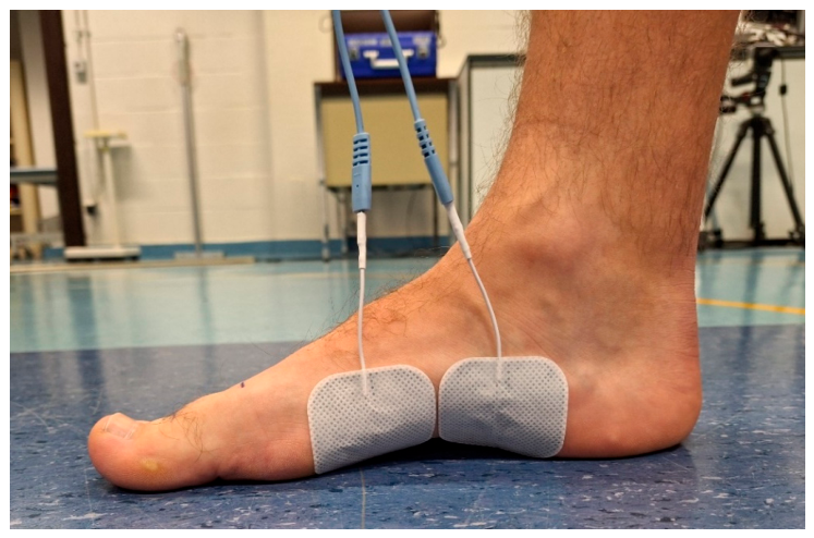
To address limitations of protocols intended to induce foot-muscle fatigue, both interventions were performed while standing for 16 min, with additional load provided by a weighted vest of 20 kg for men and 15.2 kg for women, corresponding to 26.4 ± 3.2% of body weight. Intervention A involved repeated concentric hallux and toe-flexion movements against an Airex® balance pad (Airex AG®, Sins, Switzerland), with participants performing eight sets of 75 metronome-paced contractions at 40 repetitions/min, consistent with previous toe-flexion protocols [8,9,10] (Figure 1). Intervention B consisted of repeated isometric hallux and toe-flexor contractions combined with electrical stimulation applied to the medial longitudinal arch (MLA), as shown in Figure 2. Electrical stimulation was used because previous work suggests that it can facilitate foot muscle recruitment during exercise [19] and induce neuromuscular fatigue when applied as a standalone intervention [20,21]. Additional procedural and familiarization details are provided in Supplementary Text S1.
Before interventions A and B, sit-to-stand navicular drop was measured using a caliper, followed by running ground-reaction-force recording at 9 and 12 km·h−1 and hallux flexion strength assessment. After each intervention, assessments were performed in the following order: hallux flexion strength, navicular drop, and running ground reaction forces. Based on this standardized assessment sequence, hallux flexion strength assessment began immediately after the intervention, while the ground-reaction-force recording window at 9 km·h−1 began approximately 3–4 min after completion of the intervention.
The same investigator (J-L. P-D.) carried out all measurements. Ground reaction forces were recorded at 1000 Hz using the Gaitway 3D treadmill (h/p/cosmos sports & medical GmbH, Nussdorf-Traunstein, Germany; Arsalis SRL, Glabais, Belgium) during the last 20 s of each running speed and filtered using an 8th-order Bessel low-pass filter with a 25 Hz cut-off frequency for the vertical, mediolateral, and anteroposterior force components.

2.5. Outcome Measures

2.5.1. Primary Outcome

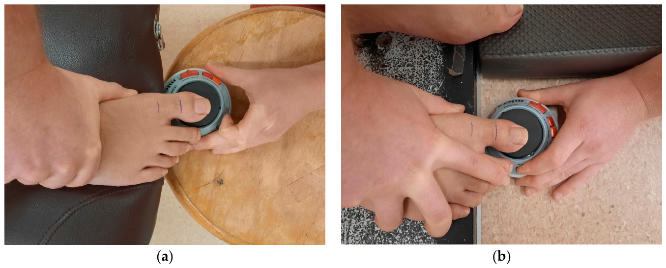
Long-sitting hallux flexion strength was used as the primary outcome to quantify neuromuscular fatigue, defined as a pre–post reduction in maximal force-generating capacity (Figure 3a) [22,23]. This task involves both IFM and EFM contributions [24]. To increase the relative contribution of IFMs compared with EFMs [25], participants were positioned supine on an examination table, supporting themselves on their elbows, with the assessed foot at the edge of the table, knee flexed, and ankle in maximal pain-free plantarflexion. A microFET®2 hand-held dynamometer (Hoggan Scientific, LLC, Salt Lake City, UT, USA) was placed on a stool, laterally secured by the examiner, and its transducer aligned under the distal hallux phalanx, while the examiner stabilized the ankle. This set-up was selected to reduce examiner-related resistance variability compared with conventional hand-held dynamometry [23,26]. Participants pushed the hallux downward maximally for 5 s while keeping trunk and pelvis on the table, followed by a rest period. They performed attempts until the relative range across the last three trials was less than 20%. The mean of the two highest values, normalized by body mass, was then used in the analyses. The number of attempts required to meet the acceptance criterion was recorded, and within-session consistency was characterized using the coefficient of variation of the last three accepted trials, calculated as CV (%) = SD/mean × 100.

2.5.2. Secondary Outcomes

Navicular drop was the difference in navicular height (marked with a pen) relative to the ground between sitting and standing [27].
Ground reaction forces during running were used to compute the hypothesis-driven biomechanical outcomes for the dominant limb: contact duration (% of stride), impact and active vertical ground-reaction-force peaks (% BW), and vertical stiffness (% BW·mm−1). Additional pre-planned secondary exploratory running biomechanical outcomes were computed. Full definitions of all biomechanical outcomes are provided in Table S1. Briefly, leg stiffness was calculated as the ratio of maximal vertical force to leg compression, vertical impulse as the area under the vertical force–time curve during foot contact, and propulsive impulse as the area under the anteroposterior force–time curve during propulsion.
Perceived fatigue was evaluated every 4 min during both interventions using a verbal numeric rating scale. Zero indicated no fatigue (same force as pre-test); ten indicated maximal fatigue with an inability to exert force. Perceived fatigue was analyzed exploratorily to describe participants’ responses to the protocols.
Hallux flexion strength (N·kg−1) was measured while standing before and at 2 min intervals during intervention A (Figure 3b). The dynamometer was placed on the ground, with the transducer horizontally aligned to the steel support and positioned under the distal phalanx of the hallux. An investigator ensured an upright posture, with the ankles and shoulders aligned in the frontal plane. Electrical stimulation current intensity (mA) was monitored every 4 min during intervention B.

2.6. Statistical Analyses

Analyses used SPSS v27 (IBM, Chicago, IL, USA). Continuous demographic variables were compared between sequence orders using Mann–Whitney U tests because QQ-plots indicated violations of normality. Repeated-measures general linear models with four measurement times were used for each primary and secondary outcome. We reported partial eta squared effect sizes, interpreted as the percentage of variance in each dependent variable explained by the main effect of time. In the case of a significant time effect (alpha = 0.05), post hoc multiple comparisons were applied across the four measurement occasions using Bonferroni correction, with the main interpretation focused on pre–post comparisons within each intervention. Within-group Hedges’ g effect sizes and corresponding 95% confidence intervals were calculated and interpreted using published thresholds [28]. Period, sequence/order, and carryover effects were not formally modeled; therefore, between-protocol pairwise comparisons were interpreted cautiously and were not considered equivalent to a crossover-adjusted comparison of intervention effectiveness.

3. Results

3.1. Participants

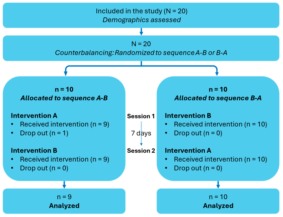
One participant withdrew from sequence A-B at the first visit due to dizziness (Figure 4). No significant between-sequence differences were found for quantitative demographic outcomes, although sex distribution was imbalanced between sequences (Table S2).

3.2. Neuromuscular Fatigue

Repeated concentric toe-flexor contractions induced a large reduction in long-sitting hallux flexion strength, whereas isometric contractions did not, partially confirming our hypothesis. Specifically, hallux flexion strength decreased by 20% after intervention A (p < 0.001, Hedges’ g = −1.07 [−1.62; −0.50]), but not after intervention B (p = 0.30, Hedges’ g = −0.47 [−0.93; −0.01]). Navicular drop was not significantly affected by the interventions (Table 1). Standing hallux flexion strength during intervention A remained stable from familiarization to 16 min (2.0 ± 0.6 to 2.0 ± 0.7 N·kg−1; p = 0.44; Table S3).
The median number of trials required to meet the acceptance criterion was 4 [Q1–Q3: 3–4.5] before intervention A, 4 [3–4] after intervention A, 5 [4–5] before intervention B, and 4 [3–5] after intervention B. Median coefficients of variation in the last three accepted trials across measurement occasions ranged from 5% [Q1–Q3: 3–9%] to 8% [Q1–Q3: 4–14%].

3.3. Perceived Fatigue and Current Intensity

Perceived fatigue increased during both interventions, reaching a median of 7 in intervention A and 6 in intervention B. Median electrical stimulation current intensity increased from 14 mA during familiarization to 16 mA at the end of intervention B (p < 0.001; Table S3). Qualitatively, electrical stimulation produced visible toe movements consistent with contraction, mainly hallux abduction and plantarflexion, and in some participants, flexion of the metatarsophalangeal joints of the lesser toes.

3.4. Running Biomechanics

None of the hypothesis-driven running outcomes, including contact duration, impact peak force, active peak force, and vertical stiffness, changed significantly after either intervention (Table 2).
Pre-planned secondary exploratory running outcomes are detailed in Table S4. Among the statistically significant exploratory findings, leg stiffness increased after both protocols, although at different running speeds, whereas impulse-related changes were protocol- and speed-specific. Leg stiffness increased by 5.2% with a large effect after intervention A at 12 km·h−1 (p < 0.001) and by 3.6% with a medium effect after intervention B at 9 km·h−1 (p = 0.02). Propulsive impulse decreased by 2.9% with a large effect after intervention A at 12 km·h−1 (p = 0.01), and vertical impulse decreased after intervention B at both 9 km·h−1 (−1.5%, medium effect, p = 0.03) and 12 km·h−1 (−1.8%, medium effect, p = 0.03).

4. Discussion

This study examined acute pre–post changes in hallux flexion strength and ground-reaction-force-derived running outcomes after two repeated toe-flexor contraction protocols. The main finding was that repeated concentric contractions induced a 20% decrease in hallux flexion strength, whereas repeated isometric contractions combined with MLA electrical stimulation did not significantly reduce hallux flexion strength. None of the hypothesis-driven running biomechanical outcomes changed significantly after either intervention. Exploratory secondary analyses suggested increases in leg stiffness and reductions in selected impulse variables after the protocols.
Prior protocols investigating foot muscle fatigue were generally shorter, lasting 8 min at most, and were often performed in seated or unloaded conditions [8,9,10,11,12]. These designs produced only modest or inconsistent reductions in force production [8,9,10,12], supporting the development of more demanding protocols in the present study. Accordingly, both interventions lasted 16 min and were performed while standing with additional load to increase foot-muscle demand, consistent with previous evidence that greater lower-limb loading increases IFM activation [29].
Following the concentric protocol, hallux flexion strength decreased by 20% and participants reported a median perceived fatigue of 7/10. These results are consistent with a reduction in force-generating capacity and thus with fatigue. However, the functional significance of this reduction at the individual level remains uncertain because published minimal detectable change values for toe flexion dynamometry are protocol-dependent and may exceed the absolute change observed in the present study [22,26]. These findings broadly align with previous reports of a 36% reduction in toe flexion strength after repeated toe flexions [9] and a 17% reduction in abductor hallucis activity after repeated hallux flexion movements [8]. Given the paucity of reference values for hallux and toe flexion strength in the general population, comparison remains difficult, and future studies should address this gap. The stable standing hallux flexion strength observed during the concentric protocol does not necessarily contradict the decrease in long-sitting hallux flexion strength. This discrepancy may be explained by differences in testing position and related task demands. Indeed, ankle position affects hallux force generation and may modify the relative contribution of intrinsic and extrinsic toe flexors [30], supporting the use of ankle plantarflexion in long-sitting to reduce EFM contribution, although this position does not isolate IFM function. In contrast, the standing test likely allowed greater contributions from extrinsic toe flexors and ankle plantar flexors, as well as lower-limb postural adjustments.
For the 16 min isometric protocol, we opted for intermittent contractions to avoid premature task failure or muscle cramps. Indeed, Kelly and colleagues reported that participants were only able to maintain a continuous contraction for 4 min, with an associated 26% reduction in strength [12]. Contrary to expectations, our isometric protocol did not significantly reduce hallux flexion strength, despite a median perceived fatigue of 6/10. Evidence from knee-extensor studies suggests that repeated concentric contractions can induce greater fatigue than repeated isometric contractions [31], which may partly explain the difference between our two protocols. However, direct comparison is difficult because our isometric protocol used cycles consisting of 8 s contraction and 4 s relaxation, which was intended to increase time under contraction while avoiding premature task failure. Performing isometric foot muscle contractions can be challenging because there is limited visible movement to guide effort. Electrical stimulation was therefore combined with voluntary contractions because it has been proposed to facilitate foot muscle recruitment during exercises [19,32] and has produced fatigue when applied as a standalone intervention [21]. One possible explanation for the absence of significant strength loss is that the electrical stimulation intensity remained limited by participants’ tolerance. However, this interpretation is indirect, because motor threshold and evoked force were not quantified. Low-frequency stimulation may also have contributed to discomfort, as it can be less comfortable than higher-frequency stimulation [33]. Since the protocol required stimulation to remain non-painful, participants’ tolerance may have limited intensity progression. Future protocols should consider quantifying motor threshold or evoked force and progressing stimulation intensity toward maximal tolerance when ethically acceptable.
Navicular drop was not significantly modified by either intervention. This finding contrasts with previous evidence that reduced IFM activity or fatigue can increase navicular drop during static stance [8,34], but is consistent with studies reporting preserved MLA height after fatigue of selected intrinsic foot muscles [9]. One possible explanation is that the protocols did not sufficiently affect the muscles responsible for MLA support or that other arch-supporting structures compensated. In addition, navicular drop is a static measure and may not capture subtle changes in dynamic foot function during running, as static arch measures only moderately reflect dynamic MLA deformation [35,36]. Therefore, the absence of a change in navicular drop does not exclude altered dynamic foot function, but it limits claims that the interventions impaired static MLA support.
This study is the first to examine running biomechanics after repeated toe-flexor contractions intended to induce fatigue. Because the isometric protocol did not significantly reduce hallux flexion strength, the observed biomechanical changes should be interpreted as responses to exercise exposure, perceived fatigue, or electrical stimulation rather than as consequences of confirmed neuromuscular fatigue. Moreover, no correction was applied across the full set of statistical tests for secondary exploratory outcomes, and between-protocol comparisons were not based on a crossover-adjusted model. These exploratory findings should therefore be interpreted as descriptive and as hypothesis-generating, rather than as confirmatory evidence of differential intervention effectiveness. Exploratory analyses showed increased leg stiffness after both protocols at selected speeds, with changes ranging from 3.6% to 5.2%. The impulse-related changes followed different descriptive patterns: propulsive impulse decreased after concentric contractions, whereas vertical impulse decreased after the isometric protocol. These findings suggest that repeated toe-flexor contractions may alter selected aspects of spring-like lower-limb behavior during running. Given the small and predominantly male sample, the recreationally active status of participants, and the barefoot treadmill-running condition, these exploratory biomechanical findings should be interpreted as preliminary and should be generalized cautiously.
The practical implications of these exploratory biomechanical changes remain uncertain. From a performance perspective, greater leg or vertical stiffness may support elastic energy storage and return and has been associated with better running economy in some studies [37,38,39]. However, stiffness is not universally beneficial, and the present study did not assess running economy, performance, or injury outcomes. Prospective evidence suggests that moderate changes in leg or joint stiffness may not substantially modify running-related injury risk [40,41]. The decrease in propulsive impulse after concentric contractions is consistent with previous tibial nerve block research, in which propulsive impulse was also reduced [3], suggesting that the concentric protocol may have impaired foot-muscle function relevant to propulsion. However, because the present intervention and strength assessment likely involved both intrinsic and extrinsic toe flexors, this effect cannot be attributed specifically to isolated IFM fatigue.
Several limitations should be acknowledged. First, the crossover design was not analyzed using a full crossover model. Although sequence order was randomized and a one-week washout period was used, period, sequence, and carryover effects were not formally modeled. Therefore, the findings should primarily be interpreted as acute pre–post changes within each intervention rather than as definitive crossover-adjusted evidence of comparative effectiveness between protocols. In addition, the Bonferroni correction was applied across pairwise comparisons generated from the four measurement occasions, making the reported within-intervention pre–post comparisons conservative but potentially increasing the risk of type II error. Moreover, the sample-size calculation was based on hallux flexion strength. Therefore, the study may have been insufficiently powered to detect small changes in the secondary running biomechanical outcomes.
Second, the interventions and strength assessments could not isolate IFMs from EFMs. Although the ankle position during hallux flexion strength testing was selected to promote IFM contribution, hallux and toe flexion are produced by both muscle groups. The modified dynamometer setup was selected to reduce examiner-related resistance variability. However, its reliability was not formally assessed, and no minimal detectable change was established for the present protocol. In addition, only hallux flexion strength was measured, excluding a comprehensive assessment of lesser-toe strength.
Third, the isometric protocol did not significantly reduce hallux flexion strength, meaning that subsequent biomechanical changes cannot be attributed to confirmed neuromuscular fatigue. In addition, the two protocols differed not only in contraction mode but also in duty cycle and the use of electrical stimulation, preventing conclusions about the isolated effect of contraction modality. Electrical stimulation intensity remained relatively low and was progressed according to participants’ tolerance, while motor threshold was not determined. This limits the interpretation of current intensity values and may have reduced the efficacy of the isometric protocol.
Fourth, contraction quality and muscle recruitment were not directly monitored. Participants did not receive real-time contraction feedback, and no electromyographic or ultrasound recordings were obtained; therefore, we could not verify maximal or consistent effort, confirm IFM activation levels, or identify the neuromuscular mechanisms underlying the changes in running biomechanics. Future protocols should integrate force, pressure, electromyographic, or ultrasound feedback to monitor contraction intensity, muscle recruitment, and performance decrements over time [42,43].
Finally, the sample characteristics and running conditions limit generalizability. The small and predominantly male sample particularly limits extrapolation to female runners, while the young and recreationally active status of participants limits extrapolation to older, less active, or trained runners. In addition, barefoot treadmill assessment reduces external validity for shod running conditions, although it standardized testing and avoided footwear-related confounding. The short treadmill familiarization and acquisition period may have limited the extent to which running biomechanics stabilized before recording. Because hallux strength and navicular drop were assessed before running, some recovery from fatigue may have occurred before the biomechanical assessment, potentially attenuating post-intervention changes. The running acquisition was intentionally kept short to minimize this delay. Future studies should include larger sex-balanced cohorts and assess shod running conditions to improve generalizability to typical running contexts.
In conclusion, in this sample of recreationally active adults, repeated concentric toe-flexor contractions performed in standing with additional load decreased hallux flexion strength, indicating measurable neuromuscular fatigue. In contrast, the isometric protocol produced perceived fatigue without confirmed hallux flexion strength loss. Neither protocol significantly modified the hypothesis-driven running biomechanical outcomes. Exploratory secondary analyses suggested changes in selected leg stiffness and impulse variables, but further studies are required to confirm these findings and clarify their underlying mechanisms.

Supplementary Materials

The following supporting information can be downloaded at https://www.mdpi.com/article/10.3390/app16178478/s1, Text S1. Additional procedural details; Table S1. Definitions of running biomechanical variables; Table S2. Between-sequence comparison of the demographic characteristics of the 19 participants who completed the study; Table S3. Perceived fatigue, current intensity and strength at four-min intervals; Table S4. Running biomechanics at 9 and 12 km·h−1. Refs. [44,45] are cited in Text S1.

Author Contributions

Conceptualization, J.-L.P.-D., V.O., C.D., K.D., S.L. and A.P.N.; Data curation, J.-L.P.-D.; Formal analysis, J.-L.P.-D.; Funding acquisition, J.-L.P.-D., C.D., K.D. and S.L.; Investigation, J.-L.P.-D. and V.O.; Methodology, J.-L.P.-D., V.O., C.D., K.D., S.L. and A.P.N.; Project administration, J.-L.P.-D.; Resources, C.D.; Software, V.O.; Supervision, C.D., K.D. and S.L.; Visualization, J.-L.P.-D.; Writing—original draft, J.-L.P.-D.; Writing—review and editing, V.O., C.D., K.D., S.L. and A.P.N. All authors have read and agreed to the published version of the manuscript.

Funding

This research was funded by the French Community of Belgium, grant number FRIA 40021590 to J-L.P-D.

Institutional Review Board Statement

The study was conducted in accordance with the Declaration of Helsinki and was approved by the Ethics Committee ‘Comité d’éthique hospitalo-facultaire des cliniques universitaires Saint-Luc’, Brussels, Belgium (protocol code B403201523492) on 17 April 2023.

Informed Consent Statement

Informed consent was obtained from all subjects involved in the study.

Data Availability Statement

The original contributions presented in the study are included in the article/Supplementary Materials, further inquiries can be directed to the corresponding author.

Acknowledgments

The authors would like to acknowledge Amélie Dupas and Alicia Ourth for their contribution to the recruitment process as part of their Master’s thesis.

Conflicts of Interest

Author Anh Phong Nguyen was employed by The Running Clinic. The remaining authors declare that the research was conducted in the absence of any commercial or financial relationships that could be construed as a potential conflict of interest.

Abbreviations

The following abbreviations are used in this manuscript:
BWBody weight
EFM(s)Extrinsic foot muscle(s)
IFM(s)Intrinsic foot muscle(s)
MLAMedial longitudinal arch
SDStandard deviation

Appendix A. Additional Methodological Details

Pilot Data Used for Sample Size Estimation

Sample size estimation was based on a pilot study including eight individuals (age: 22 ± 4 years; height: 180.7 ± 8.2 cm; body mass: 70.3 ± 13.7 kg) who completed the same interventions as those used in the main study. In the pilot study, long-sitting hallux flexion strength was 1.2 ± 0.2 N·kg−1 before and 0.9 ± 0.2 N·kg−1 after intervention A. Hallux flexion strength was 1.2 ± 0.3 N·kg−1 before and 1.1 ± 0.3 N·kg−1 after intervention B. The repeated-measures general linear model resulted in a partial eta squared effect size of 0.40, which was used for the sample size calculation.

References

  1. McKeon, P.O.; Hertel, J.; Bramble, D.; Davis, I. The foot core system: A new paradigm for understanding intrinsic foot muscle function. Br. J. Sports Med. 2015, 49, 290. [Google Scholar] [CrossRef] [Scilit]
  2. Kelly, L.A.; Lichtwark, G.; Cresswell, A.G. Active regulation of longitudinal arch compression and recoil during walking and running. J. R. Soc. Interface 2015, 12, 20141076. [Google Scholar] [CrossRef] [Scilit]
  3. Farris, D.J.; Kelly, L.A.; Cresswell, A.G.; Lichtwark, G.A. The functional importance of human foot muscles for bipedal locomotion. Proc. Natl. Acad. Sci. USA 2019, 116, 1645–1650. [Google Scholar] [CrossRef] [Scilit]
  4. Haelewijn, N.; Peters-Dickie, J.-L.; Staes, F.; Vereecke, E.; Deschamps, K. Current evidence regarding 2D ultrasonography monitoring of intrinsic foot muscle properties: A systematic review. Heliyon 2023, 9, e18252. [Google Scholar] [CrossRef] [Scilit]
  5. Smith, R.; Lichtwark, G.; Farris, D.; Kelly, L. Examining the intrinsic foot muscles’ capacity to modulate plantar flexor gearing and ankle joint contributions to propulsion in vertical jumping. J. Sport Health Sci. 2023, 12, 639–647. [Google Scholar] [CrossRef] [Scilit]
  6. Fiolkowski, P.; Bishop, M.; Brunt, D.; Williams, B. Plantar feedback contributes to the regulation of leg stiffness. Clin. Biomech. 2005, 20, 952–958. [Google Scholar] [CrossRef] [Scilit]
  7. Kent-Braun, J.A.; Fitts, R.H.; Christie, A. Skeletal muscle fatigue. Compr. Physiol. 2012, 2, 997–1044. [Google Scholar] [CrossRef] [Scilit]
  8. Headlee, D.L.; Leonard, J.L.; Hart, J.M.; Ingersoll, C.D.; Hertel, J. Fatigue of the plantar intrinsic foot muscles increases navicular drop. J. Electromyogr. Kinesiol. Off. J. Int. Soc. Electrophysiol. Kinesiol. 2008, 18, 420–425. [Google Scholar] [CrossRef] [Scilit]
  9. Okamura, K.; Kanai, S.; Oki, S.; Tanaka, S.; Hirata, N.; Sakamura, Y.; Idemoto, N.; Wada, H.; Otsuka, A. Does the weakening of intrinsic foot muscles cause the decrease of medial longitudinal arch height? J. Phys. Ther. Sci. 2017, 29, 1001–1005. [Google Scholar] [CrossRef] [Scilit]
  10. Yoshida, K.; Katsuya, M.; Takano, S.; Hayashi, K.; Hakozaki, T.; Shinohara, J. Influence of toe flexor muscle fatigue on stiffness of the intrinsic foot muscles. Isokinet. Exerc. Sci. 2024, 32, 309–313. [Google Scholar] [CrossRef] [Scilit]
  11. Keklicek, H.; Selcuk, H.; Yilmaz, A. Fatigue of the intrinsic foot core muscles had a greater effect on gait than extrinsic foot core muscles: A time-series based analyze. Foot 2024, 59, 102088. [Google Scholar] [CrossRef] [Scilit]
  12. Kelly, L.A.; Racinais, S.; Cresswell, A.G. Discharge properties of abductor hallucis before, during, and after an isometric fatigue task. J. Neurophysiol. 2013, 110, 891–898. [Google Scholar] [CrossRef] [Scilit]
  13. Kelly, L.A.; Farris, D.J.; Cresswell, A.G.; Lichtwark, G.A. Intrinsic foot muscles contribute to elastic energy storage and return in the human foot. J. Appl. Physiol. 2019, 126, 231–238. [Google Scholar] [CrossRef] [Scilit]
  14. McMahon, T.A.; Cheng, G.C. The mechanics of running: How does stiffness couple with speed? J. Biomech. 1990, 23, 65–78. [Google Scholar] [CrossRef] [Scilit]
  15. Kluitenberg, B.; Bredeweg, S.W.; Zijlstra, S.; Zijlstra, W.; Buist, I. Comparison of vertical ground reaction forces during overground and treadmill running. A validation study. BMC Musculoskelet. Disord. 2012, 13, 235. [Google Scholar] [CrossRef] [Scilit]
  16. Dwan, K.; Li, T.; Altman, D.G.; Elbourne, D. CONSORT 2010 statement: Extension to randomised crossover trials. BMJ 2019, 366, l4378. [Google Scholar] [CrossRef] [Scilit]
  17. Aquino, M.R.C.; Avelar, B.S.; Silva, P.L.; Ocarino, J.M.; Resende, R.A. Reliability of Foot Posture Index individual and total scores for adults and older adults. Musculoskelet. Sci. Pract. 2018, 36, 92–95. [Google Scholar] [CrossRef] [Scilit]
  18. van Melick, N.; Meddeler, B.M.; Hoogeboom, T.J.; Nijhuis-van der Sanden, M.W.G.; van Cingel, R.E.H. How to determine leg dominance: The agreement between self-reported and observed performance in healthy adults. PLoS ONE 2017, 12, e0189876. [Google Scholar] [CrossRef] [Scilit]
  19. Tourillon, R.; Gojanovic, B.; Fourchet, F. How to evaluate and improve foot strength in athletes: An update. Front. Sports Act. Living 2019, 1, 46. [Google Scholar] [CrossRef] [Scilit]
  20. Monjo, F.; Forestier, N. Electrically-induced muscle fatigue affects feedforward mechanisms of control. Clin. Neurophysiol. Off. J. Int. Fed. Clin. Neurophysiol. 2015, 126, 1607–1616. [Google Scholar] [CrossRef] [Scilit]
  21. James, D.C.; Solan, M.C.; Mileva, K.N. Wide-pulse, high-frequency, low-intensity neuromuscular electrical stimulation has potential for targeted strengthening of an intrinsic foot muscle: A feasibility study. J. Foot Ankle Res. 2018, 11, 16. [Google Scholar] [CrossRef] [Scilit]
  22. Fraser, J.J.; Koldenhoven, R.M.; Saliba, S.A.; Hertel, J. Reliability of ankle-foot morphology, mobility, strength, and motor performance measures. Int. J. Sports Phys. Ther. 2017, 12, 1134–1149. [Google Scholar] [CrossRef] [Scilit]
  23. Abran, G.; Schwartz, C.; Delvaux, F.; Aguilaniu, A.; Bornheim, S.; Croisier, J.-L. Foot and Ankle Muscle Isometric Strength in Nonrearfoot Compared With Rearfoot Endurance Runners. Foot Ankle Orthop. 2023, 8, 24730114231205305. [Google Scholar] [CrossRef] [Scilit]
  24. Soysa, A.; Hiller, C.; Refshauge, K.; Burns, J. Importance and challenges of measuring intrinsic foot muscle strength. J. Foot Ankle Res. 2012, 5, 29. [Google Scholar] [CrossRef] [Scilit]
  25. Hashimoto, T.; Sakuraba, K. Assessment of Effective Ankle Joint Positioning in Strength Training for Intrinsic Foot Flexor Muscles: A Comparison of Intrinsic Foot Flexor Muscle Activity in a Position Intermediate to Plantar and Dorsiflexion with that in Maximum Plantar Flexion Using Needle Electromyography. J. Phys. Ther. Sci. 2014, 26, 451–454. [Google Scholar] [CrossRef] [Scilit]
  26. Spink, M.J.; Fotoohabadi, M.R.; Menz, H.B. Foot and ankle strength assessment using hand-held dynamometry: Reliability and age-related differences. Gerontology 2010, 56, 525–532. [Google Scholar] [CrossRef] [Scilit]
  27. Zuil-Escobar, J.C.; Martínez-Cepa, C.B.; Martín-Urrialde, J.A.; Gómez-Conesa, A. Medial Longitudinal Arch: Accuracy, Reliability, and Correlation Between Navicular Drop Test and Footprint Parameters. J. Manip. Physiol. Ther. 2018, 41, 672–679. [Google Scholar] [CrossRef] [Scilit]
  28. Lakens, D. Calculating and reporting effect sizes to facilitate cumulative science: A practical primer for t-tests and ANOVAs. Front. Psychol. 2013, 4, 863. [Google Scholar] [CrossRef] [Scilit] [PubMed]
  29. Kelly, L.A.; Cresswell, A.G.; Racinais, S.; Whiteley, R.; Lichtwark, G. Intrinsic foot muscles have the capacity to control deformation of the longitudinal arch. J. R. Soc. Interface 2014, 11, 20131188. [Google Scholar] [CrossRef] [Scilit]
  30. Saeki, J.; Iwanuma, S.; Torii, S. Force Generation on the Hallux Is More Affected by the Ankle Joint Angle than the Lesser Toes: An In Vivo Human Study. Biology 2021, 10, 48. [Google Scholar] [CrossRef] [Scilit]
  31. Ducrocq, G.P.; Al Assad, S.H.; Kouzkouz, N.; Hureau, T.J. The Role of Contraction Mode in Determining Exercise Tolerance, Torque-Duration Relationship, and Neuromuscular Fatigue. Med. Sci. Sports Exerc. 2023, 55, 1218–1231. [Google Scholar] [CrossRef] [Scilit]
  32. Peters-Dickie, J.-L.; Detrembleur, C.; Guallar-Bouloc, M.; Rastelli, M.; Lobet, S.; Hidalgo, B.; Deschamps, K. The effects of foot core exercises and minimalist footwear on foot muscle sizes, foot strength, and biomechanics: A systematic review and meta-analysis. Clin. Biomech. 2025, 122, 106417. [Google Scholar] [CrossRef] [Scilit]
  33. Doucet, B.M.; Lam, A.; Griffin, L. Neuromuscular electrical stimulation for skeletal muscle function. Yale J. Biol. Med. 2012, 85, 201–215. [Google Scholar]
  34. Fiolkowski, P.; Brunt, D.; Bishop, M.; Woo, R.; Horodyski, M. Intrinsic pedal musculature support of the medial longitudinal arch: An electromyography study. J. Foot Ankle Surg. 2003, 42, 327–333. [Google Scholar] [CrossRef] [Scilit]
  35. Bencke, J.; Christiansen, D.; Jensen, K.; Okholm, A.; Sonne-Holm, S.; Bandholm, T. Measuring medial longitudinal arch deformation during gait. A reliability study. Gait Posture 2012, 35, 400–404. [Google Scholar] [CrossRef] [Scilit]
  36. Langley, B.; Cramp, M.; Morrison, S.C. Selected static foot assessments do not predict medial longitudinal arch motion during running. J. Foot Ankle Res. 2015, 8, 56. [Google Scholar] [CrossRef] [Scilit]
  37. Struzik, A.; Karamanidis, K.; Lorimer, A.; Keogh, J.W.L.; Gajewski, J. Application of Leg, Vertical, and Joint Stiffness in Running Performance: A Literature Overview. Appl. Bionics Biomech. 2021, 2021, 9914278. [Google Scholar] [CrossRef] [Scilit] [PubMed]
  38. Liu, B.; Wu, J.; Shi, Q.; Hao, F.; Xiao, W.; Yu, J.; Yu, F.; Ren, Z. Running economy and lower extremity stiffness in endurance runners: A systematic review and meta-analysis. Front. Physiol. 2022, 13, 1059221. [Google Scholar] [CrossRef] [Scilit]
  39. Van Hooren, B.; Jukic, I.; Cox, M.; Frenken, K.G.; Bautista, I.; Moore, I.S. The Relationship Between Running Biomechanics and Running Economy: A Systematic Review and Meta-Analysis of Observational Studies. Sports Med. 2024, 54, 1269–1316. [Google Scholar] [CrossRef] [Scilit] [PubMed]
  40. Brazier, J.; Maloney, S.; Bishop, C.; Read, P.J.; Turner, A.N. Lower Extremity Stiffness: Considerations for Testing, Performance Enhancement, and Injury Risk. J. Strength Cond. Res. 2019, 33, 1156–1166. [Google Scholar] [CrossRef] [Scilit]
  41. Davis, J.J.; Gruber, A.H. Leg Stiffness, Joint Stiffness, and Running-Related Injury: Evidence From a Prospective Cohort Study. Orthop. J. Sports Med. 2021, 9, 23259671211011213. [Google Scholar] [CrossRef] [Scilit]
  42. Okamura, K.; Egawa, K.; Okii, A.; Oki, S.; Kanai, S. Intrinsic foot muscle strengthening exercises with electromyographic biofeedback achieve increased toe flexor strength in older adults: A pilot randomized controlled trial. Clin. Biomech. 2020, 80, 105187. [Google Scholar] [CrossRef] [Scilit]
  43. Haelewijn, N.; Peters-Dickie, J.L.; de Ridder, R.; Deschamps, K.; Detrembleur, C.; Lobet, S.; Spanhove, V. Quantitative ultrasonography of the foot muscles: A comprehensive perspective on reliability. Quant. Imaging Med. Surg. 2025, 15, 203–216. [Google Scholar] [CrossRef] [Scilit]
  44. Gosselin, G.; Fagan, M. Foam pads properties and their effects on posturography in participants of different weight. Chiropr. Man. Ther. 2015, 23, 2. [Google Scholar] [CrossRef] [Scilit]
  45. Tourillon, R.; Bothorel, H.; McKeon, P.O.; Gojanovic, B.; Fourchet, F. Effects of a single electrical stimulation session on foot force production, foot dome stability and dynamic postural control. J. Athl. Train. 2022, 58, 51–59. [Google Scholar] [CrossRef] [Scilit]
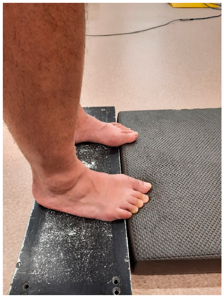
Figure 1. Position during intervention A. The metatarsophalangeal joint was aligned with the edge of the steel platform. Participants performed toe flexions against the mat.
Applsci 16 08478 g001
Figure 2. Electrode placement for electrical stimulation applied to the medial longitudinal arch.
Applsci 16 08478 g002
Figure 3. Assessment of hallux flexion strength. (a) The participant lies in a long-sitting position, and the metatarsophalangeal joint of the hallux is vertically aligned with the edge of the table. (b) The participant stands on both feet, and the metatarsophalangeal joint of the hallux is vertically aligned with the edge of the steel platform. The dynamometer transducer is placed under the distal hallux phalanx.
Applsci 16 08478 g003
Figure 4. Flowchart of the interventions and data collection. Participants performed the interventions in a randomized order, separated by a 1-week washout period, according to their sequence allocation. Intervention A consisted of concentric toe-flexor contractions against a mat while intervention B involved isometric toe-flexor contractions with electrical stimulation applied to the medial longitudinal arch of the foot.
Applsci 16 08478 g004
Table 1. Strength and navicular drop measurements before and after interventions.
Intervention AIntervention BInference
BeforeAfterBeforeAfterGeneral Linear ModelPost Hoc p-Values,
g [95% CI]
Strength sitting (N·kg−1)0.9 (0.3)0.7 (0.3) #0.9 (0.3)0.8 (0.2)p < 0.001 *
η2p = 0.29
Pre–post A: p < 0.001 #;
g = −1.07 [−1.62; −0.50]
Pre–post B: p = 0.30;
g = −0.47 [−0.93; −0.01]
Navicular drop (mm)6.5 (2.8)5.9 (2.7)6 (3.6)5.7 (4.0)p = 0.65
η2p = 0.02
/
Data are presented as mean (standard deviation). Statistical significance: * significant main effect of time at p < 0.05; # significant pre–post difference after Bonferroni correction. Hedges’ g effect sizes (g) were interpreted as very small (<0.2), small (<0.5), medium (<0.8), large (<1.2), very large (<2) and huge (≥2). Abbreviations used: g (Hedges’ g effect size), GLM (general linear model), η2p (partial eta squared effect size of the GLM), 95% CI (95% confidence interval).
Table 2. Hypothesis-driven biomechanical outcomes.
Intervention AIntervention BInference (GLM)
PrePostPrePost
9 km·h−1Contact duration
(% of stride)
38.5 (2.8)38.5 (2.8)38.8 (2.6)38.6 (2.7)p = 0.74; η2p = 0.02
Impact peak force
(% BW)
143.2 (27.4)142.8 (29.2)136.2 (29.1)141.7 (27.1)p = 1.00; η2p = 0.12
Active peak force
(% BW)
229.9 (21.3)230.5 (21.6)227.4 (17.9)229.3 (20.1)p = 0.47; η2p = 0.04
Vertical stiffness
(% BW·mm−1)
2.9 (0.3)2.9 (0.3)2.9 (0.2)2.9 (0.3)p = 0.196; η2p = 0.082
12 km·h−1Contact duration
(% of stride)
35.0 (2.6)34.7 (2.8)34.9 (2.7)34.8 (2.7)p = 0.56; η2p = 0.04
Impact peak force
(% BW)
160.6 (26.2)163.9 (28.5)163.0 (28.6)163.9 (27.2)p = 0.37; η2p = 0.06
Active peak force
(% BW)
245.5 (25.4)247.3 (25.1)245.0 (21.4)245.7 (22.5)p = 0.51; η2p = 0.03
Vertical stiffness
(% BW·mm−1)
3.2 (0.4)3.3 (0.4)3.2 (0.3)3.3 (0.4)p = 0.055; η2p = 0.13
Data are presented as mean (standard deviation). The p-value and partial eta squared are reported for the main effect of time. Statistical significance was set at p < 0.05. Abbreviations used: BW (body weight), GLM (repeated-measures general linear model), η2p (partial eta squared effect size of the GLM).
Disclaimer/Publisher’s Note: The statements, opinions and data contained in all publications are solely those of the individual author(s) and contributor(s) and not of MDPI and/or the editor(s). MDPI and/or the editor(s) disclaim responsibility for any injury to people or property resulting from any ideas, methods, instructions or products referred to in the content.