1. Introduction
Periodontal diseases are among the most prevalent chronic inflammatory oral conditions and represent a major cause of tooth loss worldwide. They are characterized by a dysbiotic microbial biofilm that triggers an inflammatory response, leading to the progressive destruction of the gingiva, periodontal ligament, cementum, and alveolar bone [
1,
2].
Periodontal wound healing is a complex biological process involving control of inflammation, angiogenesis, extracellular matrix remodeling, and tissue regeneration. Conventional therapies, including scaling and root planing, are effective in controlling infection but often provide limited outcomes, particularly in advanced defects where regenerative procedures are required [
2,
3].
In recent years, biomaterial-based therapies have gained increasing attention in periodontal regeneration. Among them, hydrogels stand out due to their ability to mimic the native extracellular matrix, high biocompatibility, injectability, biodegradability, and capacity for controlled drug delivery. Hydrogels can function as scaffolds for tissue regeneration while simultaneously delivering antimicrobial agents, growth factors, stem cells, or anti-inflammatory molecules directly to periodontal defects [
2,
4,
5].
Recent advances have also enabled the development of “smart” hydrogels responsive to environmental stimuli such as pH, temperature, or reactive oxygen species, allowing more targeted and controlled therapeutic effects with promising results in reducing inflammation, promoting angiogenesis and osteogenesis, and enhancing periodontal tissue regeneration. However, despite encouraging preclinical evidence, challenges related to long-term stability, mechanical resistance, and clinical translation remain significant. Therefore, this narrative review aims to present their potential as an adjunct in periodontal therapy.
2. Materials and Methods
2.1. Review Design
This narrative review was conducted following the principles of the Preferred Reporting Items for Systematic Reviews and Meta-Analyses (PRISMA 2020) to enhance methodological transparency and reproducibility. Although the review was not designed as a formal systematic review, a structured literature search and study selection process were implemented to minimize selection bias and ensure comprehensive coverage of the available evidence.
The objective of this review was to critically evaluate the current evidence regarding hydrogel-based biomaterials for periodontal wound healing and regeneration, emphasizing their physicochemical characteristics, biological mechanisms, antimicrobial properties, regenerative potential, clinical applications, translational challenges, and future perspectives.
2.2. Literature Search Strategy
A comprehensive electronic literature search was independently conducted by two reviewers using PubMed/MEDLINE, Scopus, and Web of Science. The search included articles published between October 2016 and May 2025, limited to studies published in English.
The search strategy combined Medical Subject Headings (MeSH) and free-text keywords using Boolean operators as follows:
“hydrogels” AND “periodontal regeneration”;
“periodontal wound healing” AND “biomaterials”;
“injectable hydrogels” AND “periodontitis”;
“smart hydrogels” AND “drug delivery”
“hydrogel scaffolds” AND “bone regeneration”;
“periodontal tissue engineering” AND “hydrogels”.
Reference lists of eligible articles were manually screened to identify additional relevant publications not retrieved during the electronic search.
2.3. Inclusion and Exclusion Criteria
Studies were selected according to predefined eligibility criteria.
Inclusioncriteria
Original research articles;
Randomized clinical trials;
Pilot clinical studies;
Animal studies;
In vitro investigations;
Systematic reviews and meta-analyses;
Narrative reviews providing relevant background information;
Studies evaluating natural, synthetic, hybrid, injectable or smart hydrogels for periodontal applications;
Articles investigating drug delivery systems, antimicrobial activity, tissue engineering, stem cell delivery or periodontal regeneration.
Exclusion criteria
Non-English publications;
Conference abstracts;
Editorials;
Letters to the editor;
Opinion papers;
Studies unrelated to periodontal regeneration;
Studies lacking sufficient methodological information.
2.4. Study Selection
Following duplicate removal, titles and abstracts were independently screened by two reviewers.
Potentially eligible studies underwent full-text evaluation according to the predefined inclusion and exclusion criteria.
Disagreements regarding study eligibility were resolved by discussion until consensus was reached.
The study selection process was summarized using a PRISMA 2020 flow diagram.
2.5. Data Extraction
The following variables were extracted from each included study:
Hydrogel composition;
Polymer classification;
Crosslinking strategy;
Drug incorporation;
Stem cell incorporation;
Antimicrobial activity;
Osteogenic potential;
Clinical application;
Main outcomes;
Advantages;
Limitations;
Translational relevance.
2.6. Quality Assessment and Risk of Bias
To improve methodological rigor, the methodological quality of the included studies was critically evaluated according to study design.
Randomized clinical trials were assessed according to the Cochrane Risk of Bias 2 (RoB 2) framework.
Animal studies were evaluated using the SYRCLE Risk of Bias Tool.
In vitro studies, pilot clinical studies, and case reports were critically appraised using the Joanna Briggs Institute (JBI) Critical Appraisal Checklists appropriate for each study design.
Methodological quality assessment considered the following domains whenever applicable:
Randomization;
Allocation concealment;
Blinding;
Outcome assessment;
Sample size adequacy;
Completeness of outcome reporting;
Reproducibility of experimental procedures;
Risk of selective reporting.
Overall methodological quality was categorized as High, Moderate, or Low, while the overall risk of bias was classified as Low, Moderate, or High.
Because this review included heterogeneous study designs, the quality assessment was used to support qualitative interpretation rather than quantitative synthesis.
2.7. Evidence Synthesis
The selected studies were qualitatively synthesized according to the principal research themes:
Periodontal wound healing;
Hydrogel classification;
Natural hydrogels;
Synthetic hydrogels;
Injectable hydrogels;
Smart hydrogels;
Drug delivery systems;
Stem cell delivery;
Antimicrobial applications;
Clinical evidence;
Translational challenges;
Future perspectives.
Greater emphasis was given to human clinical studies while considering preclinical evidence to explain biological mechanisms and emerging therapeutic strategies.
2.8. Certainty of Evidence
Because of the heterogeneity of study designs, hydrogel formulations, experimental models, and outcome measures, a quantitative meta-analysis was not feasible.
Clinical evidence was interpreted according to the GRADE framework whenever applicable, whereas preclinical evidence was synthesized descriptively.
The certainty of evidence was considered during data interpretation to avoid overestimation of preclinical findings and to distinguish experimental observations from clinically validated outcomes. The study selection process is summarized in
Table 1.
The initial search yielded 280 records across all databases. After duplicate removal and title and abstract screening, 77 full-text articles were assessed for eligibility. Following the eligibility assessment process, 41 studies met the inclusion criteria and were included in the final qualitative synthesis. Reasons for full-text exclusion included lack of relevance to periodontal regeneration, insufficient methodological detail, absence of hydrogel-based interventions, and studies unrelated to oral tissue engineering. A PRISMA flow diagram was constructed to summarize the study identification, screening, eligibility, and inclusion process. The methodological quality, risk of bias, level of evidence, and certainty of the included studies are summarized in
Table 2.
Most of the currently available evidence originates from in vitro and animal studies, whereas clinical evidence remains limited. Although the overall findings support the regenerative potential of hydrogel-based therapies, some studies reported variable outcomes attributable to differences in hydrogel composition, drug-loading strategies, experimental models, and follow-up periods. Consequently, the certainty of evidence supporting clinical translation remains moderate at best, highlighting the need for well-designed randomized controlled trials.
3. Pathophysiology of Periodontal Wound Healing and Engineered Hydrogels
Periodontal wound healing is a highly coordinated biological process involving inflammatory regulation, cell migration, angiogenesis, extracellular matrix remodeling, and tissue maturation. Unlike cutaneous healing, periodontal regeneration is particularly challenging because it requires the simultaneous restoration of multiple specialized tissues, including gingiva, periodontal ligament, cementum, and alveolar bone, all within a microenvironment constantly exposed to microbial biofilm and mechanical stress [
1,
2,
3].
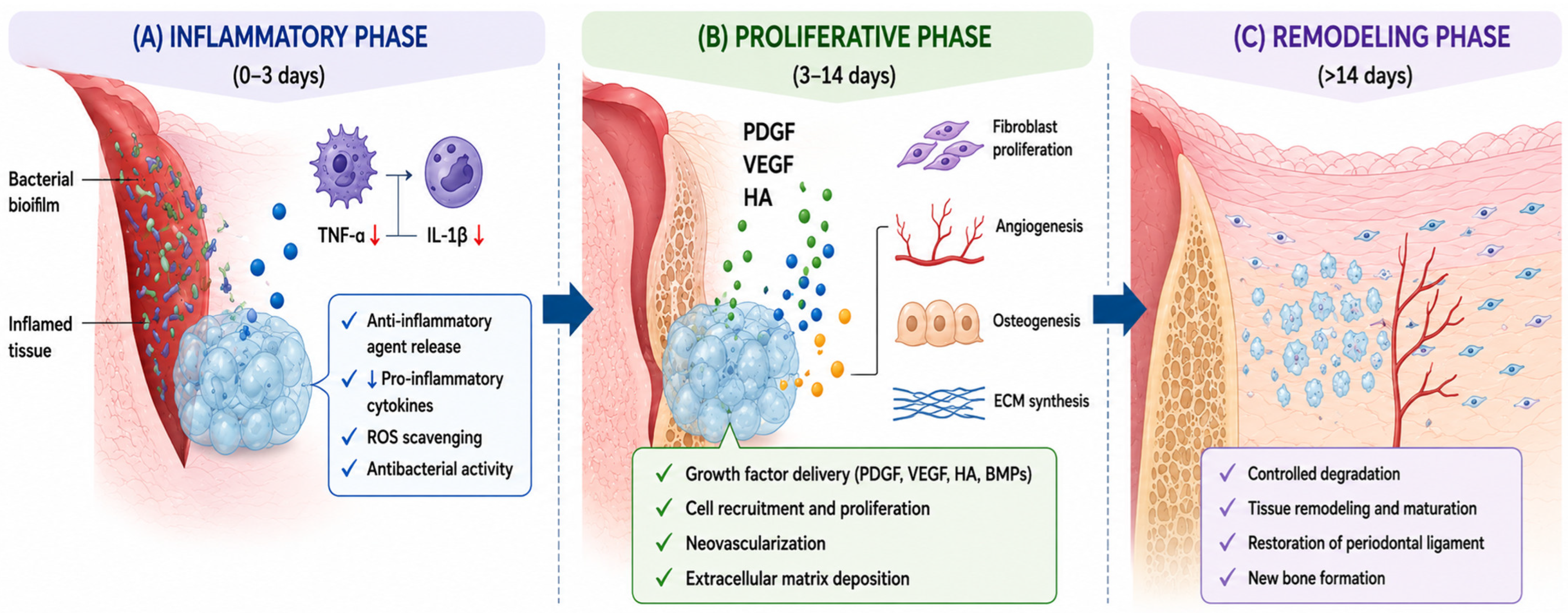
The healing process is traditionally divided into three overlapping phases: inflammatory, proliferative, and remodeling phases. During the inflammatory phase, neutrophils and macrophages migrate to the injured site and release cytokines such as TNF-α, IL-1β, and IL-6, which are essential for host defense but may also contribute to tissue destruction when excessively expressed. Matrix metalloproteinases (MMPs), highly activated in periodontal disease, further promote extracellular matrix degradation and alveolar bone loss. Recent studies suggest that engineered hydrogels can modulate this inflammatory microenvironment through the localized and sustained delivery of anti-inflammatory molecules such as doxycycline, curcumin, propolis or antioxidant nanoparticles, thereby reducing cytokine expression and oxidative stress while minimizing systemic side effects [
6,
7,
8,
9,
10].
The proliferative phase is characterized by fibroblast migration, angiogenesis, granulation tissue formation, and re-epithelialization. At this stage, hydrogels function as biomimetic extracellular matrix substitutes, providing a hydrated three-dimensional scaffold that supports cell adhesion, proliferation, and nutrient diffusion. Natural polymers such as hyaluronic acid, chitosan, collagen, and alginate are particularly advantageous due to their intrinsic biocompatibility and bioactivity. In addition, hydrogels can be enriched with growth factors such as Platelet-Derived Growth Factor (PDGF), Vascular Endothelial Growth Factor (VEGF), and Bone Morphogenetic Proteins (BMPs), promoting vascularization, osteogenic differentiation, and periodontal ligament regeneration. Experimental evidence has demonstrated that hydrogel-based delivery systems improve stem cell viability and enhance the regeneration of mineralized periodontal tissues [
11,
12].
During the remodeling phase, collagen fibers reorganize and mature while newly formed tissues acquire structural and functional stability. Hydrogels with controlled degradation profiles are especially important at this stage because they provide temporary mechanical support while being gradually replaced by newly regenerated tissue. The degradation kinetics of the hydrogel directly influence tissue maturation, cell differentiation, and extracellular matrix deposition. Smart or stimuli-responsive hydrogels have recently emerged as advanced therapeutic systems capable of responding to pH changes, reactive oxygen species, enzymatic activity, or temperature variations within the periodontal environment, allowing site-specific and on-demand release of therapeutic agents [
13,
14].
Figure 1 summarizes the principal biological mechanisms through which engineered hydrogels support periodontal regeneration across the inflammatory, proliferative, and remodeling phases.
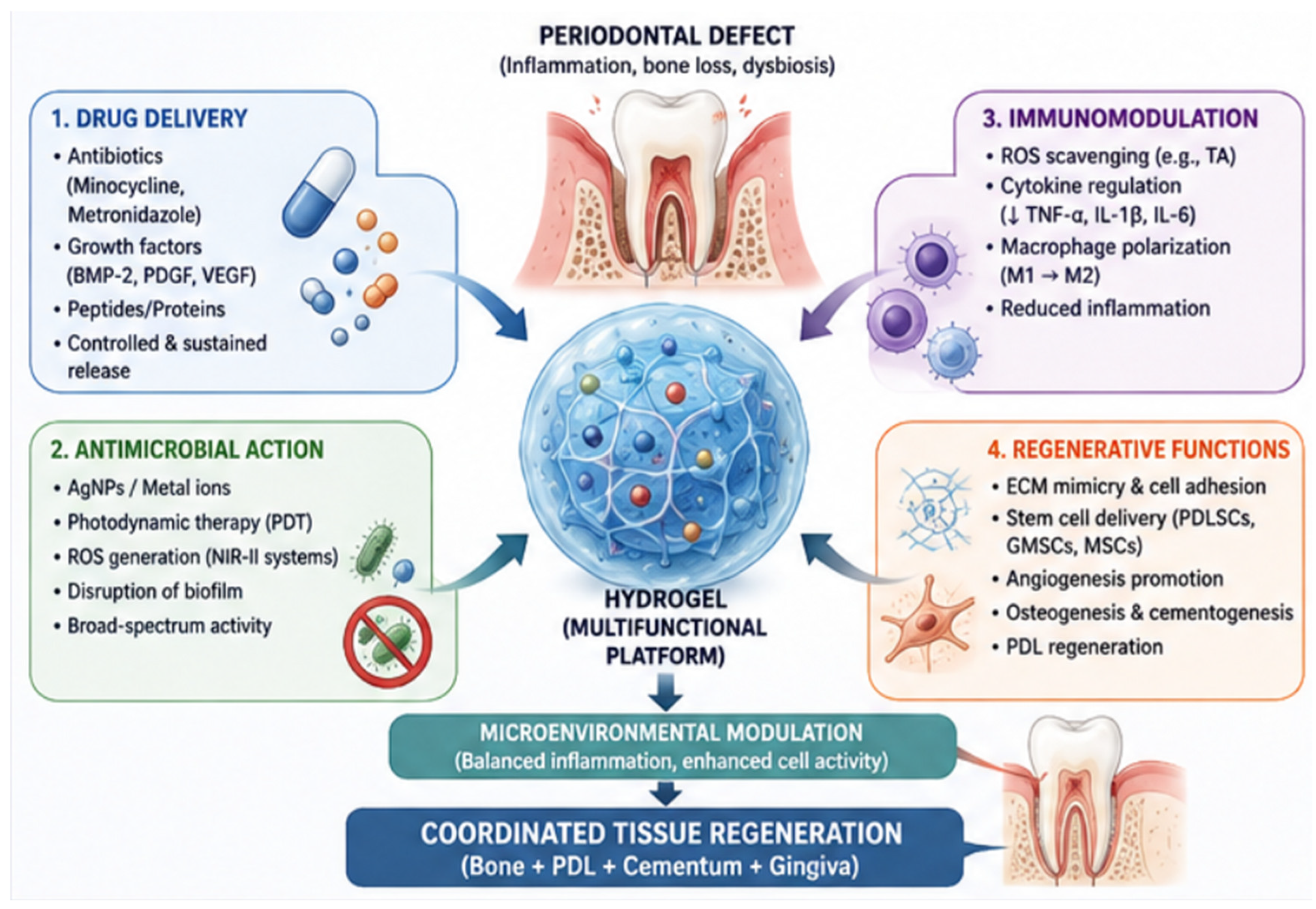
Engineered hydrogels therefore represent more than passive scaffolds; they function as multifunctional therapeutic platforms capable of orchestrating the periodontal healing cascade through spatiotemporal control of bioactive molecules and ability to simultaneously regulate inflammation, prevent bacterial proliferation, support angiogenesis, and promote osteogenesis.
4. Hydrogel Technology Overview
The three-dimensional feature of the hydrogels as hydrophilic polymer networks replicating the native extracellular matrix (ECM) architecture are critical for tissue engineering, targeted drug delivery, and periodontal regeneration due to their biocompatibility, tunable physicochemical profiles, and high loading capacities for bioactive sustained-release therapeutics [
15,
16].
The structural integrity and clinical efficacy of these systems depend on their cross-linking strategy and interconnected performance parameters, including porosity, swelling, and degradation kinetics [
4]. Physically cross-linked networks utilize reversible, non-covalent interactions but are highly susceptible to rapid dissolution, whereas chemically cross-linked hydrogels feature covalent bonds that grant superior mechanical stability, durability, and resistance to enzymatic cleavage [
14,
15]. These structural dynamics govern porosity, which balances nutrient transport and cell infiltration against antimicrobial barrier function, and must align with tissue neoformation timelines to provide adequate structural support [
16,
17].
To optimize spatiotemporal control within the inflammatory microenvironment of the periodontal pocket, stimuli-responsive (“smart”) systems leverage these parameters to trigger localized cargo release in response to fluctuations in pH, temperature, reactive oxygen species (ROS), or enzymatic activity [
4,
13]. Consequently, hydrogel design requires careful selection based on polymer origin. Natural polymers (e.g., hyaluronic acid, chitosan, alginate, collagen) provide inherent ECM homologies that mediate cell adhesion, migration, and immunomodulation, yet their biologically labile architectures suffer from low mechanical strength, rapid enzymatic degradation, and batch variability under masticatory forces and bacterial biofilms [
16,
18].
Conversely, synthetic polymers like poly(ethylene glycol) (PEG) and poly(vinyl alcohol) (PVA) offer precise, reproducible control over stiffness and degradation but lack intrinsic cell-signaling motifs, requiring functionalization with peptides (e.g., RGD sequences) or growth factors to elicit cellular interactions [
16,
19]. Ultimately, hybrid hydrogels merge these systems through blending or co-polymerization, yielding synergistic matrices that simultaneously exploit the biological cues of natural substrates and the mechanical resilience of synthetic backbones to withstand the complex challenges of the oral microenvironment [
17].
4.1. Natural Hydrogels
Hyaluronic Acid (HA)
Hyaluronic acid (HA) is a naturally occurring glycosaminoglycan and a major component of the extracellular matrix, recognized for its excellent biocompatibility, hydrophilicity, and viscoelastic properties [
18]. HA plays a critical physiological role in wound healing by regulating inflammation, angiogenesis, and cell migration. Following tissue injury, high-molecular-weight hyaluronic acid (HMW-HA) released by platelets contributes to clot stabilization and inflammatory cell recruitment, thereby initiating the healing cascade [
20].
HA-based hydrogels are particularly attractive for periodontal regeneration because they provide a hydrated microenvironment favorable for fibroblast proliferation, stem cell migration, and extracellular matrix deposition. Additionally, HA exhibits intrinsic anti-inflammatory properties and promotes tissue hydration and vascular permeability, contributing to improved soft tissue healing [
20]. Due to these characteristics, HA hydrogels have been extensively explored as wound dressings, injectable scaffolds, and carriers for growth factors and stem cells in regenerative dentistry [
21].
Chitosan
Chitosan is a cationic polysaccharide derived from the deacetylation of chitin and is widely used in biomedical applications because of its biodegradability, low toxicity, mucoadhesiveness, and antimicrobial activity [
18,
21]. Its positive surface charge allows electrostatic interaction with negatively charged bacterial membranes, disrupting membrane integrity and promoting bacterial cell death. This property is particularly valuable in periodontal therapy, where bacterial biofilm control is essential [
16].
In addition to its antimicrobial effects, chitosan possesses hemostatic properties by enhancing platelet aggregation and stabilizing fibrin clots, thereby supporting early wound-healing events. Chitosan hydrogels have demonstrated effectiveness in reducing bacterial load, controlling bleeding, and serving as local drug delivery systems for antibiotics and anti-inflammatory agents [
18]. However, despite their favorable biological profile, chitosan-based hydrogels may exhibit limited mechanical strength, often requiring combination with other polymers to improve stability and durability.
Gelatin and Alginate
Alginate, a naturally derived anionic polysaccharide extracted from brown algae, and gelatin, a denatured collagen derivative, are commonly used for hydrogel fabrication because of their ability to form highly hydrated three-dimensional networks that support cellular adhesion, proliferation, and migration [
22,
23]. When combined, gelatin–alginate hydrogels exhibit enhanced mechanical performance, tunable porosity, high water retention, and self-healing capabilities.
The physicochemical properties of these hydrogels can be adjusted by modifying the gelatin-to-alginate ratio, allowing regulation of scaffold stiffness, degradation kinetics, swelling behavior, and bioactivity [
23]. Moreover, these hydrogels demonstrate antibacterial effects, efficient exudate absorption, and favorable conditions for nutrient diffusion and tissue repair [
24,
25]. Their versatility has enabled applications in 3D bioprinting, stem cell encapsulation, wound dressings, and regenerative scaffolds for periodontal and bone tissue engineering [
4,
26].
Another strategy involves the development of injectable ternary hydrogels composed of gelatin, dialdehyde cellulose (DAC), and thiolated cellulose. In these systems, the aldehyde groups of DAC crosslink with the amino groups of gelatin through dynamic Schiff-base chemistry. Concurrently, thiolated cellulose introduces thiol-disulfide exchange pathways. This combined chemistry creates a tough, flexible network that effectively overcomes the low structural elasticity and rapid enzymatic breakdown traditionally seen in unmodified gelatin hydrogels [
27].
4.2. Synthetic Hydrogels
Polyethylene glycol
Poly(ethylene glycol) (PEG) is one of the most extensively studied synthetic polymers for biomedical applications due to its hydrophilicity, biocompatibility, and highly tunable mechanical properties [
17,
19]. PEG-based hydrogels provide excellent structural stability and can be engineered with controlled degradation rates and drug release profiles. Their chemical versatility also allows conjugation with peptides, proteins, or growth factors to improve cellular interactions and regenerative potential.
Nevertheless, PEG lacks intrinsic biological activity, limiting cell adhesion and proliferation when used alone. Consequently, PEG hydrogels are frequently combined with natural polymers or bioactive molecules to enhance biological performance in wound healing and periodontal regeneration [
28]. These hybrid systems can provide both mechanical integrity and improved biofunctionality.
Polyvinyl alcohol
Poly(vinyl alcohol) (PVA) hydrogels are synthetic polymer systems characterized by excellent mechanical strength, flexibility, hydrophilicity, and biocompatibility [
29,
30]. Depending on the fabrication method, PVA hydrogels can exhibit high toughness and elasticity, making them suitable for applications subjected to mechanical stress.
Because of their structural stability and bioadhesive behavior, PVA-based hydrogels have been investigated for wound dressings, tissue engineering scaffolds, and controlled drug delivery systems. However, similar to PEG, PVA lacks intrinsic bioactivity and may require incorporation of bioactive compounds or blending with natural polymers to improve cellular responses and regenerative capacity [
29].
4.3. Composite and “Smart” Hydrogels
Recent advances in hydrogel engineering have focused on the development of multifunctional composite and “smart” hydrogels capable of simultaneously providing antimicrobial activity, tissue regeneration, and controlled drug release. Composite hydrogels can incorporate antimicrobial agents such as chlorhexidine, metronidazole, or hydrogen peroxide, allowing sustained local release and reducing systemic side effects [
6].
Hydrogen peroxide-containing hydrogels have demonstrated prolonged antibacterial effects while also contributing to wound debridement and tissue repair processes [
31,
32]. Additionally, hydrogels combined with hydroxyapatite or calcium phosphate nanoparticles have shown promising osteoconductive and osteoinductive properties, supporting alveolar bone regeneration and mineralized tissue formation [
33,
34]. Injectable composite hydrogels are particularly advantageous for periodontal defects because they can adapt to irregular anatomical structures and permit minimally invasive application [
35].
Smart hydrogels represent a major advancement in periodontal biomaterials because they can respond dynamically to environmental stimuli such as pH, ROS levels, temperature, or electrical signals. pH-sensitive hydrogels modify their swelling behavior and drug release kinetics according to the acidity of inflamed tissues, enabling targeted therapy in periodontal pockets [
13]. Similarly, electrically responsive hydrogels have demonstrated “on–off” drug release mechanisms controlled by external electrical stimulation, providing precise regulation of therapeutic delivery [
14].
Although these technologies show remarkable promise, important challenges remain regarding long-term mechanical stability, reproducibility, large-scale manufacturing, and clinical translation.
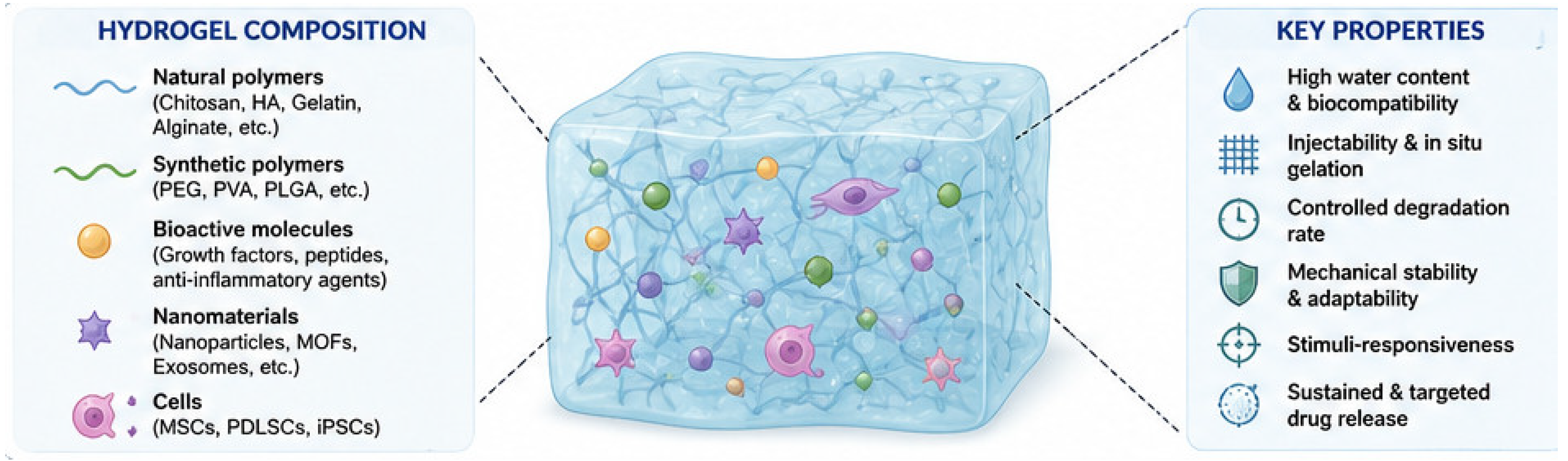
Figure 2 summarizes the main hydrogel components and their key physicochemical and biological properties relevant to periodontal regeneration.
An example of an advanced hybrid/composite smart platform is combining a natural animal protein (gelatin) with modified plant-derived polysaccharides (such as dialdehyde cellulose and thiolated cellulose). This dual-chemical cross-linking strategy significantly enhances the structural integrity, elasticity, and tunable degradation of the hydrogel, making it adaptable for intra-oral usage [
26,
36].
4.4. Mechanisms of Stimuli-Responsive (“Smart”) Hydrogels
Hydrogels capable of responding to pH changes, reactive oxygen species (ROS), temperature, or enzymatic activity have demonstrated the potential to release therapeutic agents selectively in inflammatory periodontal environments.
Stimuli-responsive hydrogels dynamically alter their physicochemical properties in response to localized periodontal microenvironmental triggers, undergoing structural modification, degradation, swelling, or therapeutic release mediated by reactive oxygen species (ROS), pH, temperature, enzymatic activity, or electrical signals. This responsiveness depends on network architecture and dynamic covalent bonds, including Schiff-base linkages, boronate ester bonds, disulfide bridges, or host–guest supramolecular interactions. These reversible linkages stabilize the matrix under physiological conditions but dissociate within pathological microenvironments [
13]. Molecule release is governed by diffusion through hydrated pores, polymer relaxation, network swelling, or scaffold degradation, with dual-responsive systems combining these modalities to achieve sequential therapeutic delivery. Such systems may improve local therapeutic efficacy while reducing systemic exposure and undesirable side effects [
8].
Because periodontal inflammation generates excessive ROS that drives tissue destruction, extracellular matrix degradation, and osteoclastogenesis, hydrogels incorporating ROS-cleavable linkers (e.g., thioketal bonds, boronic esters, or selenium moieties) undergo oxidative degradation for site-specific drug delivery. For example, ROS-triggered degradation can achieve coordinated co-release of minocycline and anti-inflammatory nanoparticles to reduce bacterial load and inflammatory cytokine expression [
8]. Recent advances have further expanded this concept through the incorporation of catalytic nanomaterials within hydrogel matrices. Nanozymes, which mimic the activity of natural antioxidant enzymes while exhibiting greater stability and tunability, have emerged as promising components for periodontal applications. Lu et al. [
37] demonstrated that oxygen-vacancy-engineered reduced Co
3O
4 nanocomposites exhibited enhanced peroxidase-like catalytic activity, highlighting the potential of nanomaterial-based catalytic systems to regulate oxidative processes. When embedded within hydrogel networks, such nanozymes may actively scavenge excess ROS, mitigate oxidative stress, and create a more favorable microenvironment for periodontal tissue regeneration.
Furthermore, integrating nanozymes with peroxidase-, catalase-, or superoxide dismutase-like activities allows hydrogels to neutralize local ROS, mitigating oxidative stress [
36]. Similarly, metal–organic framework (MOF)-integrated hydrogels utilize high porosity and tunable chemistry to combine pH- or oxidative-triggered drug delivery, immunomodulation, and tissue regeneration. To target acidic conditions caused by inflammation and bacterial metabolism, pH-responsive hydrogels utilize ionizable functional groups like carboxyl, amino, or phosphate moieties. Local pH shifts alter network ionization, osmotic pressure, and electrostatic interactions, accelerating drug release under acidic conditions while remaining stable in healthy tissues [
14]. Alternatively, electro-responsive systems incorporate conductive components, including polyaniline, polypyrrole, graphene oxide, or metallic nanoparticles, allowing external electrical stimulation to trigger matrix rearrangement and enable reversible “on–off” therapeutic release profiles [
14].
Saliva contamination, masticatory forces, and tissue movement frequently cause premature hydrogel detachment in the oral cavity. To counter this, asymmetric Janus structures and catechol- or mussel-inspired interfacial chemistries ensure stable wet-tissue bioadhesion and prevent postoperative tissue adhesion [
17]. Additionally, self-healing hydrogels leverage reversible dynamic bonds to spontaneously restore structural integrity after mechanical disruption during mastication and speech.
Next-generation platforms prioritize multifunctional hydrogels that integrate antimicrobial activity, ROS scavenging, osteoinduction, immunomodulation, and bioadhesion. Similarly, the incorporation of 3D bioprinting technologies has expanded the possibility of fabricating customized hydrogel scaffolds with controlled architecture and spatial organization for periodontal tissue engineering applications [
38].
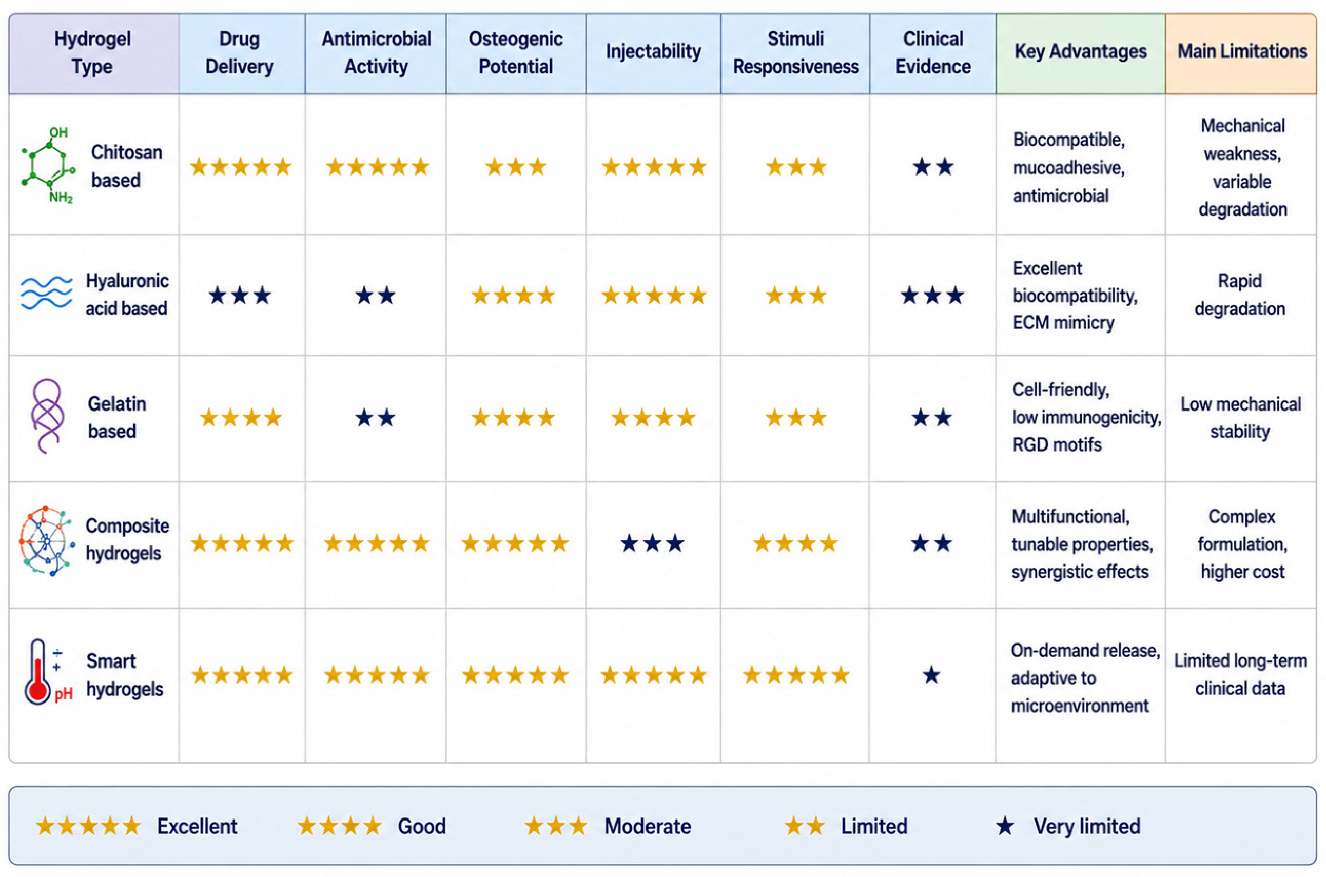
Figure 3 provides a comparative overview of the principal hydrogel platforms investigated for periodontal regeneration, highlighting their biological functions, therapeutic potential, and current level of clinical evidence.
5. Application of Hydrogels in Periodontal Healing
Hydrogels have emerged as multifunctional biomaterials in periodontal therapy due to their ability to combine localized drug delivery, structural support, antimicrobial activity, and tissue regenerative potential within a single platform.
5.1. Drug Delivery Vehicles
One of the most extensively investigated applications of hydrogels in periodontology is their use as localized drug delivery systems. Conventional systemic administration of antimicrobials in periodontal disease is often associated with low drug concentration at the target site, systemic side effects, and the potential development of bacterial resistance. Hydrogels overcome these limitations by enabling controlled and sustained release of therapeutic agents directly within the periodontal pocket, maintaining effective local concentrations while minimizing systemic exposure [
6,
7,
39].
Injectable thermosensitive hydrogels based on chitosan/gelatin/β-glycerophosphate have demonstrated promising results as carriers for metronidazole delivery. These systems undergo sol–gel transition at body temperature, allowing minimally invasive injection followed by in situ gelation inside periodontal defects. Zhu et al. [
8] reported sustained metronidazole release profiles, with cumulative release reaching approximately 67% in vitro, maintaining antimicrobial activity over prolonged periods while preserving biocompatibility.
Recent developments have focused on “smart” hydrogels capable of responding to the inflammatory microenvironment. Reactive oxygen species (ROS)-responsive hydrogels represent an important innovation because inflamed periodontal tissues exhibit elevated ROS levels. In a hyaluronic acid/poly(vinyl alcohol)-based injectable hydrogel, borate bonds degraded selectively in ROS-rich environments, triggering the controlled release of minocycline and anti-inflammatory nanoparticles. This strategy simultaneously reduced bacterial colonization, attenuated oxidative stress, and modulated local inflammation, illustrating the multifunctional therapeutic potential of responsive hydrogels [
8].
Beyond antibiotics, hydrogels have also been investigated as carriers for growth factors, anti-inflammatory molecules, probiotics, and nanoparticles. Their tunable porosity and degradation kinetics enable precise modulation of release profiles according to therapeutic requirements, making them highly versatile systems for periodontal applications.
5.2. Scaffold for Tissue Regeneration
Periodontal regeneration requires the coordinated formation of cementum, periodontal ligament, and alveolar bone, making scaffold design a critical challenge in regenerative dentistry. Hydrogels have attracted considerable interest because they can mimic the physicochemical and biological properties of the native ECM while providing a three-dimensional microenvironment favorable for cell migration, proliferation, and differentiation [
3,
4,
5,
39].
Natural polymer-based hydrogels composed of hyaluronic acid, collagen, chitosan, or alginate exhibit excellent biocompatibility and bioactivity, facilitating stem cell adhesion and tissue integration. Aligned porous hydrogels fabricated from chitosan and oxidized chondroitin sulfate supported periodontal ligament stem cells (PDLSCs) and gingival mesenchymal stem cells (GMSCs), promoting organized ligament formation and increased expression of osteogenic markers such as osteopontin (OPN), Runx-2, and collagen type I (COL-I) in periodontal defects [
27].
The regenerative potential of hydrogels can be further enhanced through incorporation of bioactive peptides and signaling molecules. Peptide-functionalized hydrogels containing Arg-Gly-Asp (RGD) motifs provide cell-adhesion sites that activate intracellular signaling pathways associated with osteogenic differentiation and tissue repair. Additionally, hydrogel stiffness, viscoelasticity, porosity, and degradation rate can be tailored to influence cellular behavior and vascularization during healing [
40].
Designing platforms that pair cell-signaling proteins with stable structural polysaccharides is crucial for proper anatomical reconstruction. A ternary gelatin/dialdehyde cellulose/thiolated cellulose configuration preserves the crucial RGD peptide motifs of gelatin necessary for initial periodontal ligament stem cell (PDLSC) adhesion. Simultaneously, the modified nanocellulose network provides a highly porous structural template that guides organized ligament alignment while facilitating hard tissue regeneration along the adjacent bone interface [
17].
Another important advancement involves combining hydrogels with osteoconductive materials such as hydroxyapatite or calcium phosphate nanoparticles. These hybrid systems better replicate the mineralized structure of alveolar bone and improve osteoinduction and mechanical stability, making them particularly suitable for periodontal intrabony defects and alveolar bone regeneration [
12].
Despite encouraging preclinical outcomes, most evidence regarding stem cell-loaded or growth factor-enriched hydrogels remains limited to animal models. Translation into clinical practice still faces important challenges related to manufacturing reproducibility, long-term stability, regulatory approval, and cost-effectiveness [
40].
5.3. Antimicrobial Activity
Persistent bacterial biofilm is the primary etiological factor in periodontal disease progression, making antimicrobial strategies essential for successful therapy. Hydrogels provide an effective platform for localized antimicrobial activity because they can encapsulate and gradually release antibacterial compounds directly into periodontal lesions [
24,
39,
41]. Silver nanoparticle-loaded hydrogels have shown broad-spectrum antibacterial properties against major periodontal pathogens. A pHEMA-based hydrogel containing silver nanoparticles and chlorhexidine demonstrated sustained antimicrobial release and significant inhibition of
Porphyromonas gingivalis. Silver ions exert bactericidal activity through multiple mechanisms, including disruption of membrane integrity, oxidative stress induction, and interference with bacterial DNA and protein synthesis [
41].
Hydrogels have also been integrated with antimicrobial peptides and photodynamic therapy systems. Chitosan-based hydrogels containing toluidine blue O (TBO) act as photosensitizer carriers capable of generating reactive oxygen species upon light activation. Peng et al. [
26] demonstrated substantial reductions in
P. gingivalis and
Aggregatibacter actinomycetemcomitans in three-dimensional gingival models following photodynamic activation.
More recently, multifunctional injectable hydrogels incorporating NIR-II photosensitizers have combined photothermal and photodynamic antibacterial effects with regenerative capacity. Under laser irradiation, these systems produce localized heat and ROS generation, enhancing bacterial elimination while simultaneously promoting bone regeneration [
42].
Beyond conventional antibiotic-loaded systems, emerging metal-based therapeutic strategies have attracted increasing interest for the management of persistent periodontal infections. In particular, chemodynamic therapy (CDT) utilizes transition metal ions to catalyze the generation of reactive oxygen species (ROS), thereby enhancing antibacterial activity within pathological microenvironments. A Mn–Cu bimetallic complex was reported to promote chemodynamic activity while simultaneously depleting intracellular glutathione, amplifying oxidative stress against pathogenic cells. Although originally explored in other biomedical contexts, similar approaches may be adapted to hydrogel-based platforms for periodontal applications, where localized ROS generation could contribute to the disruption of resilient biofilms and improve antimicrobial efficacy [
43].
In addition to their therapeutic effects, metallic nanomaterials may also contribute to improving the structural performance of hydrogel systems. The enhanced stability of gold and silver nanofluids through cationic gemini surfactant stabilization has been demonstrated, highlighting strategies for maintaining nanoparticle dispersion and functionality. The incorporation of stabilized metallic nanoparticles into hydrogel matrices may provide complementary benefits, including improved mechanical reinforcement, enhanced antimicrobial activity, and greater durability under challenging oral conditions [
44]. Such multifunctional systems represent a promising direction for the next generation of periodontal biomaterials.
Figure 4 illustrates the multifunctional mechanisms by which hydrogel-based platforms modulate the periodontal microenvironment and promote coordinated periodontal tissue regeneration.
5.4. Comparative Analysis and Evidence Hierarchy of Hydrogel Systems
Although the studies included in this review were heterogeneous in design, composition, and outcome measures, a structured comparative analysis of the available evidence allows for the identification of meaningful patterns regarding the therapeutic performance and clinical readiness of distinct hydrogel systems (
Table 3 and
Table 4).
In terms of evidence hierarchy, the highest level of clinical evidence currently available corresponds to natural and composite hydrogel systems. The randomized controlled trial conducted by Eshwar et al. [
46] represents the strongest clinical evidence, demonstrating that a fucoidan–chitosan injectable hydrogel yielded statistically significant improvements in probing pocket depth (PPD), clinical attachment level (CAL), and radiographic bone defect fill compared to conventional therapy over a 9-month follow-up period. Supporting clinical evidence from Miani et al. [
45] further established the adjunctive benefit of a metronidazole-containing hydrogel in reducing periodontal bacterial counts, reinforcing the utility of natural-polymer carriers for localized drug delivery. In contrast, synthetic hydrogels (PEG, PVA) and smart/responsive systems currently lack clinical trial data, with their evidence base confined to in vitro and animal models, placing them at the lowest tier of the evidence hierarchy.
From a comparative standpoint, natural hydrogels offer superior intrinsic bioactivity and biocompatibility, but their mechanical weakness and susceptibility to enzymatic degradation in the oral environment represent consistent limitations across studies. Synthetic hydrogels address these mechanical deficiencies but require bioactive functionalization to support cellular responses, as demonstrated by PEG and PVA-based systems. Composite and hybrid hydrogels emerge as the most balanced category, combining the biological cues of natural polymers with the structural resilience of synthetic backbones, and encompassing the only available RCT evidence. Smart hydrogels, while mechanistically the most sophisticated, remain at an early preclinical stage, with high therapeutic potential but substantial translational barriers including manufacturing complexity, oral environment stability, and absence of clinical validation.
Quantitatively, the sole RCT (Eshwar et al. [
46];
n = 40) reported a mean bone defect fill of 1.20 mm at 9 months (
p < 0.001) and significant CAL gain compared to concentrated growth factor alone. Preclinical studies using chitosan-based scaffolds. Zhang et al. [
24] demonstrated upregulation of osteogenic markers (OPN, Runx-2, COL-I), while antimicrobial systems [
26,
41] achieved significant pathogen inhibition against
P. gingivalis and
A. actinomycetemcomitans. ROS-responsive hydrogels [
8] achieved approximately 67% cumulative metronidazole release in vitro with simultaneous anti-inflammatory effects. These quantitative findings, while promising, derive primarily from preclinical settings and cannot be directly extrapolated to clinical practice without further validation through adequately powered randomized trials with standardized outcome measures and long-term follow-up.
Collectively, these data support a hierarchical classification of hydrogel systems by clinical readiness: (1) natural and composite hydrogels with clinical evidence (highest readiness); (2) natural hydrogels with preclinical evidence only (moderate readiness); (3) synthetic hydrogels requiring bioactive functionalization (low readiness); (4) smart/responsive and MOF-based systems at the frontier of preclinical research (lowest readiness, highest future potential). This hierarchy underscores the urgent need for well-designed, multicenter randomized clinical trials evaluating composite and smart hydrogel systems to bridge the current gap between preclinical promise and clinical adoption.
5.5. Advantages and Limitations
Advantages:
Hydrogels serve as biomimetic matrices in periodontal therapy due to their tunable physicochemical properties and multifunctional capacity. These networks enable localized, sustained therapeutic delivery directly into periodontal defects, maintaining localized drug concentrations while minimizing systemic adverse effects. Furthermore, stimuli-responsive hydrogels utilize localized microenvironmental cues, such as reactive oxygen species (ROS) or pH fluctuations, to achieve on-demand drug release and enhance therapeutic precision [
9].
The mechanical and degradation profiles of hydrogels are highly customizable. Modifying polymer composition, cross-linking density, or porosity regulates hydrogel stiffness, swelling kinetics, and degradation rates to match specific tissue regeneration timelines. Consequently, slowly degrading networks provide prolonged structural support, whereas rapidly degrading matrices facilitate short-term delivery.
Additionally, natural-polymer hydrogels—such as hyaluronic acid, gelatin, chitosan, and alginate—possess intrinsic biological activity. These substrates present biochemical motifs that promote fibroblast attachment, stem cell proliferation, angiogenesis, and osteogenic differentiation [
12]. Current research targets multifunctional platforms that combine these inductive properties with antimicrobial and anti-inflammatory functions [
32].
To make them easy to use, injectable hydrogels will permit minimally invasive clinical application. These formulations adapt fluidly to irregular periodontal defect geometries and undergo in situ gelation, reducing surgical trauma and enhancing adaptability within narrow periodontal pockets and complex intrabony defects where pre-formed biomaterials are structurally restricted [
7,
14,
47].
Limitations:
Despite their significant therapeutic potential, hydrogels still present important limitations that restrict their widespread clinical application. One of the major challenges is their relatively low mechanical strength. Many hydrogels are inherently soft and lack sufficient toughness to withstand the biomechanical forces generated during mastication or functional loading in the oral cavity. In periodontal regions exposed to constant mechanical stress, premature deformation or collapse of the hydrogel scaffold may compromise tissue regeneration and structural stability [
1].
Another critical limitation is the unpredictability of in vivo degradation. Hydrogel degradation can be strongly influenced by local environmental conditions, including enzymatic activity, inflammatory mediators, bacterial colonization, oxidative stress, and pH fluctuations. Excessively rapid degradation may result in premature loss of scaffold support and uncontrolled drug release, whereas excessively slow degradation can interfere with tissue remodeling and integration [
2]. Achieving an optimal balance between stability and biodegradability therefore remains a major challenge in hydrogel engineering.
Biological and translational limitations must also be considered. Although many studies report excellent results in vitro and in animal models, clinical evidence remains relatively limited. Most current studies involve small sample sizes, short follow-up periods, and heterogeneous methodologies, making direct comparison difficult. Consequently, the long-term clinical predictability and cost-effectiveness of hydrogel-based therapies are not yet fully established.
Manufacturing and regulatory barriers also represent substantial obstacles to clinical translation. Producing hydrogels under Good Manufacturing Practice (GMP) conditions while ensuring sterility, reproducibility, and batch-to-batch consistency can be technically complex and economically demanding [
48]. In addition, many advanced hydrogels are classified as combination products because they incorporate scaffolds, drugs, nanoparticles, growth factors, or living cells. This complexity requires more rigorous regulatory evaluation regarding safety, toxicity, biodegradation, and therapeutic efficacy, significantly prolonging approval processes and delaying commercialization [
3].
Another limitation involves storage stability and handling characteristics. Certain hydrogels may undergo premature degradation, dehydration, or physicochemical instability during storage, reducing their shelf life and clinical applicability. Injectable and stimuli-responsive hydrogels, although highly innovative, may also require sophisticated preparation methods or specialized equipment, increasing treatment costs and limiting accessibility in routine clinical practice.
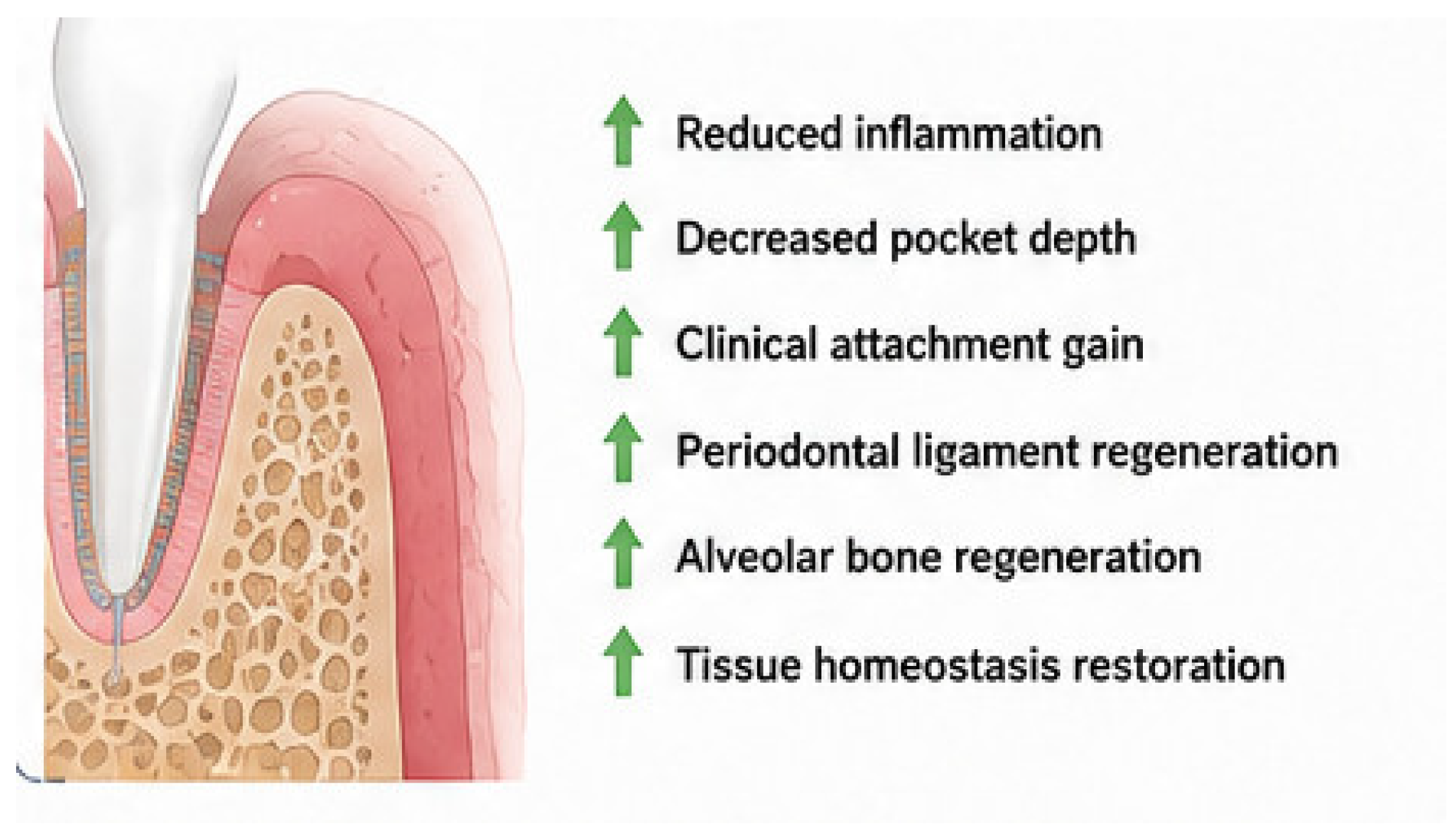
Finally, ethical and economic considerations may influence future implementation. The incorporation of stem cells, growth factors, or nanotechnology substantially increases production costs and regulatory complexity, potentially limiting the availability of these therapies in low-resource clinical settings. Therefore, future research should focus not only on improving biological performance but also on simplifying manufacturing processes, reducing costs, and generating robust long-term clinical evidence to facilitate broader clinical adoption. The main clinical outcomes associated with hydrogel-based periodontal regeneration are summarized in
Figure 5.
5.6. Bench-to-Bedside Translation and Clinical Implementation Challenges
Although most hydrogel investigations remain preclinical, clinical evaluation for periodontal therapy focuses on localized infection control and tissue regeneration. For antimicrobial delivery, an injectable, metronidazole-loaded gelatin–alginate hydrogel demonstrated high biocompatibility and sustained release kinetics within the periodontal pocket [
7,
45]. Clinically, intrapocket metronidazole gel administration significantly reduced bacterial counts in chronic periodontitis patients compared to mechanical debridement alone [
48]. For regenerative applications, a randomized controlled trial (RCT) of an injectable fucoidan–chitosan hydrogel in intrabony defects showed significantly improved bone defect fill and clinical attachment level gains, confirming its utility as a bioactive physical scaffold [
46].
Despite these outcomes, a translational gap persists between preclinical success and clinical adoption. Current clinical trials are constrained by small sample sizes, short follow-up durations, and limited methodological standardization. Furthermore, clinical translation is hindered by the dynamic oral microenvironment, where hydrogels face continuous exposure to saliva, pH fluctuations, bacterial biofilms, and cyclic mechanical loading. Salivary and tissue-derived enzymes—such as lysozyme, hyaluronidase, esterases, and matrix metalloproteinases (MMPs)—accelerate the degradation of natural polymer matrices (e.g., hyaluronic acid, gelatin, collagen, chitosan). Within the inflamed periodontal pocket, elevated oxidative stress and proteolytic activity exacerbate this degradation, causing premature structural collapse and uncoordinated drug release before complete tissue maturation occurs [
1,
2,
3,
39].
Biomechanical stress from mastication and occlusal forces imposes further constraints. Periodontal defects experience complex compressive, tensile, and shear forces that can induce hydrogel deformation, delamination, or displacement. Highly hydrated, low-modulus networks often lack the fracture toughness and fatigue resistance required to withstand long-term functional loading, a limitation especially pronounced in injectable or minimally cross-linked systems. To enhance structural stability and resistance to enzymatic cleavage, current research focuses on chemically cross-linked, composite, and hybrid networks incorporating synthetic polymers (e.g., PEG, PVA), ceramic nanoparticles, or multi-arm cross-linking agents. Additionally, self-healing and stimuli-responsive matrices are being developed to adapt dynamically to oral conditions [
22,
29].
Sterilization represents another important translational challenge. Many hydrogel systems are sensitive to conventional sterilization methods, including steam sterilization, gamma irradiation, and chemical agents, which may alter polymer structure, mechanical properties, degradation behavior, or drug-release kinetics. Consequently, maintaining hydrogel functionality while ensuring sterility remains a critical requirement for successful commercialization and clinical implementation [
2,
48].
Nevertheless, the rapid convergence of biomaterials science, nanotechnology, artificial intelligence, and regenerative medicine strongly suggests that Hydrogel-based therapies have the potential to become an important component of future periodontal treatment strategies, although further clinical validation and long-term outcome studies remain necessary. Their capacity to integrate antimicrobial action, immune modulation, tissue engineering, and personalized therapy positions hydrogels as one of the most promising platforms for achieving predictable and minimally invasive periodontal regeneration in the future.
6. Future Perspectives and Emerging Frontiers in Hydrogel Engineering for Periodontal Regeneration
Recent advances in biomaterials science have accelerated the development of next-generation hydrogel systems with increasingly sophisticated therapeutic capabilities. In particular, emerging technologies involving reactive oxygen species (ROS)-responsive hydrogels, metal–organic framework (MOF)-based systems, artificial intelligence-assisted biomaterial design, and advanced three-dimensional (3D) bioprinting are redefining the future of periodontal tissue engineering [
7,
8].
ROS-responsive hydrogels have gained considerable attention because oxidative stress plays a central role in periodontal tissue destruction and chronic inflammation. These smart systems are capable of selectively responding to elevated ROS concentrations present within inflamed periodontal pockets, enabling site-specific and on-demand release of antibacterial, anti-inflammatory, and osteogenic agents. Recent studies demonstrated that ROS-sensitive injectable hydrogels containing nanoparticle-based drug delivery platforms can simultaneously reduce bacterial colonization, modulate inflammatory cytokine expression, and promote alveolar bone regeneration [
8]. Such approaches represent an important advancement over conventional passive drug delivery systems by dynamically adapting therapeutic release according to disease activity.
Another rapidly emerging field involves hydrogel systems incorporating metal–organic frameworks (MOFs). MOFs possess highly porous structures, tunable surface chemistry, and large drug-loading capacity, making them highly attractive for periodontal applications. Recent investigations have shown that MOF-based hydrogels can provide controlled ion release, antimicrobial effects, immunomodulation, and enhanced osteogenesis. In particular, magnesium-based MOF hydrogels responsive to pH and ROS fluctuations have demonstrated promising regenerative potential by simultaneously promoting bone formation and reducing inflammatory tissue damage [
11]. These multifunctional systems may help overcome the limitations of conventional biomaterials that typically address isolated aspects of periodontal therapy.
Three-dimensional bioprinting technologies also represent a transformative advancement in regenerative periodontology. Unlike traditional scaffold fabrication techniques, 3D bioprinting allows precise spatial organization of cells, growth factors, and biomaterials within anatomically customized scaffolds. Recent biofabrication strategies using hydrogel-derived bioinks have enabled the development of multilayered periodontal constructs capable of mimicking the hierarchical organization of gingiva, periodontal ligament, cementum, and alveolar bone [
38]. Furthermore, advances in extracellular matrix-derived bioinks and stem cell-laden hydrogels may facilitate patient-specific regenerative therapies with improved structural and biological integration.
Artificial intelligence (AI) and machine learning are also beginning to influence hydrogel engineering and biomaterial optimization. AI-assisted biomaterial design may accelerate the identification of ideal polymer compositions, crosslinking conditions, degradation profiles, and drug-release kinetics according to specific clinical requirements. Predictive computational models are increasingly being explored to optimize hydrogel mechanical behavior, biocompatibility, and regenerative performance while reducing experimental variability and development time. In the future, integration of AI with biofabrication technologies and precision medicine may enable fully personalized hydrogel systems adapted to individual inflammatory profiles, microbiome composition, and tissue regenerative capacity.
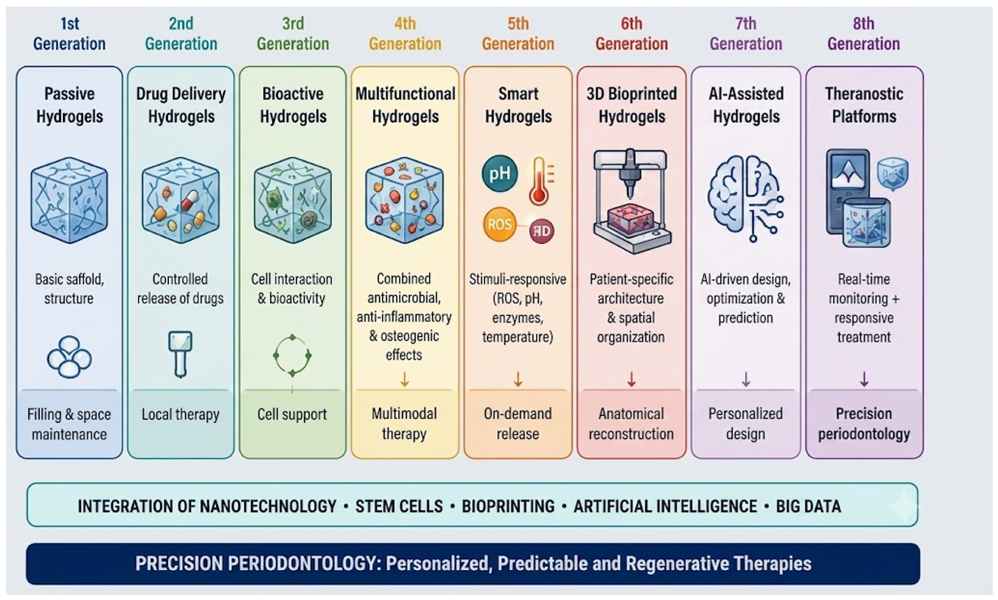
Collectively, these emerging technologies suggest that hydrogel-based therapies are evolving from passive biomaterials into intelligent, adaptive, and multifunctional therapeutic platforms. Continued integration of nanotechnology, artificial intelligence, biofabrication, and regenerative biology will likely play a central role in the development of next-generation periodontal therapies capable of achieving more predictable and personalized tissue regeneration outcomes.
Figure 6 summarizes the technological evolution of hydrogel systems from passive biomaterials to intelligent, precision-guided regenerative platforms for periodontal therapy.
6.1. Multifunctional Hydrogels
One of the most promising directions involves multifunctional hydrogels that combine bioactive ceramics, nanoparticles, peptides, or metallic ions within a single biomaterial platform [
49]. These systems aim to provide synergistic therapeutic effects by integrating antimicrobial, anti-inflammatory, angiogenic, and osteogenic properties. For example, sericin-based nanoparticles mineralized with ion-doped hydroxyapatite and coated with epigallocatechin-gallate (EGCG) demonstrated antioxidant, immunomodulatory, and bone-regenerative capabilities, significantly accelerating periodontal bone formation in vivo [
39].
Similarly, hydrogel–metal–organic framework (MOF) systems have attracted considerable attention due to their ability to provide controlled and stimuli-responsive release of bioactive molecules. Luo et al. [
11] developed magnesium–gallic acid MOF-containing hydrogels capable of responding to pH and ROS fluctuations associated with periodontal inflammation, enabling simultaneous immunomodulation and osteogenesis. These multifunctional approaches may overcome one of the major limitations of conventional therapies, which typically address either infection control or regeneration independently rather than both simultaneously.
Future research is expected to focus on integrating multiple therapeutic agents within the same hydrogel platform, allowing synchronized delivery of antibiotics, antioxidants, growth factors, and regenerative molecules according to the evolving periodontal microenvironment [
24].
Santos et al. [
5] emphasized that the modularity of natural and synthetic hydrogel components enables the design of customized systems capable of adapting to distinct inflammatory profiles and regenerative demands. In the future, hydrogel formulations may incorporate microbiome-guided antimicrobial strategies or personalized release profiles based on patient-specific biomarkers. Such precision approaches could improve therapeutic predictability while minimizing overtreatment and adverse effects.
Although personalized hydrogel therapies remain largely experimental, they represent an important step toward precision periodontology and individualized regenerative medicine.
6.2. Stem Cell Integration
The integration of stem cells within hydrogel matrices represents another major frontier in periodontal regeneration. Mesenchymal stem cells (MSCs), periodontal ligament stem cells (PDLSCs), and gingival mesenchymal stem cells (GMSCs) possess significant regenerative potential due to their ability to differentiate into osteogenic, fibroblastic, and cementoblastic lineages. Hydrogels provide an ideal microenvironment for stem cell survival because they mimic the native extracellular matrix and protect cells during implantation.
Recent advances in tissue engineering have demonstrated that stem cell-laden hydrogels can enhance tissue regeneration by improving cell retention, proliferation, vascularization, and differentiation [
18]. In periodontology, bioprinted hydrogels containing progenitor cells and bioactive molecules are being investigated to reconstruct both soft and hard periodontal tissues simultaneously. Chen et al. [
38] described recent progress in 3D bioprinting technologies capable of generating cell-laden bioinks for periodontal regeneration with improved spatial organization and tissue specificity.
Despite these advances, important translational challenges remain, including stem cell sourcing, long-term safety, immunogenicity, ethical considerations, and manufacturing costs. Therefore, future studies should prioritize standardized protocols and long-term clinical evaluation before widespread clinical implementation becomes feasible.
6.3. Bioprinting Applications
Three-dimensional bioprinting is expected to play a transformative role in the future of periodontal regeneration. Conventional scaffolds often lack the anatomical precision required to reconstruct the complex architecture of periodontal tissues, including gingiva, periodontal ligament, cementum, and alveolar bone. Bioprinting technologies address this limitation by enabling the fabrication of patient-specific scaffolds with highly controlled geometry, porosity, and spatial distribution of cells and biomolecules.
Yin et al. [
39] highlighted that extracellular matrix-derived bioinks and hydrogel-based bioprinting systems can reproduce the hierarchical organization of periodontal tissues more accurately than traditional scaffold fabrication techniques. In addition, bioprinting allows the incorporation of multiple cell populations and growth factors within different regions of the scaffold, potentially enabling simultaneous regeneration of multiple tissue compartments.
Future advancements may involve chairside bioprinting technologies, image-guided scaffold fabrication based on CBCT imaging, and hybrid bioinks capable of dynamic remodeling after implantation. However, challenges related to vascularization, mechanical stability, printing resolution, and clinical scalability must still be addressed before routine clinical application.
6.4. Responsive (“Smart”) Hydrogels
Stimuli-responsive or “smart” hydrogels represent one of the most innovative developments in periodontal biomaterials research. Unlike conventional passive delivery systems, these advanced platforms are engineered to dynamically respond to specific environmental cues associated with periodontal inflammation, including pH fluctuations, reactive oxygen species (ROS) accumulation, enzymatic activity, temperature variations, and inflammatory biomarkers. Such responsiveness allows the hydrogel matrix to undergo controlled structural changes that regulate the release of therapeutic agents according to local pathological conditions [
7,
8,
13,
50].
Recent advances have further expanded the functionality of these systems through the integration of nanotechnology and multifunctional biomaterials. For example, Zhu et al. [
8] developed ROS-sensitive injectable hydrogels incorporating minocycline-loaded ZIF-8 nanoparticles, which enabled selective release of antibacterial and anti-inflammatory agents within inflamed periodontal pockets. Similarly, Luo et al. [
11] reported self-healing metal–organic framework (MOF)-based hydrogels with dual responsiveness to pH and ROS variations, providing prolonged immunomodulatory effects together with controlled ion release throughout the healing process.
Looking ahead, the next generation of smart hydrogels may evolve into fully integrated “theranostic” platforms capable of combining real-time disease monitoring with responsive therapeutic intervention. By continuously sensing local inflammatory biomarkers and automatically adjusting drug-release profiles according to disease activity, these intelligent biomaterials could significantly improve treatment precision, therapeutic efficiency, and long-term periodontal maintenance.
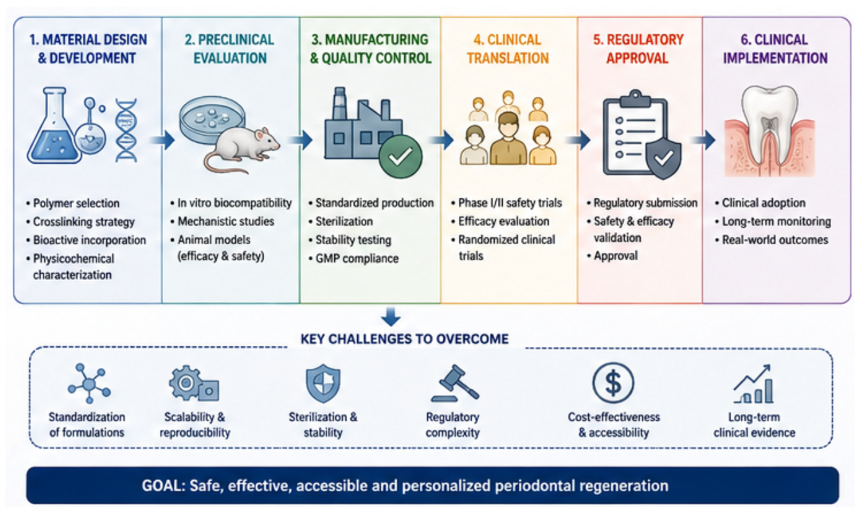
Figure 7 illustrates the key stages involved in the clinical translation of hydrogel-based periodontal therapies, from biomaterial design and preclinical evaluation to regulatory approval and routine clinical implementation.
7. Discussion
The present review confirms that hydrogels constitute one of the most promising biomaterial platforms for periodontal regeneration due to their ability to simultaneously modulate inflammation, deliver therapeutic agents, and support tissue reconstruction. Unlike conventional periodontal therapies, which mainly aim to control infection and halt disease progression, hydrogel-based systems provide a regenerative microenvironment capable of actively promoting the reconstruction of periodontal ligament, cementum, and alveolar bone. This distinction is clinically significant because true periodontal regeneration requires coordinated healing of multiple tissue compartments rather than simple tissue repair [
39,
51].
One of the major strengths identified in the literature is the biomimetic nature of hydrogels [
18]. These properties allow hydrogels to function not only as passive scaffolds but also as dynamic biological environments capable of influencing cellular behavior during wound healing. Compared with traditional regenerative materials, such as membranes or graft particles, hydrogels provide greater adaptability to irregular periodontal defects and can be engineered to deliver multiple therapeutic functions simultaneously [
16].
The evidence analyzed also indicates that hydrogels can interact with all phases of periodontal wound healing. During the inflammatory phase, hydrogels capable of releasing anti-inflammatory agents or antioxidants contribute to the reduction in cytokines such as TNF-α and IL-1β, thereby limiting tissue destruction and oxidative stress [
1]. This is particularly relevant because chronic inflammation is one of the principal factors responsible for periodontal tissue breakdown. In the proliferative phase, hydrogels enriched with growth factors such as PDGF and VEGF promote angiogenesis, fibroblast proliferation, and stem cell recruitment, accelerating tissue formation and vascularization [
16]. During the remodeling phase, controlled degradation kinetics enable the scaffold to gradually transfer mechanical and biological functions to newly formed tissues, facilitating collagen organization and maturation [
3].
Another important finding is the multifunctionality of modern hydrogel systems. In addition to acting as structural scaffolds, hydrogels can simultaneously serve as localized drug delivery systems, antimicrobial platforms, and stem cell carriers. This multifunctional behavior is especially advantageous in periodontology because periodontal defects are biologically complex environments characterized by bacterial contamination, inflammation, and loss of both soft and hard tissues. Stimuli-responsive hydrogels, particularly ROS-sensitive and pH-sensitive systems, represent an important advancement because they enable on-demand drug release according to the pathological microenvironment [
8]. Such systems may improve therapeutic precision while reducing systemic drug exposure and side effects.
The literature reviewed demonstrates that both natural and synthetic hydrogels can actively participate in all phases of periodontal wound healing. Their hydrated three-dimensional structure mimics the extracellular matrix, supporting cell adhesion, migration, angiogenesis, and tissue remodeling. In addition, advances in biomaterial engineering have enabled the development of multifunctional and stimuli-responsive hydrogels capable of delivering antimicrobial, anti-inflammatory, and osteogenic agents in a controlled and localized manner.
Despite these advances, the literature reveals a substantial discrepancy between promising preclinical findings and limited clinical translation. Most published studies are based on in vitro experiments or animal models, which consistently demonstrate improved osteogenesis, antimicrobial activity, angiogenesis, and tissue regeneration. However, relatively few randomized clinical trials have evaluated hydrogel-based therapies in humans. Existing clinical studies generally involve small sample sizes and short follow-up periods, limiting the ability to establish long-term predictability and evidence-based clinical protocols. Therefore, although preclinical evidence strongly supports the regenerative potential of hydrogels, additional multicenter clinical trials with standardized methodologies are necessary before these materials can be widely incorporated into routine periodontal therapy.
Mechanical stability remains one of the principal limitations discussed in the literature. Many hydrogels possess inherently soft and highly hydrated structures, which compromise their resistance to biomechanical forces generated during mastication [
1]. In periodontal defects exposed to functional loading, insufficient mechanical strength may lead to premature collapse or degradation of the scaffold, negatively affecting tissue regeneration. To address this limitation, recent research has focused on hybrid hydrogels incorporating ceramics, nanoparticles, or synthetic polymers to improve mechanical performance while maintaining biocompatibility.
Another critical issue involves the unpredictability of in vivo degradation. The degradation profile of hydrogels may vary according to enzymatic activity, pH changes, bacterial colonization, and inflammatory conditions within the periodontal pocket [
3]. Excessively rapid degradation may result in loss of structural support before tissue maturation occurs, whereas prolonged persistence may interfere with natural tissue remodeling.
Recent innovations involving stem cell therapy, nanoparticle delivery systems, and 3D bioprinting further expand the regenerative potential of hydrogels and may allow the fabrication of more personalized and anatomically precise scaffolds for periodontal therapy. An additional trend involves the development of “theranostic” hydrogels capable of combining diagnosis and therapy within the same platform. However, important challenges remain, including limited mechanical resistance, variability in biodegradation behavior, manufacturing scalability, regulatory approval, and the scarcity of long-term clinical trials in humans.
8. Conclusions
Overall, hydrogels represent a versatile and rapidly advancing class of biomaterials for periodontal therapeutic applications, offering controlled delivery, tunable mechanics, and cell-instructive properties that support coordinated regeneration of soft and hard periodontal tissues; however, their clinical translation requires harmonized preclinical models, comprehensive long-term safety and biodegradation data, dose- and delivery-optimization studies, and well-powered randomized clinical trials, alongside consideration of manufacturability, regulatory pathways, and cost-effectiveness to confirm their sustained safety, efficacy, and practical applicability in evidence-based periodontal practice.





 ,
,