Systematic Review

The Use of 308-nm Excimer Laser Therapy in Alopecia Areata and Primary Cicatricial Alopecia: A Systematic Review


1
Department of Dermatology, Indiana University School of Medicine, Indianapolis, IN 46202, USA
2
Ronald O. Perelman Department of Dermatology, Grossman School of Medicine, New York University, New York, NY 10016, USA
*
Author to whom correspondence should be addressed.
Appl. Sci. 2026, 16(14), 7337; https://doi.org/10.3390/app16147337
Submission received: 21 June 2026 / Revised: 17 July 2026 / Accepted: 19 July 2026 / Published: 22 July 2026

Featured Application

The 308 nm excimer laser is a light-based therapy that targets overactive immune cells in the skin without affecting the rest of the body, making it a useful option for patients with hair loss conditions who cannot tolerate repeated steroid injections or long-term medications. This review updates the evidence base for excimer therapy in alopecia areata, where response rates fall within the range reported for standard steroid injections in small heterogeneous studies, with additive benefit suggested in combination regimens. It provides, to our knowledge, the first systematic assessment of its use in primary cicatricial alopecia, finding early evidence of inflammatory suppression in lichen planopilaris and insufficient but non-negligible findings in frontal fibrosing alopecia. These findings support excimer therapy as a well-tolerated, locally acting treatment option and identify scarring alopecia as a priority area for future clinical trials.

Abstract

Background: Lichen planopilaris (LPP) and frontal fibrosing alopecia (FFA) are primary cicatricial alopecias with no FDA-approved treatments and a limited evidence base for any intervention. Alopecia areata (AA), while better studied, similarly lacks a well-tolerated, locally acting non-pharmacologic option. The 308 nm excimer laser is established in inflammatory dermatoses, yet its role in hair loss conditions has not been systematically characterized. This review provides an updated appraisal of the AA evidence base and the first systematic appraisal of excimer therapy in LPP and FFA. Methods: We searched PubMed/Medline, Embase, Web of Science, Scopus, and Cochrane CENTRAL and synthesized findings narratively, given substantial heterogeneity across included studies. Results: In total, 33 studies reporting 634 unique patients met the inclusion criteria across AA subtypes (patchy/localized, totalis, universalis, ophiasic, and pediatric), LPP, and FFA. Most evidence (30/33 studies, 613/634 patients) pertains to AA, where excimer therapy produced meaningful hair regrowth in a large proportion of patients. Cicatricial data was more limited (3/33 studies, 21/634 patients). LPP data suggest improvement in inflammatory symptoms; FFA results were mixed. Adverse effects were mild and transient, primarily erythema and hyperpigmentation. Conclusions: The use of 308 nm excimer therapy may be a well-tolerated non-pharmacologic option when pharmacologic therapy is contraindicated, poorly tolerated, or inadequately effective, though limited evidence, especially for LPP and FFA, warrants further investigation across these and other conditions not yet studied.

1. Introduction

Alopecia is divided into two general categories based on whether the condition leads to permanent destruction of hair follicles. Nonscarring types, in which hair follicles are not permanently destroyed and regrowth remains possible, include diffuse conditions like telogen effluvium and patterned loss (e.g., androgenetic alopecia), as well as localized conditions like alopecia areata (AA) [1,2]. Scarring alopecia, which includes conditions like lichen planopilaris (LPP), frontal fibrosing alopecia (FFA), discoid lupus erythematosus, and folliculitis decalvans (FD), is characterized by the destruction of the hair follicle cells and stem cells, leading to fibrotic replacement of follicular architecture with permanent hair loss [2,3,4]. Histopathologically, LPP and FFA share features of lymphocytic infiltration targeting the isthmus and apoptotic cells in the external root sheath [2,5]. In scarring forms, hair loss may progress subclinically, and substantial follicular destruction may occur before alopecia becomes clinically apparent [3]. In several major inflammatory subtypes (e.g., AA, LPP, and FFA), a shared pathogenic thread is T-cell-mediated follicular injury and genetic susceptibility [4,6]. However, downstream consequences differ, as immune privilege collapse is reversible in nonscarring AA, while the follicular destruction is permanent in scarring alopecia. In AA specifically, collapse of hair follicle immune privilege is considered a critical initiating event, exposing follicular autoantigens to autoreactive CD8 + NKG2D + T cells driven by interferon-γ (IFN-γ) [6,7,8].
Therapeutic options for hair loss conditions, especially inflammatory subtypes, remain limited compared to other major dermatologic conditions (e.g., atopic dermatitis and psoriasis) [4]. Although systemic immunosuppression is often utilized, the tolerability of available treatments is a recognized challenge [4,9]. Intralesional corticosteroid (ILCS) injection is the most commonly used treatment of localized AA in the United States, with moderate efficacy, stimulating localized regrowth in approximately 60–67% of cases [10,11]. However, side effects involve injection-site pain and localized skin atrophy; additionally, relapses are frequent after treatment discontinuation [10]. These limitations flag further caution in pediatric populations, where use is limited by injection-associated pain and in patients with extensive disease [11,12]. Recently, biologics and Janus kinase (JAK) inhibitors have been investigated as systemic therapies for severe AA and scarring alopecia; however, the limited long-term safety data support the continued rationale for targeted, local therapies [13,14,15,16]. In this context, the 308 nm excimer laser represents a targeted phototherapeutic alternative. Originally approved by the US Food and Drug Administration (FDA) for the treatment of psoriasis [17,18], the 308 nm excimer laser has since demonstrated efficacy in conditions including vitiligo [17,18], atopic dermatitis [19,20], prurigo nodularis [21], mycosis fungoides [22], localized scleroderma [23], and chronic hand and foot eczema [24]. It requires fewer treatment sessions than conventional narrowband ultraviolet B (NB-UVB) phototherapy, delivers a lower cumulative UV dose, and limits irradiation to lesional skin, allowing for high fluences at the treatment site without exposing surrounding healthy tissue [25,26].
The rationale for 308 nm excimer laser therapy is supported by its immunological mechanism. Specifically, at 308 nm, UVB is absorbed by infiltrating T cells and keratinocytes, inducing DNA photodamage, upregulating p53, and downregulating Bcl-2, driving pathogenic T cells toward apoptosis [26,27]. In AA, disruption of hair follicle immune privilege is thought to make anagen follicles more susceptible to CD8+ T cell attack and sustained perifollicular inflammation [4,28]. Given this mechanism, the 308 nm excimer laser has been evaluated as a therapeutic option in AA [29].
Despite growing interest in excimer laser therapy for hair loss, prior reviews have examined excimer therapy almost exclusively in AA. Its role in primary cicatricial alopecia, a condition with no FDA-approved treatments where the consequences of undertreated inflammation are irreversible, has never been systematically examined. We conducted a systematic review of 308 nm excimer therapy across hair loss conditions with two explicit goals: First, to provide an updated synthesis of the AA evidence base, including its performance relative to intralesional corticosteroids and its additive value in combination regimens. Second, to provide the first rigorous accounting of what evidence exists, and what does not, for excimer therapy in LPP and FFA to establish whether a proof-of-concept signal is present and to define the research agenda for prospective investigation.

2. Materials and Methods

This systematic review and narrative synthesis was conducted in accordance with the Preferred Reporting Items for Systematic Reviews and Meta-Analyses (PRISMA) guidelines. However, this review was not prospectively registered (e.g., with PROSPERO).
We included studies enrolling patients with any form of alopecia treated with 308 nm excimer laser therapy. Eligible conditions included, but were not limited to, alopecia areata (patchy, totalis, universalis, and ophiasis subtypes), androgenetic alopecia, lichen planopilaris (LPP), frontal fibrosing alopecia (FFA), central centrifugal cicatricial alopecia (CCCA), folliculitis decalvans, telogen effluvium, anagen effluvium, trichotillomania, and dissecting cellulitis. The index intervention was monotherapy or adjunctive use of the 308 nm excimer laser at any reported fluence, treatment frequency, or duration. No restrictions were placed on concurrent therapies; however, the nature and verifiability of co-interventions were considered when interpreting study findings. No restrictions on comparators were applied. Studies with active comparators, sham or untreated controls, intra-individual split designs, and single-arm before-after designs were all eligible for inclusion. The primary outcomes of interest were objective measures of hair regrowth or arrest of hair loss, assessed by any validated or investigator-reported instrument, such as the Severity of Alopecia Tool (SALT) score for AA. Secondary outcomes included patient-reported outcomes (e.g., satisfaction, quality of life), inflammatory symptom reduction (e.g., pruritus, erythema, perifollicular scale), and safety and tolerability data, including reported adverse events. We included randomized controlled trials (RCTs), non-randomized studies with concurrent comparator groups (including quasi-experimental and cohort designs), single-arm non-randomized studies, case series, and case reports. Review articles, editorials, letters without original data, conference abstracts without full-text data, and animal or in vitro studies were excluded. Inclusion was restricted to studies published in English. No restriction on publication date was applied for the lower bound; the search was conducted through 31 December 2025.
A systematic search was conducted in PubMed, Embase, Scopus, Web of Science, and the Cochrane Central Register of Controlled Trials (CENTRAL) on 15 January 2026. The search strategy was developed using controlled vocabulary terms (e.g., MeSH) and free-text keywords encompassing 308 nm excimer laser therapy and alopecia in all forms. The full search strategy for each database is provided in Appendix A. Reference lists of all included studies and relevant review articles were hand-searched to identify any additional eligible records.
All identified records were imported into Rayyan (Rayyan Systems Inc.; Cambridge, MA, USA; available at https://www.rayyan.ai/; accessed [January 2026], a web-based systematic review management platform, for deduplication and screening. Two reviewers independently screened titles and abstracts, followed by full-text review of potentially eligible records. Disagreements at any stage were resolved through discussion and consensus. Extracted variables from included studies included: study design and setting; patient demographics and alopecia subtype; disease duration and severity at baseline; excimer laser parameters (manufacturer, fluence, treatment interval, total number of sessions); co-interventions; outcome measures and assessment timepoints; reported efficacy data; and adverse events.
Meta-analysis was not performed due to clinical, methodological, and statistical heterogeneity across included studies. Instead, findings were synthesized narratively, organized by alopecia subtype. Effect estimates from individual studies are reported descriptively alongside their risk of bias ratings. Given the predominance of small, heterogeneous studies across diverse alopecia subtypes, narrative synthesis was the appropriate approach for characterizing the available evidence.
RCTs were assessed using the Cochrane Risk of Bias 2 (RoB 2) tool. Non-randomized studies with comparators were assessed using the Risk of Bias in Non-randomized Studies of Interventions (ROBINS-I) tool. Case series and non-randomized studies without comparators or baseline comparators were appraised using the Joanna Briggs Institute (JBI) Critical Appraisal Checklist for Case Series. Case Reports were appraised using the JBI Critical Appraisal Checklist for Case Reports.

3. Results

3.1. Study Selection

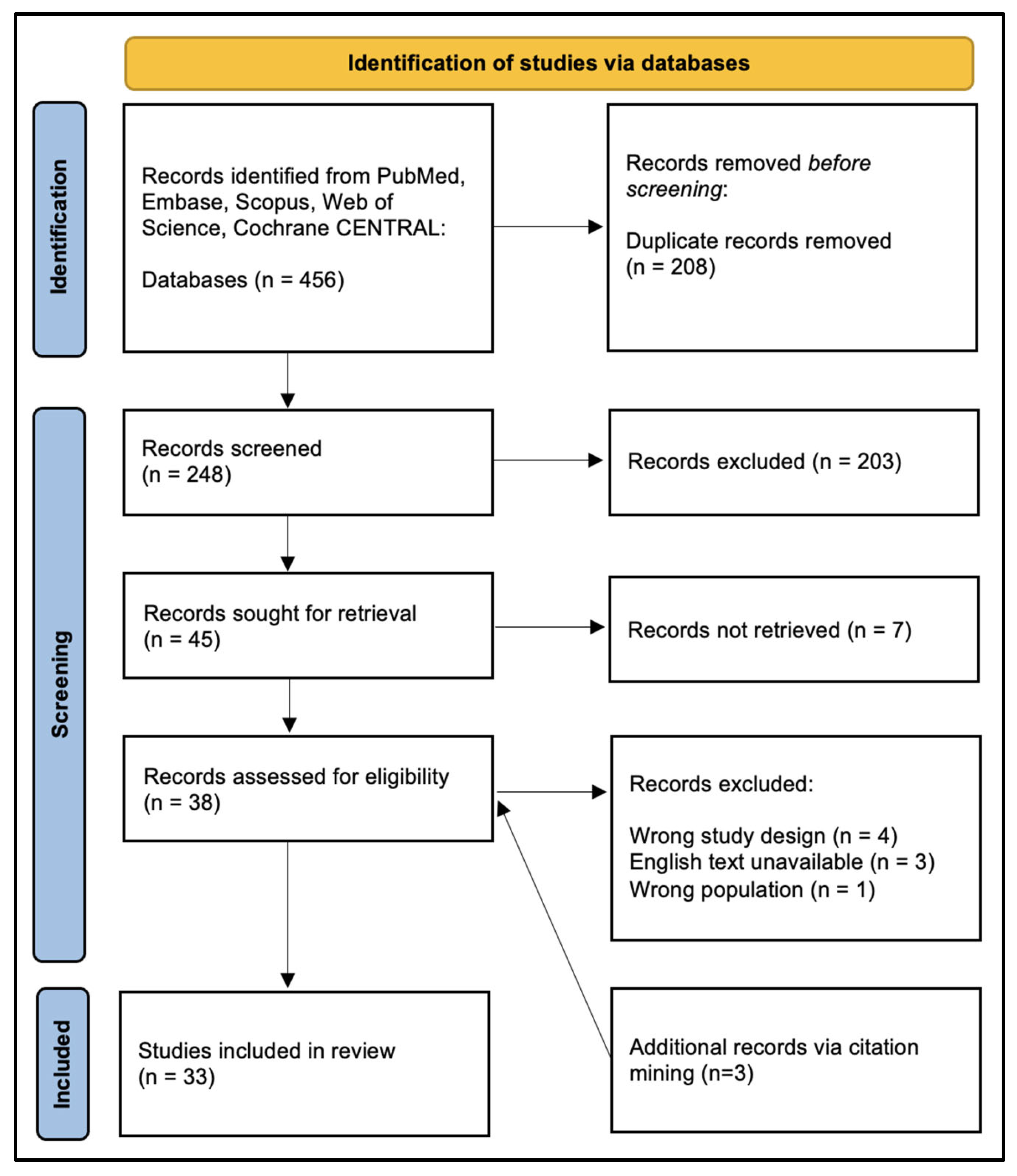
The database search retrieved 456 records across the five databases. The PubMed, Embase, Scopus, Web of Science, and Cochrane CENTRAL databases returned 53, 172, 119, 91, and 21 records, respectively. Following deduplication, 248 unique records underwent title and abstract screening. Of these, 45 were sought for retrieval for full-text review. Of these 45 articles, 7 were unable to be retrieved due to the unavailability of the full-text manuscript. Of the 38 remaining articles, 8 were excluded due to wrong study design [29,30,31,32], unavailability of an English version of the manuscript [33,34,35], or wrong population [36]. Three additional records were identified through backward citation mining of included studies. A total of 33 studies met the inclusion criteria and were included in this review (Figure 1).

3.2. Characteristics of Included Studies

The 33 included studies were published between 2004 and 2025 and were conducted across 18 countries, enrolling a total of 634 unique patients. The evidence base is substantially concentrated in AA: 30 of 33 studies enrolled AA patients, representing 613 of 634 patients (96.6%). Three studies addressed primary cicatricial alopecia: one case series in LPP (n = 13) and one RCT plus one retrospective survey in FFA (n = 8 combined). One study investigating 308 nm excimer therapy in folliculitis decalvans was identified but was excluded due to the unavailability of the full text. No eligible studies were identified for any other hair loss conditions. Dosing protocols varied across studies, with initial fluences ranging from 50–600 mJ/cm2, treatment frequencies ranging from twice weekly to once monthly, and the number of sessions ranging from 3 to 67. Full study characteristics and primary outcomes are presented in Table 1.

3.3. Synthesis of Findings

3.3.1. Mild-to-Moderate Alopecia Areata (AA)

Mild-to-moderate AA, defined as AA resulting in <50% hair loss of the scalp [68], represented the largest body of evidence, encompassing 24 of the 33 included studies, including 8 RCTs, 13 non-randomized studies, and 3 case reports. The evidence suggests that 308 nm excimer therapy produces clinically meaningful hair regrowth in a substantial proportion of mild-to-moderate AA, including single AA and multiple AA without ophiasic involvement. However, effect estimates varied considerably, and head-to-head comparisons with standard-of-care alternatives yielded mixed results.
Against intralesional corticosteroids (ILCSs), the most widely used first-line treatment for mild-to-moderate AA, 308 nm excimer laser therapy performs comparably rather than superiorly. Khan et al. (2024), the largest RCT in this review, found no significant difference in the proportion of patients achieving ≥75% regrowth between excimer therapy and ILCSs (56.5% vs. 67.4%, p = 0.19), with comparable mean SALT score reductions [42]. Kianfar et al. (2022) found ILCSs to produce greater regrowth at the end of treatment at 12 weeks (p = 0.003), though the gap narrowed by week 16 (p = 0.148), suggesting a delayed excimer therapy response [48]. Excimer therapy may be an effective alternative for patients in whom repeated corticosteroid injections are poorly tolerated, particularly children, or for whom corticosteroid-related local adverse effects are a concern [37,63].
The strongest evidence for excimer therapy’s additive value is in combination with topical therapies. Multiple studies demonstrate that excimer therapy combined with a topical agent, including minoxidil, ILCSs, or tacrolimus, outperforms either modality alone in terms of regrowth rate, trichoscopic disease activity, or immunological markers [9,40,43,49]. Li et al.’s intra-patient split patch design provides the most internally valid estimate of this incremental benefit: adding excimer therapy to topical minoxidil doubled the proportion achieving ≥50% regrowth (44.1% vs. 23.1%) and produced significantly greater hair diameter and count (p < 0.01) [9]. This reflects the incremental benefit of adding excimer therapy to a topical regimen. However, Di Filippo et al. (2022) found that patients using concurrent treatment (e.g., ILCSs or methotrexate) did not show better outcomes than patients only treated with excimer therapy [47].
When used as monotherapy, response rates across several RCTs, non-randomized studies, and case reports ranged broadly, from approximately 40% to 100% achieving any regrowth [19,39,41,44,51,52,53,57,59,60,62,63,64,65,66,67]. This variation is largely explained by differences in patient selection and disease subtype. Studies enrolling patients with treatment-naive or short-duration disease consistently reported higher rates than those enrolling patients with refractory or long-standing patches. One study identified a potential dose-response relationship: Kianfar et al. (2022) concluded that hair regrowth was positively correlated with the cumulative dose of laser treatment (Spearman’s ρ = 0.685, p = 0.005) [48].
Across the literature, we identified several predictors of response with varying consistency. First, scalp involvement responds better than beard or extremity disease, with multiple studies reporting absent response at non-scalp sites [53,63,65]. Second, atopic diathesis was identified as a negative prognostic factor in two studies [63,65], which found that most non-responders were atopic, and this finding was held in both adult and pediatric populations. In contrast, Di Filippo et al. (2022) found that the six included atopic patients unexpectedly experienced favorable responses, and Kakeji et al. (2025) found a complete response to excimer therapy only after adding concurrent delgocitinib 0.5% ointment in a patient with concurrent atopic dermatitis [38,47]. However, the small sample size in these studies warrants caution, and larger studies stratifying by atopic status are needed to confirm this observation. Shorter disease duration, younger patient age, lack of associated comorbidities, and less severe scalp involvement were found to be associated with higher rates of complete response in some studies [9,39,44,50,56,58] but not in other studies [42,47,48,58]. One study noted vertex and posterior scalp lesions to be more responsive than parietal ones, though this result has not been independently replicated [53]. There were no identified correlations between treatment response and patient sex.
A notable observation from Sanga & Mittal (2015) is that the excimer therapy response may substantially lag behind treatment completion: only 3.3% of treated patches achieved ≥50% regrowth at the end of the eight-week treatment period, rising to 53.3% at four months post-treatment (vs. 16.7% in untreated control halves, p < 0.05) [59]. This delayed response pattern, if confirmed, has implications for how therapy response should be assessed in clinical practice and future trials and may reveal that some studies with shorter evaluation periods underestimate excimer therapy’s true efficacy.

3.3.2. Severe Alopecia Areata

Evidence specific to ophiasic and extensive patchy AA (defined as ≥50% scalp involvement but less than 94%) is limited to subgroup analysis within larger trials and one case report and is consistently less favorable than for mild-to-moderate disease [43,47,48,53,55,58]. Di Filippo et al. (2022) found that only one of four ophiasic patients achieved SALT75 (75% reduction in SALT score), compared to 56% of those with patch-type disease [47]. Hsu et al. (2015) reported an overall response rate of 41.2% in a mixed cohort that included patients with extensive disease [58]. These figures are not directly comparable given differences in patient mix and outcome definitions, but the directional signal is consistent: ophiasic disease is harder to treat with excimer therapy [69].
In contrast, Fenniche et al. (2018) documented a case of complete regrowth of hair in a 5-year-old child with extensive ophiasic involvement treated over three months, with no relapse at one year, although pre-treatment with khellin, a photosensitizer, may have contributed [55]. In aggregate, excimer therapy may produce partial responses in some patients with severe AA, especially with concurrent treatment, but the evidence specifically targeting this subtype should not be considered a reliable monotherapy option in this population without further controlled investigation.

3.3.3. Very Severe Alopecia Areata (Alopecia Totalis and Universalis)

Ten studies enrolled patients with alopecia totalis (AT) and alopecia universalis (AU). The overall picture is more heterogeneous and less optimistic than for patch-type disease, but several findings are clinically instructive [38,43,45,47,50,56,58,63,64,65,67]. Spontaneous remission is rarer in AT/AU than in patch-type AA, which lends slightly greater weight to uncontrolled observations, though confounding remains a concern.
The most consistent finding across AT/AU studies is that disease duration is the strongest predictor of response. For example, Al Hamzawi (2021) reported a 90% overall response rate in 10 AT patients, with complete SALT normalization exclusively in those with the shortest disease durations [50]. Conversely, Zakaria et al. (2004) found no regrowth in any patient with AU or AT in a well-controlled split-patch study [67]. Given that early intervention improves treatment efficacy across AA modalities, and that chronic AT/AU is notably treatment-resistant with only 8.5% achieving complete recovery [70], excimer therapy, like other immunomodulatory treatments, may be most effective when administered during periods of active inflammation rather than in chronic, quiescent disease.
Two case reports illustrate the potential of excimer therapy within combination strategies for refractory AT/AU. Murakami et al. (2023) reported sustained response to excimer monotherapy over 12 months following induction with topical delgocitinib in a patient with concurrent vitiligo, and Kakeji et al. (2025) documented complete regrowth (SALT 0) within 1 year of adding delgocitinib to an existing excimer regimen after excimer therapy alone had failed [38,45].

3.3.4. Cicatricial Alopecias

Three studies addressed lichen planopilaris (LPP) and frontal fibrosing alopecia (FFA), which are conditions in which irreversible follicular destruction is driven by lymphocytic inflammation [71]. In these diseases, the therapeutic goal is the arrest of disease activity rather than regrowth.
The signal from LPP is most compelling in this subgroup. Navarini et al. (2011) used a split-scalp design in 13 patients with biopsy-confirmed active LPP and demonstrated a statistically significant reduction in erythema (p < 0.008), inflammatory lesion count (p < 0.003), hyperkeratosis (p < 0.02), pain (p < 0.008), and pruritus (p < 0.001) on treated vs. untreated half-scalps [61]. The finding of limited hair regrowth in 3/13 patients was expected given irreversible follicular destruction. The primary value of this intervention lies in inflammatory suppression and arrest of disease progression.
Evidence in FFA is less encouraging and more limited. Thuangtong et al. (2023), the only RCT in the cicatricial subgroup, found no significant difference in the Frontal Fibrosing Alopecia Severity Index (FFASI) scores between excimer-plus-ILCSs and ILCSs-alone sides at 16 weeks (p = 0.374), with 60% of patients reporting no improvement on either arm [46]. However, only five patients were enrolled with four weeks of post-treatment follow-up. Zhang et al. (2019), a retrospective survey study, identified three patients with FFA who received excimer therapy, and all three reported treatment response, though survey methodology and concurrent treatments limit interpretation [54].

3.3.5. Adverse Events and Tolerability

The safety profile and tolerability of 308 nm excimer therapy across the hair loss literature are favorable and generally consistent in the short term (Table 2). Transient erythema was near-universal in the 28 studies where adverse event data were reported. In fact, in most studies, transient erythema was used as the titration endpoint for dose escalation rather than being regarded as a complication. Post-inflammatory hyperpigmentation was the most clinically significant common event but resolved without sequelae in all cases where follow-up was documented. Desquamation, pruritus, and mild pain were reported across multiple studies and were uniformly self-limiting.
More significant events were infrequent and generally dose-dependent, limited to transient vesicle formation and crusting in one AA case report, blistering in one AA study, persistent erythema leading to discontinuation in one AA study, and burning in one FFA study. No systemic adverse events, photosensitizing complications, or malignant sequelae were reported across any included study. However, these findings reflect short-term, inconsistently reported outcomes rather than an established long-term safety profile. No study assessed the safety of repeated ultraviolet exposure over extended follow-up, and adverse event reporting practices varied in granularity and duration across studies.

3.3.6. Relapse and Durability of Response

Durability of excimer-induced remission is incompletely characterized across the literature (Table 3). Most prospective studies reported follow-up of three to six months, which may be insufficient to capture the natural relapse kinetics of a chronically relapsing condition such as AA. Where relapse data were available, rates among initial responders ranged from 0% (in several shorter follow-up studies) to approximately 36% at six months [63] and 22% at 8–12 months [50]. This variability likely reflects differences in disease severity, follow-up duration, treatment protocols (Table S1), and concurrent treatment rather than true heterogeneity in excimer therapy’s biological durability.
However, two observations stand out. First, Di Filippo et al. (2022) identified a mean follow-up period of 5.7 years and found that, among the 16.7% of patients who relapsed, excimer re-treatment was used successfully [47]. However, potential confounders include the retrospective nature of the study and the potential loss to follow-up. Second, Murakami et al. (2023) documented 12 months of sustained remission on excimer monotherapy following combination induction with delgocitinib [45]. Relapse definitions were not standardized across studies, limiting cross-study comparison.

3.4. Risk of Bias Assessment

The results of the risk of bias assessment can be found in the Supplementary Materials. Risk of bias was assessed using the Cochrane RoB 2 tool for RCTs (Table S2), ROBINS-I for non-randomized studies (Table S3), and JBI Critical Appraisal Checklists for case series and case reports (Table S4a,b). Among the nine RCTs, two (Sirichotiyakul et al., 2020; Thuangtong et al., 2023) were rated low risk [46,52], and the remaining seven were rated Some Concerns, primarily due to a lack of participant and assessor blinding. Among non-randomized studies assessed with ROBINS-I, one study (Li et al., 2020) was rated low risk [9], while the majority were rated moderate risk, with deviation from intended intervention (Domain 4) and measurement of outcomes (Domain 6) representing the predominant concerns. One non-randomized study with comparators (Hsu et al., 2015) [58] received a serious risk rating, primarily due to a lack of adjustment for confounding variables. Among the ten single-arm studies appraised using the JBI Critical Appraisal Checklist for Case Series, five were rated low risk, four moderate risk, and one (Zhang et al., 2019) [54] high risk, reflecting incidental subgroup reporting without defined inclusion criteria or formal statistical analysis. Case reports (n = 6) were generally well-reported, with five rated low risk and one (Ohtsuki et al., 2010) [62] rated moderate risk owing to unclear consecutive and complete inclusion and demographic reporting. These risk-of-bias findings should be considered when interpreting the effect estimates reported in Section 3.3, particularly for non-randomized studies where spontaneous remission cannot be excluded as a contributor to observed outcomes.

4. Discussion

Excimer therapy may fulfill a distinct niche in the alopecia treatment landscape as a well-tolerated, targeted, locally acting option that avoids the systemic effects of pharmacological therapy, including immunosuppression, metabolic abnormalities, and thrombosis [72,73,74,75,76]. This positions it as a relevant alternative or adjunct in patients for whom repeated corticosteroid injections are poorly tolerated, systemic immunosuppression is contraindicated, or long-term pharmacologic therapy carries unacceptable risk.

4.1. Excimer Therapy in Primary Cicatricial Alopecia

This systematic review’s central contribution is the first rigorous characterization of excimer therapy evidence in primary cicatricial alopecia: conditions with no FDA-approved treatments where undertreated lymphocytic inflammation causes irreversible follicular destruction. For LPP and FFA, the clinical question is not whether excimer therapy produces regrowth but whether it can arrest the inflammatory process driving permanent hair loss, and no prior review has yielded eligible studies for appraisal. This review identifies a proof-of-concept signal in LPP and an insufficient but non-absent signal in FFA, and it supports an argument for prospective investigation in these conditions.
LPP and FFA are characterized by immune privilege collapse of the hair follicle bulge region, leading to lymphocytic inflammation targeting epithelial hair follicle stem cells, followed by permanent follicular destruction and fibrosis [77]. The pathophysiology centers on a Th1-biased cytotoxic immune response with contributions from the JAK/STAT pathway [77,78]. By analogy with AA, excimer therapy in cicatricial conditions may arrest lymphocytic follicular destruction through UVB-mediated T-cell apoptosis [79,80]. Currently, evidence-based guidelines for the treatment of LPP and FFA are lacking, and there are no FDA-approved treatments for these conditions [81,82]. The available cicatricial evidence suggests a proof-of-concept warranting dedicated prospective investigation. Until then, excimer therapy remains investigational in LPP and insufficient to guide practice in FFA.
A 2024 systematic review of laser and phototherapy across alopecia subtypes (Jafari et al.) found no excimer-specific studies in cicatricial conditions and concluded the available data were too sparse for generalization [83]. This review extends this work by identifying and critically appraising the three available studies in LPP and FFA. For a pair of conditions where clinicians currently navigate treatment decisions without any FDA-approved therapy or high-quality RCT evidence, even a preliminary signal from a well-tolerated, locally acting intervention carries clinical relevance. Importantly, this review also quantifies the evidence vacuum in other hair loss conditions. The absence of eligible studies in other alopecia subtypes, identified through a five-database systematic search, defines a clear research agenda for phototherapy-focused investigation in conditions where targeted, non-pharmacologic options remain unexplored.
One potential explanation for the preliminary findings of excimer therapy’s success in LPP but not in FFA may relate to the treatment window of opportunity for these diseases. Given that the primary goal in cicatricial alopecia treatment is the arrest of hair loss and reduction in inflammatory symptoms, treatment must be initiated during the inflammatory stage rather than the fibrotic stage of the disease [84]. LPP is driven by a sustained, intense inflammatory response that provides a larger and more persistent window of opportunity compared to FFA, which is driven by a milder inflammatory response and a larger apoptotic response [85]. Because excimer therapy results in T-cell suppression, it may have a larger effect in LPP.
Two additional studies published after our formal search window offer further post hoc context for the cicatricial alopecia findings. Spindler et al. (2026), in a retrospective cohort of 15 patients with scarring alopecia, found that excimer therapy, which was used as an adjunctive therapy in most patients, was associated with subjective improvement in hair or hairline appearance in 60% of patients after a mean of 3.4 months, with improvement in perifollicular hyperkeratosis and erythema in 83% of affected patients [86]. Ferrer Guillén et al. (2026), a retrospective observational study of 30 FFA patients using excimer laser as an adjuvant to ongoing systemic and topical therapy, reported stabilization of frontotemporal hairline recession in 53.3% of patients and improvement in 20% at 6 months [87]. Additionally, perifollicular hyperkeratosis and erythema improved in 70% and 83.3% of patients, respectively. Transient erythema was reported only in four patients. The outcomes of these studies are consistent with this review’s characterization of inflammatory suppression as the primary achievable outcome in cicatricial alopecia. Together, they reinforce cicatricial alopecia as an area of active, expanding clinical interest.

4.2. Excimer Therapy in Alopecia Areata

Current evidence suggests that excimer therapy may perform comparably to ILCSs in mild-to-moderate AA, with no significant difference in the proportion achieving ≥75% regrowth, and may demonstrate additive value when combined with topical agents, including minoxidil and corticosteroids.
We identified predictors for treatment response to excimer therapy in AA. Scalp-limited disease responds substantially better than beard or extremity involvement, with multiple studies reporting absent responses at non-scalp sites. Atopic diathesis was identified as a negative prognostic factor across two studies [63,65], although another study and a case report made contrasting observations [38,47]. One potential explanation for this paradox is a distinct immunophenotype of atopic AA with heightened Th2 inflammation that may be particularly amenable to excimer therapy’s documented Th2-suppressive and Treg-inducing effects [88,89]. Alternatively, although atopic dermatitis is predominantly Th2-driven, it still exhibits a robust Th1 signature that can be preferentially targeted by excimer therapy [89]. Given these mutually exclusive hypotheses, prospective studies stratifying by atopic status and immunophenotyping at baseline are needed to resolve them. Shorter disease duration and less severe involvement are associated with higher rates of complete response across multiple study designs, presumably because the underlying inflammatory target is more accessible earlier in the disease course. This is consistent with the broader literature establishing disease duration as a general predictor across modalities [90,91,92,93].
A particularly interesting finding from the AA synthesis is the observation by Sanga & Mittal (2015) and Kianfar et al. (2022) that the peak excimer therapy response may substantially lag treatment completion [48,59]. If this hypothesis for a delayed biological response pattern is confirmed in larger studies, it is consistent with responses seen with other phototherapy options [94,95,96]. Future excimer therapy trials should pre-specify follow-up assessments extending at least four to six months beyond treatment completion, and clinical response monitoring should not be curtailed at the end of a treatment course.
Importantly, excimer therapy has been reported to show some safety and efficacy in pediatric AA patients as young as 4 years old [37,63]. This is clinically relevant given the psychosocial burden of needle-based procedures in pediatric dermatology, parental preference for non-invasive treatments, and injection-associated pain [10,97,98,99,100,101,102,103]. Excimer therapy may serve as maintenance or an adjunct alongside JAK inhibitors in this population [104,105,106,107].

4.3. Research Gaps and Limitations

Several important gaps in the evidence base emerge from this review that should inform the design of future studies. A notable gap is the complete absence of any identified evidence for 308 nm excimer therapy in other alopecia subtypes for which targeted phototherapy has at least theoretical relevance, such as androgenetic alopecia and central centrifugal cicatricial alopecia. Second, protocol heterogeneity across included studies also prevents any conclusions about optimal dosing, and no study was designed primarily to characterize the dose-response relationship or identify a minimum effective cumulative dose. Finally, the relapse literature is inadequate: most studies followed patients for three to six months, relapse definitions were not standardized, and no study was powered to assess durability as a primary outcome.
This review has limitations. First, the clinical and methodological heterogeneity across studies precluded quantitative pooling, and findings are necessarily qualitative without pooled effect estimates. Second, restriction to English-language publications may have introduced language bias, potentially excluding relevant studies published in other languages, particularly given the geographic diversity of the included literature. Third, the predominance of non-randomized observational studies with concerns for bias and case series/reports across the evidence base limits the certainty with which excimer therapy’s efficacy can be attributed independently of co-interventions and natural disease courses. Fourth, this review was not prospectively registered (e.g., with PROSPERO), which we acknowledge as a limitation of the reporting process. Fifth, we did not perform a formal certainty-of-evidence assessment, as the marked heterogeneity across three distinct conditions and the very limited evidence base for LPP and FFA offer little basis for domain-level certainty ratings.

5. Conclusions

308 nm excimer therapy occupies a clinically meaningful niche in the alopecia treatment landscape as a locally acting, tolerable, non-pharmacologic option. Its strongest evidence is in mild-to-moderate scalp-limited AA, where response rates fall within the range reported for intralesional corticosteroids in small heterogeneous studies, with additive benefit suggested in combination with topical agents, and with relevance for pediatric patients and those for whom repeated injections are contraindicated or poorly tolerated. More broadly, this review contributes to the phototherapy literature by characterizing the conditions under which targeted UVB-mediated T-cell apoptosis may be therapeutically applicable beyond excimer therapy’s established indications, and by identifying primary cicatricial alopecia as a candidate indication for prospective phototherapy investigation. This evidence base, however, is overwhelmingly concentrated in AA (30 of 33 included studies), and comparative-effectiveness claims should be interpreted with this imbalance in mind. This review also identifies an absence of eligible studies across a broad range of hair loss conditions, including androgenetic alopecia and telogen effluvium, which provides an explicit research agenda for phototherapy-focused investigation. Future trials should prioritize standardized protocols, extended post-treatment follow-up of at least four to six months, and prospective investigation of cicatricial alopecia.

Supplementary Materials

The following supporting information can be downloaded at https://www.mdpi.com/article/10.3390/app16147337/s1: Table S1: Excimer therapy treatment protocols of included studies. Table S2: Cochrane RoB 2.0 Risk of Bias Assessment (Randomized Controlled Trials). Table S3: ROBINS-I Risk of Bias Assessment (Non-Randomized Studies). Table S4a: JBI Critical Appraisal for Case Reports. Table S4b: JBI Critical Appraisal for Case Series.

Author Contributions

Conceptualization, J.M. and V.R.; methodology, J.M.; validation, M.B. and N.K.J.; formal analysis, J.M. and M.B.; investigation, J.M. and M.B.; resources, J.M.; data curation, J.M., M.B., N.K.J. and V.R.; writing—original draft preparation, J.M. and M.B.; writing—review and editing, J.M., M.B., N.K.J., S.K.T.Q. and V.R.; visualization, J.M. and M.B.; supervision, N.K.J., S.K.T.Q. and V.R.; project administration, J.M. All authors have read and agreed to the published version of the manuscript.

Funding

This research received no external funding.

Institutional Review Board Statement

Not applicable.

Informed Consent Statement

Not applicable.

Data Availability Statement

No new data were created or analyzed in this study. Data sharing is not applicable to this article.

Conflicts of Interest

The authors declare no conflicts of interest.

Abbreviations

The following abbreviations are used in this manuscript:
AAAlopecia Areata
FFAFrontal Fibrosing Alopecia
FFASIFrontal Fibrosing Alopecia Severity Index
IFN-γInterferon-γ
ILCSIntralesional Corticosteroid
JAKJanus Kinase
JBIJoanna Briggs Institute
LPPLichen Planopilaris
NRSNon-Randomized Study
PRISMAPreferred Reporting Items for Systematic Reviews and Meta-Analyses
RCTRandomized Controlled Trial
RoB 2Revised Cochrane Risk of Bias Tool for Randomized Trials
ROBINS-IRisk of Bias in Non-Randomized Studies of Interventions
SALTSeverity of Alopecia Tool
UVBUltraviolet B

Appendix A

The following search strategies were applied across five electronic databases. All searches combined controlled vocabulary terms (MeSH or database-equivalent Emtree terms) with free-text keywords in title and abstract fields, restricted to English-language records published through 31 December 2025.
PubMed/MEDLINE: Searched via the National Library of Medicine PubMed interface (pubmed.ncbi.nlm.nih.gov). MeSH terms were combined with free-text title/abstract terms. Date and language restrictions were applied as native PubMed limits.
(“excimer laser”[Title/Abstract] OR “excimer lasers”[Title/Abstract] OR “308 nm”[Title/Abstract] OR “308-nm”[Title/Abstract] OR “308nm”[Title/Abstract] OR “308 nanometer”[Title/Abstract] OR “xenon chloride laser”[Title/Abstract] OR “XeCl laser”[Title/Abstract] OR “monochromatic excimer”[Title/Abstract] OR “Lasers, Excimer”[MeSH Terms]) AND (alopecia[Title/Abstract] OR alopecias[Title/Abstract] OR “hair loss”[Title/Abstract] OR “alopecia areata”[Title/Abstract] OR “androgenetic alopecia”[Title/Abstract] OR “pattern hair loss”[Title/Abstract] OR “male pattern baldness”[Title/Abstract] OR “female pattern hair loss”[Title/Abstract] OR “lichen planopilaris”[Title/Abstract] OR “frontal fibrosing alopecia”[Title/Abstract] OR “cicatricial alopecia”[Title/Abstract] OR “scarring alopecia”[Title/Abstract] OR “folliculitis decalvans”[Title/Abstract] OR “telogen effluvium”[Title/Abstract] OR “anagen effluvium”[Title/Abstract] OR trichotillomania[Title/Abstract] OR “dissecting cellulitis”[Title/Abstract] OR “Alopecia”[MeSH Terms] OR “Alopecia Areata”[MeSH Terms])
Embase: Searched via Elsevier Embase through the Ovid interface. Emtree-controlled vocabulary terms (/exp) were combined with title/abstract field tags (:ti,ab). Date and language limits were applied as Ovid search limits.
(‘excimer laser’:ti,ab OR ‘excimer lasers’:ti,ab OR ‘308 nm’:ti,ab OR ‘308-nm’:ti,ab OR ‘308nm’:ti,ab OR ‘308 nanometer’:ti,ab OR ‘xenon chloride laser’:ti,ab OR ‘XeCl laser’:ti,ab OR ‘monochromatic excimer’:ti,ab OR ‘excimer laser’/exp) AND (alopecia:ti,ab OR alopecias:ti,ab OR ‘hair loss’:ti,ab OR ‘alopecia areata’:ti,ab OR ‘androgenetic alopecia’:ti,ab OR ‘pattern hair loss’:ti,ab OR ‘male pattern baldness’:ti,ab OR ‘female pattern hair loss’:ti,ab OR ‘lichen planopilaris’:ti,ab OR ‘frontal fibrosing alopecia’:ti,ab OR ‘cicatricial alopecia’:ti,ab OR ‘scarring alopecia’:ti,ab OR ‘folliculitis decalvans’:ti,ab OR ‘telogen effluvium’:ti,ab OR ‘anagen effluvium’:ti,ab OR trichotillomania:ti,ab OR ‘dissecting cellulitis’:ti,ab OR ‘alopecia’/exp OR ‘alopecia areata’/exp).
Cochrane Central Register of Controlled Trials (CENTRAL): Searched via the Cochrane Library (cochranelibrary.com), Trials section. Search fields covered title, abstract, and keywords (:ti,ab,kw). Publication year and language limits were applied via Cochrane search filters.
(“excimer laser” OR “excimer lasers” OR “308 nm” OR “308-nm” OR “308nm” OR “xenon chloride laser” OR “XeCl laser” OR “monochromatic excimer”):ti,ab,kw AND (alopecia OR “hair loss” OR “alopecia areata” OR “androgenetic alopecia” OR “lichen planopilaris” OR “frontal fibrosing alopecia” OR “cicatricial alopecia” OR “scarring alopecia” OR “folliculitis decalvans”):ti,ab,kw
Web of Science: Searched via the Clarivate Web of Science Core Collection. Topic field search (TS=) queries title, abstract, author keywords, and Keywords Plus fields. Language and publication year limits were applied as Web of Science search filters.
TS=(“excimer laser” OR “excimer lasers” OR “308 nm” OR “308-nm” OR “308nm” OR “xenon chloride laser” OR “XeCl laser” OR “monochromatic excimer”) AND TS=(alopecia OR alopecias OR “hair loss” OR “alopecia areata” OR “androgenetic alopecia” OR “pattern hair loss” OR “male pattern baldness” OR “female pattern hair loss” OR “lichen planopilaris” OR “frontal fibrosing alopecia” OR “cicatricial alopecia” OR “scarring alopecia” OR “folliculitis decalvans” OR “telogen effluvium” OR “anagen effluvium” OR “dissecting cellulitis”)
Scopus: Searched via Elsevier Scopus (scopus.com). TITLE-ABS-KEY searches title, abstract, and author keyword fields simultaneously. Language and publication year limits were applied as Scopus document search filters.
TITLE-ABS-KEY(“excimer laser” OR “excimer lasers” OR “308 nm” OR “308-nm” OR “308nm” OR “xenon chloride laser” OR “XeCl laser” OR “monochromatic excimer”) AND TITLE-ABS-KEY(alopecia OR alopecias OR “hair loss” OR “alopecia areata” OR “androgenetic alopecia” OR “pattern hair loss” OR “male pattern baldness” OR “female pattern hair loss” OR “lichen planopilaris” OR “frontal fibrosing alopecia” OR “cicatricial alopecia” OR “scarring alopecia” OR “folliculitis decalvans” OR “telogen effluvium” OR “anagen effluvium” OR “dissecting cellulitis”).

References

  1. Dakkak, M.; Forde, K.M.; Lanney, H. Hair Loss: Diagnosis and Treatment. Am. Fam. Physician 2024, 110, 243–250. [Google Scholar] [PubMed]
  2. Stefanato, M.C. Histopathology of alopecia: A clinicopathological approach to diagnosis. Histopathology 2010, 56, 24–38. [Google Scholar] [CrossRef] [Scilit] [PubMed]
  3. Bolduc, C.; Sperling, C.L.; Shapiro, J. Primary cicatricial alopecia. J. Am. Acad. Dermatol. 2016, 75, 1081–1099. [Google Scholar] [CrossRef] [Scilit] [PubMed]
  4. Kinoshita-Ise, M.; Fukuyama, M.; Ohyama, M. Recent Advances in Understanding of the Etiopathogenesis, Diagnosis, and Management of Hair Loss Diseases. J. Clin. Med. 2023, 12, 3259. [Google Scholar] [CrossRef] [Scilit] [PubMed]
  5. Poblet, E.; Jiménez, F.; Pascual, A.; Piqué, E. Frontal fibrosing alopecia versus lichen planopilaris: A clinicopathological study. Int. J. Dermatol. 2006, 45, 375–380. [Google Scholar] [CrossRef] [Scilit] [PubMed]
  6. Xing, L.; Dai, Z.; Jabbari, A.; Cerise, E.J.; Higgins, A.C.; Gong, W.; Jong, D.A.; Harel, S.; Destefano, M.G.; Rothman, L.; et al. Alopecia areata is driven by cytotoxic T lymphocytes and is reversed by JAK inhibition. Nat. Med. 2014, 20, 1043–1049. [Google Scholar] [CrossRef] [Scilit] [PubMed]
  7. Bertolini, M.; McElwee, K.; Gilhar, A.; Bulfone-Paus, S.; Paus, R. Hair follicle immune privilege and its collapse in alopecia areata. Exp. Dermatol. 2020, 29, 703–725. [Google Scholar] [CrossRef] [Scilit] [PubMed]
  8. Gilhar, A.; Laufer-Britva, R.; Keren, A.; Paus, R. Frontiers in alopecia areata pathobiology research. J. Allergy Clin. Immunol. 2019, 144, 1478–1489. [Google Scholar] [CrossRef] [Scilit] [PubMed]
  9. Li, A.; Meng, X.; Xing, X.; Tan, H.; Liu, J.; Li, C. Efficacy and Influence Factors of 308-nm Excimer Lamp with Minoxidil in the Treatment of Alopecia Areata. Lasers Surg. Med. 2020, 52, 761–767. [Google Scholar] [CrossRef] [Scilit] [PubMed]
  10. Gilhar, A.; Etzioni, A.; Paus, R. Alopecia Areata. N. Engl. J. Med. 2012, 366, 1515–1525. [Google Scholar] [CrossRef] [Scilit] [PubMed]
  11. Price, H.V. Treatment of Hair Loss. N. Engl. J. Med. 1999, 341, 964–973. [Google Scholar] [CrossRef] [Scilit] [PubMed]
  12. Barton, R.V.; Toussi, A.; Awasthi, S.; Kiuru, M. Treatment of pediatric alopecia areata: A systematic review. J. Am. Acad. Dermatol. 2022, 86, 1318–1334. [Google Scholar] [CrossRef] [Scilit] [PubMed]
  13. Agarwal, P.; Ajuchi, O.; Lukowiak, M.T.; Rao, K.B. Off-label use of biologics and janus kinase (JAK) inhibitors for scarring alopecias: A narrative review. Arch. Dermatol. Res. 2025, 317, 803. [Google Scholar] [CrossRef] [Scilit] [PubMed]
  14. Dahabreh, D.; Jung, S.; Renert-Yuval, Y.; Bar, J.; Duca, D.E.; Guttman-Yassky, E. Alopecia Areata: Current Treatments and New Directions. Am. J. Clin. Dermatol. 2023, 24, 895–912. [Google Scholar] [CrossRef] [Scilit] [PubMed]
  15. Lensing, M.; Jabbari, A. An overview of JAK/STAT pathways and JAK inhibition in alopecia areata. Front. Immunol. 2022, 13, 955035. [Google Scholar] [CrossRef] [Scilit] [PubMed]
  16. Passeron, T.; King, B.; Seneschal, J.; Steinhoff, M.; Jabbari, A.; Ohyama, M.; Tobin, J.D.; Randhawa, S.; Winkler, A.; Telliez, J.-B.; et al. Inhibition of T-cell activity in alopecia areata: Recent developments and new directions. Front. Immunol. 2023, 14, 1243556. [Google Scholar] [CrossRef] [Scilit] [PubMed]
  17. Beggs, S.; Short, J.; Rengifo-Pardo, M.; Ehrlich, A. Applications of the Excimer Laser. Dermatol. Surg. 2015, 41, 1201–1211. [Google Scholar] [CrossRef] [Scilit] [PubMed]
  18. Passeron, T.; Ortonne, J.-P. Use of the 308-nm excimer laser for psoriasis and vitiligo. Clin. Dermatol. 2006, 24, 33–42. [Google Scholar] [CrossRef] [Scilit] [PubMed]
  19. Aubin, F.; Vigan, M.; Puzenat, E.; Blanc, D.; Drobacheff, C.; Deprez, P.; Humbert, P.; Laurent, R. Evaluation of a novel 308-nm monochromatic excimer light delivery system in dermatology: A pilot study in different chronic localized dermatoses. Br. J. Dermatol. 2005, 152, 99–103. [Google Scholar] [CrossRef] [Scilit] [PubMed]
  20. Brenninkmeijer, E.E.A.; Spuls, P.I.; Lindeboom, R.; Wal, D.V.A.C.; Bos, J.D.; Wolkerstorfer, A. Excimer laser vs. clobetasol propionate 0·05% ointment in prurigo form of atopic dermatitis: A randomized controlled trial, a pilot. Br. J. Dermatol. 2010, 163, 823–831. [Google Scholar] [CrossRef] [Scilit] [PubMed]
  21. Saraceno, R.; Nisticò, P.S.; Capriotti, E.; Felice, D.C.; Rhodes, E.L.; Chimenti, S. Monochromatic excimer light (308-nm) in the treatment of prurigo nodularis. Photodermatol. Photoimmunol. Photomed. 2008, 24, 43–45. [Google Scholar] [CrossRef] [Scilit] [PubMed]
  22. Olsen, A.E.; Hodak, E.; Anderson, T.; Carter, B.J.; Henderson, M.; Cooper, K.; Lim, W.H. Guidelines for phototherapy of mycosis fungoides and Sézary syndrome: A consensus statement of the United States Cutaneous Lymphoma Consortium. J. Am. Acad. Dermatol. 2016, 74, 27–58. [Google Scholar] [CrossRef] [Scilit] [PubMed]
  23. Takahashi, T.; Takahashi, T.; Segawa, Y.; Oka, K.; Takahashi, T.; Asano, Y. Circumscribed Morphea Successfully Treated With Excimer Laser: Analysis of Histopathological Changes. J. Dermatol. 2026, 53, 691–695. [Google Scholar] [CrossRef] [Scilit] [PubMed]
  24. Shroff, A.; Malajian, D.; Czarnowicki, T.; Rose, S.; Bernstein, M.D.; Singer, K.G.; Lebwohl, G.M.; Hadi, S.; Guttman-Yassky, E. Use of 308 nm excimer laser for the treatment of chronic hand and foot eczema. Int. J. Dermatol. 2016, 55, e447–e453. [Google Scholar] [CrossRef] [Scilit] [PubMed]
  25. Elmets, A.C.; Lim, W.H.; Stoff, B.; Connor, C.; Cordoro, M.K.; Lebwohl, M.; Armstrong, W.A.; Davis, M.R.D.; Elewski, E.B.; Gelfand, M.J.; et al. Joint American Academy of Dermatology–National Psoriasis Foundation guidelines of care for the management and treatment of psoriasis with phototherapy. J. Am. Acad. Dermatol. 2019, 81, 775–804. [Google Scholar] [CrossRef] [Scilit] [PubMed]
  26. Ly, K.; Smith, P.M.; Thibodeaux, G.Q.; Beck, M.K.; Liao, W.; Bhutani, T. Beyond the Booth. Dermatol. Clin. 2020, 38, 157–163. [Google Scholar] [CrossRef] [Scilit] [PubMed]
  27. Bianchi, B.; Campolmi, P.; Mavilia, L.; Danesi, A.; Rossi, R.; Cappugi, P. Monochromatic excimer light (308 nm): An immunohistochemical study of cutaneous T cells and apoptosis-related molecules in psoriasis. J. Eur. Acad. Dermatol. Venereol. 2003, 17, 408–413. [Google Scholar] [CrossRef] [Scilit] [PubMed]
  28. Connell, J.S.; Crotts, S.; Reis, R.; Lensing, M.M.; Kahl, P.; Henderson, N.; Ayush, O.; Zhu, Z.; Ortolan, S.L.; Ko, C.A.; et al. TH1 effector CD4 T cells rely on IFN-γ production to induce alopecia areata. Sci. Adv. 2025, 11, eadz2257. [Google Scholar] [CrossRef] [Scilit] [PubMed]
  29. McMichael, A.J. Excimer Laser: A Module of the Alopecia Areata Common Protocol. J. Investig. Dermatol. Symp. Proc. 2013, 16, S77–S79. [Google Scholar] [CrossRef] [Scilit] [PubMed]
  30. Gamret, A.C.; Potluri, V.S.; Krishnamurthy, K.; Fertig, R.M. Frontal fibrosing alopecia: Efficacy of treatment modalities. Int. J. Women’s Health 2019, 11, 273–285. [Google Scholar] [CrossRef] [Scilit]
  31. Graham, P.M.; Gupta, N.; Altman, D.A. En coup de sabre. Cutis 2019, 103, 34–36. [Google Scholar] [PubMed]
  32. Nie, X.; Hao, B.; Sun, M.; Li, Y. Clinical observation of 308 nm excimer light on alopecia areata in adolescents and children. Minerva Medica 2020, 113, 751–752. [Google Scholar] [CrossRef] [Scilit] [PubMed]
  33. Raulin, C.; Gündogan, C.; Greve, B.; Gebert, S. Die Excimer-Laser-Therapie der Alopecia areata–Halbseitige Evaluation eines repräsentativen Areals: Excimer laser therapy of alopecia areata–side-by-side evaluation of a representative area. JDDG J. Dtsch. Dermatol. Ges. 2005, 3, 524–526. [Google Scholar] [CrossRef] [Scilit]
  34. Wada, M.; Satomura, A.; Itou, T.; Takahara, M.; Furue, M. A case of alopecia ophiasis effectively treated with 308 nm excimer lamp. Nishinihon J. Dermatol. 2014, 76, 459–464. [Google Scholar] [CrossRef] [Scilit]
  35. Wollenschläger, I.; Hermann, J.; Ockenfels, H. UVB-308-nm-(NUVB-) Therapie mittels Excimer-Laser bei atopischer Dermatitis und weiteren inflammatorischen Dermatosen. Der Hautarzt 2009, 60, 898–906. [Google Scholar] [CrossRef] [Scilit]
  36. Rattanakaemakorn, P.; Triyangkulsri, K.; Iamsumang, W.; Suchonwanit, P. 308-nm excimer lamp vs. combination of 308-nm excimer lamp and 10% liquor carbonis detergens in patients with scalp psoriasis: A randomized, single-blinded, controlled trial. Front. Med. 2021, 8, 677948. [Google Scholar] [CrossRef] [Scilit]
  37. Dineshkumar, R.; Manthri, S.; Duraisamy, S.; Malvi, A. Use of Excimer Lamp and Topical Tofacitinib 2% Gel in the Treatment of Pediatric Alopecia Areata: A Case Report. Cureus 2025, 17, e83492. [Google Scholar] [CrossRef] [Scilit] [PubMed]
  38. Kakeji, Y.; Kogame, T.; Yagi, Y.; Kambe, N.; Kabashima, K. A case of refractory alopecia areata successfully treated by combining delgocitinib ointment with excimer laser. Ski. Health Dis. 2025, 5, 154–157. [Google Scholar] [CrossRef] [Scilit] [PubMed]
  39. Nagui, A.N.; Nada, R.H.; Anwar, M.D.; Mahran, M.N. Evaluation of the efficacy of excimer light versus topical trichloroacetic acid (TCA) 35% in the treatment of patchy alopecia areata of the scalp. Arch. Dermatol. Res. 2025, 317, 453. [Google Scholar] [CrossRef] [Scilit] [PubMed]
  40. Tawfik, M.Y.; Hofny, M.R.E.; Zidan, M.F.; Ghazally, A. 308 nm–Excimer light together with topical betamethasone valerate in treating alopecia areata. Arch. Dermatol. Res. 2025, 317, 342. [Google Scholar] [CrossRef] [Scilit] [PubMed]
  41. Kalegowda, D.; Madegowda, S.B.; Rajegowda, H.M.; Rathod, M.P. Dermoscopic evaluation of efficacy of 308 nm Excimer Laser in treatment of alopecia areata: A prospective study. J. Pak. Assoc. Dermatol. 2024, 34, 857–863. [Google Scholar]
  42. Khan, J.; Gul, H.; Ullah, I.; Khan, M. Comparison of efficacy of intralesional triamcinolone versus 308nm Excimer laser in the treatment of localized alopecia areata. J. Pak. Assoc. Dermatol. 2024, 34, 851–856. [Google Scholar] [CrossRef] [Scilit]
  43. Mohammed, M.N.; Hassan, R.M.; Samy, A.N.; Aladl, S.A.; Dawa, A.F.A.; Ibrahim, A.M.S. Excimer Light versus Combination of Tacrolimus and Excimer Light in the Treatment of Alopecia Areata. Dermatol. Ther. 2024, 2024, 8864825. [Google Scholar] [CrossRef] [Scilit]
  44. Rodríguez-Acosta, D.E.; Nieto, F.D.; Berna-Rico, E.; Galván-García, R.H.; Meza-Castro, G.R.; Rosales-Lerma, K.A.; Guerrero-Cervantes, V.A.; Ramírez-Marín, A.H. Assessing the Efficacy of Monthly 308 nm Excimer Lamp Double-Stacked Pulse Adjusted Therapy for Alopecia Areata. Ski. Appendage Disord. 2024, 10, 18–25. [Google Scholar] [CrossRef] [Scilit] [PubMed]
  45. Murakami, K.; Saito, Y.; Takeuchi, S.; Sujino, K.; Tanikawa, A.; Tanese, K.; Fukuda, K. Successful management of severe alopecia areata and vitiligo using topical delgocitinib and a 308-nm excimer laser. Eur. J. Dermatol. 2023, 33, 428–430. [Google Scholar] [CrossRef] [Scilit] [PubMed]
  46. Thuangtong, R.; Wongdama, S.; Jantanapornchai, N.; Yan, C.; Thanomkitti, K.; Triwongwaranat, D. Efficacy and Safety of Combination 308-nm Excimer Laser and Intralesional Corticosteroid versus Intralesional Corticosteroid Monotherapy in the Treatment of Frontal Fibrosing Alopecia: A Pilot Study. Siriraj Med. J. 2023, 75, 835–837. [Google Scholar] [CrossRef] [Scilit]
  47. Di Filippo, Y.; Montaudie, H.; Passeron, T. Efficacy of 308-nm excimer therapy in alopecia areata: A retrospective study with long-term follow-up. Photodermatol. Photoimmunol. Photomed. 2022, 38, 507–510. [Google Scholar] [CrossRef] [Scilit] [PubMed]
  48. Kianfar, N.; Dasdar, S.; Mahmoudi, H.; Abedini, R.; Fahim, S.; Hosseini, A.S.; Daneshpazhooh, M. Comparison of the efficacy and safety of 308-nm excimer laser with intralesional corticosteroids for the treatment of alopecia areata: A randomized controlled study. Lasers Surg. Med. 2022, 54, 502–510. [Google Scholar] [CrossRef] [Scilit] [PubMed]
  49. Ramadan, O.; Elshahat, O.; Abd-Allah, E.-E.; Gomaa, T.; Abd-Allah, F.; Darwish, M.; Moustafa, A.; El-Din, S.; Elsemary, S.; El, A.; et al. Comparative efficacy of topical minoxidil versus topical minoxidil combined with 308-nm excimer laser therapy in the treatment of scalp alopecia areata: A dermoscopic, histological and biochemical study. Eur. Chem. Bull. 2022, 11, 544–554. [Google Scholar]
  50. Al Hamzawi, N.K. Combining 308-monochromatic excimer phototherapy with monthly IM triamcinolone acetonide for the treatment of resistant alopecia totalis. Am. J. Dermatol. Res. Rev. 2021, 4, 37. [Google Scholar] [CrossRef] [Scilit]
  51. Elnagar, A.A.R.; Nada, W.D.; Hassan, M.A.; Hegab, S.D. Evaluation of the Efficacy of Excimer Light 308 nm Wavelength Versus Low Level Laser in Treatment of Alopecia Areata. J. Adv. Med. Med. Res. 2021, 33, 120–129. [Google Scholar] [CrossRef] [Scilit]
  52. Sirichotiyakul, P.; Meephansan, J.; Suchonwanit, P. UV308 excimer lamp phototherapy for the treatment of alopecia areata: A randomized, self-controlled study. Photodermatol. Photoimmunol. Photomed. 2020, 36, 405–407. [Google Scholar] [CrossRef] [Scilit] [PubMed]
  53. Al Hamzawi, N.K. Evaluation of the efficacy and safety of 308-nm monochromatic excimer lamp in the treatment of resistant alopecia areata. Int. J. Trichology 2019, 11, 199. [Google Scholar] [CrossRef] [Scilit] [PubMed]
  54. Zhang, M.; Zhang, L.; Rosman, I.S.; Mann, C.M. Frontal fibrosing alopecia demographics: A survey of 29 patients. Cutis 2019, 103, E16–E22. [Google Scholar] [PubMed]
  55. Fenniche, S.; Hammami, H.; Zaouak, A. Association of khellin and 308-nm excimer lamp in the treatment of severe alopecia areata in a child. J. Cosmet. Laser Ther. 2018, 20, 156–158. [Google Scholar] [CrossRef] [Scilit] [PubMed]
  56. Arakawa, Y.; Nomiyama, T.; Katoh, N. Three hundred and eight nanometer excimer light therapy for alopecia universalis that is resistant to other treatments: A clinical study of 11 patients. J. Dermatol. 2016, 43, 1412–1416. [Google Scholar] [CrossRef] [Scilit] [PubMed]
  57. Byun, W.J.; Moon, H.J.; Bang, Y.C.; Shin, J.; Choi, S.G. Effectiveness of 308-nm Excimer Laser Therapy in Treating Alopecia Areata, Determined by Examining the Treated Sides of Selected Alopecic Patches. Dermatology 2015, 231, 70–76. [Google Scholar] [CrossRef] [Scilit] [PubMed]
  58. Hsu, T.-C.; Lin, T.-K.; Hsu, C.-K.; Jou, H.-J.; Yang, C.-C. Excimer lamp as an effective alternative treatment for severe alopecia areata. Dermatol. Sin. 2015, 33, 151–153. [Google Scholar] [CrossRef] [Scilit]
  59. Sanga, N.Z.; Mittal, A. Original papers A study on efficacy of 308 nm excimer light therapy in alopecia areata. Dermatol. Rev. 2015, 3, 198–205. [Google Scholar] [CrossRef] [Scilit]
  60. Ohtsuki, A.; Hasegawa, T.; Komiyama, E.; Takagi, A.; Kawasaki, J.; Ikeda, S. 308-nm excimer lamp for the treatment of alopecia areata: Clinical trial on 16 cases. Indian J. Dermatol. 2013, 58, 326. [Google Scholar] [CrossRef] [Scilit] [PubMed]
  61. Navarini, A.A. Low-Dose Excimer 308-nm Laser for Treatment of Lichen Planopilaris. Arch. Dermatol. 2011, 147, 1325. [Google Scholar] [CrossRef] [Scilit] [PubMed]
  62. Ohtsuki, A.; Hasegawa, T.; Ikeda, S. Treatment of alopecia areata with 308-nm excimer lamp. J. Dermatol. 2010, 37, 1032–1035. [Google Scholar] [CrossRef] [Scilit] [PubMed]
  63. Al-Mutairi, N. 308-nm Excimer Laser for the Treatment of Alopecia Areata in Children. Pediatr. Dermatol. 2009, 26, 547–550. [Google Scholar] [CrossRef] [Scilit] [PubMed]
  64. Nisticò, P.S.; Saraceno, R.; Schipani, C.; Costanzo, A.; Chimenti, S. Different Applications of Monochromatic Excimer Light in Skin Diseases. Photomed. Laser Surg. 2009, 27, 647–654. [Google Scholar] [CrossRef] [Scilit] [PubMed]
  65. Al-Mutairi, N. 308-nm Excimer Laser for the Treatment of Alopecia Areata. Dermatol. Surg. 2007, 33, 1483–1487. [Google Scholar] [CrossRef] [Scilit] [PubMed]
  66. Gundogan, C.; Greve, B.; Raulin, C. Treatment of alopecia areata with the 308-nm xenon chloride excimer laser: Case report of two successful treatments with the excimer laser. Lasers Surg. Med. 2004, 34, 86–90. [Google Scholar] [CrossRef] [Scilit] [PubMed]
  67. Zakaria, W.; Passeron, T.; Ostovari, N.; Lacour, J.-P.; Ortonne, J.-P. 308-nm excimer laser therapy in alopecia areata. J. Am. Acad. Dermatol. 2004, 51, 837–838. [Google Scholar] [CrossRef] [Scilit] [PubMed]
  68. Moussa, A.; Bennett, M.; Wall, D.; Meah, N.; York, K.; Bokhari, L.; Asfour, L.; Rees, H.; Abraham, S.L.; Asz-Sigall, D.; et al. The Alopecia Areata Severity and Morbidity Index (ASAMI) Study. JAMA Dermatol. 2024, 160, 341. [Google Scholar] [CrossRef] [Scilit] [PubMed]
  69. Li, F.; Zhang, W.; Wang, C.; Zhang, J.; Li, X.; Zhou, C. Clinical Characteristics, Subtype Classification and Prognostic Factors of Ophiasis: A Retrospective Study of 150 Patients with Therapeutic Implications. J. Am. Acad. Dermatol. 2026, 95, 400–408. [Google Scholar] [CrossRef] [Scilit] [PubMed]
  70. Burroway, B.; Griggs, J.; Tosti, A. Alopecia totalis and universalis long-term outcomes: A review. J. Eur. Acad. Dermatol. Venereol. 2020, 34, 709–715. [Google Scholar] [CrossRef] [Scilit] [PubMed]
  71. Rivera-Ruiz, I.; Ungar, B.; Dávila-Flores, V.; Gay-Mimbrera, J.; Gómez-Arias, J.P.; Juan-Cencerrado, M.; Mochón-Jiménez, C.; Parra-Peralbo, E.; Isla-Tejera, B.; López, L.-V.T.; et al. Unravelling the transcriptomic landscape of primary lymphocytic scarring alopecias: Systematic review and meta-analysis. Front. Immunol. 2025, 16, 1651019. [Google Scholar] [CrossRef] [Scilit] [PubMed]
  72. FDA. Janus Kinase (JAK) Inhibitors: Drug Safety Communication—FDA Requires Warnings about Increased Risk of Serious Heart-Related Events, Cancer, Blood Clots, and Death; FDA: Silver Spring, MD, USA, 2021.
  73. Hoisnard, L.; Lebrun-Vignes, B.; Maury, S.; Mahevas, M.; Karoui, E.K.; Roy, L.; Zarour, A.; Michel, M.; Cohen, L.J.; Amiot, A.; et al. Adverse events associated with JAK inhibitors in 126,815 reports from the WHO pharmacovigilance database. Sci. Rep. 2022, 12, 7140. [Google Scholar] [CrossRef] [Scilit] [PubMed]
  74. Martinez-Molina, C.; Tey, G.M.J.; Ruiz-Ramos, J.; Feliu, A.; Puig-Campmany, M.; Vidal, S.; Corominas, H. Reacciones adversas a inhibidores de quinasa Janus: Estudio de su incidencia y de factores predictivos en los pacientes con artritis reumatoide. Med. Clín. 2024, 163, 391–396. [Google Scholar] [CrossRef] [Scilit] [PubMed]
  75. Olivera, A.P.; Lasa, S.J.; Bonovas, S.; Danese, S.; Peyrin-Biroulet, L. Safety of Janus Kinase Inhibitors in Patients With Inflammatory Bowel Diseases or Other Immune-mediated Diseases: A Systematic Review and Meta-Analysis. Gastroenterology 2020, 158, 1554–1573.e1512. [Google Scholar] [CrossRef] [Scilit] [PubMed]
  76. Sidbury, R.; Alikhan, A.; Bercovitch, L.; Cohen, E.D.; Darr, M.J.; Drucker, M.A.; Eichenfield, F.L.; Frazer-Green, L.; Paller, S.A.; Schwarzenberger, K.; et al. Guidelines of care for the management of atopic dermatitis in adults with topical therapies. J. Am. Acad. Dermatol. 2023, 89, e1–e20. [Google Scholar] [CrossRef] [Scilit] [PubMed]
  77. Harries, J.M.; Meyer, K.; Chaudhry, I.; Kloepper, E.J.; Poblet, E.; Griffiths, E.C.; Paus, R. Lichen planopilaris is characterized by immune privilege collapse of the hair follicle’s epithelial stem cell niche. J. Pathol. 2013, 231, 236–247. [Google Scholar] [CrossRef] [Scilit] [PubMed]
  78. Dubin, C.; Glickman, W.J.; Duca, D.E.; Chennareddy, S.; Han, J.; Dahabreh, D.; Estrada, D.Y.; Zhang, N.; Kimmel, W.G.; Singer, G.; et al. Scalp and serum profiling of frontal fibrosing alopecia reveals scalp immune and fibrosis dysregulation with no systemic involvement. J. Am. Acad. Dermatol. 2022, 86, 551–562. [Google Scholar] [CrossRef] [Scilit] [PubMed]
  79. Tanha, E.A.; Ghane, Y.; Jafarzadeh, A.; Goodarzi, A. A systematic review of procedural modalities in the treatment of lichen planopilaris, frontal fibrosing alopecia, and discoid lupus erythematosus. Lasers Med. Sci. 2025, 40, 431. [Google Scholar] [CrossRef] [Scilit] [PubMed]
  80. Zhang, F.W.; Wu, H. Full-Spectrum phototherapy in hair loss management: A systematic review of wavelength-dependent mechanisms, clinical efficacy, and future directions. Lasers Med. Sci. 2025, 40, 367. [Google Scholar] [CrossRef] [Scilit] [PubMed]
  81. Toro, D.P.N.; Strunk, A.; Garg, A.; Han, G. Prevalence and Treatment Patterns in Patients With Lichen Planopilaris. JAMA Dermatol. 2024, 160, 865. [Google Scholar] [CrossRef] [Scilit] [PubMed]
  82. Willaert, M.; Dongen, V.T.; Dikrama, P.; Nijsten, T.; Hijnen, D.; Waalboer-Spuij, R. Systemic Treatments for Lichen Planopilaris and Frontal Fibrosing Alopecia: A Retrospective Study of 315 Patients. Acta Derm.-Venereol. 2025, 105, adv42465. [Google Scholar] [CrossRef] [Scilit] [PubMed]
  83. Jafari, A.M.; Bazgir, G.; Hosseini-Baharanchi, S.F.; Jafarzadeh, A.; Goodarzi, A. Efficacy and Safety of Laser Therapy and Phototherapy in Cicatricial and NonCicatricial Alopecia: A Systematic Review Study. Health Sci. Rep. 2024, 7, e70180. [Google Scholar] [CrossRef] [Scilit] [PubMed]
  84. Ezemma, O.; Devjani, S.; Kelley, J.K.; Senna, M.M. Treatment modalities for lymphocytic and neutrophilic scarring alopecia. J. Am. Acad. Dermatol. 2023, 89, S33–S35. [Google Scholar] [CrossRef] [Scilit] [PubMed]
  85. Gálvez-Canseco, A.; Sperling, L. Lichen planopilaris and frontal fibrosing alopecia cannot be differentiated by histopathology. J. Cutan. Pathol. 2018, 45, 313–317. [Google Scholar] [CrossRef] [Scilit] [PubMed]
  86. Spindler, A.; Maas, D.; Boroumand, Y.; Zappi, I.; Shapiro, J.; Lo Sicco, K.I. Clinical response and time to improvement with 308-nm excimer laser therapy in scarring alopecia: A retrospective cohort study. J. Dermatol. Treat. 2026, 37, 2697405. [Google Scholar] [CrossRef] [Scilit]
  87. Ferrer Guillén, B.; Rodríguez-Villa Lario, A.; Ricart Vaya, J.M.; Gómez Zubiaur, A. Excimer 308-nm laser for the treatment of frontal fibrosing alopecia: An observational study in 30 patients. Front. Med. 2026, 13, 1736980. [Google Scholar] [CrossRef] [Scilit]
  88. Bastos, T.; Ito, M.; Kamata, M.; Shimizu, T.; Uchida, H.; Okada, Y.; Watanabe, A.; Tomura, Y.; Takeshima, R.; Hiura, A.; et al. A 308-nm Excimer Lamp Ameliorates MC903-Induced Atopic Dermatitis With Reductions of Intraepidermal Nerve Fiber Density and Expression of Thymic Stromal Lymphopoietin. J. Dermatol. 2026, 53, 810–817. [Google Scholar] [CrossRef] [Scilit] [PubMed]
  89. Kim, M.; Duca, D.E.; Dahabreh, D.; Lozano-Ojalvo, D.; Carroll, B.; Manson, M.; Bose, S.; Gour, D.; Nandymazumdar, M.; Liu, Y.; et al. Alopecia areata exhibits cutaneous and systemic OX40 activation across atopic backgrounds. Allergy 2024, 79, 3401–3414. [Google Scholar] [CrossRef] [Scilit] [PubMed]
  90. Fukuyama, M.; Ito, T.; Ohyama, M. Alopecia areata: Current understanding of the pathophysiology and update on therapeutic approaches, featuring the Japanese Dermatological Association guidelines. J. Dermatol. 2022, 49, 19–36. [Google Scholar] [CrossRef] [Scilit] [PubMed]
  91. Lee, S.; Kim, J.B.; Lee, C.-H.; Lee, W.-S. Topographic Phenotypes of Alopecia Areata and Development of a Prognostic Prediction Model and Grading System. JAMA Dermatol. 2019, 155, 564. [Google Scholar] [CrossRef] [Scilit] [PubMed]
  92. Lyakhovitsky, A.; Aronovich, A.; Gilboa, S.; Baum, S.; Barzilai, A. Alopecia areata: A long-term follow-up study of 104 patients. J. Eur. Acad. Dermatol. Venereol. 2019, 33, 1602–1609. [Google Scholar] [CrossRef] [Scilit] [PubMed]
  93. Wenjie, Y.; Yue, W.; Mengjiao, W.; Yumei, Z.; Xi, H. Longitudinal clinical course, treatment outcomes, and relapse patterns in alopecia areata: A prospective cohort study. Front. Med. 2025, 12, 1695618. [Google Scholar] [CrossRef] [Scilit] [PubMed]
  94. Herz-Ruelas, E.M.; Gomez-Flores, M.; Miranda-Maldonado, I.; Welsh, E.; Ocampo-Candiani, J.; Welsh, O. Escalating dosimetry of UVA-1 in the treatment of alopecia areata. Int. J. Dermatol. 2017, 56, 653–659. [Google Scholar] [CrossRef] [Scilit] [PubMed]
  95. Taieb, E.A.M.; Hegazy, M.E.; Ibrahim, M.H.; Osman, B.A.; Abualhamd, M. Topical calcipotriol vs narrowband ultraviolet B in treatment of alopecia areata: A randomized-controlled trial. Arch. Dermatol. Res. 2019, 311, 629–636. [Google Scholar] [CrossRef] [Scilit] [PubMed]
  96. Yamamoto, A.; Enomoto, Y.; Sakurai, M.; Watanabe, O.; Sakaida, T.; Kanayama, Y.; Morita, A. Efficacy of Local and Whole-Body Phototherapy for the Treatment of Various Types of Alopecia Areata. Photodermatol. Photoimmunol. Photomed. 2026, 42, e70061. [Google Scholar] [CrossRef] [Scilit] [PubMed]
  97. Armenta, M.A.; Jaquez, D.S.; Levy, L.M.; Diaz, Z.L. Use of psychologic strategies to reduce pain and anxiety related to dermatology procedures. Pediatr. Dermatol. 2019, 36, 416–417. [Google Scholar] [CrossRef] [Scilit] [PubMed]
  98. Blume-Peytavi, U.; Wahn, U. Optimizing the treatment of atopic dermatitis in children: A review of the benefit/risk ratio of methylprednisolone aceponate. J. Eur. Acad. Dermatol. Venereol. 2011, 25, 508–515. [Google Scholar] [CrossRef] [Scilit] [PubMed]
  99. Karlsson, K.; Rydström, I.; Nyström, M.; Enskär, K.; Englund, D.A.-C. Consequences of Needle-Related Medical Procedures: A Hermeneutic Study With Young Children (3–7 Years). J. Pediatr. Nurs. 2016, 31, e109–e118. [Google Scholar] [CrossRef] [Scilit] [PubMed]
  100. Maleki-Yazdi, A.K.; Heen, F.A.; Zhao, X.I.; Guyatt, H.G.; Suzumura, A.E.; Makhdami, N.; Chen, L.; Winders, T.; Wheeler, E.K.; Wang, J.; et al. Values and Preferences of Patients and Caregivers Regarding Treatment of Atopic Dermatitis (Eczema). JAMA Dermatol. 2023, 159, 320. [Google Scholar] [CrossRef] [Scilit] [PubMed]
  101. Ritota, C.P.; Lo, M.K.A. Cushing’s Syndrome in Postburn Children Following Intralesional Triamcinolone Injection. Ann. Plast. Surg. 1996, 36, 508–511. [Google Scholar] [CrossRef] [Scilit] [PubMed]
  102. Segers, W.E.; Ketelaar, M.; Man, D.P.C.A.M.; Schoonhoven, L.; Putte, D.V.M.E.; Hoogen, D.V.A. How to support children to develop and express their coping preferences around minor invasive medical procedures: Children’s and parents’ perspectives. Eur. J. Pediatr. 2023, 182, 5553–5563. [Google Scholar] [CrossRef] [Scilit] [PubMed]
  103. Waśkiel-Burnat, A.; Kołodziejak, M.; Sikora, M.; Stochmal, A.; Rakowska, A.; Olszewska, M.; Rudnicka, L. Therapeutic management in paediatric alopecia areata: A systematic review. J. Eur. Acad. Dermatol. Venereol. 2021, 35, 1299–1308. [Google Scholar] [CrossRef] [Scilit] [PubMed]
  104. Kibbie, J.; Kines, K.; Norris, D.; Dunnick, A.C. Oral tofacitinib for the treatment of alopecia areata in pediatric patients. Pediatr. Dermatol. 2022, 39, 31–34. [Google Scholar] [CrossRef] [Scilit] [PubMed]
  105. King, B.; Zhang, X.; Harcha, G.W.; Szepietowski, C.J.; Shapiro, J.; Lynde, C.; Mesinkovska, A.N.; Zwillich, H.S.; Napatalung, L.; Wajsbrot, D.; et al. Efficacy and safety of ritlecitinib in adults and adolescents with alopecia areata: A randomised, double-blind, multicentre, phase 2b–3 trial. Lancet 2023, 401, 1518–1529. [Google Scholar] [CrossRef] [Scilit] [PubMed]
  106. Rudnicka, L.; Arenbergerova, M.; Grimalt, R.; Ioannides, D.; Katoulis, C.A.; Lazaridou, E.; Olszewska, M.; Ovcharenko, S.Y.; Piraccini, M.B.; Prohic, A.; et al. European expert consensus statement on the systemic treatment of alopecia areata. J. Eur. Acad. Dermatol. Venereol. 2024, 38, 687–694. [Google Scholar] [CrossRef] [Scilit] [PubMed]
  107. Wei, D.; Huang, D.; Wang, S.; Li, W.; Xiong, H.; Wen, S. Efficacy and Safety of Baricitinib in the Treatment of Severe Alopecia Areata in Pediatric Patients: Data from Real-Clinical Practice. Dermatology 2025, 241, 447–452. [Google Scholar] [CrossRef] [Scilit] [PubMed]
Figure 1. PRISMA 2020 diagram outlining record selection process. In total, 456 records were initially retrieved, and 33 records were included in the final review.
Applsci 16 07337 g001
Table 1. Study characteristics and primary outcomes of included studies.
Author (Year)DesignPopulation (n)InterventionComparatorPrimary Outcome
Dineshkumar (2025) [37] CRAA (1)Excimer + topical JAKiBaselineComplete regrowth at 6 months
Kakeji (2025) [38]CRSevere AA (1)Excimer → +JAKiBaselineInitial worsening → complete regrowth
Nagui (2025) [39]RCTMild-to-Moderate AA (20)Excimer + topical minoxidil + topical clobetasolTCA + minoxidil + clobetasol50% vs. 70% showed ≥75% improvement (NS); 90% vs. 90% showed ≥50% improvement (NS)
Tawfik (2025) [40]RCTMultiple AA (30)Excimer ± topical betamethasoneTopical betamethasoneRegrowth: Combo 60%, Excimer 56%, Steroid 28%
Kalegowda (2024) [41]NRSSingle AA (18), Multiple AA (6)ExcimerBaseline~60% ≥ 50% regrowth
Khan (2024) [42]RCTSingle AA (24), Multiple AA (68)ExcimerILCS56.5% vs. 67.4% showed ≥75% improvement (NS)
Mohammed (2024) [43]RCTSingle AA (14), Multiple AA (17), Ophiasic AA (2), AT (5), AU (2)ExcimerExcimer + tacrolimusMean SALT change at 6 months: Excimer 13.5 → 3.3, Combo 11.2 → 0.5 (NS)
Rodríguez-Acosta (2024) [44]NRSSingle/Multiple AA (39); Ophiasic AA (1)Excimer + topical minoxidil + topical clobetasolBaseline77.8% showed ≥50% regrowth, median SALT reduction 70.7%
Murakami (2023) [45]CRVery Severe AA (1)Excimer + delgocitinibBaselineSALT 99 → 30
Thuangtong (2023) [46]RCTFFA (5)Excimer + ILCSILCSNS, 60% reported no effect for either
Di Filippo (2022) [47]NRSMild-to-Moderate AA (25), Ophiasic AA (4), Severe AA (5), AT (2)Excimer ± previous concurrent therapiesBaseline52% SALT75
Kianfar (2022) [48]RCTMultiple AA (14), Ophiasic (2)ExcimerILCSInferior response at week 12, NS at week 16; 47% vs. 66% showed ≥50% improvement (NS), regrowth positively correlated with cumulative dose
Ramadan (2022) [49]RCTMild-to-Moderate AA (60)Excimer + topical minoxidilTopical minoxidilCombination superior for dermoscopic improvement and patient-reported improvement
Al Hamzawi (2021) [50]NRSAT (10)Excimer + IM TACBaseline70% showed ≥75% regrowth, 90% showed any response
Elnagar (2021) [51]RCTMultiple AA (30)ExcimerLLLT80% vs. 20% showed ≥50% regrowth
Li (2020) [9]NRSSingle or Multiple AA (38)Excimer + topical minoxidilTopical minoxidil44.1% vs. 23.1% showed ≥50% regrowth, greater hair diameter/count on excimer side
Sirichotiyakul (2020) [52]RCTSingle or Multiple AA (4)ExcimerControl patchSignificant hair regrowth score and trichoscopic activity marker improvement
Al Hamzawi (2019) [53]NRSMultiple AA (16), Ophiasic AA (2)ExcimerBaseline≥50% regrowth in 55.6%, successfully treated patches only on vertex and scalp
Zhang (2019) [54]NRSFFA (3)Excimer + previous concurrent therapiesBaselinePatient-reported improvement in 3/3 patients (stabilization or regrowth)
Fenniche (2018) [55]CROphiasic AA (1)Excimer + topical khellinBaselineComplete regrowth
Arakawa (2016) [56]NRSAU (11)ExcimerBaseline36% showed >70% regrowth, 18.2% showed <10% regrowth
Byun (2015) [57]NRSMild AA (10)ExcimerControl patchMean regrowth score 3.5 on 0–4 scale; 50% increased hair thickness; limited count change
Hsu (2015) [58]NRSMild-to-Moderate AA (7), Severe AA (5), AT or AU (5)Excimer ± previous concurrent therapiesBaseline OR control patch41.2% overall response; 29.4% showed ≥50% regrowth
Sanga (2015) [59]NRSSingle AA (40)ExcimerControl patchAt end of treatment: 3.3% vs. 0% showed ≥50% regrowth; At 4-month follow-up: 53.3% vs. 16.7% showed ≥50% regrowth
Ohtsuki (2013) [60]NRSSingle AA (7), Multiple AA (9)Excimer ± previous concurrent therapiesBaseline62.5% showed ≥50% regrowth
Navarini (2011) [61]NRSLPP (13)ExcimerControl patchSignificant reduction in erythema, inflammatory lesions, hyperkeratosis, pain, and pruritus
Ohtsuki (2010) [62]CRSingle AA (3)ExcimerBaselineComplete regrowth in all 3 patients
Al-Mutairi (2009) [63]NRSMultiple AA (9), AU (2)ExcimerControl patch63.6% of scalp patches showed complete regrowth; no response on extremities, atopic patients, or AT lesions
Nisticò (2009) [64]NRSSingle AA (6), AU (2)ExcimerBaseline37.5% showed any regrowth; 37.5% discontinued due to unsatisfactory results
Al-Mutairi (2007) [65]NRSMultiple AA (17), AU (1)ExcimerControl patch76.5% of scalp patches showed complete regrowth; no regrowth on extremities and atopic patients
Aubin (2005) [19]NRSAA, unspecified (8)ExcimerBaseline50% showed any regrowth, mean regrowth of 47.5%
Gundogan (2004) [66]CRSingle AA (2)ExcimerBaselineComplete regrowth
Zakaria (2004) [67]NRSMild-to-Moderate AA (5), AT (1), AU (3)ExcimerControl patch>50% regrowth in mild-to-moderate AA patients, no regrowth in AT/AU patients
AA = alopecia areata; AT = alopecia totalis; AU = alopecia universalis;CR = case report; FFA = frontal fibrosing alopecia; LPP = lichen planopilaris; SALT = Severity of Alopecia Tool; JAKi = Janus kinase inhibitor; ILCS = intralesional corticosteroid; IM TAC = intramuscular triamcinolone acetonide; LLLT = low-level laser therapy; NS = not significant; NRS = non-randomized study; RCT = randomized controlled trial; TCA = trichloroacetic acid.
Table 2. Adverse event data for included studies.
Author (Year)Adverse Events
Dineshkumar (2025) [37]None
Kakeji (2025) [38]Erythema
Nagui (2025) [39]Pain, erythema (30%)
Tawfik (2025) [40]Erythema, hyperpigmentation (100%)
Kalegowda (2024) [41]Erythema, pruritus, desquamation
Khan (2024) [42]NR
Mohammed (2024) [43]NR
Rodríguez-Acosta (2024) [44]Post-inflammatory hyperpigmentation (100%), erythema (100%), desquamation, pruritus
Murakami (2023) [45]NR
Thuangtong (2023) [46]Erythema (60%), burning sensation (20%)
Di Filippo (2022) [47]Erythema, hyperpigmentation
Kianfar (2022) [48]Erythema, pruritus, desquamation, burns (25%)
Ramadan (2022) [49]Erythema (20%), pruritus (36.6%)
Al Hamzawi (2021) [50]Tender erythema, desquamation (20%)
Elnagar (2021) [51]Erythema, pruritus, dryness
Li (2020) [9]Erythema, desquamation (26.5%), hyperpigmentation, pruritus (29.4%), pain (8.8%)
Sirichotiyakul (2020) [52]Erythema, pruritus, desquamation
Al Hamzawi (2019) [53]Painful erythema (16.6%), post-inflammatory hyperpigmentation (27.8%), desquamation (27.8%)
Zhang (2019) [54]NR
Fenniche (2018) [55]Transient erythema
Arakawa (2016) [56]Erythema, hyperpigmentation
Byun (2015) [57]Mild pain (12.5%), erythema (62.5%)
Hsu (2015) [58]Painful erythema (17.6%), desquamation (17.6%), pruritus (17.6%)
Sanga (2015) [59]Persistent erythema resulting in dropout (12.5%)
Ohtsuki (2013) [60]Erythema, hyperpigmentation, pruritus
Navarini (2011) [61]None
Ohtsuki (2010) [62]Erythema
Al-Mutairi (2009) [63]Erythema, hyperpigmentation, pruritus, desquamation
Nisticò (2009) [64]NR
Al-Mutairi (2007) [65]Erythema, hyperpigmentation, pruritus, desquamation
Aubin (2005) [19]Intense erythema, blistering
Gundogan (2004) [66]Erythema, vesicles/crusting
Zakaria (2004) [67]Erythema, hyperpigmentation
NR = not reported.
Table 3. Follow-up and relapse data of included studies.
Author (Year)Follow-Up DurationRelapse
Dineshkumar (2025) [37]NR0%
Kakeji (2025) [38]2 years0%
Nagui (2025) [39]3 monthsNR
Tawfik (2025) [40] a3 months12% (3/25 combo patches); 20% (5/25 excimer-alone patches)
Kalegowda (2024) [41]3 months0%
Khan (2024) [42]12 weeksNR
Mohammed (2024) [43]6 monthsNR
Rodríguez-Acosta (2024) [44]24 weeks0%
Murakami (2023) [45]12 months0%
Thuangtong (2023) [46]4 weeksNR
Di Filippo (2022) [47]5.7 years (mean)16.7% (6/36)
Kianfar (2022) [48]1 monthNR
Ramadan (2022) [49]3 monthsNR
Al Hamzawi (2021) [50]8–12 months22.2% (2/9 responders)
Elnagar (2021) [51]3 monthsNR
Li (2020) [9]3–11 months0%
Sirichotiyakul (2020) [52]3 months0%
Al Hamzawi (2019) [53]6 months0%
Zhang (2019) [54]NRNR
Fenniche (2018) [55]1 year0%
Arakawa (2016) [56] bNR9.1% (1/11; occurred during treatment, subsequently recovered after dose escalation)
Byun (2015) [57]NRNR
Hsu (2015) [58]6 months25% (1/4 responders with ≥6-month follow-up)
Sanga (2015) [59]4 months0%
Ohtsuki (2013) [60]1–25 months6.25% (1/16)
Navarini (2011) [61]NRNR
Ohtsuki (2010) [62]4 weeks0%
Al-Mutairi (2009) [63] c6 months36.4% (4/11)
Nisticò (2009) [64]4 monthsNR
Al-Mutairi (2007) [65]6 months11.1% (2/18)
Aubin (2005) [19]6 monthsNR
Gundogan (2004) [66]5–18 months0%
Zakaria (2004) [67]3 months0%
NR = not reported. a Relapse data for Tawfik (2025) [40] are reported at the patch level rather than the patient level and are therefore not directly comparable to other entries in this column. b Arakawa (2016) [56]: The reported event (9.1%) occurred during the active treatment course and subsequently resolved with dose escalation; it is included here for completeness but does not represent post-treatment relapse in the conventional sense. c Al-Mutairi (2009) [63]: The denominator reflects all 11 patients (9 scalp AA + 2 AU), including non-responders. Relapse occurred exclusively among scalp AA responders; no AU patient achieved an initial response.
Disclaimer/Publisher’s Note: The statements, opinions and data contained in all publications are solely those of the individual author(s) and contributor(s) and not of MDPI and/or the editor(s). MDPI and/or the editor(s) disclaim responsibility for any injury to people or property resulting from any ideas, methods, instructions or products referred to in the content.