1. Introduction
In recent years, a dynamic development of advanced carrier systems used in pharmacy, medicine, and cosmetology has been observed. The growing interest in this field stems from the need to improve the bioavailability of active compounds, enhance their physicochemical stability, and enable more controlled and targeted delivery to the site of action. Their application facilitates more effective transport of active substances into the deeper layers of the skin, thereby increasing the efficacy of cosmetic formulations. At the same time, these systems reduce the risk of potential irritation through the precise dosing of active ingredients. Among the most extensively studied systems are liposomes [
1], which were the first to find practical applications. Liposomes, composed of one or several phospholipid bilayers, have long been the subject of intensive research due to their biocompatibility and their ability to encapsulate both hydrophilic and lipophilic substances [
2]. However, their limitations include relatively low stability and a limited loading capacity for hydrophobic compounds. In response to the latter drawback, polymer-based systems and nanoemulsion-based carriers were developed [
3]. The latter are distinguished by their high capacity for solubilizing hydrophobic compounds, large interfacial surface area, and favorable application properties [
3,
4,
5]. Solid lipid nanoparticles (SLN) and nanostructured lipid carriers (NLC), based on solid lipids or mixtures of solid and liquid lipids, represent a more advanced group of carrier systems designed to overcome the limitations of conventional formulations while also improving drug loading capacity and the control of active substance release. The importance of these systems continues to grow with the advancement of nanotechnology and the increasing demand for more effective and safer forms of active ingredient delivery.
The above technology and approach have also been used to form solid lipid nanoparticles (SLNs) or nanostructured lipid carriers (NLCs). The core of lipid nanoparticles can be composed either of only solid lipid (SLN), which has a highly ordered crystalline structure, or of a blend of a solid and a liquid lipid (NLC), which forms a less ordered crystalline structure or even an amorphous solid structure. The formed lipid matrix is still solid at body temperature, which is of great importance for any pharmaceutical and cosmetic related applications, where the most frequent administration route is dermal [
6,
7]. Lipid nanoparticles are made from physiological and compatible lipids, coated by amphiphilic surfactants as the outer shell [
6,
8]. Surfactants form integral parts of colloidal systems by their role in imparting the zeta potential required for the physical stability of formulation through the avoidance of particle aggregation [
8,
9]. The fabrication of lipid nanoparticles for targeted applications often requires close monitoring of the particle size along with its distribution, charge, physical state, shape, and other interfacial properties such as, e.g., rheology or density [
10,
11]. According to the foregoing, the stability of such nanoparticles remains a very challenging issue during design, fabrication and further product development. In cosmetology, lipid-based carrier systems such as solid lipid nanoparticles (SLN) and nanostructured lipid carriers (NLC) are gaining increasing importance. Both systems are based on biocompatible lipids, which ensure good skin tolerance and safety in cosmetic formulations. Both SLN and NLC are used in moisturizing, anti-aging, protective, and dermatological products [
12,
13,
14]. They enable improved bioavailability of active substances, prolonged activity, and a reduction in potential side effects, making them promising carrier systems in modern cosmetic formulations [
15,
16,
17].
Recently there has been growing interest in designing novel multifunctional surfactants for targeted applications in engineering nanoparticles or functional interfaces [
10,
18]. Surfactants are being designed and synthesized to obtain products with specific physicochemical properties for targeted applications (so-called custom-designed products). Due to their outstanding properties, these surfactants and colloidal systems derived from them find many practical applications, e.g., as environmentally and dermatologically acceptable surface-active agents in the cosmetic industry, as carriers for active substances. The key issues in surfactant-based formulations are the structure–performance relationships and the chemical compatibility. Surfactants containing more than one hydrophilic head group and one hydrophobic chain in their structure are known to exhibit a wide range of interesting surface and performance properties, making them multipurpose tools in biological applications [
19,
20,
21,
22]. Important issues in surfactant-based formulations are the chemical compatibility and the structure–performance relationships. This is why many investigations in our group are devoted to these areas, principally in relation to so-called dicephalic (also described as double-headed or bifunctional) surfactants [
20,
21,
23,
24,
25].
N-oxide surfactants are increasingly used in the design of nanocarriers, where they act as stabilizers and structure-forming agents in systems such as micelles, nanoemulsions, and liposomes [
19,
26,
27,
28]. They are also distinguished by their good compatibility with other types of surfactants [
28]. Their relatively low toxicity and biological compatibility make them attractive components of modern nanotransport systems, particularly in pharmaceutical and cosmetic applications [
29,
30,
31]. Moreover, they exhibit antioxidant and antiradical activity, which may make them useful in protecting human cells and products from oxidative damage [
32,
33].
The primary role of surfactants in cosmetics is to remove impurities and sebum. They bind to dirt and sebum, breaking them down into smaller particles and thereby enabling them to be rinsed away with water. Although cleansing is essential, surfactants can affect the skin in various ways. Their excessive use may disrupt the skin’s hydrolipid barrier and cause irritation. Macrophages play a significant role in skin inflammation, as they are among the main cells of the innate immune system present at sites of tissue damage or infection [
34]. Excessive activation of pro-inflammatory macrophages may lead to prolonged inflammation and tissue damage, whereas an impaired regenerative response can disrupt the restoration of the skin barrier [
35]. Therefore, there is a growing interest in identifying compounds with mild properties that also exhibit additional health-promoting effects.
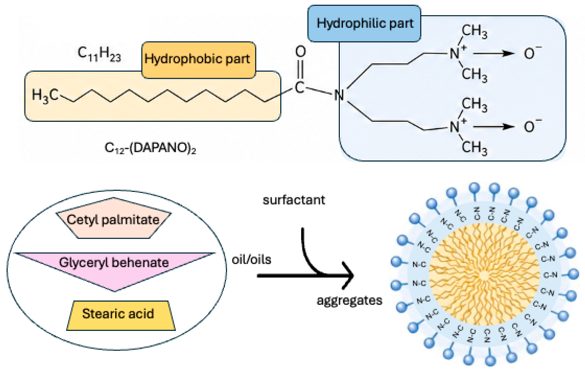
In the present contribution, we describe the potential of dicephalic di-N-oxide surfactants, N,N-bis [3,3′-(dimethylamino)propyl]dodecylamide (C12-(DAPANO)2), which comprises one hydrophobic tail and two hydrophilic head groups connected via a linker to a nitrogen bridge, in enhancing the stability of lipid nanoparticles. In this work, we investigate the effect of carrier oil type and di-N-oxide surfactants on the polymorphic behavior and aggregation stability of nanoparticles. The main objectives of this study were to examine nanoparticles prepared with three different solid lipids (cetyl palmitate, glyceryl behenate, and stearic acid) and to evaluate the influence of C12-(DAPANO)2 on the physicochemical properties of both the core and surface of the lipid nanoparticles. The size distribution of lipid nanoparticles was assessed by dynamic light scattering (DLS), while their shape and morphology were characterized using transmission electron microscopy (TEM) and atomic force microscopy (AFM). The size and shape of lipid nanoparticles are of fundamental importance for understanding surfactant behavior at the molecular level and are also critical for their practical applications. For analyzing the structure and interactions between components, differential scanning calorimetry (DSC) and thermogravimetric analysis (TGA) were employed. These techniques provide valuable structural information on dispersed particles and were used to investigate the crystalline structure of solid lipid nanoparticles (SLNs) and nanostructured lipid carriers (NLCs). The safety of the carriers was also verified against the J774.E macrophage cell line. No toxic effects were demonstrated.
The present work was undertaken to address the growing demand of the cosmetics and pharmaceutical industry for safe and non-toxic surfactants. We believe that this study represents one of the first comprehensive investigations of the effects of carrier oil and N-oxide surfactants on the stability of NLC and SLN dispersions and constitutes an important step toward the rational design of such systems.
2. Materials and Methods
2.1. Materials
Crodamol CP-PA-(SG) (cetyl palmitate) was obtained as a generous gift sample from Croda (Goole, UK). Compritol© 888 ATO (glyceryl behenate), a mixture of mono, di, and triglycerides of behenic acid (C22), was kindly donated by Gattefossè (Saint-Priest, France). Stearic acid was obtained from Sigma-Aldrich (Poznan, Poland). Mirgyol 812 (caprylic/capric triglycerides) was provided by CREMER OLEO GmbH & Co. KG (Hamburg, Germany). 3,3′-Iminobis(N,N-dimethylpropylamine) and lauroyl chloride were purchased from Sigma-Aldrich, (Poznan, Poland). Other reagents and solvents were of commercial grade and were not additionally purified before use. The water used in all the experiments was doubly distilled and purified with the Millipore Milli-Q purification system (Bedford, MA, USA) [
36,
37,
38].
2.2. Synthesis of Surfactant and Chemical Composition of Nanoparticles
The dicephalic surfactant N,N-bis [3,3′-(dimethylamino)-propyl]dodecylamide di-N-oxide (C
12-(DAPANO)
2) (structure and abbreviation are shown in
Figure 1) was synthesized and characterized according to Skrzela et al. [
36]. The synthesis was carried out in a reaction of stoichiometric amounts of dodecanoyl chloride with 3,3′-iminobis(N,N-dimethylpropylamine) in a chloroform solution at room temperature in the presence of a saturated NaHCO
3 aqueous solution. Crude liquid semiproduct was isolated by extraction in chloroform. To purify the semiproduct, the conversion to crystalline hydrochlorides was carried out in a reaction with gaseous HCl and thus the product was purified by means of crystallization from an acetate/chloroform mixture. The N,N-bis(3,3′-dimethylaminopropyl)dodecylamide hydrochlorides were treated with the saturated NaHCO
3 aqueous solution to obtain pure N,N-bis(3,3′-dimethylaminopropyl) dodecylamide di-N-oxide. The last step involved a reaction of the pure semiproduct with hydrogen peroxide (30% solution) in isopropyl alcohol at 50–60 °C for about 30 h. Then the solvent was evaporated and pure N,N-bis [3,3′-(dimethylamino)-propyl]dodecylamide di-N-oxide (C
12-(DAPANO)
2) was obtained. The structure and purity of the compounds were verified using
1H-NMR (
Figure 1) spectra in CDCl
3 solution: 0.86 (t, 3H, 3JHH = 6.6 Hz, CH
3(CH
2)
8CH
2CH
2CON-); 1.25 (m, 16H, CH
3(CH
2)
8CH
2CH
2CON-); 1.62–1.71 (k, 4H, -N[(CH
2CH
2CH
2N(CH
3)
2)
2]); 1.64 (k, 2H, CH
3(CH
2)
8CH
2CH
2CON-); 2.17–2.21 (s, 12H, -N[(CH
2CH
2CH
2N(CH
3)
2)
2]); 2.25 (t, 2H, CH
3(CH
2)
8CH
2CH
2CON-); 2.25–2.30 (t, 4H, -N[(CH
2CH
2CH
2N(CH
3)
2)
2]); 3.27–3.33 (t, 4H, -N[(CH
2CH
2CH
2N(CH
3)
2)
2]). Elemental analyses calculated C
22H
47N
3O
3, C, 65.86%; H, 11.81%; N 10.47%. Found: C, 65.76%; H, 11.86%; N 10.45%.
Solid lipid nanoparticles (SLNs) and nanostructured lipid carriers (NLCs) were produced by high-speed homogenization and ultrasound techniques. Briefly, the lipid phase consisted of only solid lipid for SLNs and solid and liquid lipid for NLCs melted approximately 10 °C above the melting point of solid lipid. An aqueous phase was prepared by dissolving C12-(DAPANO)2 in double-distilled water and heated to the same temperature as the oil phase. Hot aqueous phase was added to the oil phase and homogenization was carried out by high-speed stirring, using an Ultra-Turrax homogenizer (Ultra-Turrax T-25, IKA, Staufen im Breisgau, Germany) at 11,000 rpm for 15 min for SLNs and 20 min for NLCs. Coarse hot oil in water emulsion obtained was sonicated at 75–80 °C using a Sonic-1 ultrasonic bath (Polsonic, Warsaw, Poland, 100 W, 40 kHz) for 10 min to obtain SLNs and 15 min for NLCs. The obtained suspension was cooled down to room temperature in an ice bath to obtain the nanosized (<350 nm) lipid nanoparticles for further investigations.
The total amount of lipid phase (solid lipid, oil) was kept constant in all lipid nanoparticle suspensions (8%,
w/
w). SLNs contained only solid lipids (cetyl palmitate, glyceryl behenate, stearic acid) while in NLCs a portion of the solid lipid has been replaced by oil (Miglyol
® 812) at a level of 2.4%. The lipid nanoparticles in suspension were stabilized using 1% (
w/
w) of C
12-(DAPANO)
2.
Table 1 provides the composition of the formulations.
2.3. Physicochemical Parameters and Stability
The average size (i.e., the hydrodynamic diameter-DH), polydispersity index (PdI) and zeta potential (ZP) of the studied lipid nanoparticles were determined by the DLS (dynamic light scattering) method. The measurements were performed using a Zetasizer Nano Series from Malvern Instruments (Malvern, UK) with the detection angle of 173° in optically homogeneous square polystyrene cells. To remove any impurities, before the measurements, the samples were filtered through a filter with a 0.22 μm pore size directly to the optical cell. All measurements were performed at 25 °C. Each value was obtained as an average of three runs with at least 10 measurements. The DTS (Nano) program was applied for data evaluation. The study was performed for freshly prepared nanocapsules and for those stored for 90 days. The corresponding DH and PdI were calculated. ZP was measured by the microelectrophoretic method using a Malvern Zetasizer Nano ZS apparatus. The field strength applied was 20 V/cm. The zeta potential of the analyzed samples was obtained by calculating the average of 10 runs. All measurements were performed at 25 °C. Each value was obtained as an average of three subsequent runs of the instrument with at least 10 measurements.
SLN and NLC formulations were stored in polyethylene microtubes at room temperature (25 ± 1 °C) in a dark environment for 90 days. The formulations were analyzed with respect to particle size. The samples were analyzed after preparation (day 0) and after 90 days of storage at room temperature.
2.4. Microscopy Visualization
The transmission electron microscopy (TEM) measurements were performed to measure the morphology and size distribution of lipid nanoparticles. Images were taken using a FEI Tecnai G2 20 XTWIN electron microscope (FEI, Hillsboro, OR, USA). The size distribution of the nanoparticles for each sample was determined by counting the size of approximately 250 nanoparticles from several TEM images obtained from different parts of the TEM grids. A few drops of the diluted suspension were placed on the grid, stained with 2% uranyl acetate, and then the image was captured. The size distribution plots were fitted by using a Gauss curve approximation.
The morphology of the lipid nanoparticles was examined using the Veeco NanoScope Dimension V AFM (Bruker, Billerica, MA, USA) with an RT ESP Veeco tube scanner. The scanning speed was 0.5 Hz and a low-resonance-frequency pyramidal silicon cantilever resonating at 250–331 kHz was employed (at a constant force of 20–80 N/m). The amplitude of the resonance was set manually to the lowest possible amplitude for stable imaging within the contamination layer present on the surface. The SLNs and NLCs solutions were allowed to adsorb on a freshly cleaned mica surface for 12 h and then the excess substrate was removed by rinsing in double-distilled water for 1 min and dried at room temperature.
2.5. Thermal Analysis
The melting behavior and crystallinity of the lipid nanoparticles were investigated by a PerkinElmer DSC 8500 calorimeter (PerkinElmer, Shelton, CT, USA) equipped with a PerkinElmer CLN2 liquid nitrogen cooling system (PerkinElmer, Shelton, CT, USA), which was used to provide an inert gas atmosphere in the measurement cell. Aluminum pans were loaded with lyophilized lipid suspension and hermetically sealed; empty pans were used as a reference. Samples were submitted to two heating and two cooling runs from 0 °C to 100 °C and a rate of 5 °C/min was used. The onset temperature, melting point (peak maximum), and melting enthalpy (ΔH) were calculated.
To compare the crystallinity of the developed formulations, the recrystallization index (RI) is a useful parameter. It is defined as the percentage of the lipid matrix that recrystallizes during storage. The RI is calculated based on the melting enthalpy (ΔH), according to values reported in the literature [
37,
38] as follows:
where Δ
HSLN/NLC and Δ
HBulk Material are the melting enthalpy (J/g) of lyophilized SLNs or NLCs and lipid in bulk state, respectively. The concentration of the lipid phase is given in the percentage of the solid lipid in the total dispersion, and in lyophilized SLNs it is 100%, whereas in the case of NLC the solid lipid in the matrix is equal to 70% (e.g., a 70% (
w/
w) dispersion is 10/100 parts = 0.7).
Thermogravimetric analysis (TG-DTA) was carried out using a Setaram SETSYS 16/18 thermogravimetric analyzer (Setaram Instrumentation, Caluire, France), operated under a nitrogen atmosphere, with a heating rate of 5 °C/min and in a range of 20–500 °C (sample mass ∼ 4 mg).
2.6. Cytotoxicity Assessment of SLN and NLC in Macrophage (J774.E)
Cytotoxicity assessment was carried out using murine macrophages (J774.E). The choice of this in vitro model was made since, under in vivo conditions, macrophages constitute the primary line of defense against particulate matter. Thus, they are responsible for the distribution and clearance of nanoparticles and their agglomerates. Cells were cultured in RPMI-1640 medium (Institute of Immunology and Experimental Therapy, Wrocław, Poland) supplemented with 10% fetal bovine serum (FBS, Sigma-Aldrich, (Poznan, Poland)), L-glutamine (Sigma-Aldrich, (Poznan, Poland)) and antibiotics (penicillin and streptomycin, Sigma-Aldrich, Poznan, Poland)). For the cytotoxicity assessment, cells were seeded in 96-well plates (Thermo Fisher Scientific, Waltham, MA, USA) at a density of 7 × 10³ (J774.E) cells per well and pre-incubated at 37 °C for 24 h in a humidified atmosphere of 5% CO2. After that, nanoparticle dispersions were added. Stock dispersions of SLN and NLC were prepared in water and sonicated for 30 s before mixing with the complete culture medium. Cells were exposed to dispersions at concentrations ranging from 1 to 200 µg/mL for 48 h (5% CO2, 37 °C). After that, the MTT assay was carried out. The test is based on the enzymatic reduction in the tetrazolium salt MTT [3-(4,5-dimethylthiazol-2-yl)-2,5-diphenyl-tetrazoliumbromide] (Sigma-Aldrich, Poznan, Poland) in living, metabolically active cells. The metabolite, purple-colored formazan is measured colorimetrically, using a multiwell plate reader. Preliminary experiments showed no interference of the investigated dispersions with either MTT or formazan in a cell-free system. After 2 h of incubation at 37 °C, 80 μL of lysis buffer was added. The buffer consisted of 225 mL dimethylformamide (Sigma-Aldrich, Poznan, Poland), 67.5 g sodium dodecyl sulfate (Sigma-Aldrich, Poznan, Poland)) and 275 mL distilled water. The optical density (OD) was measured after 24 h using a spectrophotometric microplate reader (ELx800, BioTek, Winooski, VT, USA) at the wavelength of 570 nm (reference 630 nm). The OD of control cells was taken as 100%. Cell viability was determined as follows: % viability = (mean OD in the test wells/mean OD for control wells) × 100. The results were obtained from 3 independent experiments.
2.7. Statistical Analysis
Unless otherwise indicated, all the data are mean values ± s.d. calculated from at least three independent experiments. Student’s t-test was used to check the significance level between independent variables. The level of significance was set to p < 0.05.
3. Results
Nano drug delivery systems have significant applications in cosmetics, where they are used to more effectively transport active ingredients into the deeper layers of the skin. Thanks to nanotechnology, substances such as vitamins, antioxidants, and peptides can be encapsulated in nanocarriers, which increases their stability and protects them from degradation. This allows for better absorption of the ingredients as well as their gradual release, prolonging the product’s effectiveness. As a result, skincare products can act more precisely, for example, in reducing wrinkles or improving skin hydration. The use of nanotechnology also helps reduce irritation, as active ingredients are released in a controlled manner and can be used at lower concentrations while maintaining their effectiveness.
3.1. Functionalized N-Oxide Surfactants in the Fabrication of Lipid Nanoparticles with Different Lipids
The choice of surfactants in the structural design of the lipid nanoparticles—solid lipid nanoparticles (SLNs) and nanostructured lipid carriers (NLCs)—is one of the most important approaches for obtaining nanoproducts with desirable properties. Surfactants play a key role not only in controlling particle size and stabilizing dispersions, but also in influencing crystallization and polymorphic transitions. In the present study, we examined a dicephalic di-N-oxide surfactant, N,N-bis [3,3′-(dimethylamino)propyl]dodecylamine, C
12-(DAPANO)
2, and its ability to form stable, small-sized SLN dispersions containing different types of solid lipids at body temperature: a wax (cetyl palmitate), a triglyceride (glyceryl behenate), and a fatty acid (stearic acid) as the lipid matrix. NLCs were also prepared, in which the lipid matrix consisted of mixtures of the solid lipids used in SLNs with a constant amount of Miglyol 812 as the liquid oil. As is well known, the selected surfactant exhibits a strong tendency to form micelles in aqueous solution and can be considered a nonionic species at pH values close to or above 7 [
20]. The SLN and NLC products stabilized by the investigated N-oxide surfactants under study were successfully prepared by using high-speed homogenization followed by ultrasonication (see
Section 2.3). The total lipid phase content was kept constant at 8% (
w/
w), and the oil content in the lipid matrix of the NLCs was 30% (
w/
w) relative to the solid lipid. The composition of the formulations is presented in
Table 1.
3.2. Particle Size, ZP and Shape of SLNs and NLCs
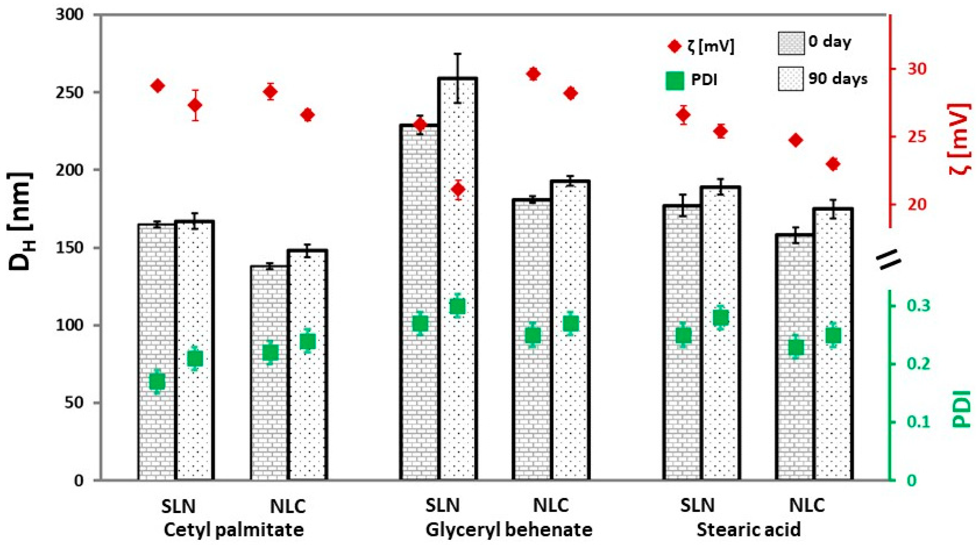
Since each individual system must be characterized, it is also desirable to improve the general understanding of how the choice of surfactant and lipid affects these properties in order to optimize the development process. Therefore, a thorough characterization of the physicochemical properties of the lipid nanoparticles was carried out. The prepared SLNs and NLCs were comprehensively evaluated in terms of their stability, particle morphology, and crystal structure. Therefore, the mean particle size and PdI of SLNs and NLCs were evaluated immediately after preparation using dynamic light scattering (DLS). Nanoparticles prepared with different lipids exhibited size distributions within the nanometer range (
Figure 2). All formulations showed mean particle diameters in the range of 130–250 nm and PdI values between 0.17 and 0.30. The particle size of lipid nanoparticles prepared with different lipids decreased in the following order: glyceryl behenate > stearic acid > cetyl palmitate. The particle sizes of all NLC systems prepared with medium-chain triglycerides (MCT) were more than 15 nm smaller than their SLN counterparts. This effect can be attributed to the higher liquid lipid content, which reduces the internal viscosity of NLCs and, consequently, lowers surface tension, facilitating the formation of smaller particles [
39]. A similar dependence was observed in other studies, described in the literature [
40,
41,
42].
ZP was used to measure the particle charge and electrostatic repulsion; it is the potential at the surface of shear, and it is measured in millivolts [
8,
43]. ZP is commonly used to assess the stability of emulsion systems, as high values are expected to protect particles from aggregation (in the absence of other stabilizing factors, such as steric stabilizers or hydrophilic surface groups) [
8,
43,
44]. For low-molecular-weight surfactants and pure electrostatic stabilization, ZP values above 30 mV provide good physical stability; the value of near |20| mV provides only a short-term physical stability whereas the values in the range of −5 mV to +5 mV indicate fast aggregation [
38,
43]. The colloid system with a steric stabilizer, could be stable even if its ZP value was higher than |20| mV [
43,
44].
The performed ZP measurements demonstrated that all obtained SLNs and NLCs carried a positive charge, with values in water exceeding +20 mV (
Figure 2), indicating a well-charged particle surface and, in principle, good physical stability. ZP of the SLNs prepared with different lipids decreased as follows: cetyl palmitate > stearic acid > glyceryl behenate. In the case of NLCs, the general trend of ZP was glyceryl behenate > cetyl palmitate > stearic acid. There was no correlation between the ZP magnitude and lipid nanoparticles. Most of the prepared nanoparticle dispersions are expected to be stable. These results also suggest that not only electrostatic repulsion determines nanoparticle stability, but that steric stabilization—provided here by the dicephalic surfactant C
12-(DAPANO)
2—may also contribute significantly to the formation of stable dispersions. The actual stability of the optimized SLN and NLC dispersions was further evaluated through stability studies.
The potential advantage of surfactants containing two polar head groups or with a dimeric/gemini architecture has been demonstrated in several colloidal systems. Bazylińska et al. demonstrated that dicephalous ionic surfactants can be successfully used to prepare biocompatible oil-in-water nanoemulsions, in which the type of surfactant, the surfactant-to-oil ratio, and the oil composition influence droplet size, surface charge, and stability [
21]. Takács et al. directly investigated the structure-stability relationship in latex particle dispersions stabilized with gemini surfactants and showed that surfactant architecture strongly influences adsorption, particle charging, and aggregation; in particular, gemini surfactants with appropriate alkyl chain length promoted colloidal stability more effectively than simple monomeric analogs under comparable conditions [
45]. Similarly, Nagao et al. reported that the gemini surfactant, sodium dilauramidoglutamide lysine, improved both the colloidal and structural stability of phytantriol-based cubosomes, confirming the role of gemini surfactants as effective stabilizers of nanostructured lipid systems [
46]. Furthermore, Wang et al. showed that nonionic gemini surfactants exhibited significantly lower critical micelle concentrations and better surface activity than their monomeric counterparts, further supporting the theory that surfactant architecture can improve interface performance [
47]. These studies provide a mechanistic basis for the assumption that the dicephalic structure of C
12-(DAPANO)
2 may contribute to the stability of the studied nanoparticles. The presence of two polar head groups may improve interfacial anchoring and increase the steric-electrosteric barrier at the particle surface, thereby reducing the likelihood of aggregation.
The stability of SLNs and NLCs (
Figure 2) was experimentally verified by DLS over a period of 90 days. The results showed that formulations containing cetyl palmitate and stearic acid as solid lipids remained stable against aggregation when stored as aqueous suspensions at room temperature. For formulations based on glyceryl behenate, a small and negligible increase in particle size (less than 50 nm) was observed; however, PdI remained around 0.3, indicating relatively homogeneous particle populations. After three months of storage, no significant decrease in ZP values was detected, further confirming the good stability of these preparations.
3.3. TEM and AFM
TEM and AFM are rapid, powerful and relatively non-invasive visual techniques to obtain information about the mean size and the actual surface and morphology characteristics of the prepared SLNs and NLCs [
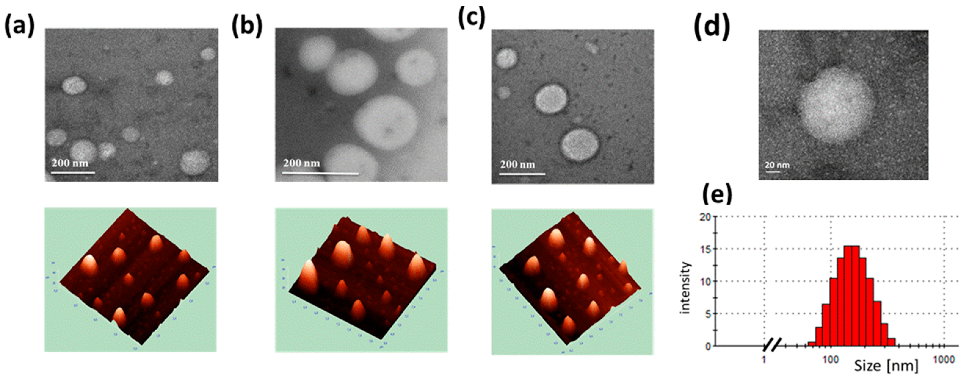
48]. The TEM images revealed that NLCs (
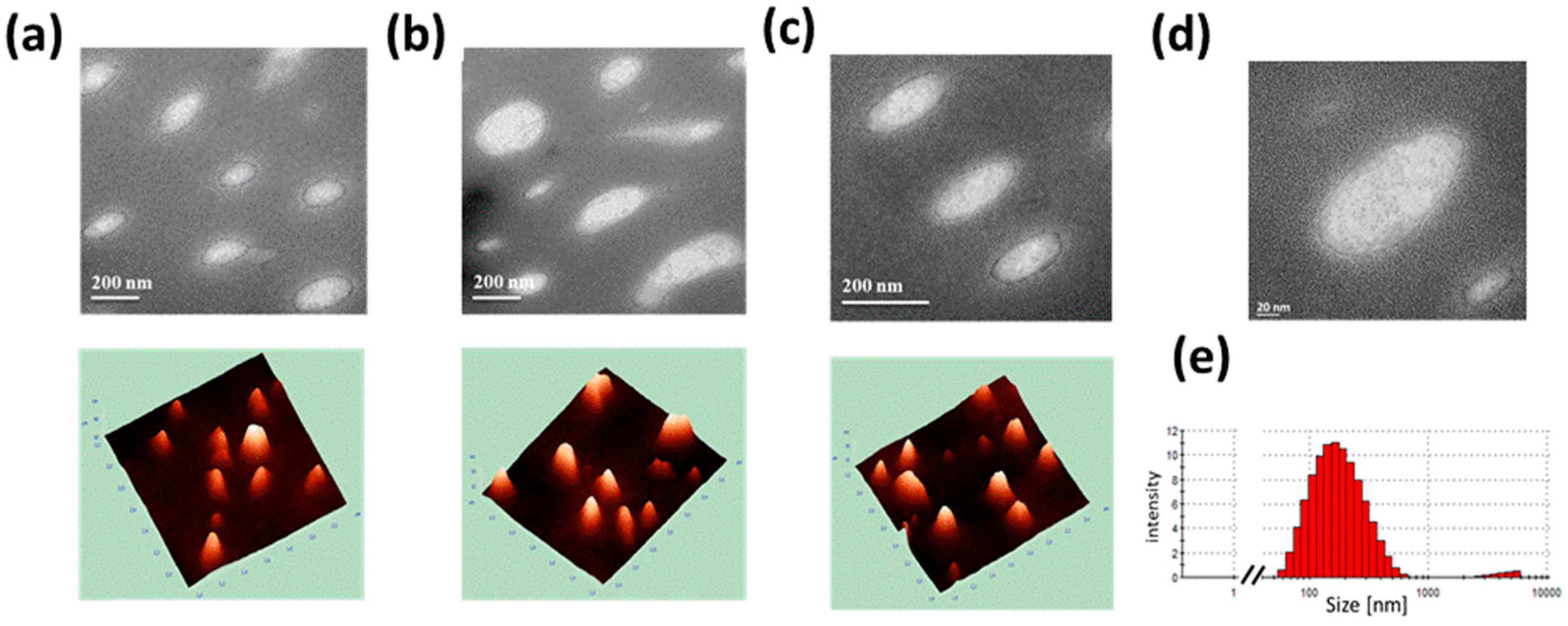
Figure 3) exhibited a spherical morphology with a small diameter range of 100–200 nm, while SLNs (
Figure 4) appeared in quasi-spherical, cylindrical, and irregular shapes, independent of the solid lipid used in the matrix, and with larger particle sizes (150–250 nm). The addition of oil to the solid lipid core of the nanoparticles reduced particle size, likely due to a decrease in viscosity and, consequently, surface tension, which facilitates the formation of smaller particles with smoother surfaces [
39]. The size and morphology observed by TEM correlated well with the results measured by AFM (
Figure 3 and
Figure 4). However, in contrast to TEM figures of SLNs and NLCs, AFM visualized the topography of the particles by investigating the three-dimensional structures.
Moreover, changing the solid lipid in formulations stabilized by the non-ionic dicephalic surfactant C
12-(DAPANO)
2 did not appear to affect the particle morphology, as confirmed by both microscopic techniques applied. In addition to particle size and shape, TEM imaging also indicated that the particle surface is coated with surfactant. The staining agent, uranyl acetate, has bonded to the carboxylic group [
48]. The density of carboxylic groups is higher in the lipid core than in the surfactant layer, resulting in differences in staining intensity at the outer layer, which indicates the presence of a soft surfactant layer around the SLNs. The diameters of SLNs and NLCs obtained by DLS (
Table 1), TEM, and AFM (
Figure 3 and
Figure 4) were in reasonable agreement, with minor differences arising from the distinct sample preparation procedures and measurement principles. It should be noted that DLS does not directly measure particle size but rather analyzes light scattering, from which particle size is calculated. For this reason, sizes obtained by TEM or AFM are generally considered to provide higher spatial resolution and may be regarded as more direct measurements of nanoparticle dimensions [
39,
44].
3.4. Thermal Analysis
Formulation development of the lipid nanoparticle (LN) aims to maintain physical stability in both particle size and crystalline state of the lipid matrix. The particle size is the most important property of LN due to the desired fast drug release and high bioavailability. Beside these prior factors, for different types of fat, it has already been shown that it is possible to stabilize desired and unstable polymorphic forms by using the different emulsifiers [
40,
41,
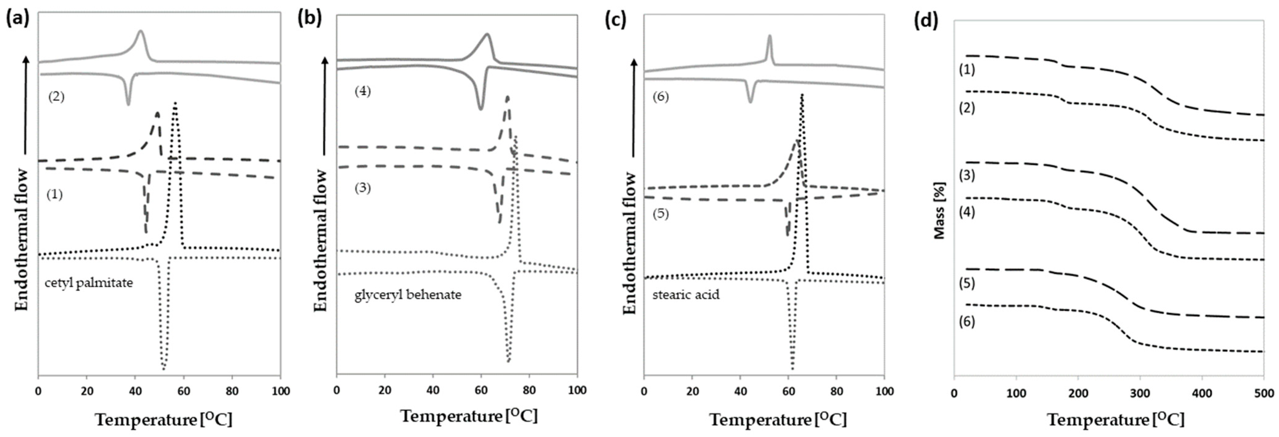
49]. The melting and crystallization behavior and polymorphic transformation kinetics of all 6 formulations and bulk material (
Figure 5) were examined by differential scanning calorimetry (DSC).
In this research, we investigated the effect of the non-ionic dicephalic C
12-(DAPANO)
2 surfactant on the thermotropic behavior of SLNs and NLCs containing the three different solid lipids (cetyl palmitate, glyceryl behenate, stearic acid). Knowledge of the thermal behavior of bulk lipid is useful for interpreting its behavior in emulsified phases. Therefore, we characterized the thermal transitions of cetyl palmitate, glyceryl behenate, and stearic acid. Lipid phases were initially heated to 100 °C to completely melt them and then subjected to controlled cooling–heating cycles with the heat flow being recorded as a function of temperature (
Figure 5). Cetyl palmitate (
Figure 5a) revealed two peaks: the first peak with a lower melting point (45.85 °C) which based on literature data [
18] was attributed to the α-polymorphic form (thermodynamically unstable modification), whereas the second peak (56.50 °C) was attributed to the β-polymorphic form (stable modification). Glyceryl behenate (
Figure 5b) melts between 64 °C and 76 °C with the melting point at 74.92 °C. Based on the high content of diglycerides (>50%) it can be concluded that bulk material crystallizes in the β’ modification [
37]. Stearic acid (
Figure 5c) exhibited a single melting peak at 65.67 °C, which, in agreement with previous studies, corresponds to the β polymorphic form [
50].
The SLN thermal analysis allows evaluating the thermodynamic parameters change associated with the physical state of the lipid core [
8,
37]. Comparing the data obtained by different samples, information about the distribution of components and interaction can be gained. All SLNs thermograms were compared with their bulk lipid (
Figure 5,
Table 2). The melting temperature of nanoparticles was 1–6 °C lower than that of the bulk lipid, respectively, and these results agree with the data reported in the literature [
8,
37,
51]. This shift in peak temperature is attributed to the colloidal size, which increases the specific surface area, as well as to interactions between lipid and surfactant molecules that lead to a less ordered phase [
18,
52,
53]. The effect of the surfactants was explained by the surface tension generated near the particle surface, which shifts the onset and melting temperatures and melting enthalpy of the lipid towards lower values compared to bulk lipid [
18,
53]. It is worth noting that the DSC thermograms (
Figure 5a) for SLN containing cetyl palmitate show that the separate peak of the α-modification disappears which suggests that the hydrophobic chain of surfactant is dispersed in the lipid blend, which creates distortion in the lipid matrix.
In the case of NLC, DSC experiments are useful to understand the behavior of solid lipids mixing with liquid lipids, such as oils. Cetyl palmitate, glyceryl behenate and stearic acid exhibit good miscibility with medium-chain triglycerides (Miglyol
® 812) and thus, a homogeneous lipid matrix can be obtained in the NLCs. In the case of the studied NLCs, a lower and wider melting point was recorded (
Figure 5). Also, the melting enthalpy of the lipid nanocarriers decreased when the liquid lipid was incorporated in the lipid core (
Table 2). Obviously, the oil, functioning as an impurity for the main lipid, has an additional and greater effect with respect to the colloidal size and matrix crystallization [
18,
38]. The presence of shoulders in the calorimetric curves of SLNs containing stearic acid (
Figure 5c) indicated that the distribution of surfactants in this lipid nanoparticle frame was not homogeneous; probably surfactant molecules were mainly arranged on the particle surface [
53].
As mentioned previously, the surfactant’s action in the lipid nanoparticles can be attributed to a decrease in the melting temperature and enthalpy, and to the colloidal low dimensions of the particles, in particular to their high surface area to volume ratio described by the Thomson equation [
52]. This state is the reason for lattice defects in the lipid matrices, and in consequence, a decrease in their crystallinity in comparison to their bulk counterparts [
37,
52]. There are several reports that investigate the influence of the surfactants on the melting and crystallization behavior of the lipid nanoparticles [
18,
43,
54]. The effect of the surfactants is seen in the crystallization behavior. To confirm this statement, we calculated the recrystallization index of the SLNs and NLC formulations. Recalling the experimental section, the index RI was calculated from the total melting enthalpy of the lipid nanoparticle dispersions divided by the total enthalpy of the physical mixture and percentage composition of solid lipid. The SLNs are formed only by the solid lipid whereas the NLCs core is composed of the solid (70%) and liquid (30%) lipids. The RI value of the bulk material was set at 100% and this was used as a reference to determine the degree of crystallinity of the nanoformulations. The RI values for all 6 formulations were below 100% (
Table 2). The highest RI (more than 50%) was received for lipid nanoparticles composed of glyceryl behenate; however, this value is smaller for about 10% than that observed in other studies, described in the literature [
37]. The RI value for lipid SLNs and NLCs formulation made by cetyl palmitate and stearic acid is smaller than 30% and the values of the recrystallization index for the NLC were almost 3 times smaller than SLN formulations. The recrystallization index could affect the long-term stability of aqueous SLN dispersions. In general, dispersions with a highly recrystallized lipid phase (high recrystallization index) showed an increased particle size growth [
37]. Our result indicates that the non-ionic dicephalic C
12-(DAPANO)
2 surfactant significantly reduces the ability of the matrix to recrystallize.
The TG curves (
Figure 5) showed the thermal decomposition steps for all lipid nanoparticles. The thermal behaviors of the SLN and NLC formulations containing the same solid lipids were very similar to each other. The first stage was observed by TG in the temperature range from 35 to 110 °C with a ∆m of <3.0%. It is due to the release of water molecules from lipid nanoparticles. Thermal events observed in the second stage at about 140–180 °C were accompanied by a mass loss (∆m = 5–10%) curve probably because of volatiles evaporation (N
2, O
2, CO
2, and the rest of H
2O) or low-temperature degradation of unstable chemical fragments in the samples [
50]. Subsequently, all samples were stable in a very wide temperature range, from 180 to 250 °C (∆m around 5.0%), and in the next stage above 250 °C underwent rapid decomposition (∆m more than 50%). During continuous heating, the samples in the last stage were carbonized (∆m > 3%). By the comparison of the TG curves of all formulations (1–6), it was concluded that up to 150 °C, all nanosystems have had thermal stability.
3.5. Cytotoxicity Assessment of SLN and NLC in Macrophage
Macrophages are key players in skin inflammation, acting as major innate immune cells at sites of injury or infection. They recognize pathogens, remove damaged cells, and release inflammatory mediators such as cytokines and chemokines, thereby both driving and resolving inflammation [
34]. They can adopt different functional states: pro-inflammatory M1 macrophages produce factors like TNF-α, IL-1β, IL-6, and reactive oxygen species, enhancing inflammation and pathogen clearance, while anti-inflammatory M2 macrophages support tissue repair, wound healing, and inflammation resolution through mediators such as IL-10 and TGF-β [
55,
56]. Maintaining a balance between these phenotypes is essential for proper skin regeneration. Disruption of this balance may lead to chronic inflammatory conditions, including atopic dermatitis, psoriasis, acne, and non-healing wounds [
57]. Therefore, macrophages are considered an important target in developing new dermatological and cosmetic therapies.
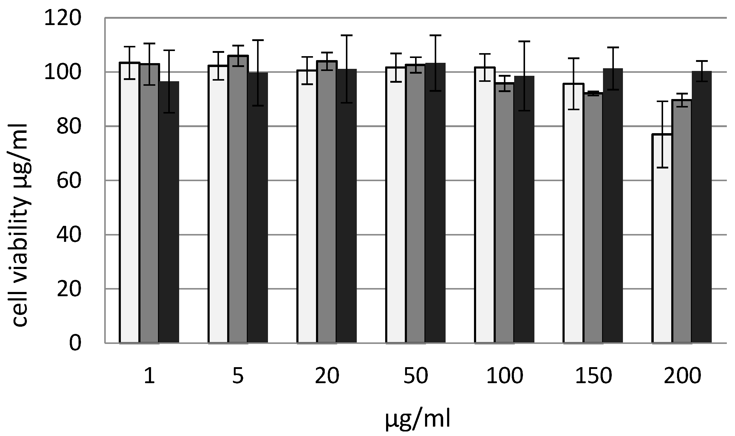
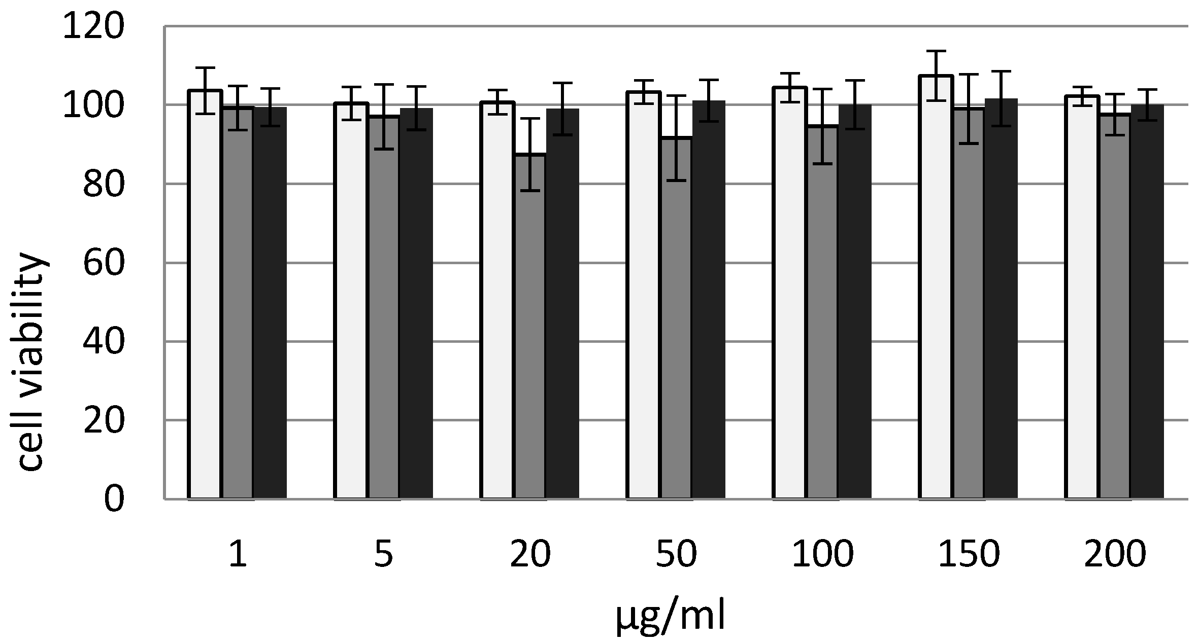
The effect of the prepared SLN and NLCs on cell viability of the J774.E cell line was studied over a range of 1–200 µg/mL and is presented in
Figure 6 and
Figure 7. At most tested concentrations for SLN systems, cell viability remained around 95–105%, suggesting a lack of significant cytotoxicity at lower and intermediate doses. For concentrations of 1–150 µg/mL, the values approached 100%, and the differences between the bars were small and within the range of measurement error. Only Formula 1 and, to a lesser extent, Formula 3 induced a slight to moderate, dose-dependent decrease in cell viability at higher concentrations. For NLC, the lowest value was observed at a concentration of 20 µg/mL for Formula 4, where viability decreased to approximately 85–90%. However, due to the relatively large error bar, this decrease may not be biologically or statistically significant. For the remaining formulations, even at higher concentrations, no deterioration in viability was observed. Overall, the tested nanoparticles showed little to no cytotoxicity in the cell models used. All tested NLCs were found to be biocompatible in the in vitro model and could be further investigated as potential delivery platforms.
Since the tested NLC and SLN carriers demonstrate safety toward the examined macrophage cell line, it can be concluded that they do not induce a pro-inflammatory response at the cellular level. The lack of macrophage activation—cells that play a key role in the initiation and maintenance of inflammatory processes—suggests that the studied systems do not trigger undesirable immune reactions.
Therefore, it can be assumed that the use of NLC and SLN carriers in cosmetic or dermatological formulations will be safe for the skin and will not lead to the exacerbation of inflammatory processes. This indicates their potential as mild and well-tolerated delivery systems for active substances.
4. Conclusions
The studied dicephalic di-N-oxide surfactant was proven to be a suitable stabilizer for lipid nanoparticles; it allowed fine control of the LNP formation process and improved their thermal stability. The SLNs and NLCs were successfully prepared with three different solid lipids, i.e., cetyl palmitate, glyceryl behenate, and stearic acid, with a small particle size (DH < 250 nm) and a narrow particle size distribution (PdI < 0.3) obtained. All formulations preserved good physical stability of SLNs and NLCs after 90 days of storage at room temperature. Morphology studies by TEM and AFM showed unsymmetrical shape SLNs and almost spherical NLCs with smooth surfaces. The NLCs showed less ordered crystalline structure than the SLNs, which was conferred by the inclusion of the oil; as a result they had lower values for phase transition temperature, melting enthalpy, and recrystallization index. Since the tested NLC and SLN carriers are safe for macrophages, they do not induce a pro-inflammatory response at the cellular level. The absence of macrophage activation suggests they do not trigger undesirable immune reactions. Therefore, these carriers appear to be safe and well-tolerated delivery systems for cosmetic or dermatological use without exacerbating inflammation.
Our study established that bifunctional N-oxide surfactants proved suitable for the stabilization of SLN and NLC dispersions, and they could provide a good opportunity for fabricating a new functional class of stabilizers for nanoparticles for future dermal products. Future work will aim at investigating how surfactants influence the properties of drug-loaded lipid nanoparticles, such as physicochemical stability, as well as the drug release.



 ,
,