Review

Reducing Stress Shielding in Cementless Total Hip Arthroplasty: A Translational Review of the Gradient-Modulus Ti-Nb-Sn Femoral Stem


1
Department of Orthopaedic Surgery, Tohoku University Graduate School of Medicine, 1-1 Seiryo-machi, Aoba-ku, Sendai 980-8574, Japan
2
Department of Radiology, Akita University Graduate School of Medicine, 1-1-1 Hondo, Akita 010-8543, Japan
*
Author to whom correspondence should be addressed.
Appl. Sci. 2026, 16(11), 5630; https://doi.org/10.3390/app16115630
Submission received: 2 May 2026 / Revised: 23 May 2026 / Accepted: 26 May 2026 / Published: 4 June 2026

Featured Application

The β-type Ti-33.6Nb-4Sn (TNS) alloy femoral stem represents a clinically applicable solution to stress shielding in cementless total hip arthroplasty, offering direct relevance to orthopedic surgeons selecting implant materials for patients at risk of proximal femoral bone loss—particularly younger, active patients and those with poor bone quality. The gradient Young’s modulus design principle reviewed here is further applicable to the development of low-modulus fixation devices for fracture management and to β-type titanium dental implants, providing a broad material design platform for load-bearing skeletal interfaces.

Abstract

Stress shielding (SS) after cementless total hip arthroplasty arises from the stiffness mismatch between conventional Ti-6Al-4V femoral stems (110 GPa) and cortical bone (10–30 GPa). The β-type Ti-33.6Nb-4Sn (TNS) alloy femoral stem addresses this limitation through a continuous Young’s modulus gradient (~70 GPa proximally to ~40 GPa distally) achieved by localized heat treatment of a single homogeneous alloy. This review synthesizes a translational research program encompassing material characterization, finite element modeling (FEM), preclinical animal studies, and prospective clinical follow-up of up to seven years. FEM demonstrated favorable proximal micromotion well below the osseointegration threshold, with physiological proximal stress concentration concordant with clinical outcomes. At seven years, SS grade distribution was significantly lower in the TNS group than in Ti-6Al-4V controls, with SS frequency reduced in Gruen Zones 2, 3, and 6, and no stem-related failures; however, third-degree SS was still observed in 11 of 34 evaluable cases (32%), indicating that modulus-gradient optimization alone is insufficient to fully prevent SS. TNS alloy is currently the only β-type titanium alloy clinically applied in joint prostheses. Remaining challenges include stem geometry optimization, additive manufacturing-based porous structures, and dual-energy X-ray absorptiometry-based bone density quantification. Future directions encompass long-term follow-up, cyclic fatigue FEM simulations, and expansion to fracture fixation devices and dental implants.

1. Introduction

1.1. Global Increase in THA and the Revision Burden

Total hip arthroplasty (THA) is a highly successful procedure that significantly improves pain, function, and quality of life [1]. Its global volume has increased markedly due to population aging and higher functional demands among younger patients [1,2]. In the United States, primary THA is projected to reach 850,000 cases by 2030 and 1,429,000 by 2040 (129% and 284% increases from 2014), while Germany is expected to see a 62% rise by 2060 [2,3]. Accordingly, revision THA is also increasing, with projected rises of 43–70% by 2030 in the United States and approximately 40% by 2060 in Germany [3,4]. This trend highlights the need to optimize primary THA to reduce complications and revision demand. Revision THA is technically more challenging, particularly in the presence of poor bone quality. Stress shielding (SS) after cementless THA leads to proximal femoral bone loss, increasing the risk of periprosthetic fracture and complicating re-implantation [5,6,7,8]. Therefore, preventing SS and preserving bone stock are essential for improving primary THA durability and facilitating future revision procedures.

1.2. Stress Shielding: Pathophysiology and Clinical Impact

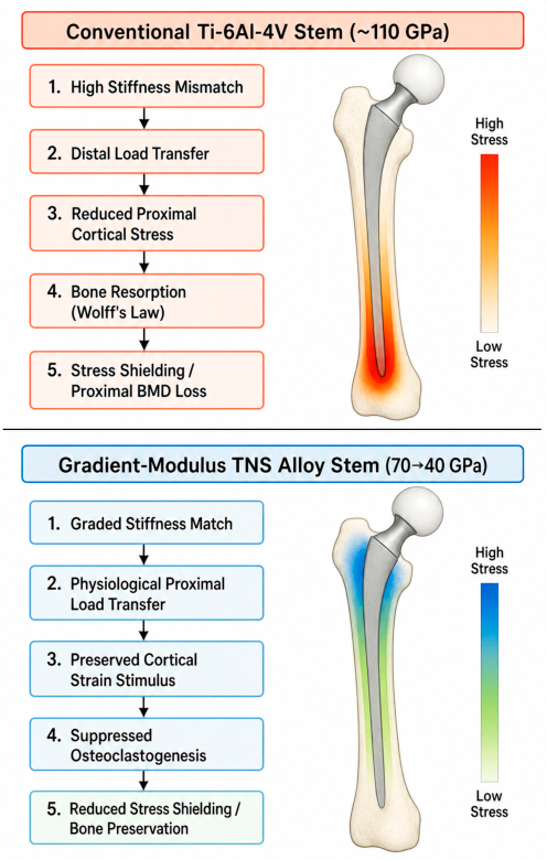
The fundamental cause of SS is the stiffness mismatch between conventional Ti-6Al-4V stems (~110 GPa) and cortical bone (10–30 GPa) [5], which shifts mechanical load away from the proximal femur. Finite element analyses have demonstrated that fully ingrown stems reduce proximal cortical loading by approximately 30% and increase distal stresses up to fourfold compared to cemented configurations [9,10]. Under these non-physiological conditions, bone remodels according to Wolff’s law [5]—resorption occurs where strain stimulus is reduced—and computational simulations confirm that press-fit stems drive a progressive “stress bypass,” continuously reducing proximal cortical bone stock over time [11,12]. Longitudinally, high-degree SS has been shown to increase progressively, with a preoperative canal bone ratio ≥ 0.49 identified as an independent risk factor [13]. The clinical consequences operate at multiple levels. Radiographically, SS is graded according to the classification of Engh et al. [6]; structurally, it predisposes patients to aseptic loosening and periprosthetic fracture [5,6]; and functionally, it is associated with reduced Harris Hip Score, impaired walking ability, and thigh pain [6,13]. Recent dual-energy X-ray absorptiometry (DXA) studies further confirm BMD reductions exceeding 20% in Gruen Zone 7 within short follow-up periods [14]. The contrasting biomechanical consequences of high-modulus versus gradient-modulus stem design are summarized schematically in Figure 1.

1.3. Limitations of Conventional Ti-6Al-4V Stems

Despite their proven mechanical reliability, Ti-6Al-4V alloy stems exhibit a Young’s modulus of approximately 110 GPa—five to eightfold higher than cortical bone (20.7 GPa) and trabecular bone (14.8 GPa)—a mismatch that cannot be resolved by surface treatment, geometry modification, or coating alone, as the bulk stiffness remains unchanged [15,16]. Finite element-based bone remodeling simulations have predicted periprosthetic BMD reductions of up to 45.8% over 10 years with Ti-6Al-4V stems [17], a finding corroborated by longitudinal DXA studies demonstrating that proximal femoral bone loss continues into the second and third postoperative decades [18,19]. These data underscore that the intrinsic stiffness of Ti-6Al-4V represents a fundamental material limitation, motivating the development of alternative alloy systems with lower, bone-compatible elastic moduli.

1.4. Historical Development of Low-Modulus Femoral Stems

Efforts to reduce stem stiffness span more than four decades. The first-generation RM isoelastic stem (Bombelli and Mathys, 1982) confirmed the conceptual validity of stiffness-matched implants but demonstrated unacceptably poor clinical performance: a 14–17-year follow-up of 149 hips reported a 10-year survival rate of only 70%, with high failure rates attributable to poor primary fixation and excessive debris production [20,21]. This established a central design paradox: reducing stiffness risks compromising the structural integrity required for stable fixation. β-type titanium alloys offered a second-generation approach with lower elastic moduli and adequate mechanical strength [22,23]; however, the Ti-Mo-Zr-Fe Accolade stem (~74 GPa)—the most widely used clinical example—reported a revision rate of 66.7% at 12 years, primarily from trunnion failure and elevated metal ion levels [24], demonstrating that alloy substitution alone is insufficient without attention to geometry and bearing design [23]. A comparative analysis of β-alloy failures reveals a consistent pattern: modulus reduction alone is insufficient without concurrent optimization of stem geometry, bearing design, and surface fixation strategy. The Ti-Mo-Zr-Fe Accolade stem failed primarily due to trunnion corrosion and elevated metal ion levels—a consequence of modular neck design rather than alloy stiffness per se [24]. The RM isoelastic stem failed due to inadequate primary fixation and excessive polyethylene debris [20,21]. These failure modes are mechanistically distinct from SS and underscore that the TNS alloy’s clinical success reflects not only its material properties but also its metaphyseal-filling geometry, sandblasted proximal surface, and polished distal shaft—design features that collectively address the limitations of prior low-modulus stems. The key theoretical advance came from Kuiper and Huiskes [25], whose mathematical optimization showed that a spatially graded, non-homogeneous Young’s modulus could reduce bone–stem interface stresses by more than 50% [25,26,27,28]—providing the foundation for the functionally graded stem concept and, ultimately, the TNS alloy stem [23,25]. Unlike prior reviews of low-modulus β-Ti alloys—which have largely remained at the preclinical or material characterization stage—the present review is distinguished by its synthesis of a complete translational evidence chain: from alloy development and FEM validation to prospective clinical outcomes at 7 years [29,30,31]. To our knowledge, this constitutes the first critical review to evaluate a functionally graded β-Ti femoral stem against a matched Ti-6Al-4V control cohort with mid-term radiographic follow-up, while simultaneously appraising the limitations that must be addressed before this design paradigm can be recommended for broad clinical adoption.

1.5. Objectives of This Review

The Ti-33.6Nb-4Sn (TNS) alloy femoral stem directly realizes the Kuiper–Huiskes design ideal through localized heat treatment of a single homogeneous alloy, achieving a continuous Young’s modulus gradient (approximately 70 GPa proximally to 40 GPa distally) without compositional change [17,31,32,33]. A translational research program encompassing material characterization, patient-specific finite element modeling, preclinical animal studies, and prospective clinical follow-up of up to seven years has now been completed for this implant. The present review synthesizes this evidence across four domains: (1) material properties and design principles of TNS alloy; (2) biomechanical finite element analyses and their concordance with clinical radiographic outcomes; (3) preclinical animal studies of TNS alloy implants; and (4) clinical outcomes across short- to mid-term follow-up with comparison to conventional Ti-6Al-4V stems. Remaining design limitations and future perspectives—including additive manufacturing, advanced surface modification, and application to diverse femoral morphologies—are also discussed.
Literature searches were conducted in PubMed, Scopus, and Web of Science for publications from database inception through March 2025. The following search term combinations were used: (“Ti-Nb-Sn” OR “TNS alloy” OR “Ti-33.6Nb-4Sn”) AND (“stress shielding” OR “total hip arthroplasty” OR “femoral stem”); (“low-modulus titanium” OR “beta-titanium alloy”) AND (“stress shielding” OR “bone remodeling” OR “cementless”); and (“finite element” OR “FEM”) AND (“hip arthroplasty” OR “femoral stem” OR “stress shielding”). Only English-language publications were included. Studies were considered eligible for inclusion if they reported original data on: (1) material characterization or biomechanical testing of TNS alloy or related β-type titanium alloys; (2) finite element modeling of femoral stem stress distribution; (3) preclinical animal studies of low-modulus implant fixation or fracture healing; or (4) clinical outcomes of cementless THA femoral stems with radiographic or functional follow-up. Review articles were included when they provided relevant comparative context or theoretical framework; conference abstracts and case reports were excluded. Additional references were identified through backward citation searching of included articles and forward citation searching of key primary sources. Final article selection was performed by the corresponding author (Y.M.), with ambiguous cases resolved by consensus with co-authors (H.T., R.K.).

2. TNS Alloy: Material Properties and Design

2.1. Composition and Basic Properties of β-Type Titanium Alloys

β-type titanium alloys—characterized by a body-centered cubic crystal structure stabilized by elements such as Nb, Zr, Ta, Mo, Fe, and Sn—have emerged as the most promising class of metallic biomaterials for load-bearing orthopedic implants, offering a uniquely favorable combination of low elastic modulus, high specific strength, excellent corrosion resistance, and inherent biocompatibility [34]. The reported Young’s modulus of β-type Ti alloys ranges from approximately 37 to 85 GPa depending on composition and processing—substantially lower than the ~110 GPa of Ti-6Al-4V and considerably closer to the 10–30 GPa of cortical bone—thereby reducing the driving force for SS at the bone–implant interface [35,36,37]. Representative alloy systems include Ti-Nb-Zr-Ta (~55–65 GPa), Ti-Mo-Zr-Fe (~74 GPa), and Ti-Nb-Sn (TNS, ~40 GPa), each with distinct balances of modulus reduction, mechanical strength, and biological response [35,36,37] (Table 1). In terms of biocompatibility, β-type Ti alloys comprising non-toxic elements (Nb, Zr, Ta, Sn) demonstrate favorable osseointegration capacity, corrosion resistance in simulated body fluids, and cytotoxicity profiles superior to conventional alloys containing Al and V [23,38].

2.2. TNS Alloy: Composition, Microstructure, and Mechanical Properties

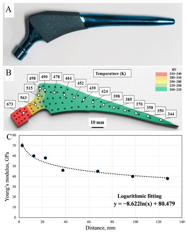
The Ti-33.6Nb-4Sn (TNS) alloy was developed by Hanada et al. at Tohoku University through severe cold rolling and cold swaging of β-phase rods, yielding a characteristic fiber structure of stress-induced α″ martensite that simultaneously achieves a low Young’s modulus (~40–52 GPa) and a high tensile strength exceeding 800 MPa [32,33]. The key to TNS alloy’s low-modulus properties lies in the role of Sn addition: while Nb stabilizes the β-phase by suppressing the martensitic transformation, Sn suppresses the formation of the ω-phase—a metastable precipitate that would otherwise increase the elastic modulus and embrittle the alloy—thereby preserving both low stiffness and ductility during heat treatment [33,40]. A defining characteristic of TNS alloy is the tunability of its mechanical properties through heat treatment alone, without compositional change: localized application of heat at 673 K along the longitudinal axis of the stem creates a continuous gradient in Young’s modulus from approximately 70 GPa proximally to 40 GPa distally, with the proximal high-modulus zone ensuring structural integrity at the neck while the distal low-modulus zone promotes physiological load transfer [17,32,33]. Biocompatibility studies of Ti-Nb-Sn alloys have demonstrated cytotoxicity profiles comparable to Ti-6Al-4V and commercially pure titanium in direct-contact cell culture assays, with bone tissue compatibility confirmed by in vivo insertion into rabbit femoral medullary canals showing no adverse tissue reactions [41]. TNS alloy is currently the only β-type titanium alloy to have been clinically applied in joint prostheses—a distinction that reflects both the maturity of its translational research program and the robustness of its preclinical safety validation [31].

2.3. Gradient Young’s Modulus via Localized Heat Treatment

Using a custom gradient furnace, the proximal region of the TNS stem is heated at 673 K under vacuum while the distal region is shielded by an insulating plate, inducing a continuous Young’s modulus gradient—~70 GPa proximally, ~50 GPa mid-stem, and ~40 GPa distally—without altering alloy composition [29,33] (Figure 2). The proximal high-modulus zone ensures mechanical strength and resistance to neck fracture, while the distal low-modulus zone promotes physiological load transfer and reduces the driving force for SS [27,42]. This approach achieves functional optimization through heat treatment alone, in contrast to conventional functionally graded materials that require compositional changes and risk heterogeneous biocompatibility [17].

2.4. Surface Modification

Anodic oxidation of the TNS alloy generates a TiO2 surface layer that enhances bone affinity through photocatalytic activity, improving osteoconductivity and providing antibacterial performance—properties unachievable by the low-modulus alloy substrate alone [43,44]. Surface characterization by scanning electron microscope, X-ray photoelectron spectroscopy, and X-ray diffraction has demonstrated that anodic oxidation combined with hot water treatment produces abundant apatite formation and a nanotubular TiO2 morphology, resulting in significantly higher bone-bonding strength and new bone formation in pull-out and histomorphometric assays compared to untreated TNS alloy [45]. These surface modifications complement the low-modulus design of the TNS stem: by improving osseointegration at the bone–implant interface, anodic oxidation may further suppress SS beyond what modulus reduction alone can achieve [45].

3. Biomechanical Analysis: FEM Studies

3.1. Theoretical Background

The theoretical foundation for modulus-graded stem design derives from the mathematical optimization work of Kuiper and Huiskes [25], who demonstrated that a non-homogeneous distribution of Young’s modulus along the femoral stem could reduce bone–stem interface stress by more than 50% compared to a homogeneously stiff stem—establishing the quantitative target that the TNS alloy stem was designed to fulfill [17,30]. Under physiological loading, bone maintains its mass and architecture through the mechanostat: osteocytes, the primary mechanosensory cells embedded within the lacunocanalicular network, detect deviations in strain stimulus from an adaptive setpoint and orchestrate four distinct mechanoadaptive pathways—formation modeling, targeted remodeling, resorption modeling, and disuse-mediated remodeling—to restore mechanical homeostasis [46,47]. The mechanostat response depends not only on strain magnitude but also on duration, rate, spatial distribution, and rest intervals between loading cycles, underscoring that restoring physiological strain stimulus around a femoral stem requires a continuous gradient of stiffness rather than a simple bulk modulus reduction [46,48].

3.2. In Vitro Biomechanical Validation and Strain-Adaptive Remodeling FEM

Yamako et al. confirmed in vitro that the gradient bending stiffness of the TNS stem produced favorable proximal load transfer without compromising initial press-fit stability compared to a morphologically similar Ti-6Al-4V stem [27]. A subsequent strain-adaptive bone remodeling FEM incorporating CT-derived bone density mapping and the Huiskes remodeling algorithm predicted that, while the TNS stem reduces proximal BMD by 45.8% at 10 years, bone density in the TNS group remains 42.6% higher than that of Ti-6Al-4V, with a lower stress-strength ratio throughout [17,42].

3.3. Patient-Specific FEM

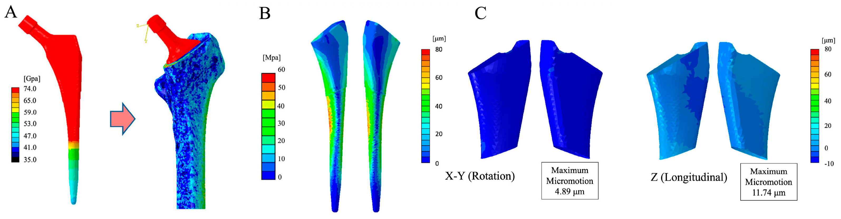
A patient-specific FEM was constructed by implanting the validated gradient Young’s modulus TNS stem model into a CT-derived femoral model of a female patient in her sixties with champagne-flute canal morphology (Dorr A/B), with bone mineral density mapped element-by-element from CT Hounsfield units via calibration phantom; boundary and loading conditions followed physiological peak walking forces [17,49,50]. FEM analysis demonstrated proximal micromotion of 4.89 μm (rotational) and 11.74 μm (longitudinal)—well below both the 40 μm osseointegration threshold and the 150 μm loosening threshold—confirming favorable conditions for bone ingrowth and long-term fixation [42,51]. Von Mises stress was concentrated in the proximal medial and lateral cortex, with low stress distribution distally, a pattern consistent with physiological load transfer and confirmatory of SS suppression predicted by prior FEM simulations [17,42] (Figure 3).

3.4. Comparison with Other THA FEM Studies

Alternative strategies to reduce SS have been evaluated through FEM, providing a comparative framework for positioning the TNS stem. Porous/lattice 3D-printed Ti stems—exemplified by Arabnejad et al.—demonstrated up to 75% reduction in bone loss secondary to SS compared to solid implants by locally mimicking cancellous bone stiffness [52]; however, concerns regarding fatigue failure under cyclic loading and the absence of long-term clinical data remain unresolved [53,54]. Geometry-based approaches—including anatomically grooved stems and short metaphyseal-fixation stems—achieved proximal strain improvements of 10–20% locally, though distal load transfer patterns remained suboptimal in certain configurations [55,56]. Among geometry-based approaches, grooved stem cross-sections have been shown to reduce proximal SS by 31–50% through enhanced metaphyseal load transfer, and short metaphyseal-fixation stems reduce diaphyseal SS by limiting distal cortical engagement; however, both strategies remain geometry-dependent and lose efficacy in Dorr type C femora with poor metaphyseal bone quality [57,58,59,60]. Porous/lattice 3D-printed stems achieve up to 75% reduction in SS-related bone loss in FEM, but the porosity required for meaningful stiffness reduction simultaneously compromises fatigue strength under long-term cyclic loading. In contrast, the TNS stem achieves stiffness reduction through material design alone (Section 2), preserving bulk mechanical integrity—a distinction that underpins its unique clinical translatability. PEEK composite stems offer ultra-low elastic modulus (~3–4 GPa) but face persistent concerns over fatigue strength and osseointegration, limiting clinical translation [61].

3.5. Limitations of Current FEM Approaches and Future Simulation Directions

The FEM analyses reviewed here share four principal limitations acknowledged by Mori et al.: static rather than cyclic loading conditions, ideal neutral stem alignment, a single champagne-flute femoral morphology (Dorr A/B), and simplified muscle loading without comprehensive musculoskeletal modeling [17]. The restriction to Dorr A/B morphology is clinically significant, as Dorr type C femora—characterized by a wide canal and thin cortex—are associated with greater risk of high-degree SS due to reduced cortical mechanical support, and represent a population in which the biomechanical advantages of the TNS stem have yet to be computationally validated [57,58,59,60]. A next-generation FEM framework for the TNS stem should address each of these limitations in a stepwise manner. First, cyclic fatigue simulations incorporating 106 or more physiological loading cycles—replicating one to two years of walking activity—are required to evaluate progressive micromotion accumulation at the bone–stem interface and to assess the risk of late fatigue fracture at the low-modulus distal stem, a scenario that static analysis cannot detect [52,53]. Second, extension of the patient-specific model to Dorr type C femora—which exhibit thin cortical bone, wide medullary canals, and the highest susceptibility to high-degree SS—is essential to determine whether the modulus-gradient strategy generalizes beyond the current Dorr A/B population and to guide implant sizing decisions in this high-risk morphotype [57,58,59,60]. Third, integration of validated musculoskeletal models incorporating patient-specific hip joint reaction forces, abductor muscle loading, and gait kinematics would substantially improve the physiological fidelity of boundary conditions beyond the simplified static peak-load assumptions used to date [49]. Fourth, parametric malalignment analysis—systematically varying varus/valgus stem positioning by ±5°—would quantify the sensitivity of the modulus-gradient effect to surgical technique variability, a clinically critical factor given that varus positioning has been shown to increase distal load transfer and thigh pain incidence in conventional stems [62]. Together, these advances would transform the TNS FEM program from a single-case proof-of-concept into a robust computational platform capable of supporting personalized implant selection across the full spectrum of patient-specific femoral morphologies and surgical scenarios.
More fundamentally, it should be acknowledged that the FEM model is constructed on the mechanobiological premise that reducing implant stiffness improves proximal stress transfer; agreement between FEM predictions and clinical observations therefore reflects the internal consistency of this concept rather than constituting independent experimental verification. FEM provides indispensable theoretical and mechanistic support for the modulus-gradient design rationale, but definitive proof of clinical efficacy requires prospective controlled clinical studies with objective bone density outcomes.

4. Preclinical Evidence

4.1. Rationale for Low-Modulus Implants from Animal Models

The foundational in vivo evidence that stem stiffness directly drives periprosthetic bone resorption derives from the canine porous-coated THA model of Bobyn et al., which demonstrated that titanium stems produced significantly less SS and femoral bone resorption than cobalt-chromium stems of identical geometry, establishing a direct causal link between bulk implant stiffness and adaptive bone loss [63]. These findings are mechanobiologically explained by strain-adaptive remodeling: reduced stem stiffness preserves the periosteal and endosteal strain stimulus within the adaptive window, preventing the resorption-initiating reduction in mechanosensory signal that stiffer implants impose on proximal cortical osteocytes [11,46]. Together, these observations provided the mechanobiological rationale for developing the TNS alloy stem: if a clinically translatable alloy could reduce implant stiffness toward the bone-compatible range while maintaining structural integrity, the adaptive bone loss cascade could be attenuated from its material origin.

4.2. TNS Alloy Plates and Nails: Fracture Healing Studies

In a rabbit tibial osteotomy model, Kogure et al. demonstrated that elastic intramedullary nails composed of TNS alloy promoted fracture healing compared to Ti-6Al-4V nails, with histomorphometric analysis showing accelerated callus formation attributable to preserved interfragmentary micromotion within the osteogenic range [64]. Ito et al. extended these findings to a rabbit tibial plate fixation model, demonstrating that β-type Ti-Nb-Sn alloy plates with low Young’s modulus accelerated osteosynthesis compared to Ti-6Al-4V plates, with greater callus bridging and higher bone mineral density at the fracture site at 8 weeks [65]. In mouse tibiae, Mori et al. confirmed that TNS intramedullary nails accelerated fracture healing via modulation of the strain stimulus at the cortical callus level, while Koyama et al. subsequently showed in a rat femoral model that TNS plates modulated interfragmentary movement to promote osteosynthesis—collectively demonstrating that the biomechanical benefit of low stiffness extends across multiple species, bone geometries, and fixation constructs [66,67].

4.3. Mechanobiological Link: Fracture Healing Studies to THA Stress Shielding

Reducing implant stiffness maintains the local strain stimulus within the osteogenic adaptive window: osteocytes detect the preserved mechanical signal, suppress sclerostin expression to activate Wnt/β-catenin-mediated bone formation, and downregulate RANKL-driven osteoclastogenesis—an anabolic cascade equally operative at both fracture callus and periprosthetic cortical interfaces [47,68,69]. The optimal interfragmentary micromotion range for callus stimulation—approximately 0.2–1.0 mm axially—is readily abolished by overly rigid fixation, redirecting healing toward fibrous non-union; conversely, micromotion exceeding ~1 mm shifts tissue differentiation toward fibrocartilage rather than direct bone formation, and the TNS alloy’s compliance uniquely maintains fixation constructs within this therapeutic window [70,71,72]. This mechanobiological framework directly underpins the TNS femoral stem rationale: the material property that accelerates fracture healing—preservation of physiological strain stimulus at the bone–implant interface—is the identical mechanism by which the gradient-modulus TNS stem suppresses SS in the periprosthetic femur [17,70].

5. Clinical Outcomes

5.1. TNS Stem Design and Surgical Technique

The TNS femoral stem is a metaphyseal-filling, cementless tapered stem manufactured from low-modulus Ti-33.6Nb-4Sn alloy, classified under the fit-and-fill geometry category (Type C in the Radaelli system) designed to achieve rigid initial fixation, osseointegration, and balanced femoral stress distribution simultaneously [73,74]. The proximal one-third of the stem carries a sandblasted, roughened surface to promote press-fit fixation and long-term bone ingrowth via osseointegration, a surface strategy well-validated across multiple cementless designs. The distal two-thirds is polished to permit microaxial displacement within the canal, allowing for physiological load transfer to the proximal cortex and avoiding distal load monopolization that would exacerbate SS. This proximal-dominant fixation philosophy reflects the foundational principle described by Moore, in which the intramedullary stem achieves stability through cortical engagement at the metaphyseal-diaphyseal junction rather than cement bonding [75,76,77]. The TNS stem is implanted via the posterolateral approach (PLA), which affords extensive femoral canal access and reliable anatomical landmarks for reproducible implantation with a relatively short learning curve. The acetabular component used in combination is the ARCHA dual-mobility cup; pairing a dual-mobility cup with the posterolateral approach is a recognized strategy for minimizing postoperative dislocation risk. Compared to the direct anterior approach, the PLA provides superior femoral exposure and visualization for metaphyseal-filling stem placement, while both approaches yield comparable mid-term functional outcomes [78,79].

5.2. Short-Term Results (1–3 Years)

Chiba et al. reported the initial mid-term results of the TNS stem, demonstrating SS suppression and JOA scores comparable to conventional Ti-6Al-4V metaphyseal-filling stems at approximately 3 years, with no cases of Engh Grade 3 or 4 SS [29] (Table 2). Mori et al. subsequently confirmed these findings in 40 patients with radiographic follow-up at 1 and 3 years: no cases exceeded Engh Grade 2 SS at 3 years, substantially lower than the 35–50% Grade 3+ rates reported for conventional Ti-6Al-4V stems [17]. SS was primarily observed in Gruen Zones 1 and 7, consistent with the proximal load distribution of a metaphyseal-filling design. Interobserver reliability was κ = 0.85 (1-year) and κ = 0.75 (3-year) by Engh’s classification, and κ = 0.78 for Gruen zone assessment, all indicating substantial agreement [17,29].

5.3. Mid-Term Results with Comparative Control Group

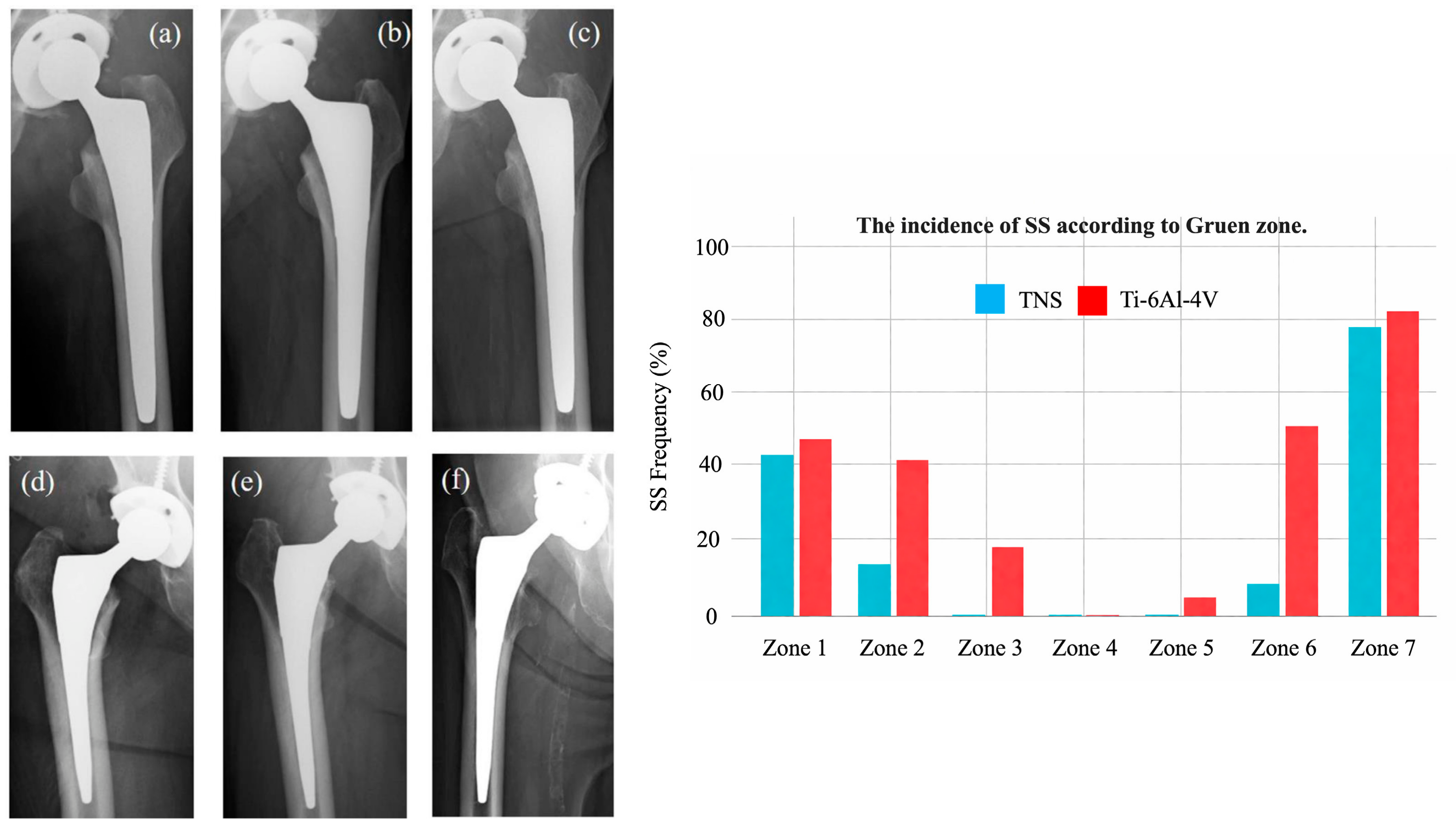
Baba et al. directly compared the TNS stem with Ti-6Al-4V metaphyseal-filling stems of similar morphology (VerSys HA TCP Fiber Metal Taper and Synergy Select II) in a prospective cohort [30] (Table 2). Final JOA scores were 84.6 ± 10.8 in the TNS group and 82.2 ± 9.8 in the control group, with no significant difference (p = 0.41), suggesting comparable clinical recovery in this cohort, though larger studies are needed to confirm equivalence. The overall SS grade distribution was significantly lower in the TNS group (p = 0.03), with SS frequency significantly reduced specifically in Gruen Zones 2, 3, and 6—the diaphyseal-to-metaphyseal transitional cortex—compared to the Ti-6Al-4V controls.

5.4. Mid-Term Results: 7-Year Follow-Up

In a prospective study of 35 TNS and 21 Ti-6Al-4V metaphyseal-filling stem patients (median 87 months follow-up), the overall SS grade distribution was significantly lower in the TNS group (p = 0.03), with SS frequency significantly reduced in Gruen Zones 2, 3, and 6 (p = 0.02, 0.01, 0.001) [31] (Table 2). However, third-degree SS still occurred in 11 TNS cases, indicating that modulus-gradient optimization alone is insufficient and that further refinements in stem geometry and surface modification are required (Figure 4). JOA scores were comparable at final follow-up (84.6 ± 10.8 vs. 82.2 ± 9.8; p = 0.41), with no implant-related failures, and interobserver reliability was weighted κ = 0.77.

5.5. Comparison with Conventional Ti-6Al-4V Stems

Conventional Ti-6Al-4V stems have been associated with Grade 3/4 SS in 35–50% of cases within 2–3 years postoperatively, a substantially higher burden than the zero Grade 3+ cases observed at 3 years with the TNS stem [6]. Yamako et al. predicted through strain-adaptive FEM simulation that Ti-6Al-4V stems would reduce calcar-region BMD by up to 45.8% at 10 years, whereas the TNS gradient-modulus stem was projected to preserve significantly greater proximal bone stock over the same period [42] (Table 2). Engh et al. further demonstrated that SS carries direct clinical consequences: patients with higher SS grades exhibited significantly reduced walking scores and lower Harris Hip Scores compared to those without SS, underscoring the functional relevance of radiographic bone loss [6].

5.6. Application in Dental Implantology

At present, the clinical application of β-type low-modulus titanium alloys in skeletal implants is limited to the TNS system; however, a similar design concept has been rapidly expanding in the dental implantology. A recent randomized controlled trial evaluated a β-type Ti-Nb-Zr (TNZ) alloy dental implant against modified Grade 4 cpTi controls in 77 patients over 12 months, achieving 100% implant success and survival in both groups, with TNZ demonstrating superior fatigue resistance at a comparable elastic modulus [39]. Fractal dimension analysis of periapical radiographs showed time-dependent bone remodeling without pathological adaptation, and marginal bone loss did not exceed 1 mm in any case, confirming that low-modulus β-Ti alloys preserve crestal bone in the dental setting through the same mechanocompatibility principle operative in the periprosthetic femur. A comprehensive review of β-Ti alloys for dental implants further documented that Nb- and Sn-containing β-stabilized systems consistently yield elastic moduli of 37–85 GPa—bridging the gap between cpTi (110 GPa) and cortical bone (~27 GPa)—while maintaining corrosion resistance superior to Ti-6Al-4V [23]. These cross-disciplinary findings reinforce the mechanobiological rationale underlying the TNS orthopedic stem design and suggest that modulus-graded β-Ti implants represent a broader platform technology applicable across load-bearing skeletal interfaces.

5.7. Concordance Between FEM Predictions and Clinical Observations

FEM analysis of the TNS stem demonstrated that the proximal gradient-modulus region concentrates von Mises stress in the proximal medial and lateral cortex, with comparatively low stress distribution in the distal region—a pattern that mechanistically predicts reduced SS in diaphyseal Gruen zones [17]. This prediction was directly corroborated by clinical radiographic data: SS frequency was significantly reduced specifically in Gruen Zones 2, 3, and 6 in the TNS group compared to Ti-6Al-4V controls (p = 0.02, 0.01, 0.001), while Zones 1 and 7—the proximal-most cortex—remained comparably affected in both groups [31]. This spatial concordance between computational stress distribution and zone-specific SS suppression represents a notable finding of the TNS research program, providing preliminary translational support that warrants confirmation in larger independent cohorts.
It should be noted that these three evidence streams represent fundamentally different levels of the translational research hierarchy and carry distinct inferential weights: FEM provides mechanistic and theoretical support under idealized conditions; preclinical animal studies demonstrate biological plausibility under controlled experimental conditions; and prospective clinical studies provide the highest level of clinical evidence, albeit constrained by small sample sizes, non-randomized design, and limited follow-up. The convergence of predictions and observations across these levels is interpreted as translational consistency rather than equivalent independent validation.

5.8. Safety Profile

Across the 35 TNS patients followed for a median of 87 months, no cases of periprosthetic fracture, dislocation, or periprosthetic joint infection were recorded [31]. One patient underwent revision THA for liner breakage; this complication was attributable to polyethylene wear rather than stem failure, and was therefore excluded from SS assessment due to potential osteolysis-related confounding. Mild thigh pain was reported in 2 patients (5.7%), both of which were self-limiting and did not require further intervention, consistent with the low-modulus stem’s favorable mechanical compatibility at the stem-bone interface [17].

5.9. Critical Appraisal of the Clinical Evidence Base

Before translational conclusions can be drawn from the TNS stem clinical data, several important limitations must be explicitly acknowledged. First, all published TNS stem studies originate from Tohoku University and its affiliated institutions within a single academic network, and the largest comparative cohort comprises only 35 TNS and 21 control cases—sample sizes that preclude multivariate adjustment for clinically relevant confounders such as femoral morphology (Dorr type), preoperative bone mineral density, body mass index, and activity level [30,31]. This single academic network design limits the generalizability of the findings to broader patient populations and surgical practices. Second, the maximum follow-up of 87 months, while providing meaningful mid-term evidence of SS suppression, is insufficient to exclude late-onset complications, including fatigue fracture at the low-modulus distal stem, progressive Grade 3 SS beyond 10 years, or aseptic loosening secondary to cumulative micromotion at the bone–implant interface. Third, the absence of dual-energy X-ray absorptiometry (DXA)-based periprosthetic bone mineral density quantification means that radiographic SS grading—an inherently observer-dependent metric—remains the sole measure of bone remodeling, limiting direct comparability with the international literature in which DXA-derived Gruen zone BMD is increasingly used as the primary endpoint. Fourth, the non-randomized prospective design introduces potential selection bias; in particular, differences in femoral canal morphology between the TNS and control groups cannot be excluded as a confounding factor in the observed SS grade differences. These limitations do not negate the translational significance of the TNS stem data, but they underscore the need for multicenter randomized controlled trials with DXA-based endpoints and follow-up exceeding 10 years before definitive efficacy conclusions can be established.

6. Current Limitations and Future Perspectives

6.1. Remaining Clinical Challenges

As discussed in Section 5.9, the current clinical evidence base for the TNS stem is constrained by cohorts from a single academic network, limited sample sizes, and a maximum follow-up of 87 months [31]. Although the TNS stem significantly suppressed SS progression over 7 years, third-degree SS was still observed in 11 of 34 evaluable cases, demonstrating that a Young’s modulus gradient alone is insufficient to fully prevent SS and that complementary design modifications—including cross-sectional geometry optimization, stem shortening, and porous surface treatment—remain necessary [31] (Table 3). Quantitative bone density assessment by DXA was not performed in any of the published clinical studies, limiting the ability to objectively characterize periprosthetic BMD changes over time and to correlate radiographic SS grading with actual bone mass loss [17]. Consequently, the current evidence supports the conclusion that the TNS stem reduces and attenuates SS compared to conventional Ti-6Al-4V stems, but is insufficient to claim complete prevention or resolution of this phenomenon. Furthermore, the existing clinical evidence base is constrained by small sample sizes, a non-randomized prospective design, and a maximum follow-up of 87 months; studies exceeding 10 years are required to assess the long-term durability of SS suppression and to exclude the risk of late-onset fatigue fracture at the stem neck or distal tip. It should further be noted that while radiographic SS grade distribution was significantly reduced in the TNS group, the current evidence does not yet demonstrate a corresponding reduction in revision rate, periprosthetic fracture incidence, or patient-reported outcome measures; establishing these clinical endpoints as downstream benefits of SS reduction remains a priority for future prospective investigation.
SS in cementless THA is a multifactorial phenomenon, and Young’s modulus optimization—while the defining principle of the TNS stem—represents one component of a broader determinant framework [5,9,17,30]. Stem geometry, including cross-sectional shape and length, independently modulates proximal-to-distal load distribution [55,56]. Fixation strategy determines the primary site of load transfer: metaphyseal-filling designs preferentially load the proximal cortex, while diaphyseal-fixation stems bypass proximal bone regardless of alloy stiffness [75,76,77]. Porous surface treatment and anodic oxidation further amplify SS suppression by enhancing proximal osseointegration [44,45]. The occurrence of third-degree SS in 11 TNS cases at 7 years underscores that future stem development must integrate geometry optimization, controlled porosity, and enhanced surface bioactivity as complementary design dimensions [31]. Furthermore, because the TNS stem differs from the Ti-6Al-4V control stems not only in alloy composition and modulus distribution but also in surface treatment and proximal geometry, the relative contribution of the modulus gradient to the observed reduction in SS cannot be definitively isolated in the current non-randomized clinical evidence; randomized controlled trials with geometry-matched implants are required to establish the independent effect of the modulus gradient on SS prevention.

6.2. Stem Design Optimization

The current TNS stem design is relatively long and bulky in the proximal region, limiting compatibility with minimally invasive surgery and contributing to suboptimal metaphyseal fill in some patients. Stem shortening to shift load transfer closer to the metaphysis—as demonstrated with short tapered-wedge stems—would improve both MIS compatibility and proximal cortical strain distribution, while cross-sectional geometry optimization and degree-of-taper adjustment are similarly warranted to achieve more uniform strain across the proximal femur [81,82]. A modular design would further allow for adaptation to individual femoral morphology, which is particularly relevant given that East Asian femora—including Japanese patients who constitute the primary TNS clinical cohort—exhibit a characteristically higher prevalence of Dorr type A anatomy with smaller distal canal dimensions and greater metaphyseal-diaphyseal mismatch than Western femora [83]. Stems with slimmer distal profiles and optimized porous coating distribution that decreases friction coefficients in the middle medial zone while increasing them in the lateral proximal and distal zones have been shown computationally to reduce proximal SS, offering a design template applicable to next-generation TNS development.

6.3. Advanced Manufacturing: 3D Printing and Porous Structures

A critical limitation of the current TNS stem fabrication process is that the localized heat treatment required to generate the Young’s modulus gradient makes simultaneous application of porous surface coatings technically challenging, restricting the degree of osseointegration enhancement achievable through surface modification alone. Arabnejad et al. demonstrated that a fully 3D-printed porous titanium femoral stem with architecture optimized to mimic local bone tissue properties could reduce bone loss secondary to SS by 75% compared to a fully solid implant, validating the potential of additive manufacturing as a complementary strategy for SS suppression [52]. Cortis et al. further showed that AM-enabled lattice redesign of hip prostheses simultaneously improves stress distribution and structural safety under physiological and surgical loading conditions [53], while Liu et al. systematically reviewed porous lattice stem designs and identified regional stiffness gradation—with higher proximal and lower distal stiffness—as the key design principle for balancing SS reduction and osseointegration [54]. The integration of TNS alloy with additive manufacturing represents a compelling next frontier: β-Ti alloys including Ti-Nb-Sn systems have been successfully processed via laser deposition and spark plasma sintering, opening pathways to produce TNS stems with spatially defined porosity without the constraint of heat treatment. Such hybrid designs combining a modulus-graded TNS matrix with a porous proximal architecture could synergistically address both the stiffness mismatch and the osseointegration limitations identified in current clinical studies.
Beyond structural optimization, additive manufacturing uniquely enables patient-specific implant design—a paradigm in which implant geometry, porosity distribution, and stiffness gradient are computationally tailored to the individual patient’s femoral morphology and bone mineral density map derived from preoperative CT data. Recent advances in topology optimization algorithms have demonstrated that region-specific lattice architectures—with higher stiffness proximally to ensure primary fixation and lower stiffness distally to reduce SS—can be computationally generated and fabricated via laser powder bed fusion within clinically acceptable tolerances [52,84,85]. Furthermore, the integration of β-type titanium alloys including Ti-Nb-based systems with selective laser melting has been demonstrated to preserve low elastic modulus properties in the as-built condition, confirming the material compatibility of TNS-type alloys with AM processing [86,87]. The convergence of patient-specific CT-based modeling, topology-optimized lattice design, and low-modulus β-Ti alloy fabrication represents the most promising near-term pathway toward a next-generation TNS stem that simultaneously addresses the geometric limitations of the current design, enhances proximal osseointegration through controlled porosity, and maintains the modulus-gradient principle that has demonstrated clinical efficacy over seven years of follow-up.

6.4. Surface Modification Advances

Anodic oxidation of TNS alloy produces a TiO2 surface layer with submicron-scale porosity that enhances bone affinity and provides antibacterial activity through photocatalytic properties; in vivo animal studies demonstrated significantly increased mature lamellar bone formation and higher pull-out failure loads compared to untreated TNS rods, suggesting that anodic oxidation represents a promising strategy to augment osseointegration of the TNS hip stem and potentially further suppress SS beyond what the modulus gradient alone achieves [43,45]. Among alternative surface treatments, hydroxyapatite/TCP coatings remain the most widely implemented approach for cementless THA, with evidence supporting lower rates of aseptic loosening, reduced thigh pain incidence, and improved proximal femoral load transfer compared to uncoated stems; bioactive glass coatings represent an emerging alternative with osteoconductive properties applicable to low-modulus β-Ti substrates [15]. Since robust osseointegration at the proximal press-fit interface reduces interfacial micromotion and maintains physiological strain stimuli in the metaphyseal cortex, surface modification strategies that accelerate and strengthen bone ingrowth are mechanistically complementary to the modulus-grading approach and are expected to synergistically reduce SS progression in next-generation TNS stems.

6.5. Advanced FEM and Simulation

The current TNS FEM was performed under static loading in a single Dorr type A femoral morphology, leaving critical biomechanical scenarios unaddressed. Cyclic fatigue loading simulations incorporating physiological loading cycles are required to evaluate progressive micromotion at the bone–stem interface and to assess late fatigue fracture risk at the lower-modulus distal stem. Extension to Dorr type C femora—which exhibit thin cortical bone, wide medullary canals, and the highest susceptibility to postoperative SS and periprosthetic fracture—is essential to determine whether the modulus-gradient strategy generalizes beyond the current patient population [88,89,90]. Malalignment scenario analysis is equally important: varus stem positioning has been shown to substantially alter strain distribution with increased distal load transfer and higher thigh pain incidence, and valgus positioning may exacerbate proximal SS—both scenarios potentially undermining the TNS modulus-gradient effect [62,80]. Comprehensive musculoskeletal modeling incorporating patient-specific muscle forces, joint reaction forces, and joint kinematics—as validated in recent lower-limb biomechanical simulation frameworks—would further improve physiological fidelity beyond current static boundary conditions [60].

6.6. Application to Other Orthopedic Devices and Dental Implantology

The problem of SS is not unique to hip arthroplasty but represents a universal biomechanical challenge at load-bearing bone–implant interfaces across the musculoskeletal system. In reverse shoulder arthroplasty, short stem malalignment has been shown to significantly alter periprosthetic bone density distribution, with varus positioning driving proximal humeral SS in a pattern mechanistically analogous to the proximal femoral bone loss observed with stiff THA stems [91]. In spinal fixation, experimental analysis of screw spacers placed in porcine vertebral tissue has demonstrated measurable SS effects at the bone–implant contact zone, confirming that stiffness mismatch drives adaptive resorption independent of joint type or loading direction [92]. In dental implantology, comparative FEM analyses of titanium, CFR-PEEK, and ceramic implants have quantified the degree to which implant material stiffness determines the magnitude of crestal bone SS—with stiffer materials consistently producing greater peri-implant bone loss [93]. These cross-joint observations underscore that the gradient-modulus design principle embodied in the TNS alloy stem addresses a fundamental material-biological incompatibility operative across all skeletal interfaces, and that the translational lessons derived from the TNS hip stem program are broadly applicable to the development of next-generation low-modulus fixation devices in shoulder arthroplasty, spinal surgery, and dental implantology. TNS alloy’s low Young’s modulus and heat-treatment tunability extend its application beyond the hip stem. In fracture fixation, TNS alloy plates modulate interfragmentary movement and accelerate osteosynthesis in both rat femoral and rabbit tibial models, outperforming conventional Ti-6Al-4V hardware by preserving physiological strain stimuli at the fracture site [65,67]. Elastic TNS intramedullary nails similarly promote tibial osteotomy healing in rabbits, confirming that stiffness-matched fixation is beneficial across implant modalities [64,94]. Notably, TNS alloy remains the only β-type titanium alloy clinically applied in orthopedic devices; low-modulus β-Ti alloys including Ti-Nb-Zr-Ta systems have demonstrated favorable preclinical osseointegration profiles in dental implant contexts, making cross-disciplinary expansion of TNS to dental implantology a logical next step pending formal clinical validation [39].

7. Conclusions

The TNS alloy femoral stem represents a meaningful advancement in cementless THA, addressing the long-standing problem of SS through a gradient Young’s modulus (70 GPa proximal → 40 GPa distal) achievable by localized heat treatment of a single homogeneous alloy—a strategy that maintains compositional consistency and clinical scalability. FEM analysis indicated favorable proximal micromotion (4.89 μm rotational, 11.74 μm longitudinal—well below the 40 μm osseointegration threshold) and physiological proximal stress concentration, findings that are consistent with radiographic outcomes showing significantly suppressed SS at 1, 3, and 7 years postoperatively. Clinical evidence indicated that SS grade distribution was significantly lower in the TNS group than in conventional Ti-6Al-4V fit-and-fill stem controls (p = 0.03), with no stem-related implant failures over a median 7-year follow-up.
TNS alloy is currently the only β-type titanium alloy to have achieved clinical application in joint prostheses and fracture fixation devices, underscoring its translational maturity relative to other low-modulus β-Ti candidates. Nonetheless, the occurrence of Grade 3 SS in a subset of TNS patients indicates that modulus grading alone is insufficient, and remaining challenges include stem geometry optimization (shortening, cross-sectional redesign, taper adjustment), incorporation of proximal porous structures via additive manufacturing, objective DXA-based periprosthetic bone density quantification, and extension to diverse femoral morphologies including Dorr type C and osteoporotic bone. Future research should prioritize long-term follow-up exceeding 10 years, cyclic fatigue FEM simulations, comprehensive musculoskeletal modeling, and cross-disciplinary expansion of TNS alloy applications to fracture fixation plates, nails, and dental implants.

Author Contributions

Y.M. and H.T. conceptualized this study and designed the review framework. Y.M. conducted the literature search and analysis and wrote the original draft of the manuscript. Y.M., H.T., K.B., R.K. and N.M. contributed to data interpretation and manuscript preparation. T.A. supervised the project. Y.M., H.T., K.B., R.K., N.M. and T.A. reviewed and edited the manuscript. All authors have read and agreed to the published version of the manuscript.

Funding

This research received no external funding.

Institutional Review Board Statement

Not applicable.

Informed Consent Statement

Not applicable.

Data Availability Statement

No new data were generated or analyzed in this study.

Conflicts of Interest

The authors declare no conflicts of interest.

Abbreviations

The following abbreviations are used in this manuscript:
SSStress shielding
TNSTi-33.6Nb-4Sn
THATotal hip arthroplasty
FEMFinite element modeling
BMDBone mineral density
DXADual-energy X-ray absorptiometry
TNZTi-Nb-Zr

References

  1. Pallante, G.D.; Statz, J.M.; Milbrandt, T.A.; Trousdale, R.T. Primary Total Hip Arthroplasty in Patients 20 Years Old and Younger. J. Bone Jt. Surg. Am. 2020, 102, 519–525. [Google Scholar] [CrossRef] [PubMed]
  2. Singh, J.A.; Yu, S.; Chen, L.; Cleveland, J.D. Rates of Total Joint Replacement in the United States: Future Projections to 2020-2040 Using the National Inpatient Sample. J. Rheumatol. 2019, 46, 1134–1140. [Google Scholar] [CrossRef]
  3. Klug, A.; Pfluger, D.H.; Gramlich, Y.; Hoffmann, R.; Drees, P.; Kutzner, K.P. Future burden of primary and revision hip arthroplasty in Germany: A socio-economic challenge. Arch. Orthop. Trauma Surg. 2021, 141, 2001–2010. [Google Scholar] [CrossRef]
  4. Schwartz, A.M.; Farley, K.X.; Guild, G.N.; Bradbury, T.L., Jr. Projections and Epidemiology of Revision Hip and Knee Arthroplasty in the United States to 2030. J. Arthroplast. 2020, 35, S79–S85. [Google Scholar] [CrossRef]
  5. Huiskes, R.; Weinans, H.; van Rietbergen, B. The relationship between stress shielding and bone resorption around total hip stems and the effects of flexible materials. Clin. Orthop. Relat. Res. 1992, 274, 124–134. [Google Scholar] [CrossRef]
  6. Engh, C.A., Jr.; Young, A.M.; Engh, C.A., Sr.; Hopper, R.H., Jr. Clinical consequences of stress shielding after porous-coated total hip arthroplasty. Clin. Orthop. Relat. Res. 2003, 417, 157–163. [Google Scholar] [CrossRef]
  7. Burchard, R.; Graw, J.A.; Soost, C.; Schmitt, J. Stress shielding effect after total hip arthroplasty varies between combinations of stem design and stiffness-a comparing biomechanical finite element analysis. Int. Orthop. 2023, 47, 1981–1987. [Google Scholar] [CrossRef]
  8. Kwon, D.G.; Lee, T.J.; Kang, J.S.; Moon, K.H. Correlation between stress shielding and clinical outcomes after total hip arthroplasty with extensively porous coated stems. J. Arthroplast. 2013, 28, 1728–1730. [Google Scholar] [CrossRef] [PubMed]
  9. Huiskes, R. The various stress patterns of press-fit, ingrown, and cemented femoral stems. Clin. Orthop. Relat. Res. 1990, 261, 27–38. [Google Scholar] [CrossRef]
  10. Modi, S.R.; Dongare, A.; Jha, K. Strain shielding effect analysis of solid and porous Ti-6Al-4V alloy implanted femur bone using finite element analysis. J. Med. Eng. Technol. 2025, 49, 217–230. [Google Scholar] [CrossRef]
  11. Van Rietbergen, B.; Huiskes, R.; Weinans, H.; Sumner, D.R.; Turner, T.M.; Galante, J.O. ESB Research Award 1992. The mechanism of bone remodeling and resorption around press-fitted THA stems. J. Biomech. 1993, 26, 369–382. [Google Scholar] [CrossRef]
  12. Maeda, T.; Nakano, M.; Nakamura, Y.; Momose, T.; Sobajima, A.; Takahashi, J.; Nakata, K.; Nawata, M. Relationship between Stress Shielding and Optimal Femoral Canal Contact Regions for Short, Tapered-Wedge Stem Analyzed by 2D and 3D Systems in Total Hip Arthroplasty. J. Clin. Med. 2023, 12, 3138. [Google Scholar] [CrossRef]
  13. Kusano, T.; Seki, T.; Higuchi, Y.; Takegami, Y.; Osawa, Y.; Ishiguro, N. Preoperative Canal Bone Ratio is Related to High-Degree Stress Shielding: A Minimum 5-Year Follow-Up Study of a Proximally Hydroxyapatite-Coated Straight Tapered Titanium Femoral Component. J. Arthroplast. 2018, 33, 1764–1769. [Google Scholar] [CrossRef]
  14. Werneburg, F.; Herntrich, A.; Dietz, J.; Wohlrab, D.; Gutteck, N.; Karl-Stefan, D.; Zeh, A. Clinically relevant differences in stress shielding between two short-stemmed femoral prostheses. Arch. Orthop. Trauma Surg. 2025, 145, 365. [Google Scholar] [CrossRef]
  15. Apostu, D.; Lucaciu, O.; Berce, C.; Lucaciu, D.; Cosma, D. Current methods of preventing aseptic loosening and improving osseointegration of titanium implants in cementless total hip arthroplasty: A review. J. Int. Med. Res. 2018, 46, 2104–2119. [Google Scholar] [CrossRef] [PubMed]
  16. Rho, J.Y.; Ashman, R.B.; Turner, C.H. Young’s modulus of trabecular and cortical bone material: Ultrasonic and microtensile measurements. J. Biomech. 1993, 26, 111–119. [Google Scholar] [CrossRef]
  17. Mori, Y.; Tanaka, H.; Kurishima, H.; Kanabuchi, R.; Mori, N.; Sasagawa, K.; Aizawa, T. Biomechanical and Clinical Validation of a Modulus-Graded Ti-Nb-Sn Femoral Stem for Suppressing Stress Shielding in Total Hip Arthroplasty. Appl. Sci. 2025, 15, 4827. [Google Scholar] [CrossRef]
  18. Peitgen, D.S.; Innmann, M.M.; Merle, C.; Gotterbarm, T.; Moradi, B.; Streit, M.R. Periprosthetic Bone Mineral Density Around Uncemented Titanium Stems in the Second and Third Decade After Total Hip Arthroplasty: A DXA Study After 12, 17 and 21 Years. Calcif. Tissue Int. 2018, 103, 372–379. [Google Scholar] [CrossRef] [PubMed]
  19. Liu, Y.; Wei, W.X.; Zeng, Y.; Ma, J.; Yang, J.; Shen, B. Comparison of Femoral Bone Mineral Density Changes around 3 Common Designs of Cementless Stems after Total Hip Arthroplasty-A Retrospective Cohort Study. Orthop. Surg. 2022, 14, 1059–1070. [Google Scholar] [CrossRef] [PubMed]
  20. Bombelli, R.; Mathys, R. Cementless isoelastic RM total hip prosthesis. J. R. Soc. Med. 1982, 75, 588–597. [Google Scholar] [CrossRef]
  21. Bombelli, R.; Gerundini, M.; Aronson, J. Early results of the RM-isoelastic cementless total hip prosthesis: 300 consecutive cases with 2-year follow-up. Hip 1984, 133–145. [Google Scholar]
  22. Sidhu, S.S.; Singh, H.; Gepreel, M.A. A review on alloy design, biological response, and strengthening of beta-titanium alloys as biomaterials. Mater. Sci. Eng. C Mater. Biol. Appl. 2021, 121, 111661. [Google Scholar] [CrossRef] [PubMed]
  23. Calazans Neto, J.V.; Celles, C.A.S.; de Andrade, C.; Afonso, C.R.M.; Nagay, B.E.; Barao, V.A.R. Recent Advances and Prospects in beta-type Titanium Alloys for Dental Implants Applications. ACS Biomater. Sci. Eng. 2024, 10, 6029–6060. [Google Scholar] [CrossRef]
  24. McCarthy, C.J.; Moore, J.; Condon, F. Large head metal-on-metal bearing surface with a TMZF titanium alloy femoral stem with high rates of revision and trunnion failure. J. Orthop. 2023, 46, 164–168. [Google Scholar] [CrossRef]
  25. Kuiper, J.H.; Huiskes, R. The predictive value of stress shielding for quantification of adaptive bone resorption around hip replacements. J. Biomech. Eng. 1997, 119, 228–231. [Google Scholar] [CrossRef][Green Version]
  26. Weinans, H.; Sumner, D.R.; Igloria, R.; Natarajan, R.N. Sensitivity of periprosthetic stress-shielding to load and the bone density-modulus relationship in subject-specific finite element models. J. Biomech. 2000, 33, 809–817. [Google Scholar] [CrossRef]
  27. Yamako, G.; Chosa, E.; Totoribe, K.; Hanada, S.; Masahashi, N.; Yamada, N.; Itoi, E. In-vitro biomechanical evaluation of stress shielding and initial stability of a low-modulus hip stem made of beta type Ti-33.6Nb-4Sn alloy. Med. Eng. Phys. 2014, 36, 1665–1671. [Google Scholar] [CrossRef]
  28. Glassman, A.H.; Bobyn, J.D.; Tanzer, M. New femoral designs: Do they influence stress shielding? Clin. Orthop. Relat. Res. 2006, 453, 64–74. [Google Scholar] [CrossRef]
  29. Chiba, D.; Yamada, N.; Mori, Y.; Oyama, M.; Ohtsu, S.; Kuwahara, Y.; Baba, K.; Tanaka, H.; Aizawa, T.; Hanada, S.; et al. Mid-term results of a new femoral prosthesis using Ti-Nb-Sn alloy with low Young’s modulus. BMC Musculoskelet. Disord. 2021, 22, 987. [Google Scholar] [CrossRef] [PubMed]
  30. Baba, K.; Mori, Y.; Chiba, D.; Kuwahara, Y.; Kurishima, H.; Tanaka, H.; Kogure, A.; Kamimura, M.; Yamada, N.; Ohtsu, S.; et al. TiNbSn stems with gradient changes of Young’s modulus and stiffness reduce stress shielding compared to the standard fit-and-fill stems. Eur. J. Med. Res. 2023, 28, 214. [Google Scholar] [CrossRef] [PubMed]
  31. Baba, K.; Mori, Y.; Tanaka, H.; Kanabuchi, R.; Kuriyama, Y.; Kurishima, H.; Ito, K.; Kamimura, M.; Chiba, D.; Aizawa, T. Effectiveness of Stress Shielding Prevention Using a Low Young’s Modulus Ti-33.6Nb-4Sn Stem: A 7-Year Follow-Up Study. Med. Sci. 2025, 13, 51. [Google Scholar] [CrossRef] [PubMed]
  32. Hanada, S.; Masahashi, N.; Jung, T.K.; Miyake, M.; Sato, Y.S.; Kokawa, H. Effect of swaging on Young׳s modulus of beta Ti-33.6Nb-4Sn alloy. J. Mech. Behav. Biomed. Mater. 2014, 32, 310–320. [Google Scholar] [CrossRef]
  33. Hanada, S.; Masahashi, N.; Jung, T.K.; Yamada, N.; Yamako, G.; Itoi, E. Fabrication of a high-performance hip prosthetic stem using beta Ti-33.6Nb-4Sn. J. Mech. Behav. Biomed. Mater. 2014, 30, 140–149. [Google Scholar] [CrossRef] [PubMed]
  34. Niinomi, M. Mechanical biocompatibilities of titanium alloys for biomedical applications. J. Mech. Behav. Biomed. Mater. 2008, 1, 30–42. [Google Scholar] [CrossRef]
  35. Mohammed, M.T.; Khan, Z.A.; Siddiquee, A.N. Beta titanium alloys: The lowest elastic modulus for biomedical applications: A review. Int. J. Chem. Mol. Nucl. Mater. Metall. Eng. 2014, 8, 726–731. [Google Scholar]
  36. Kopova, I.; Strasky, J.; Harcuba, P.; Landa, M.; Janecek, M.; Bacakova, L. Newly developed Ti-Nb-Zr-Ta-Si-Fe biomedical beta titanium alloys with increased strength and enhanced biocompatibility. Mater. Sci. Eng. C Mater. Biol. Appl. 2016, 60, 230–238. [Google Scholar] [CrossRef]
  37. Marin, E.; Lanzutti, A. Biomedical Applications of Titanium Alloys: A Comprehensive Review. Materials 2023, 17, 114. [Google Scholar] [CrossRef]
  38. Yang, Y.; Wang, J.N.; Hu, L.X.; Qasim; Liu, X.C.; Xu, W. Density functional theory insight into the role of Al and V in Ti-6Al-4V dental implants: Structural, electronic, and mechanical properties. BMC Oral Health 2025, 25, 1447. [Google Scholar] [CrossRef]
  39. Jo, D.; Byun, S.H.; Park, S.Y.; Kim, J.H.; Kim, M.; Lee, H.J.; Kim, Y.K.; Yang, B.E.; Yi, Y.J. Structure-Property-Function Evaluation of a beta-Type Ti-Nb-Zr Alloy for Dental Implant Applications with Short-Term Clinical Validation. J. Funct. Biomater. 2026, 17, 96. [Google Scholar] [CrossRef]
  40. Brumbauer, F.; Okamoto, N.L.; Ichitsubo, T.; Sprengel, W.; Luckabauer, M. Minor additions of Sn suppress the omega phase formation in beta titanium alloys. Acta Mater. 2024, 262, 119466. [Google Scholar] [CrossRef]
  41. Miura, K.; Yamada, N.; Hanada, S.; Jung, T.K.; Itoi, E. The bone tissue compatibility of a new Ti-Nb-Sn alloy with a low Young’s modulus. Acta Biomater. 2011, 7, 2320–2326. [Google Scholar] [CrossRef]
  42. Yamako, G.; Janssen, D.; Hanada, S.; Anijs, T.; Ochiai, K.; Totoribe, K.; Chosa, E.; Verdonschot, N. Improving stress shielding following total hip arthroplasty by using a femoral stem made of beta type Ti-33.6Nb-4Sn with a Young’s modulus gradation. J. Biomech. 2017, 63, 135–143. [Google Scholar] [CrossRef]
  43. Mori, Y.; Masahashi, N.; Aizawa, T. A Review of Anodized TiNbSn Alloys for Improvement in Layer Quality and Application to Orthopedic Implants. Materials 2022, 15, 5116. [Google Scholar] [CrossRef]
  44. Masahashi, N.; Mori, Y.; Tanaka, H.; Kogure, A.; Inoue, H.; Ohmura, K.; Kodama, Y.; Nishijima, M.; Itoi, E.; Hanada, S. Bioactive TiNbSn alloy prepared by anodization in sulfuric acid electrolytes. Mater. Sci. Eng. C Mater. Biol. Appl. 2019, 98, 753–763. [Google Scholar] [CrossRef]
  45. Masahashi, N.; Mori, Y.; Inoue, H. Biocompatible functional surface of titanium-based implant materials. Int. Mater. Rev. 2025, 70, 465–496. [Google Scholar] [CrossRef]
  46. Skerry, T.M. One mechanostat or many? Modifications of the site-specific response of bone to mechanical loading by nature and nurture. J. Musculoskelet. Neuronal Interact. 2006, 6, 122–127. [Google Scholar]
  47. Hughes, J.M.; Castellani, C.M.; Popp, K.L.; Guerriere, K.I.; Matheny, R.W., Jr.; Nindl, B.C.; Bouxsein, M.L. The Central Role of Osteocytes in the Four Adaptive Pathways of Bone’s Mechanostat. Exerc. Sport Sci. Rev. 2020, 48, 140–148. [Google Scholar] [CrossRef]
  48. Warden, S.J.; Hurst, J.A.; Sanders, M.S.; Turner, C.H.; Burr, D.B.; Li, J. Bone adaptation to a mechanical loading program significantly increases skeletal fatigue resistance. J. Bone Miner. Res. 2005, 20, 809–816. [Google Scholar] [CrossRef]
  49. Heller, M.O.; Bergmann, G.; Kassi, J.P.; Claes, L.; Haas, N.P.; Duda, G.N. Determination of muscle loading at the hip joint for use in pre-clinical testing. J. Biomech. 2005, 38, 1155–1163. [Google Scholar] [CrossRef]
  50. Rancourt, D.; Shirazi-Adl, A.; Drouin, G.; Paiement, G. Friction properties of the interface between porous-surfaced metals and tibial cancellous bone. J. Biomed. Mater. Res. 1990, 24, 1503–1519. [Google Scholar] [CrossRef]
  51. Sanchez, E.; de Vries, E.; Matthews, D.; van der Heide, E.; Janssen, D. The effect of coating characteristics on implant-bone interface mechanics. J. Biomech. 2024, 163, 111949. [Google Scholar] [CrossRef] [PubMed]
  52. Arabnejad, S.; Johnston, B.; Tanzer, M.; Pasini, D. Fully porous 3D printed titanium femoral stem to reduce stress-shielding following total hip arthroplasty. J. Orthop. Res. 2017, 35, 1774–1783. [Google Scholar] [CrossRef] [PubMed]
  53. Cortis, G.; Mileti, I.; Nalli, F.; Palermo, E.; Cortese, L. Additive manufacturing structural redesign of hip prostheses for stress-shielding reduction and improved functionality and safety. Mech. Mater. 2022, 165, 104173. [Google Scholar] [CrossRef]
  54. Liu, B.L.; Wang, H.Z.; Zhang, N.Z.; Zhang, M.; Cheng, C.K. Femoral Stems With Porous Lattice Structures: A Review. Front. Bioeng. Biotech. 2021, 9, 772539. [Google Scholar] [CrossRef]
  55. Heyland, M.; Checa, S.; Kendoff, D.; Duda, G.N. Anatomic grooved stem mitigates strain shielding compared to established total hip arthroplasty stem designs in finite-element models. Sci. Rep. 2019, 9, 482. [Google Scholar] [CrossRef]
  56. Yan, S.G.; Chevalier, Y.; Liu, F.; Hua, X.; Schreiner, A.; Jansson, V.; Schmidutz, F. Metaphyseal anchoring short stem hip arthroplasty provides a more physiological load transfer: A comparative finite element analysis study. J. Orthop. Surg. Res. 2020, 15, 498. [Google Scholar] [CrossRef]
  57. Dorr, L.D.; Faugere, M.C.; Mackel, A.M.; Gruen, T.A.; Bognar, B.; Malluche, H.H. Structural and cellular assessment of bone quality of proximal femur. Bone 1993, 14, 231–242. [Google Scholar] [CrossRef]
  58. Kim, J.T.; Jeong, H.J.; Lee, S.J.; Kim, H.J.; Yoo, J.J. Does Proximally Coated Single-Wedge Cementless Stem Work Well in Dorr Type C Femurs? Minimum 10-year Followup. Indian J. Orthop. 2019, 53, 94–101. [Google Scholar] [CrossRef]
  59. Lee, K.H.; Hung, Y.T.; Chang, C.Y.; Wang, J.C.; Tsai, S.W.; Chen, C.F.; Wu, P.K.; Chen, W.M. The cementless taper wedge vs. fit-and-fill stem in primary total hip arthroplasty: Risk of stem-related complication differs across Dorr types. Arch. Orthop. Trauma Surg. 2024, 144, 2839–2847. [Google Scholar] [CrossRef]
  60. Xu, D.; Zhou, H.; Quan, W.; Ma, X.; Chon, T.E.; Fernandez, J.; Gusztav, F.; Kovacs, A.; Baker, J.S.; Gu, Y. New Insights Optimize Landing Strategies to Reduce Lower Limb Injury Risk. Cyborg Bionic Syst. 2024, 5, 0126. [Google Scholar] [CrossRef]
  61. Ceddia, M.; Trentadue, B.; De Giosa, G.; Solarino, G. Topology Optimization of a Femoral Stem in Titanium and Carbon to Reduce Stress Shielding with the FEM Method. J. Compos. Sci. 2023, 7, 298. [Google Scholar] [CrossRef]
  62. Fottner, A.; Woiczinski, M.; Kistler, M.; Schroder, C.; Schmidutz, T.F.; Jansson, V.; Schmidutz, F. Varus malalignment of cementless hip stems provides sufficient primary stability but highly increases distal strain distribution. Clin. Biomech. 2018, 58, 14–20. [Google Scholar] [CrossRef]
  63. Bobyn, J.D.; Glassman, A.H.; Goto, H.; Krygier, J.J.; Miller, J.E.; Brooks, C.E. The effect of stem stiffness on femoral bone resorption after canine porous-coated total hip arthroplasty. Clin. Orthop. Relat. Res. 1990, 261, 196–213. [Google Scholar] [CrossRef]
  64. Kogure, A.; Mori, Y.; Tanaka, H.; Kamimura, M.; Masahashi, N.; Hanada, S.; Itoi, E. Effects of elastic intramedullary nails composed of low Young’s modulus Ti-Nb-Sn alloy on healing of tibial osteotomies in rabbits. J. Biomed. Mater. Res. B Appl. Biomater. 2019, 107, 700–707. [Google Scholar] [CrossRef]
  65. Ito, K.; Mori, Y.; Kamimura, M.; Koguchi, M.; Kurishima, H.; Koyama, T.; Mori, N.; Masahashi, N.; Hanada, S.; Itoi, E.; et al. beta-type TiNbSn Alloy Plates With Low Young Modulus Accelerates Osteosynthesis in Rabbit Tibiae. Clin. Orthop. Relat. Res. 2022, 480, 1817–1832. [Google Scholar] [CrossRef] [PubMed]
  66. Mori, Y.; Fujisawa, H.; Kamimura, M.; Kogure, A.; Tanaka, H.; Mori, N.; Masahashi, N.; Aizawa, T. Acceleration of Fracture Healing in Mouse Tibiae Using Intramedullary Nails Composed of beta-Type TiNbSn Alloy with Low Young’s Modulus. Tohoku J. Exp. Med. 2021, 255, 135–142. [Google Scholar] [CrossRef]
  67. Koyama, T.; Mori, Y.; Kamimura, M.; Tanaka, H.; Tome, R.; Ito, K.; Koguchi, M.; Mori, N.; Aizawa, T. TiNbSn alloy plates with low Young’s modulus modulates interfragmentary movement and promote osteosynthesis in rat femur. J. Mech. Behav. Biomed. Mater. 2025, 161, 106820. [Google Scholar] [CrossRef] [PubMed]
  68. Wu, Y.; Gan, D.; Liu, Z.; Qiu, D.; Tan, G.; Xu, Z.; Xue, H. Osteocytes: Master orchestrators of skeletal homeostasis, remodeling, and osteoporosis pathogenesis. Front. Cell Dev. Biol. 2025, 13, 1670716. [Google Scholar] [CrossRef] [PubMed]
  69. Seddiqi, H.; Klein-Nulend, J.; Jin, J. Bone tissue regeneration: Role of osteocyte mechanosensing and mechanotransduction. Stem Cells Transl. Med. 2026, 15, szag017. [Google Scholar] [CrossRef]
  70. Mori, Y.; Kamimura, M.; Ito, K.; Koguchi, M.; Tanaka, H.; Kurishima, H.; Koyama, T.; Mori, N.; Masahashi, N.; Aizawa, T. A Review of the Impacts of Implant Stiffness on Fracture Healing. Appl. Sci. 2024, 14, 2259. [Google Scholar] [CrossRef]
  71. Rechter, G.R.; Anthony, R.T.; Rennard, J.; Kellam, J.F.; Warner, S.J. The Impact of Early Axial Interfragmentary Motion on the Fracture Healing Environment: A Scoping Review. Injury 2024, 55, 111917. [Google Scholar] [CrossRef]
  72. Mori, Y.; Tanaka, H.; Kamimura, M.; Mori, N.; Aizawa, T. Mechanobiological Implications of Low-Young’s Modulus TiNbSn Alloy Plates for Fracture Fixation: A Focused Review. Med. Sci. 2026, 14, 149. [Google Scholar] [CrossRef] [PubMed]
  73. Radaelli, M.; Buchalter, D.B.; Mont, M.A.; Schwarzkopf, R.; Hepinstall, M.S. A New Classification System for Cementless Femoral Stems in Total Hip Arthroplasty. J. Arthroplast. 2023, 38, 502–510. [Google Scholar] [CrossRef]
  74. Berdini, M.; Procaccini, R.; Zanoli, G.F.; Faini, A.; Verdenelli, A.; Gigante, A. Influence of Femoral Stem Geometry on Total Hip Replacement: A Comparison of Clinical Outcomes of a Straight and an Anatomical Uncemented Stem. J. Clin. Med. 2024, 13, 6459. [Google Scholar] [CrossRef]
  75. Moore, A.T. The self-locking metal hip prosthesis. J. Bone Jt. Surg. Am. 1957, 39, 811–827. [Google Scholar] [CrossRef]
  76. Shichman, I.; Askew, N.; Habibi, A.; Nherera, L.; Macaulay, W.; Seyler, T.; Schwarzkopf, R. Projections and Epidemiology of Revision Hip and Knee Arthroplasty in the United States to 2040–2060. Arthroplast. Today 2023, 21, 101152. [Google Scholar] [CrossRef]
  77. Kang, J.S.; Ko, S.H.; Na, Y.; Youn, Y.H. Clinical and Radiological Outcomes of Rectangular Tapered Cementless Stem According to Proximal Femoral Geometry in Elderly Asian Patients. Hip Pelvis 2019, 31, 224–231. [Google Scholar] [CrossRef] [PubMed]
  78. Ait Mokhtar, M. Postero-posterolateral approach in total hip arthroplasty. Int. Orthop. 2020, 44, 2577–2585. [Google Scholar] [CrossRef] [PubMed]
  79. Wang, K.; Zhang, T.; Xia, R.; Wang, J.; Yu, J.; Wang, W. Efficacy of direct anterior approach versus posterolateral approach in total hip arthroplasty: A systematic review and meta-analysis. Arch. Orthop. Trauma Surg. 2024, 144, 4443–4453. [Google Scholar] [CrossRef]
  80. Maeda, T.; Obayashi, O.; Ishijima, M.; Sato, T.; Musha, Y.; Ikegami, H. Finite element analysis of mechanical stress in a cementless tapered-wedge short stem in the varus position. J. Orthop. Surg. Res. 2024, 19, 385. [Google Scholar] [CrossRef]
  81. Riviere, C.; Grappiolo, G.; Engh, C.A., Jr.; Vidalain, J.P.; Chen, A.F.; Boehler, N.; Matta, J.; Vendittoli, P.A. Long-term bone remodelling around ‘legendary’ cementless femoral stems. EFORT Open Rev. 2018, 3, 45–57. [Google Scholar] [CrossRef] [PubMed]
  82. Solou, K.; Solou, A.V.; Tatani, I.; Lakoumentas, J.; Tserpes, K.; Megas, P. Increased stability of short femoral stem through customized distribution of coefficient of friction in porous coating. Sci. Rep. 2024, 14, 12243. [Google Scholar] [CrossRef]
  83. Bahk, J.H.; Han, S.B.; Rhyu, K.H.; Yoo, J.J.; Lim, S.J.; Park, K.K.; Kim, S.M.; Lim, Y.W. Identification of Essential Features in Developing a Novel Femoral Stem Reflecting Anatomical Features of East Asian Population: A Morphological Study. J. Clin. Med. 2024, 13, 6030. [Google Scholar] [CrossRef]
  84. Garcia-Avila, J.; Gonzalez-Gallegos, C.P.; Segura-Ibarra, V.; Vazquez, E.; Garcia-Lopez, E.; Rodriguez, C.A.; Vargas-Martinez, A.; Cuan-Urquizo, E.; Ramirez-Cedillo, E. Dynamic topology optimization of 3D-Printed transtibial orthopedic implant using tunable isotropic porous metamaterials. J. Mech. Behav. Biomed. Mater. 2024, 153, 106479. [Google Scholar] [CrossRef] [PubMed]
  85. van Kootwijk, A.; Moosabeiki, V.; Saldivar, M.C.; Pahlavani, H.; Leeflang, M.A.; Kazemivand Niar, S.; Pellikaan, P.; Jonker, B.P.; Ahmadi, S.M.; Wolvius, E.B.; et al. Semi-automated digital workflow to design and evaluate patient-specific mandibular reconstruction implants. J. Mech. Behav. Biomed. Mater. 2022, 132, 105291. [Google Scholar] [CrossRef]
  86. Kong, W.; Cox, S.C.; Lu, Y.; Villapun, V.; Xiao, X.; Ma, W.; Liu, M.; Attallah, M.M. The influence of zirconium content on the microstructure, mechanical properties, and biocompatibility of in-situ alloying Ti-Nb-Ta based beta alloys processed by selective laser melting. Mater. Sci. Eng. C Mater. Biol. Appl. 2021, 131, 112486. [Google Scholar] [CrossRef] [PubMed]
  87. Gao, Y.; Jiang, W.; Zeng, D.; Liang, X.; Ma, C.; Xiao, W. Additive manufacturing of titanium alloys for biomedical applications: A systematic review. Rev. Mater. Res. 2025, 1, 100011. [Google Scholar] [CrossRef]
  88. Tsubosaka, M.; Hayashi, S.; Hashimoto, S.; Takayama, K.; Kuroda, R.; Matsumoto, T. Patients with a Dorr type C femoral bone require attention for using a Summit cementless stem: Results of total hip arthroplasty after a minimum follow-up period of 5 years after insertion of a Summit cementless stem. J. Orthop. Sci. 2018, 23, 671–675. [Google Scholar] [CrossRef]
  89. Kaku, N.; Pramudita, J.A.; Yamamoto, K.; Hosoyama, T.; Tsumura, H. Stress distributions of the short stem and the tapered wedge stem at different alignments: A finite element analysis study. J. Orthop. Surg. Res. 2022, 17, 530. [Google Scholar] [CrossRef]
  90. Hosoyama, T.; Kaku, N.; Pramudita, J.A.; Shibuta, Y. Comparison of Early Postoperative Stress Distribution around Short and Tapered Wedge Stems in Femurs with Different Femoral Marrow Cavity Geometries Using Finite Element Analysis. Clin. Orthop. Surg. 2024, 16, 724–732. [Google Scholar] [CrossRef]
  91. Ceddia, M.; Romasco, T.; Pietro, N.D.; Cipollina, A.; Piattelli, A.; Lamberti, L.; Trentadue, B. Quantifying Stress Shielding in Dental Implants: A Comparative Finite Element Study of Titanium, CFR-PEEK, and Ceramic Materials. Materials 2026, 19, 869. [Google Scholar] [CrossRef] [PubMed]
  92. Alcantara-Arreola, E.A.; Silva-Garces, K.N.; Mendoza-Martinez, J.; Cardoso-Palomares, M.A.; Torres-SanMiguel, C.R. Experimental Analysis of Stress Shielding Effects in Screw Spacers Placed in Porcine Spinal Tissue. J. Funct. Biomater. 2024, 15, 238. [Google Scholar] [CrossRef]
  93. Lee, W.; Yoon, Y.J.; Kwon, J.; Kim, G.B.; Lim, T.K.; Koh, K.H. Stress Shielding Effects of Short Stem Alignment and Bone Density in Reverse Shoulder Arthroplasty. J. Orthop. Res. 2026, 44, e70140. [Google Scholar] [CrossRef] [PubMed]
  94. Fujisawa, H.; Mori, Y.; Kogure, A.; Tanaka, H.; Kamimura, M.; Masahashi, N.; Hanada, S.; Itoi, E. Effects of intramedullary nails composed of a new beta-type Ti-Nb-Sn alloy with low Young’s modulus on fracture healing in mouse tibiae. J. Biomed. Mater. Res. B Appl. Biomater. 2018, 106, 2841–2848. [Google Scholar] [CrossRef] [PubMed]
Figure 1. Schematic illustration of contrasting biomechanical pathways in cementless total hip arthroplasty. Upper panel: conventional high-modulus stem (Ti-6Al-4V, ~110 GPa): the stiffness mismatch between implant and cortical bone drives distal load transfer, reduces proximal cortical stress stimulus below the mechanostat adaptive threshold, and initiates Wolff’s law–mediated bone resorption, resulting in progressive SS and proximal femoral bone loss. Lower panel: gradient-modulus TNS alloy stem (~70 GPa proximally to ~40 GPa distally): the spatially graded stiffness restores physiological proximal load transfer, maintains cortical strain stimulus within the adaptive window, suppresses osteoclast-mediated resorption, and preserves proximal bone stock over time.
Applsci 16 05630 g001
Figure 2. (A) Photograph of the TNS alloy femoral stem. (B) Schematic illustration of the heat-treatment-induced temperature distribution and Vickers hardness of the TNS alloy stem. White circles indicate actual measured temperatures (K) at corresponding sites along the stem axis; color mapping represents the spatial distribution of Vickers hardness (HV). Black circles indicate measured Vickers hardness values at each site. HV: Vickers hardness. (C) Young’s modulus gradient curve of the Ti-Nb-Sn alloy stem derived from validation experiments, showing the relationship between distance from the insulating plate and Young’s modulus. (A,B) Reprinted with permission from Chiba et al. [29]. (C) Reprinted with permission from Mori et al. [17].
Applsci 16 05630 g002
Figure 3. (A) Schematic representation of the patient-specific FEM model. A Young’s modulus-graded Ti-Nb-Sn femoral stem was implanted into a CT-derived femoral model with bone mineral density mapped element-by-element. A champagne-flute-shaped femoral canal from a female patient in her 60s was selected for the simulation. (B) Von Mises stress distribution within the Ti-Nb-Sn femoral stem under physiological loading conditions. Stress concentration is observed in the proximal medial and lateral cortex, consistent with physiological load transfer; the distal region shows comparatively low stress, indicating suppression of distal load bypass. A color bar representing stress magnitude (MPa, red = high, blue = low) is presented. (C) FEM analysis of interface micromotion at the proximal stem region. The calculated maximum micromotion was 4.89 μm in the rotational direction and 11.74 μm in the longitudinal direction—both well below the 40 μm osseointegration threshold and the 150 μm loosening threshold, confirming favorable conditions for bone ingrowth. A color bar representing micromotion magnitude (μm) is presented. Reprinted from Mori et al. [17].
Applsci 16 05630 g003
Figure 4. Representative radiographs of two patients who underwent THA using the TNS alloy femoral stem. SS was graded according to the classification of Engh et al., in which Grade 1 indicates cortical rounding at the proximal femur, Grade 2 indicates medial cortical bone loss proximal to the lesser trochanter, and Grade 3 indicates medial cortical bone resorption extending below the lesser trochanter. Case 1 (ac): A 58-year-old male with idiopathic osteonecrosis of the left femoral head. No SS was observed at final follow-up. (a) Radiograph at 3 weeks postoperatively, demonstrating neutral stem alignment within the femoral canal. (b) Radiograph at 52 weeks postoperatively, showing maintained proximal cortical density without evidence of SS. (c) Radiograph at 94 months postoperatively, confirming absence of SS at final follow-up (Grade 0); cortical bone density is preserved in Gruen zones 1–7. Case 2 (df): A 68-year-old female with right hip osteoarthritis. Progressive SS was observed over time. (d) Radiograph at 3 weeks postoperatively, demonstrating neutral stem alignment. (e) Radiograph at 52 weeks postoperatively, showing rounding of the proximal femoral neck consistent with first-degree SS (Grade 1; Gruen zones 1 and 7). (f) Radiograph at 85 months postoperatively, showing medial cortical bone resorption extending below the lesser trochanter, consistent with third-degree SS (Grade 3; Gruen zones 5, 6, and 7); this case illustrates the residual biomechanical challenge of modulus-gradient stems in patients with compromised metaphyseal bone stock. The graph shows comparisons of the incidence of SS between the TNS group and the Ti-6Al-4V group across the different Gruen zones at 7 years postoperatively. THA: total hip arthroplasty. Reprinted from Baba et al. [31].
Applsci 16 05630 g004
Table 1. Comparison of β-Type titanium alloys for orthopedic applications.
AlloyYoung’s Modulus (GPa)Tensile Strength (MPa)Key FeatureClinical ApplicationReference
Ti-6Al-4V~110~900High strength; standard referenceWidely used in THA, fracture fixationRho et al. [16]
Ti-33.6Nb-4Sn 40–70 *>800Gradient modulus via heat treatment; only β-Ti alloy clinically applied in joint prosthesesTHA femoral stem, fracture fixation plates/nails (Japan)Hanada et al. [33]
Ti-Nb-Zr-Ta55–65~800Non-toxic elements; good biocompatibilityPreclinical; dental implant researchKopova et al. [36]
Ti-Nb-Zr-Ta-Si-Fe~37~1296Ultra-low modulus; high yield strengthPreclinical onlyKopova et al. [36]
Ti-Mo-Zr-Fe~74~1000Moderate modulus reductionAccolade stem; 66.7% revision rate at 12 yr (trunnion failure)McCarthy et al. [24]
Ti-Nb-Zr~55~900Fatigue resistance; dental focusDental implant RCT; 100% survival at 12 monthsJo et al. [39]
* The Young’s modulus of the Ti-33.6Nb-4Sn (TNS) alloy varies continuously from 70 GPa (proximal) to 40 GPa (distal) as a result of localized heat treatment, without compositional change; this functionally graded modulus distribution is the defining design feature of the TNS femoral stem.
Table 2. Summary of clinical studies on the TNS femoral stem.
StudyYearDesignN (TNS/Control)Follow-UpSS AssessmentKey SS FindingJOA ScoreImplant Failure
Chiba et al. [29]2021Prospective40/—~3 yrEngh’s classificationNo Grade 3/4 SS; SS suppressed vs. Ti-6Al-4V historicalImproved (n.s. vs. control)None
Mori et al. [17]2025Prospective + FEM40/—1, 3 yrEngh’s classification + Gruen zoneNo Grade ≥ 3 at 3 yr; SS primarily Zones 1, 784.6 ± 10.8 (final)None
Baba et al. [30]2023Prospective comparative40/21~3 yrEngh’s classification + Gruen zoneOverall SS distribution significantly lower (p = 0.03); Zones 2, 3, 6 significantly reducedTNS: 84.6 ± 10.8 vs. control: 82.2 ± 9.8 (p = 0.41)None
Baba et al. [31]2025Prospective comparative35/21Median 87 mo (7 yr)Engh’s classification + Gruen zoneOverall SS distribution significantly lower (p = 0.03); Zones 2, 3, 6 significantly reduced (p = 0.02, 0.01, 0.001); Grade 3 SS in 11 TNS casesTNS: 84.6 ± 10.8 vs. control: 82.2 ± 9.8 (p = 0.41)None
n.s. means not significant.
Table 3. Strategies to reduce stress shielding in cementless THA: comparative overview.
StrategyRepresentativeModulus Reduction MechanismSS Reduction (FEM/Clinical)OsseointegrationCurrent StatusKey LimitationReference
Gradient modulus alloy (TNS)Ti-33.6Nb-4Sn stemLocalized heat treatment; single alloy compositionSignificant (clinical, 7 yr; p = 0.03)Sandblasted proximal surface; confirmedClinically applied (Japan)Grade 3 SS in subset; small cohort; no DXA dataBaba et al. [31]; Mori et al. [17]
Porous/lattice 3D-printed Ti stemArabnejad et al. 2017Regional porosity mimicking cancellous boneUp to 75% bone loss reduction (FEM)Favorable (porous architecture)Preclinical/early clinicalFatigue failure risk; no long-term clinical dataArabnejad et al. [52]; Cortis et al. [53]; Liu et al. [54]
Short metaphyseal stemVarious (e.g., Metha, Fitmore)Reduced diaphyseal engagement10–20% proximal strain improvementComparable to standard stemsWidely used clinicallyLimited benefit in Dorr type CMaeda et al. [80]; Rivière et al. [81]; Solou et al. [82]
PEEK composite stemExperimentalUltra-low modulus (3–4 GPa)Favorable (FEM)Poor osseointegrationPreclinical onlyFatigue strength concerns; no clinical translationCeddia et al. [61]
Geometry optimization (grooved/ribbed)Heyland et al. 2019Cross-sectional design modification31–50% SS reduction (FEM)Unchanged from Ti-6Al-4VPreclinical/limited clinicalGeometry alone cannot resolve bulk stiffness mismatchHeyland et al. [55]
HA/TCP coatingStandard cementless stemsEnhanced osseointegration (indirect)Moderate (reduced aseptic loosening)Superior to uncoatedWidely used clinicallyDoes not address bulk modulusApostu et al. [15]
Disclaimer/Publisher’s Note: The statements, opinions and data contained in all publications are solely those of the individual author(s) and contributor(s) and not of MDPI and/or the editor(s). MDPI and/or the editor(s) disclaim responsibility for any injury to people or property resulting from any ideas, methods, instructions or products referred to in the content.