Article

Hyperthermia and Chemotherapy Combination in Triple-Negative Breast Cancer Cells

CESAM-Centre for Environmental and Marine Studies & Department of Biology, University of Aveiro, 3810-193 Aveiro, Portugal
*
Authors to whom correspondence should be addressed.
Appl. Sci. 2025, 15(18), 9883; https://doi.org/10.3390/app15189883
Submission received: 17 July 2025 / Revised: 29 August 2025 / Accepted: 4 September 2025 / Published: 9 September 2025
(This article belongs to the Section Applied Biosciences and Bioengineering)

Abstract

Breast cancer remains the most prevalent cancer among women worldwide and a major contributor to cancer-related mortality. Among its subtypes, triple-negative breast cancer (TNBC) is particularly aggressive, with limited therapeutic options and poor survival outcomes. In this study, we investigated the cytotoxic effects of hyperthermia in combination with the chemotherapeutic agents paclitaxel (PTX) and doxorubicin (DOX) in the TNBC cell line MDA-MB-231. Hyperthermia combined with PTX or DOX significantly reduced cell viability compared to the isolated treatments (p < 0.05). The combination with DOX was the most effective, with a 30% greater inhibition of viability compared to DOX alone. Notably, cells treated with 0.04 µM DOX plus hyperthermia (43 °C, 60 min) achieved 47.1 ± 6.8% viability, whereas 0.2 µM DOX alone at 37 °C reduced viability to 52.4 ± 5.0%, representing a fourfold lower drug dose for similar efficacy (Dose reduction index of 5.7). Mechanistic studies revealed that combined treatments impaired cell cycle progression, increased reactive oxygen species (ROS) production, and induced apoptosis. Overall, our findings demonstrate that hyperthermia is a promising adjuvant to enhance the efficacy of PTX and DOX in TNBC cells, potentially reducing required drug doses and associated side effects.

1. Introduction

Breast cancer is the most prevalent cancer among adults worldwide, with approximately 2.3 million new cases and 685,000 deaths reported in 2020 alone [1,2]. Its clinical heterogeneity, driven by cellular and molecular differences, results in distinct subtypes defined by the presence or absence of hormone receptors (HR) and human epidermal growth factor receptor 2 (HER2): luminal A (HR+/HER2–), luminal B (HR+/HER2+), HER2-enriched (HR–/HER2+), and triple-negative breast cancer (TNBC; HR–/HER2–) [3,4]. TNBC is associated with poor prognosis due to its aggressive nature, lack of targeted therapies, and resistance to hormone- and HER2-directed treatments [5,6]. Standard care remains largely restricted to chemotherapy, but high recurrence rates and a median survival of only 9–12 months in metastatic TNBC highlight the urgent need for novel therapeutic strategies [4].
One promising approach is the use of hyperthermia (HT) as an adjuvant therapy. Cancer cells are particularly susceptible to heat stress due to their acidic microenvironment, nutrient deprivation, and irregular vascularization [7]. Hyperthermia disrupts DNA repair, alters the tumor microenvironment, modulates immune responses, reduces cell survival, and sensitizes tumors to chemotherapy and radiotherapy [8,9,10,11,12]. Consequently, combining HT with chemotherapy has been shown to enhance therapeutic efficacy while allowing for reduced drug dosages, thereby mitigating toxicity and side effects [13,14,15,16].
Specifically, regarding triple-negative breast cancer, Dunne et al. [17] demonstrated that hyperthermia in combination with thermosensitive cisplatin liposomes offered a novel method for more effective treatment. In this line, other works observed an increase in the overall therapeutic efficacy with the combined treatments of hyperthermia and chemotherapy both in in vitro and in vivo breast cancer models [18,19,20] and in human patients [21,22].
Regarding the combination of hyperthermia and paclitaxel (PTX), there are some studies reporting an increase in anticancer activity compared with the isolated treatments [23,24,25,26,27,28,29,30,31]. However, triple-negative breast cancer, one of the most aggressive forms, requires further investigation.
Previous studies have shown that combining hyperthermia with DOX can increase its antitumor activity compared to each treatment alone [7,20,30,31,32,33,34,35,36,37]. Nevertheless, additional research is needed to fully understand the cytotoxic mechanisms involved, including cell cycle disruptions, types of apoptotic induction, and the generation of reactive oxygen species (ROS). Such an understanding is essential for the development of novel chemotherapeutic agents aimed at overcoming drug resistance.
In this study, we aimed to clarify the cytotoxic mechanisms involved in combining HT with PTX or DOX in TNBC cells. Using the MDA-MB-231 cell line, we evaluated not only cell viability but also alterations in cell cycle dynamics, ROS production, and apoptotic induction (Figure 1), thereby providing new insights into the therapeutic potential of these combination strategies.

2. Materials and Methods

2.1. Cell Culture

The human triple-negative breast cancer cell line MDA-MB-231 was generously provided by Dr. Fátima Duarte (CEBAL, Beja, Portugal). Cells were cultured in Dulbecco’s Modified Eagle Medium (DMEM; Gibco, Life Technologies, Grand Island, NY, USA) supplemented with 10% fetal bovine serum (FBS; Gibco), 2 mM L-glutamine (Grisp, Porto, Portugal), 1% penicillin-streptomycin (100 U/mL penicillin and 100 µg/mL streptomycin; Grisp, Porto, Portugal), and 2.5 µg/mL fungizone (Gibco, Life Technologies, Grand Island, NY, USA). Cultures were maintained at 37 °C in a humidified atmosphere containing 5% CO2.

2.2. Cell Viability Measurements

2.2.1. Hyperthermia Treatment

MDA-MB-231 cells were seeded in 96-well plates at 9 × 10⁴, 7 × 10⁴ and 5 × 10⁴ cells/mL for 24 h, 48 h and 72 h of exposure, respectively. After 24 h of attachment, the medium was replaced with a fresh medium, and cells were exposed to different temperatures (40, 42, 43 and 45 °C) during 30, 60 or 120 min and incubated at 37 °C for 24, 48, and 72 h. For the exposure, an incubator preheated to the test temperature was used. The temperature was controlled with the aid of a thermometer connected to a thermocouple (Traceable®, VWR, Carnaxide, Portugal) inserted into a test plate exposed to the same conditions as the test plates, which allowed the temperature to be measured throughout the exposure time, where the variation was ± 0.2 °C. Cell viability was assessed using 3-(4,5-dimethylthiazol-2-yl)-2,5-diphenyl-tetrazolium bromide (MTT, 98%; Sigma-Aldrich, St. Louis, MO, USA). After incubation, 50 μL of MTT solution (1 mg/mL in phosphate-buffered saline) was added to each well, and plates were further incubated for 4 h at 37 °C. The medium containing MTT was then removed and replaced with 150 μL of dimethyl sulfoxide (DMSO). Plates were shaken in the dark for 2 h, and absorbance was measured at 570 nm using a Synergy HT® Multi-Mode microplate reader (BioTek®, Vinooski, VT, USA).

2.2.2. Drug Treatment

To prepare stock solution, doxorubicin hydrochloride (DOX) (Cayman Chemical, Ann Arbor, MI, USA) and paclitaxel (PTX) (from Taxus yannanensis, Sigma-Aldrich) were dissolved in dimethyl sulfoxide (DMSO, ≥99.5%; Sigma-Aldrich, St. Louis, MO, USA). MDA-MB-231 cells were seeded in 96-well plates and allowed to adhere for 24 h at 37 °C and 5% CO2, and exposed to PTX (0, 0.06, 0.1, 0.12, 1.2, 6 and 12 µM) or DOX (0, 0.02, 0.04, 0.08, 0.2, 0.4, 0.8, 2 and 4 µM) during 24, 48 and 72 h. Cell viability was determined as described above.

2.2.3. Hyperthermia Treatments in Combination with PTX or DOX

For combination experiments, MDA-MB-231 cells were seeded in 96-well plates, allowed to adhere for 24 h and exposed to PTX (0.06, 0.12 and 1.2 µM) or DOX (0.04, 0.08 and 0.2 µM). The doses were selected considering the 3 lowest concentrations that showed significant differences in the dose–response assay. The plates were exposed to 43 °C for 30, 60, or 120 min and incubated at 37 °C during 24, 48 and 72 h for cell viability evaluation.

2.3. Cell Cycle Analysis

The culture medium was then replaced with paclitaxel (PTX, 0.12 µM) or doxorubicin (DOX, 0.04 µM), followed by exposure to 43 °C for 60 min and subsequent incubation at 37 °C for 72 h. After treatment, cells were harvested, washed, fixed in 85% ethanol, and stored at −20 °C. For cell cycle analysis, samples were washed, resuspended in PBS, and incubated with 50 µg/mL RNase and propidium iodide (≥94%; Sigma-Aldrich, St. Louis, MO, USA) for 20 min in the dark. Fluorescence intensity was measured using an Attune® Acoustic Focusing Cytometer (Applied Biosystems, Thermo Fisher Scientific, Waltham, MA, USA), and the proportions of cells in G0/G1, S, and G2/M phases were calculated with FlowJo software v10.10.0 (FlowJo LLC, Ashland, OR, USA).

2.4. Analysis of Intracellular ROS

Intracellular reactive oxygen species (ROS) were quantified using the probe 2’,7’-dichlorofluorescein diacetate (DCFH-DA). MDA-MB-231 cells were seeded in 12-well plates and allowed to adhere for 24 h. The medium was then replaced with paclitaxel (PTX, 0.12 µM) or doxorubicin (DOX, 0.04 µM), followed by exposure to 43 °C for 60 min and incubation at 37 °C for 72 h. Cells were washed with PBS and incubated with 10 µM DCFH-DA in culture medium containing 2% FBS for 30 min. DCF fluorescence was measured using an Attune® Acoustic Focusing Cytometer.

2.5. Cell Apoptosis Assay

Apoptosis was quantitatively assessed using the FITC Annexin V Apoptosis Detection Kit (BD Pharmingen™, San Diego, CA, USA) through double staining with Annexin V and propidium iodide (PI). MDA-MB-231 cells were seeded in 6-well plates and allowed to adhere at 37 °C for 24 h. The medium was then replaced with paclitaxel (PTX, 0.12 µM) or doxorubicin (DOX, 0.04 µM), followed by exposure to 43 °C for 60 min and incubation at 37 °C for 72 h. After treatment, cells were collected, counted, and washed with PBS. Cells were resuspended in 1× binding buffer and stained with 5 µL each of Annexin V-FITC and PI for 15 min. Fluorescence was measured using an Attune® Acoustic Focusing Cytometer, and data were analyzed with FlowJo software v10.10.0 (FlowJo LLC, Ashland, OR, USA).

2.6. Statistical Analysis

For each experiment, at least three replicates and two independent assays were performed. Data analysis was performed with SigmaPlot software (version 14.0), using One-way ANOVA followed by Tukey’s test for multiple comparisons (p < 0.05). Drug and hyperthermia interaction analysis was performed using CompuSyn software (ComboSyn Inc. version 1.0, Paramus, NJ, USA), based on the Chou–Talalay method with the calculation of Combination Index (CI) and Dose-Reduction Index (DRI). CI values were interpreted as follows: CI < 1, synergism; CI = 1, additivity; CI > 1, antagonism, and extreme CI or DRI values were reported as NR (non-reliable).

3. Results and Discussion

3.1. Effects of Hyperthermia Treatment on MDA-MB-231 Cell Viability

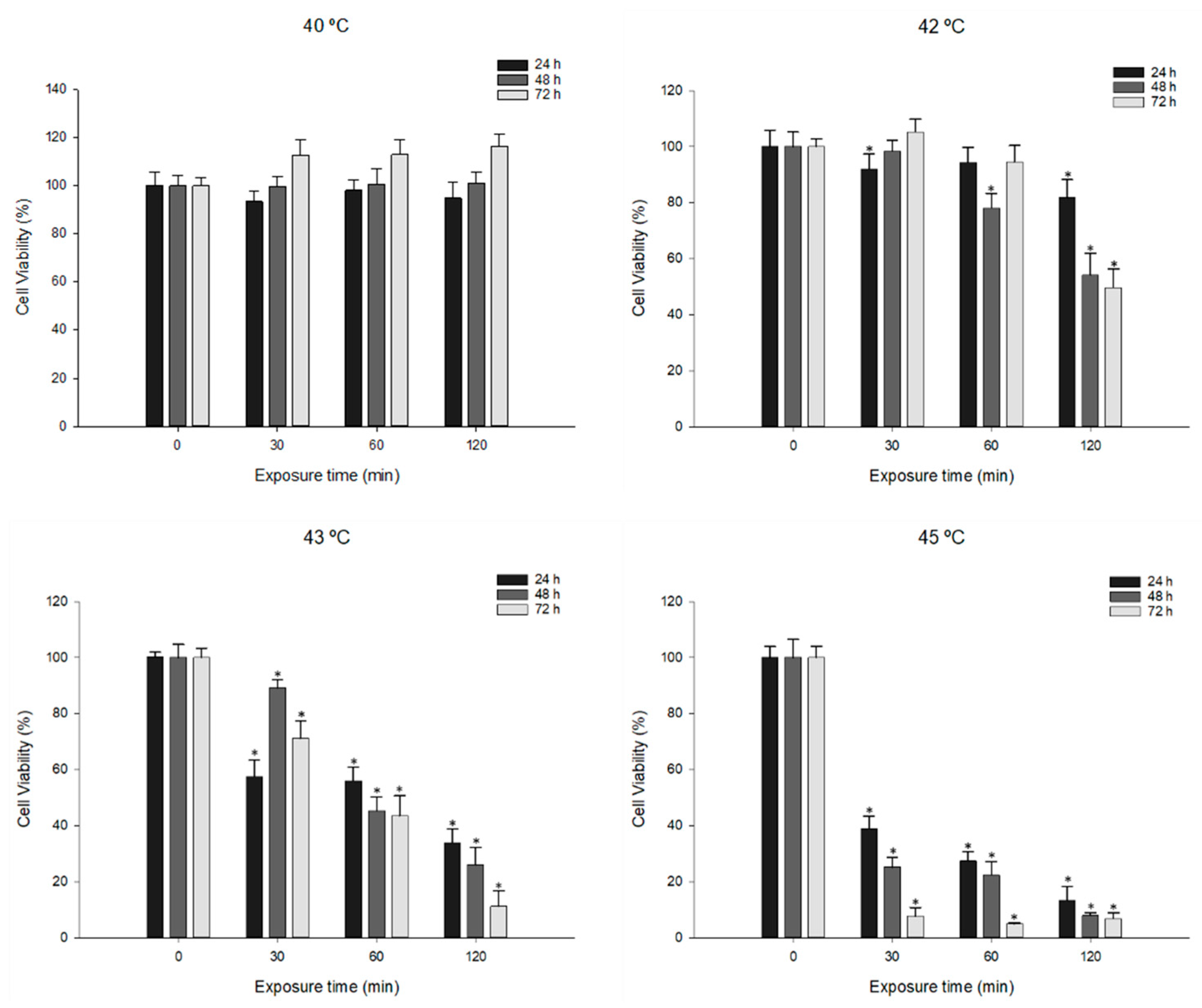
To study the effect of hyperthermia on cell viability, and understand the optimal conditions for combination therapy, MDA-MB-231 cells were exposed to temperatures of 40, 42, 43 and 45 °C, for 0, 30, 60 and 120 min. As shown in Figure 2, exposure to 40 °C did not decrease cell viability, and exposure to 42 °C only decreased cell viability for 24, 48 and 72 h after 120 min treatment. Temperatures of 43 and 45 °C induced a significant viability decrease for all the treatments and time exposures. However, after exposure to 45 °C, a very high reduction in cell viability was observed, which would hinder future assays. Thus, 43 °C was chosen as the temperature for further combination assays.
Thompson et al. [38] reported that the MDA-MB-231 cells showed reduced viability at 45 °C, while Lee et al. [39] only found a reduction in cell viability, to 77.9%, after 45 min at 47 °C. This difference in results may be related to differences in incubation times and temperature exposure conditions.

3.2. Effect of PTX and DOX Treatment on Cell Viability

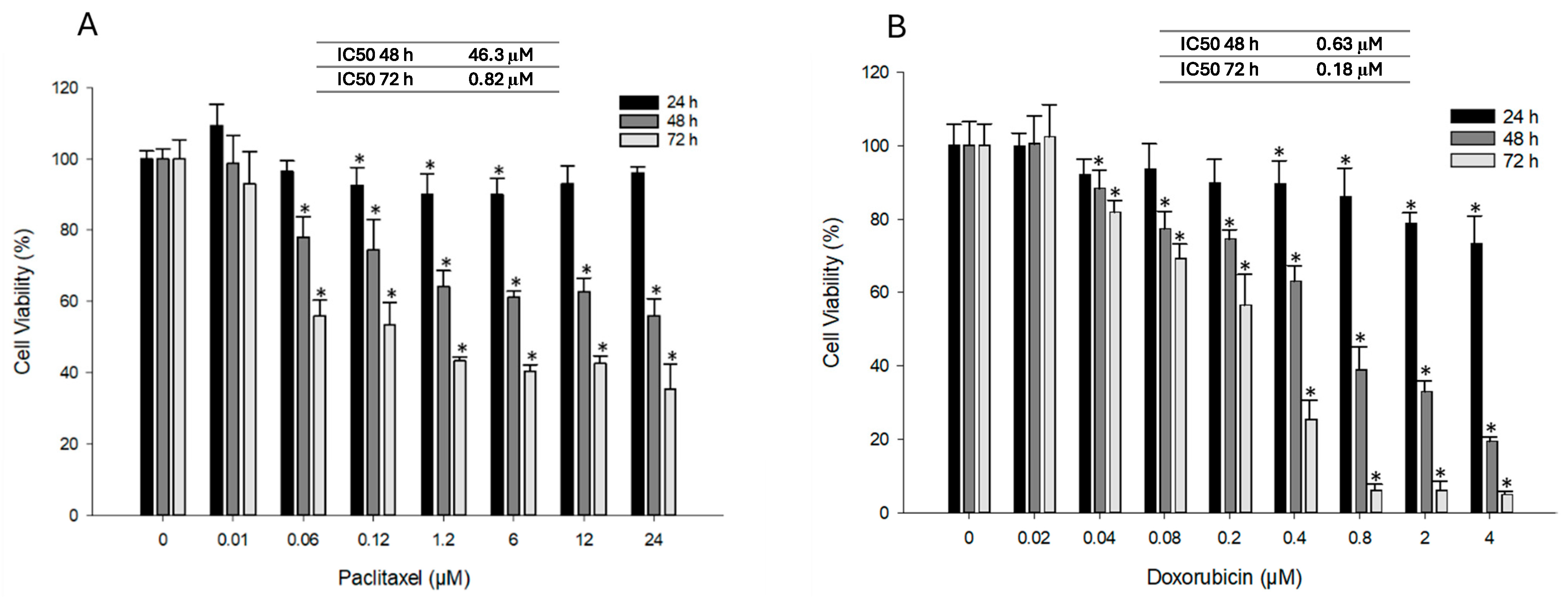
The dose–response to PTX and DOX was evaluated to select the appropriate doses for subsequent experiments. In the 24-h assay, cell viability was barely affected by PTX, with no decrease below 90% observed at any tested concentration (Figure 3A), making it impossible to determine an IC50 at this time point. For 48- and 72-h exposures, a significant decrease in viability was observed (p < 0.05), with IC50 values of 46.33 µM and 0.82 µM, respectively. This indicates that PTX-induced cytotoxicity is strongly time-dependent. Interestingly, a sharp decrease in viability was observed even at low concentrations (0.06 µM), followed by a plateau at higher concentrations for both 48 and 72 h. These findings are in agreement with Chang et al. [40], who reported a reduction in cell viability to approximately 50% after three days of treatment with 0.5 µM PTX, with limited additional reduction at higher concentrations.
For DOX, cell viability remained above 97% at 0.02 µM (Figure 3B). At 24 h, a significant reduction in viability was only observed at concentrations ≥0.4 µM, with a minimum viability of 73.1 ± 7.7% at 4 µM. For 48- and 72-h exposures, a significant dose-dependent decrease was observed, with IC50 values of 0.63 µM and 0.18 µM, respectively, demonstrating both time- and dose-dependent cytotoxicity. These results are consistent with previous studies [41,42,43].

3.3. Effect of Combined PTX and Hyperthermia Treatment on Cell Viability

To investigate the effects of combining PTX with hyperthermia, MDA-MB-231 cells were treated with the three lowest PTX concentrations that had previously shown significant effects (0.06, 0.12, and 1.2 µM). Cells were exposed to hyperthermia at 43 °C for 0, 30, 60, or 120 min and subsequently incubated for 24, 48, or 72 h. Across all conditions, significant differences (p < 0.05) were observed compared with the untreated control. However, to confirm a true enhancement of the combined treatment, statistical significance was required not only relative to the control, but also compared with the corresponding PTX condition at 37 °C and the respective hyperthermia condition without PTX.
After 24 h (Figure 4A), only one condition—1.2 µM PTX combined with 30 min hyperthermia—met this criterion, reducing cell viability to 65.3 ± 5.9% (an 11.2% improvement). CI analysis further indicated synergism for this condition (CI = 0.68) with a favourable DRI of 11.9, suggesting that the dose of PTX could be markedly reduced to achieve the same effect. Interestingly, CI analysis also suggested synergism at 0.12 µM PTX (CI = 0.69), in line with the Fa–CI plot (Figure 4D), where lower CI values coincided with higher affected fractions (Fa).
In contrast, no synergism was observed after 48 h (Figure 4B), although DRI values still indicated favourable dose reduction (2.8 for 0.06 and 0.12 µM, and 1.5 for 1.2 µM PTX after 30 min of hyperthermia). At 72 h, two conditions (0.12 and 1.2 µM PTX with 60 min hyperthermia) showed modest improvements of 8.7% and 9.8%, reducing viability to 45.0 ± 5.1% and 40.1 ± 3.2%, respectively (Figure 4C). Nevertheless, CI analysis at 72 h did not confirm synergism.
The results of exposure to the three concentrations combined with hyperthermia at 60 and 120 min after 24 h of incubation, and at 120 min after 48 h of incubation, showed antagonism. These findings seem to be consistent with the hypothesis proposed by Leal et al. [44], who suggested that elevated temperature may interfere with PTX’s binding to microtubules, thereby attenuating its cytotoxicity. This could partly explain the limited improvement observed under prolonged hyperthermia treatment (60–120 min). However, the additive effects observed at certain conditions suggest that the interaction may also depend on the duration of thermal exposure and subsequent recovery time, indicating a more complex relationship between PTX and hyperthermia.

3.4. Effect of Combined DOX and Hyperthermia Treatment on Cell Viability

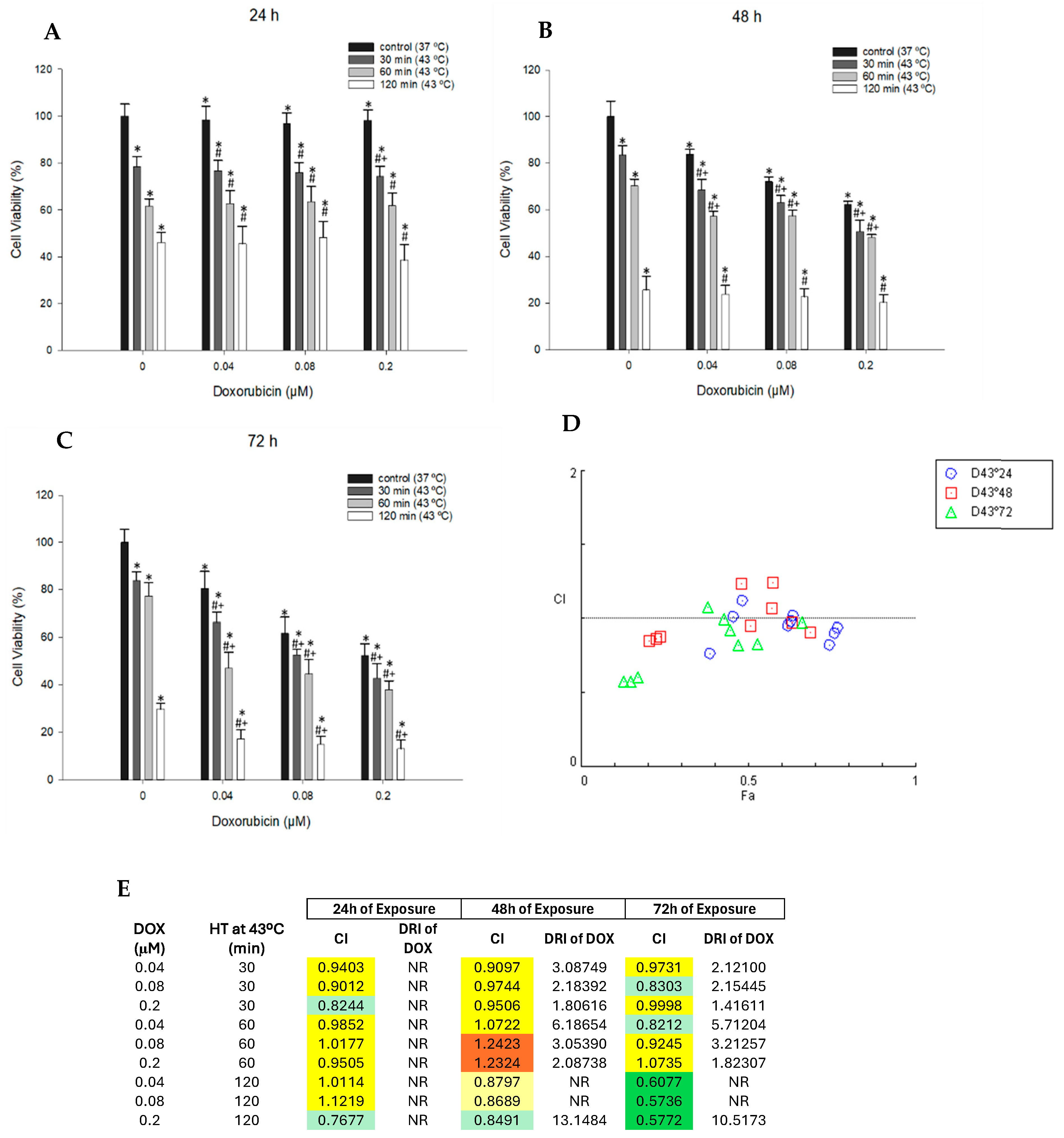
For DOX, results in Figure 5 show that a statistically significant decrease in cell viability compared with the control, the respective DOX condition at 37 °C, and the hyperthermia alone condition were observed only at 24 h for 0.2 µM DOX after 30 min of hyperthermia. This finding was consistent with CI analysis, which indicated moderate synergism for this condition, as well as for 0.2 µM DOX after 120 min of hyperthermia.
At 48 h, improvements were observed in several conditions: 0.04 and 0.2 µM at 43 °C for 30 and 60 min, and 0.08 µM at 43 °C for 30 min. However, CI analysis indicated only additive effects (Figure 5E), with moderate synergism observed for 0.2 µM after 120 min. DRI values were consistently higher than 1, indicating favorable dose reduction potential.
The strongest results were obtained at 72 h, which also showed greater consistency in maintaining CI values < 1 (Fa–CI plot, Figure 5D). Under these conditions, all combined treatments showed improvement. The most notable effect was observed with 0.04 µM DOX at 43 °C for 60 min, which reduced cell viability to 47.1 ± 6.8%—a 30.3% improvement compared with individual treatments (Figure 5C). This was supported by a CI value of 0.8 (moderate synergism) and a favorable DRI of 5.7. Similarly, 0.2 µM DOX combined with 120 min of hyperthermia showed low CI values and higher DRI. However, since hyperthermia alone at 120 min already induced a strong decrease in viability, this condition may not represent the most effective combination.
The observed improvements in viability reduction with combined DOX–hyperthermia treatments are consistent with previous studies. Kurokawa & Matsui [7] reported that MDA-MB-231 cells exposed to 1 µM DOX for 60 min at 42 °C and incubated for 24 h exhibited an ~20% decrease in viability. Likewise, Kulkarni-Dwivedi et al. [20] showed that DOX delivery via Fol-LSMO nanoparticles under hyperthermia enhanced anticancer activity in breast cancer cells. Importantly, the combined treatments with hyperthermia enabled a substantial reduction in the DOX dose required to achieve similar cytotoxic effects. For example, cells treated with 0.04 µM DOX and 43 °C hyperthermia for 60 min reached a viability of 47.1 ± 6.8%, whereas at 37 °C, only 0.2 µM DOX reduced viability to 52.4 ± 5.0%. This indicates that hyperthermia allowed a fourfold reduction in DOX concentration to achieve a comparable effect. Consistently, the Chou–Talalay method confirmed this improvement with a favorable DRI of 5.7, further supporting the dose-sparing effect of the combined treatment.

3.5. Effect of Combination of Hyperthermia and PTX/DOX on Cell Cycle Profile

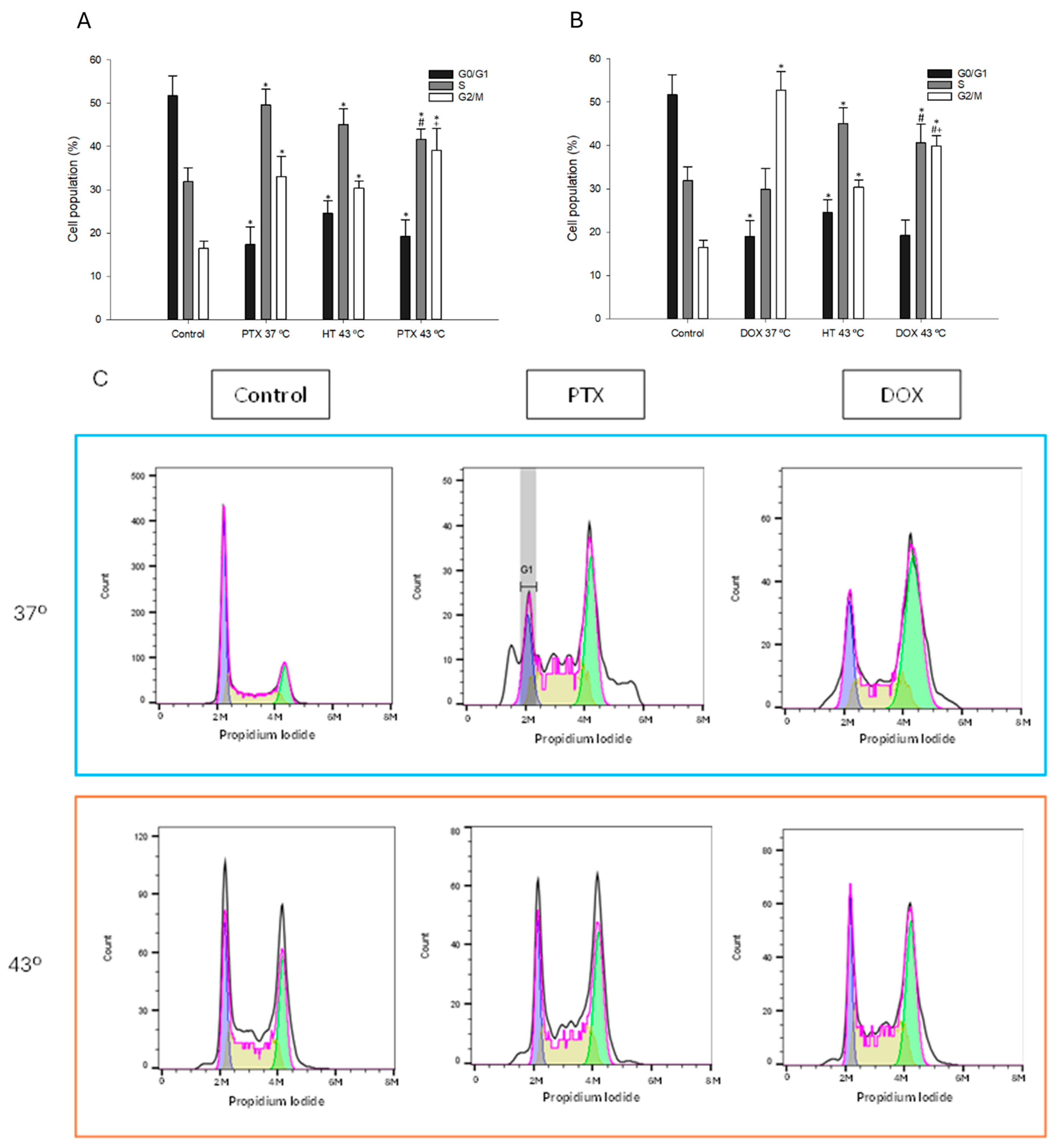
To study the changes induced by the combined treatments on the cell cycle profile, MDA-MB-231 cells were exposed to the lowest concentrations that showed the greatest improvement: 0.12 µM PTX or 0.04 µM DOX plus 43°C for 60 min, followed by incubation at 37 °C for 72 h 37 °C. Exposure to hyperthermia or PTX alone induced an increase in the percentage of cells in the G2/M phase. These results are consistent with previous reports that hyperthermia causes protein aggregation and interrupts cell cycle progression in the G2/M phase [45,46]. An increase in the S phase was also observed. The combination of PTX and temperature resulted in a decrease in cells in S phase, increasing the percentage in G2/M (Figure 6A).
Treatment with DOX led to a marked accumulation of cells in the G2/M phase (Figure 6B), in agreement with earlier findings [47,48]. When combined with hyperthermia, however, DOX caused a reduction in the proportion of cells in G2 and a concomitant rise in the S-phase population. The observed decrease in G1-phase cells may be linked to a fraction of the population entering apoptosis [49], consistent with the increased levels of apoptotic cells detected following the combined treatment.
The histograms (Figure 6C) illustrate the changes in DNA content among the different phases of the cell cycle under each condition. The treatment with PTX or DOX, both alone and in combination with hyperthermia, resulted in a distinct shift in cell populations, particularly an increase in the G2/M phase and a reduction in G1, confirming the data shown quantitatively in the bar graphs. This effect was more pronounced in the DOX + hyperthermia condition, aligning with the observed cytotoxicity and apoptotic effects discussed previously.

3.6. Effect of Combined Treatment with Chemotherapy and Hyperthermia on Intracellular ROS Levels

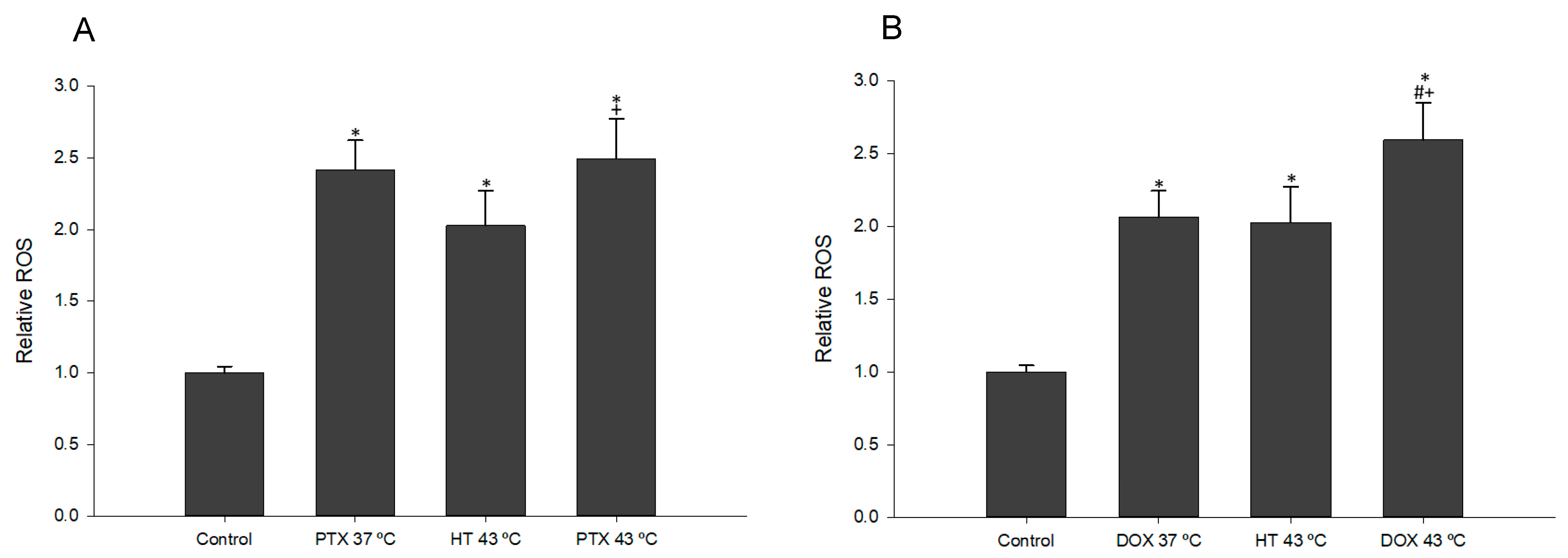
To evaluate oxidative stress following combined treatments, MDA-MB-231 cells were exposed to 0.12 µM PTX or 0.04 µM DOX in combination with hyperthermia at 43 °C for 60 min, followed by 72 h incubation at 37 °C. As shown in Figure 7A, both PTX and hyperthermia alone triggered an increase in ROS levels relative to the control. Their combination also elevated ROS, but without a significant difference compared with the individual treatments.
DOX can directly interact with the cell membrane by binding to plasma proteins, which leads to the enzymatic reduction of DOX through electron transfer. This process can result in the generation of highly reactive hydroxyl free radicals [50]. In line with this, DOX treatment alone significantly increased ROS compared to control (Figure 7B). When combined with hyperthermia, DOX induced a modest additional rise in intracellular ROS (0.53%) relative to single treatments. Similar results were also reported in Kurokawa & Matsui and Terasaki et al. [7,37].

3.7. Effect of Combined Chemotherapy and Hyperthermia Treatment in Apoptosis Induction

To measure the effect of combined treatments on cell apoptosis, Annexin V assay was performed with double labeling with Annexin V-FITC and PI. Cells were exposed to 0.12 µM PTX or 0.04 µM DOX plus 43 °C for 60 min, and incubated for 72h at 37 °C.
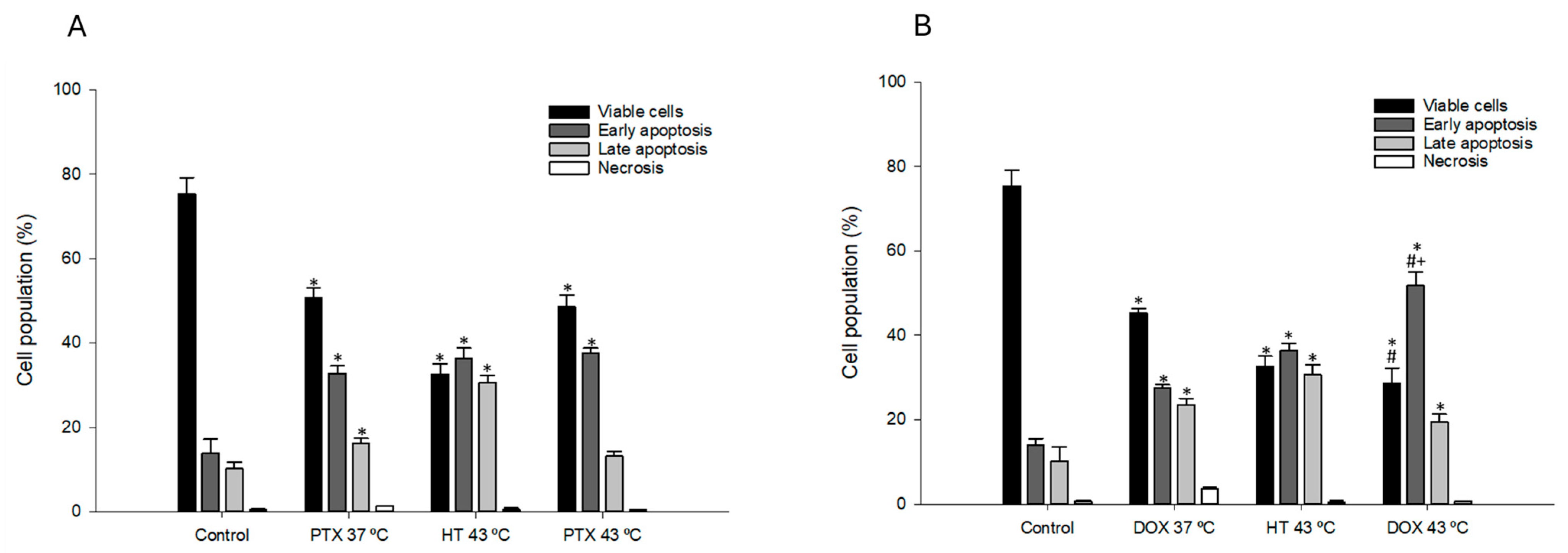
The results in Figure 8A show that hyperthermia treatment of MDA-MB-231 cells led to a sharp decrease in cell viability (42.8%) and a corresponding increase in the proportion of cells in both early and late apoptosis compared with the control. Exposure to PTX led to a decrease in viable cells, and consequently a significant (p < 0.05) increase in the percentage of cells in early and late apoptosis. However, the combination with hyperthermia did not induce significant differences when compared to PTX treatment alone. These results are consistent with what was reported in Michalakis et al. [27].
After exposure of MDA-MB-231 cells to DOX and hyperthermia, there was a significant increase (15.4%) in the percentage of cells in early apoptosis compared to exposure to DOX and hyperthermia alone (Figure 8B). Several articles reported an induction of initial apoptosis in cells after exposure to DOX [41,43]. DOX influences the Bcl-2/Bax apoptosis pathway through the activation of various AMPK (AMP activated protein kinase) molecular signals [50].

4. Conclusions

Our findings demonstrate that, under the in vitro conditions tested, hyperthermia enhanced the cytotoxic effects of chemotherapy drugs, although the magnitude of the effect varied between agents. In combination with PTX, hyperthermia produced only modest and transient improvements, with limited evidence of consistent synergism. In contrast, the combination with DOX showed a more robust effect, allowing up to a fourfold reduction in the drug dose required to achieve comparable cytotoxicity, as confirmed by favorable DRI values. This dose-sparing potential is particularly relevant, as it could help mitigate side effects and reduce the risk of drug resistance, commonly associated with chemotherapy. Mechanistically, the combined treatments impaired cell cycle progression, with accumulation in the G2/M and S phases, and induced early apoptosis accompanied by increased reactive oxygen species production.
This work presents the cytotoxic mechanisms implicated in the synergy between chemotherapy and hyperthermia to breast cancer cells, thereby enriching the understanding for future investigations into chemotherapy approaches. Nevertheless, this study is preclinical and exclusively in vitro, performed in a single cell line (MDA-MB-231). Further studies in other breast cancer models and in vivo are required to validate the clinical relevance of these findings.

Author Contributions

Conceptualization, A.C., V.B. and H.O.; methodology, A.C., V.B. and H.O.; investigation, A.C.; formal analysis, A.C. and V.B.; data curation, A.C. and V.B.; writing—original draft preparation, A.C. and V.B.; writing—review and editing, V.B. and H.O.; supervision, V.B. and H.O.; funding acquisition H.O. All authors have read and agreed to the published version of the manuscript.

Funding

This work is funded by national funds through FCT—Fundação para a Ciência e a Tecnologia I.P., under the project/grant UID/50006 + LA/P/0094/2020. V.B. was supported by FCT under contract CDL-CTTRI-54-SGRH/2022 and DOI: 10.54499/2022.05740.CEECIND/CP1720/CT0031), and H.O. under contract DOI: 10.54499/CEECIND/04050/2017/CP1459/CT0023.

Institutional Review Board Statement

Not applicable.

Data Availability Statement

The data presented in this study are available on request from the corresponding author.

Conflicts of Interest

The authors declare no conflicts of interest.

References

  1. World Health Organization. WHO Launches New Roadmap on Breast Cancer 2023. Available online: https://www.who.int/news/item/03-02-2023-who-launches-new-roadmap-on-breast-cancer (accessed on 21 April 2023).
  2. Arnold, M.; Morgan, E.; Rumgay, H.; Mafra, A.; Singh, D.; Laversanne, M.; Vignate, J.; Gralow, J.R.; Cardoso, F.; Siesling, S.; et al. Current and future burden of breast cancer: Global statistics for 2020 and 2040. Breast 2022, 66, 15–23. [Google Scholar] [CrossRef]
  3. Malhotra, G.K.; Zhao, X.; Band, H.; Band, V. Histological, molecular and functional subtypes of breast cancers 2010. Cancer Biol. Ther. 2010, 10, 955–960. [Google Scholar] [CrossRef] [PubMed]
  4. Tong, C.W.S.; Wu, M.; Cho, W.C.S.; To, K.K.W. Recent advances in the treatment of breast cancer. Front Oncol. 2018, 8, 227. [Google Scholar] [CrossRef]
  5. Bardia, A.; Mayer, I.A.; Diamond, J.R.; Moroose, R.L.; Isakoff, S.J.; Starodub, A.N.; Shah, N.C.; O’Shaughnessy, J.; Kalinsky, K.; Guarino, M.; et al. Efficacy & safety of anti-Trop-2 antibody drug conjugate sacituzumab govitecan (IMMU-132) in heavily pretreated patients with metastatic triple-negative breast cancer. J. Clin. Oncol. 2017, 35, 2141–2148. [Google Scholar] [CrossRef] [PubMed]
  6. Stockmans, G.; Deraedt, K.; Wildiers, H.; Moerman, P.; Paridaens, R. Triple-negative breast cancer. Curr. Opin Oncol. 2008, 20, 614–620. [Google Scholar] [CrossRef] [PubMed]
  7. Kurokawa, H.; Matsui, H. The Cytotoxicity of Doxorubicin Can Be Accelerated by a Combination of Hyperthermia and 5-Aminolevulinic Acid. Antioxidants 2021, 10, 1531. [Google Scholar] [CrossRef]
  8. Issels, R.; Kampmann, E.; Kanaar, R.; Lindner, L.H. Hallmarks of hyperthermia in driving the future of clinical hyperthermia as targeted therapy: Translation into clinical application. Int. J. Hyperth. 2016, 32, 89–95. [Google Scholar] [CrossRef]
  9. Vaupel, P.; Horsman, M.R. Tumour perfusion and associated physiology: Characterization and significance for hyperthermia. Int. J. Hyperth. 2010, 26, 209–210. [Google Scholar] [CrossRef]
  10. Oei, A.L.; Vriend, L.E.M.; Crezee, J.; Franken, N.A.P.; Krawczyk, P.M. Effects of hyperthermia on DNA repair pathways: One treatment to inhibit them all. Radiat. Oncol. 2015, 10, 1–13. [Google Scholar] [CrossRef]
  11. Lee, H.; Kim, S.; Choi, B.H.; Park, M.T.; Lee, J.; Jeong, S.Y.; Choi, E.; Lin, B.; Kim, C.; Park, H. Hyperthermia improves therapeutic efficacy of doxorubicin carried by mesoporous silica nanocontainers in human lung cancer cells. Int. J. Hyperth. 2011, 27, 698–707. [Google Scholar] [CrossRef]
  12. Krawczyk, P.M.; Eppink, B.; Essers, J.; Stap, J.; Rodermond, H.; Odijk, H.; Zelensky, A.; Bree, C.; Stalpers, L.J.; Buist, M.R.; et al. Mild hyperthermia inhibits homologous recombination, induces BRCA2 degradation, and sensitizes cancer cells to poly (ADP-ribose) polymerase-1 inhibition. Proc. Natl. Acad. Sci. USA 2011, 108, 9851–9856. [Google Scholar] [CrossRef]
  13. Wust, P.; Hildebrandt, B.; Sreenivasa, G.; Rau, B.; Gellermann, J.; Riess, H.; Felix, R.; Schlag, P.M. Hyperthermia in combined treatment of cancer. Lancet Oncol. 2002, 3, 487–497. [Google Scholar] [CrossRef]
  14. Hildebrandt, B.; Wust, P.; Ahlers, O.; Dieing, A.; Sreenivasa, G.; Kerner, T.; Felix, R.; Riess, H. The cellular and molecular basis of hyperthermia. Crit. Rev. Oncol. Hematol. 2002, 43, 33–56. [Google Scholar] [CrossRef] [PubMed]
  15. Sharma, A.; Özayral, S.; Caserto, J.S.; Ten Cate, R.; Anders, N.M.; Barnett, J.D.; Kandala, S.K.; Henderson, E.; Stewart, J.; Liapi, E.; et al. Increased uptake of doxorubicin by cells undergoing heat stress does not explain its synergistic cytotoxicity with hyperthermia. Int. J. Hyperth. 2019, 36, 712–720. [Google Scholar] [CrossRef] [PubMed]
  16. Harima, Y.; Ohguri, T.; Imada, H.; Sakurai, H.; Ohno, T.; Hiraki, Y.; Tuji, K.; Tanaka, M.; Terashima, H. A multicentre randomised clinical trial of chemoradiotherapy plus hyperthermia versus chemoradiotherapy alone in patients with locally advanced cervical cancer. Int. J. Hyperth. 2016, 32, 801–808. [Google Scholar] [CrossRef] [PubMed]
  17. Dunne, M.; Dou, Y.N.; Drake, D.M.; Spence, T.; Gontijo, S.M.; Wells, P.G.; Allen, C. Hyperthermia-mediated drug delivery induces biological effects at the tumor and molecular levels that improve cisplatin efficacy in triple negative breast cancer. J. Control. Release 2018, 282, 35–45. [Google Scholar] [CrossRef]
  18. Li, X.; Zhang, X.; Khan, I.U.; Guo, N.N.; Wang, B.; Guo, Y.; Xiao, B.; Zhang, Y.; Chu, Y.; Chen, J.; et al. The anti-tumor effects of the combination of microwave hyperthermia and lobaplatin against breast cancer cells in vitro and in vivo. Biosci. Rep. 2022, 42, BSR20190878. [Google Scholar] [CrossRef]
  19. Ammendola, M.; Currò, G.; Memeo, R.; Curto, L.S.; Luposella, M.; Zuccalà, V.; Pessaux, P.; Navarra, G.; Gadaleta, C.D.; Ranieri, G. Targeting Stem Cells with Hyperthermia: Translational Relevance in Cancer Patients. Oncology 2020, 98, 755–762. [Google Scholar] [CrossRef]
  20. Kulkarni-Dwivedi, N.; Patel, P.R.; Shravage, B.V.; Umrani, R.D.; Paknikar, K.M.; Jadhav, S.H. Hyperthermia and doxorubicin release by Fol-LSMO nanoparticles induce apoptosis and autophagy in breast cancer cells. Nanomedicine 2022, 17, 1929–1949. [Google Scholar] [CrossRef]
  21. Loboda, A.; Smolanka Sr, I.; Orel, V.E.; Syvak, L.; Golovko, T.; Dosenko, I.; Lyashenko, A.; Smolanka, I.; Dasyukevich, O.; Tarasenko, T.; et al. Efficacy of Combination Neoadjuvant Chemotherapy and Regional Inductive Moderate Hyperthermia in the Treatment of Patients With Locally Advanced Breast Cancer. Technol. Cancer Res. Treat 2020, 19, 1533033820963599. [Google Scholar] [CrossRef]
  22. Klimanov, M.Y.; Syvak, L.A.; Orel, V.E.; Lavryk, G.V.; Tarasenko, T.Y.; Orel, V.B.; Rykhalskyi, A.Y.; Stegnii, V.V.; Nesterenko, A.O. Efficacy of Combined Regional Inductive Moderate Hyperthermia and Chemotherapy in Patients With Multiple Liver Metastases From Breast Cancer. Technol. Cancer Res. Treat 2018, 17, 1533033818806003. [Google Scholar] [CrossRef]
  23. Othman, T.; Goto, S.; Lee, J.B.; Taimura, A.; Matsumoto, T.; Kosaka, M. Hyperthermic enhancement of the apoptotic and antiproliferative activities of paclitaxel. Pharmacology 2001, 62, 208–212. [Google Scholar] [CrossRef] [PubMed]
  24. Lin, Y.; Liu, Z.; Li, Y.; Liao, X.; Liao, S.; Cen, S.; Yang, L.; Wei, J.; Hu, X. Short-term hyperthermia promotes the sensitivity of MCF-7 human breast cancer cells to paclitaxel. Biol. Pharm Bull. 2013, 36, 376–383. [Google Scholar] [CrossRef]
  25. Liao, S.; Hu, X.; Liu, Z.; Lin, Y.; Liang, R.; Zhang, Y.; Li, Q.; Li, Y.; Liao, X. Synergistic action of microwave-induced mild hyperthermia and paclitaxel in inducing apoptosis in the human breast cancer cell line MCF-7. Oncol. Lett. 2019, 17, 603–615. [Google Scholar] [CrossRef] [PubMed]
  26. Rivera-Rodriguez, A.; Chiu-Lam, A.; Morozov, V.M.; Ishov, A.M.; Rinaldi, C. Magnetic nanoparticle hyperthermia potentiates paclitaxel activity in sensitive and resistant breast cancer cells. Int. J. Nanomed. 2018, 13, 4771–4779. [Google Scholar] [CrossRef] [PubMed]
  27. Michalakis, J.; Georgatos, S.D.; De Bree, E.; Polioudaki, H.; Romanos, J.; Georgoulias, V.; Tsiftsis, D.D.; Theodoropoulos, P.A. Short-term exposure of cancer cells to micromolar doses of paclitaxel, with or without hyperthermia, induces long-term inhibition of cell proliferation and cell death in vitro. Ann. Surg. Oncol. 2007, 14, 1220–1228. [Google Scholar] [CrossRef]
  28. Cai, D.; Liu, L.; Han, C.; Ma, X.; Qian, J.; Zhou, J.; Zhu, W. Cancer cell membrane-coated mesoporous silica loaded with superparamagnetic ferroferric oxide and Paclitaxel for the combination of Chemo/Magnetocaloric therapy on MDA-MB-231 cells. Sci. Rep. 2019, 9, 14475. [Google Scholar] [CrossRef]
  29. Su, S.; Tian, Y.; Li, Y.; Ding, Y.; Ji, T.; Wu, M.; Nie, G. “Triple-punch” strategy for triple negative breast cancer therapy with minimized drug dosage and improved antitumor efficacy. ACS Nano 2015, 9, 1367–1378. [Google Scholar] [CrossRef]
  30. Wu, S.K.; Chiang, C.F.; Hsu, Y.H.; Lin, T.H.; Liou, H.C.; Fu, W.M.; Lin, W.L. Short-time focused ultrasound hyperthermia enhances liposomal doxorubicin delivery and antitumor efficacy for brain metastasis of breast cancer. Int. J. Nanomed. 2014, 9, 4485–4494. [Google Scholar] [CrossRef]
  31. Lokerse, W.J.; Bolkestein, M.; Dalm, S.U.; Eggermont, A.M.; de Jong, M.; Grüll, H.; Koning, G.A. Comparing the therapeutic potential of thermosensitive liposomes and hyperthermia in two distinct subtypes of breast cancer. J. Control. Release 2017, 258, 34–42. [Google Scholar] [CrossRef]
  32. Dragojevic, S.; Ryu, J.S.; Hall, M.E.; Raucher, D. Targeted Drug Delivery Biopolymers Effectively Inhibit Breast Tumor Growth and Prevent Doxorubicin-Induced Cardiotoxicity. Molecules 2022, 27, 3371. [Google Scholar] [CrossRef]
  33. Liu, X.; Wang, C.; Ma, H.; Yu, F.; Hu, F.; Yuan, H. Water-Responsive Hybrid Nanoparticles Codelivering ICG and DOX Effectively Treat Breast Cancer via Hyperthermia-aided DOX Functionality and Drug Penetration. Adv. Healthc. Mater 2019, 8, 1801486. [Google Scholar] [CrossRef]
  34. Piehler, S.; Dähring, H.; Grandke, J.; Göring, J.; Couleaud, P.; Aires, A.; Cortajarena, A.L.; Courty, J.; Latorre, A.; Somoza, A.; et al. Iron Oxide Nanoparticles as Carriers for DOX and Magnetic Hyperthermia after Intratumoral Application into Breast Cancer in Mice: Impact and Future Perspectives. Nanomaterials 2020, 10, 1016. [Google Scholar] [CrossRef]
  35. Vujaskovic, Z.; Kim, D.W.; Jones, E.; Lan, L.; McCall, L.; Dewhirst, M.W.; Craciunescu, O.; Stauffer, P.; Liotcheva, V.; Betof, A.; et al. A phase I/II study of neoadjuvant liposomal doxorubicin, paclitaxel, and hyperthermia in locally advanced breast cancer. Int. J. Hyperthermia 2010, 26, 514–521. [Google Scholar] [CrossRef]
  36. de Maar, J.S.; MSuelmann, B.B.; GJABraat, M.N.; van Diest, P.J.; BVaessen, H.H.; Witkamp, A.J.; Linn, S.C.; Moonen, C.T.; van der Wall, E.; Deckers, R. Phase I feasibility study of Magnetic Resonance guided High Intensity Focused Ultrasound-induced hyperthermia, Lyso-Thermosensitive Liposomal Doxorubicin and cyclophosphamide in de novo stage IV breast cancer patients: Study protocol of the i-GO study. BMJ Open 2020, 10, 040162. [Google Scholar] [CrossRef] [PubMed]
  37. Terasaki, A.; Kurokawa, H.; Ito, H.; Komatsu, Y.; Matano, D.; Terasaki, M.; Bando, H.; Hara, H.; Matsui, H. Elevated Production of Mitochondrial Reactive Oxygen Species via Hyperthermia Enhanced Cytotoxic Effect of Doxorubicin in Human Breast Cancer Cell Lines MDA-MB-453 and MCF-7. Int. J. Mol. Sci. 2020, 219, 522. [Google Scholar] [CrossRef] [PubMed]
  38. Thompson, E.A.; Graham, E.; Macneill, C.M.; Young, M.; Donati, G.; Wailes, E.M.; Jones, B.T.; Levi-Polyachenko, N.H. Differential response of MCF7, MDA-MB-231, and MCF 10A cells to hyperthermia, silver nanoparticles and silver nanoparticle-induced photothermal therapy. Int. J. Hyperth. 2014, 30, 312–323. [Google Scholar] [CrossRef] [PubMed]
  39. Lee, T.H.; Bu, J.; Kim, B.H.; Poellmann, M.J.; Hong, S.; Hyun, S.H. Sub-lethal hyperthermia promotes epithelial-to-mesenchymal-like transition of breast cancer cells: Implication of the synergy between hyperthermia and chemotherapy. RSC Adv. 2019, 9, 52–57. [Google Scholar] [CrossRef]
  40. Chang, J.C.; Chang, H.S.; Wu, Y.C.; Cheng, W.L.; Lin, T.T.; Chang, H.J.; Kuo, S.; Chen, S.-T.; Liu, C.-S. Mitochondrial transplantation regulates antitumour activity, chemoresistance and mitochondrial dynamics in breast cancer. J. Exp. Clin. Cancer Res. 2019, 38, 1–16. [Google Scholar] [CrossRef]
  41. Ghafari, F.; Rajabi, M.R.; Mazoochi, T.; Taghizadeh, M.; Nikzad, H.; Atlasi, M.A.; Taherian, A. Comparing apoptosis and necrosis effects of Arctium lappa root extract and doxorubicin on MCF7 and MDA-MB-231 cell lines. Asian Pac. J. Cancer Prev. 2017, 18, 795–802. [Google Scholar] [CrossRef]
  42. Guerreiro, P.S.; Fernandes, A.S.; Costa, J.G.; Castro, M.; Miranda, J.P.; Oliveira, N.G. Differential effects of methoxyamine on doxorubicin cytotoxicity and genotoxicity in MDA-MB-231 human breast cancer cells. Mutat Res. Genet. Toxicol. Environ. Mutagen 2013, 757, 140–147. [Google Scholar] [CrossRef]
  43. Pilco-Ferreto, N.; Calaf, G.M. Influence of doxorubicin on apoptosis and oxidative stress in breast cancer cell lines. Int. J. Oncol. 2016, 49, 753–762. [Google Scholar] [CrossRef]
  44. Leal, B.Z.; Meltz, M.L.; Mohan, N.; Kuhn, J.; Prihoda, T.J.; Herman, T.S. Interaction of hyperthermia with taxol in human MCF-7 breast adenocarcinoma cells. Int. J. Hyperth. 1999, 15, 225–236. [Google Scholar] [CrossRef]
  45. Ahmed, K.; Tabuchi, Y.; Kondo, T. Hyperthermia: An effective strategy to induce apoptosis in cancer cells. Apoptosis 2015, 20, 11. [Google Scholar] [CrossRef] [PubMed]
  46. Furusawa, Y.; Iizumi, T.; Fujiwara, Y.; Zhao, Q.L.; Tabuchi, Y.; Nomura, T.; Kondo, T. Inhibition of checkpoint kinase 1 abrogates G2/M checkpoint activation and promotes apoptosis under heat stress. Apoptosis 2011, 17, 102–112. [Google Scholar] [CrossRef]
  47. Hevener, K.E.; Verstak, T.A.; Lutat, K.E.; Riggsbee, D.L.; Mooney, J.W. Recent developments in topoisomerase-targeted cancer chemotherapy. Acta Pharm Sin. B 2018, 8, 844–861. [Google Scholar] [CrossRef] [PubMed]
  48. Bar-On, O.; Shapira, M.; Hershko, D.D. Differential effects of doxorubicin treatment on cell cycle arrest and Skp2 expression in breast cancer cells. Anticancer. Drugs 2007, 18, 1113–1121. [Google Scholar] [CrossRef] [PubMed]
  49. Jackson, J.G.; Pereira-Smith, O.M. Primary and Compensatory Roles for RB Family Members at Cell Cycle Gene Promoters That Are Deacetylated and Downregulated in Doxorubicin-Induced Senescence of Breast Cancer Cells. Mol. Cell. Biol. 2006, 26, 2501–2510. [Google Scholar] [CrossRef]
  50. Tacar, O.; Sriamornsak, P.; Dass, C.R. Doxorubicin: An update on anticancer molecular action, toxicity and novel drug delivery systems. J. Pharm. Pharmacol. 2012, 65, 157–170. [Google Scholar] [CrossRef]
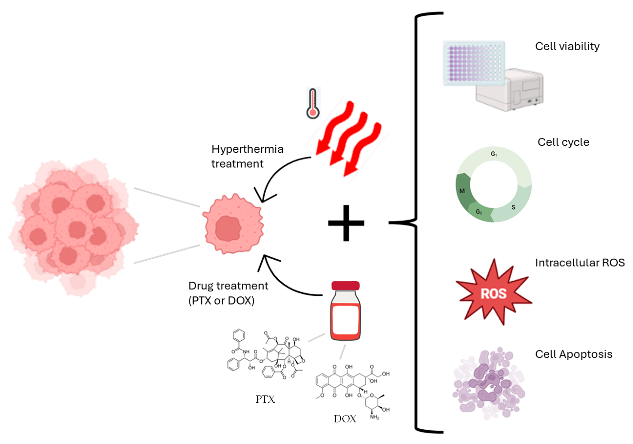
Figure 1. Schematic overview of experimental design.
Applsci 15 09883 g001
Figure 2. Effect of hyperthermia on cell viability of MDA-MB-231 cells when exposed to 40, 42, 43 and 45 °C, for 30, 60 and 120 min, after 24, 48 and 72 h. Data are shown as average ± standard deviation and * indicates statistical significance in relation to control (p < 0.05).
Applsci 15 09883 g002
Figure 3. Effect of different concentrations of (A) PTX and (B) DOX on cell viability of MDA-MB-231 cells, for 24, 48 and 72 h. Data are shown as average ± standard deviation and * indicates statistical significance in relation to control (p < 0.05).
Applsci 15 09883 g003
Figure 4. Effect of PTX on the viability of MDA-MB-231 cells after (A) 24, (B) 48 and (C) 72 h at 37 °C, or in combination with hyperthermia at 43 °C, for 30, 60 or 120 min. Data are shown as mean ± standard deviation. Statistical significance (p < 0.05) is indicated as: * vs. control; # vs. the respective PTX condition at 37 °C; + vs. the corresponding hyperthermia condition alone. (D) Fa–CI plot of PTX combined with hyperthermia for the different exposure times. (E) Combination Index (CI) values with a color gradient applied for visual interpretation (green = synergism, CI < 1; yellow = additive effect, CI ≈ 1; red = antagonism, CI > 1) and Dose-Reduction Index (DRI) values for PTX, where DRI > 1 indicates favorable dose reduction. Extreme values are reported as NR (non-reliable).
Applsci 15 09883 g004
Figure 5. Effect of DOX on the viability of MDA-MB-231 cells after (A) 24, (B) 48 and (C) 72 h at 37 °C, or in combination with hyperthermia at 43 °C, for 30, 60 or 120 min. Data are shown as mean ± standard deviation. Statistical significance (p < 0.05) is indicated as: * vs. control; # vs. the respective DOX condition at 37 °C; + vs. the corresponding hyperthermia condition alone. (D) Fa–CI plot of DOX combined with hyperthermia for the different exposure times. (E) Combination Index (CI) values with a color gradient applied for visual interpretation (green = synergism, CI < 1; yellow = additive effect, CI ≈ 1; red = antagonism, CI > 1) and Dose-Reduction Index (DRI) values for DOX, where DRI > 1 indicates favorable dose reduction. Extreme values are reported as NR (non-reliable).
Applsci 15 09883 g005
Figure 6. Effects of hyperthermia (HT) (43 °C for 60 min) combined with (A) PTX (0.12 µM) and (B) DOX (0.04 µM) on cell cycle distribution of MDA-MB-231 cells, after 72 h of exposure. Data are shown as average ± standard deviation and statistical significance (p < 0.05) is represented as * in comparison to the control at 37 °C; # when compared to the respective PTX or DOX condition at 37 °C; and + when compared to the same condition of hyperthermia alone. (C) Histograms represent DNA content distribution obtained by flow cytometry.
Applsci 15 09883 g006
Figure 7. Effects of hyperthermia (HT) (43 °C for 60 min) combined with (A) PTX (0.12 µM) or (B) DOX (0.04 µM) on production of intracellular ROS, on MDA-MB-231 cells, after 72 h of exposure. Data are shown as average ± standard deviation and statistical significance (p < 0.05) is represented as * in comparison to the control at 37 °C; # when compared to the respective PTX condition at 37 °C; and + when compared to the same condition of hyperthermia alone.
Applsci 15 09883 g007
Figure 8. Effects of hyperthermia (HT) (43 °C for 60 min) combined with (A) PTX (0.12 µM) and (B) DOX (0.04 µM) on apoptotic profile of MDA-MB-231 cells, after 72 h of exposure. Data are shown as average ± standard deviation and statistical significance (p < 0.05) is represented as * in comparison to the control at 37 °C; # when compared to the respective PTX condition at 37 °C; and + when compared to the same condition of hyperthermia alone.
Applsci 15 09883 g008
Disclaimer/Publisher’s Note: The statements, opinions and data contained in all publications are solely those of the individual author(s) and contributor(s) and not of MDPI and/or the editor(s). MDPI and/or the editor(s) disclaim responsibility for any injury to people or property resulting from any ideas, methods, instructions or products referred to in the content.