Review

Microbial Peptidases: Key Players in Reducing Gluten Immunogenicity Through Peptide Degradation

by
Africa Sanchiz
1,2,*,†,
M. Isabel San-Martín
1,2,†,
N. Navasa
1,2,
Honorina Martínez-Blanco
1,2,
Miguel Ángel Ferrero
1,2,
Leandro Benito Rodríguez-Aparicio
1,2,* and
Alejandro Chamizo-Ampudia
1,2
1
Area Biochemistry, Department of Molecular Biology, University of León, 24071 León, Spain
2
Instituto de Biología Molecular, Genómica y Proteómica (INBIOMIC), University of León, Campus de Vegazana, 24071 León, Spain
*
Authors to whom correspondence should be addressed.
These authors contributed equally to this work.
Appl. Sci. 2025, 15(14), 8111; https://doi.org/10.3390/app15148111
Submission received: 8 June 2025 / Revised: 18 July 2025 / Accepted: 19 July 2025 / Published: 21 July 2025

Abstract

Gluten-related disorders, including celiac disease (CeD) and non-celiac gluten sensitivity (NCGS), are triggered by the immune response to gluten peptides that resist complete digestion by human gastrointestinal enzymes. Microbial peptidases have emerged as promising biocatalysts capable of degrading these immunogenic peptides, offering potential therapeutic and industrial applications. This review explores the role of microbial peptidases in gluten degradation, highlighting key enzyme families, their mechanisms of action, and their effectiveness in reducing gluten immunogenicity. Additionally, we discuss advances in enzymatic therapy, food processing applications, and the challenges associated with optimizing microbial enzymes for safe and efficient gluten detoxification. Understanding the potential of microbial peptidases in mitigating gluten-related disorders paves the way for novel dietary and therapeutic strategies.

1. Introduction to Celiac Disease and Non-Celiac Gluten Sensitivity

Gluten-related disorders, including celiac disease (CeD) and non-celiac gluten sensitivity (NCGS), affect millions of people worldwide. The scientific term gluten refers to a group of water insoluble proteins present in wheat, barley, and rye [1]. CeD affects approximately 1% of the global population [2], and it is triggered by the ingestion of specific gluten protein fractions in genetically predisposed individuals expressing HLA-DQ2 or HLA-DQ8 haplotypes. Gluten-derived peptides reach the small intestinal epithelium, where they are deamidated by transglutaminase-2 (TG2). This modification initiates a gluten-specific immune response involving a CD4+ T-cell inflammatory cascade that ultimately leads to the production of antibodies against gluten peptides and TG2 [3]. Furthermore, villous atrophy and crypt hyperplasia in the duodenum are common consequences of this immune activation. In contrast, NCGS is a gluten-related disorder in which patients do not test positive for CeD but exhibit similar symptoms, such as abdominal pain, bloating, diarrhea, constipation, or headaches [4].
From an epidemiological standpoint, the incidence and prevalence of CeD have increased significantly over the past few decades, particularly in Western countries. Recent data suggest that while CeD affects about 1% of the population globally, up to 80–90% of individuals remain undiagnosed [5]. The rising global burden is also becoming evident in regions such as Asia, the Middle East, and South America, where both clinical awareness and diagnostic capabilities are improving [6]. In contrast, NCGS is not well defined epidemiologically due to the lack of biomarkers, but population-based surveys suggest a prevalence of approximately 3–6% [7]. These trends underscore the importance of developing improved diagnostic tools and exploring alternative treatment options.
Currently, the only effective management strategy for CeD and NCGS is a strict gluten-free diet (GFD). However, adherence to this diet is challenging, difficult to maintain, and must be carefully balanced to prevent nutritional deficiencies [8]. Additionally, the safety of gluten-free products remains a concern due to potential contamination and numerous product recalls [9,10]. From a regulatory standpoint, the gluten-free threshold has been established at 20 parts per million (ppm) [11].
The rising prevalence and severity of CeD and related disorders may partly stem from changes in modern agricultural and food processing practices. The intensive use of nitrogen fertilizers has significantly increased the gluten content in modern wheat varieties compared to older strains, thus increasing exposure to immunogenic gluten peptides [12]. Additionally, the widespread use of chemical pesticides and fungicides has reduced the natural microbial diversity associated with cereal grains and seeds, potentially disrupting gut microbial communities involved in immune regulation and intestinal health [13]. Moreover, the replacement of traditional microbial fermentations in many foods with chemical fermentation methods has led to an increase in gluten content and immunogenicity by eliminating secondary microbial processes that normally modify and reduce gluten’s antigenic properties during fermentation [14]. This reduction in microbial diversity and alteration of fermentation processes may contribute to the increasing incidence of gluten-related immune disorders.
The cause of CeD was identified in the 1950s, and scientific research has primarily focused on its diagnosis, pathophysiology, and treatment [15]. Subsequently, strategies have been developed to reduce gluten content in foods through detoxification and elimination techniques [16,17,18,19,20,21]. Many researchers have published new methods to reduce the immunogenicity of gluten peptides by applying several strategies to cereals or their proteins. Thus, it has been described that heating or other thermal treatments change the gliadin structure, as occurs in bread preparation [22]. Other studies have simulated in vitro digestion after bread baking or thermal processing, to quantify gluten peptides remaining by mass spectrometry, although more studies are necessary to improve biological relevance [23].
Among these strategies to reduce the immunogenicity of gluten peptides, microbial peptidases have emerged as promising and specific tools for the degradation of immunogenic gluten peptides. These strategies are based on the fact that CD4+ T-cells are specifically activated by some epitopes, which are recognized by HLA-DQ2 or 8 receptors present on antigen-presenting cells in individuals with a genetic predisposition to celiac disease [24]. These peptides are known as immunogenic peptides, with 9 to 33 amino acid residues, the most studied being the immunodominant 33-mer peptide from a2-gliadin [25]. These microbial enzymes, produced by various microorganisms, are capable of hydrolyzing peptide bonds within immunogenic epitopes, thereby reducing their ability to trigger adverse immune responses. Gluten-derived peptides are rich in proline and glutamine, making their degradation challenging for broad-spectrum peptidases [26]. In nature, the hydrolysis of proline-rich proteins is primarily carried out by a specialized class of enzymes known as proline-specific peptidases (PSPs), which exhibit unique specificity that makes them ideal for neutralizing the immunogenic potential of these peptides. PSPs play a crucial role in the fine regulation of various metabolic processes, cellular differentiation and maturation, the processing of biologically active peptides and proteins, and immune responses [27]. Their medical relevance has been extensively studied in diseases such as diabetes, cancer, Alzheimer’s disease, Parkinson’s disease, hypertension, and various neuropsychiatric disorders [28].
Since gluten immunogenic peptides contain both proline and glutamine, it is essential to consider peptidases with specificity for both amino acids. The combination of PSPs with peptidases targeting glutamine represents a potential strategy for partially or completely eliminating the immunogenicity of these peptides. In this regard, oral enzyme therapy with gluten-degrading peptidases is emerging as a promising therapeutic approach [29,30]
This review addresses the role of microbial peptidases in gluten degradation, highlighting their mechanisms of action, effectiveness in reducing immunogenicity, and potential applications in therapeutic strategies and the food industry. Additionally, advances in the development of enzymatic therapies, applications in food processing, and challenges associated with optimizing these enzymes for safe and efficient gluten detoxification will be discussed. Exploring the potential of microbial peptidases represents an innovative approach that could facilitate new strategies to mitigate the effects of gluten-related disorders and improve the quality of life of affected individuals.

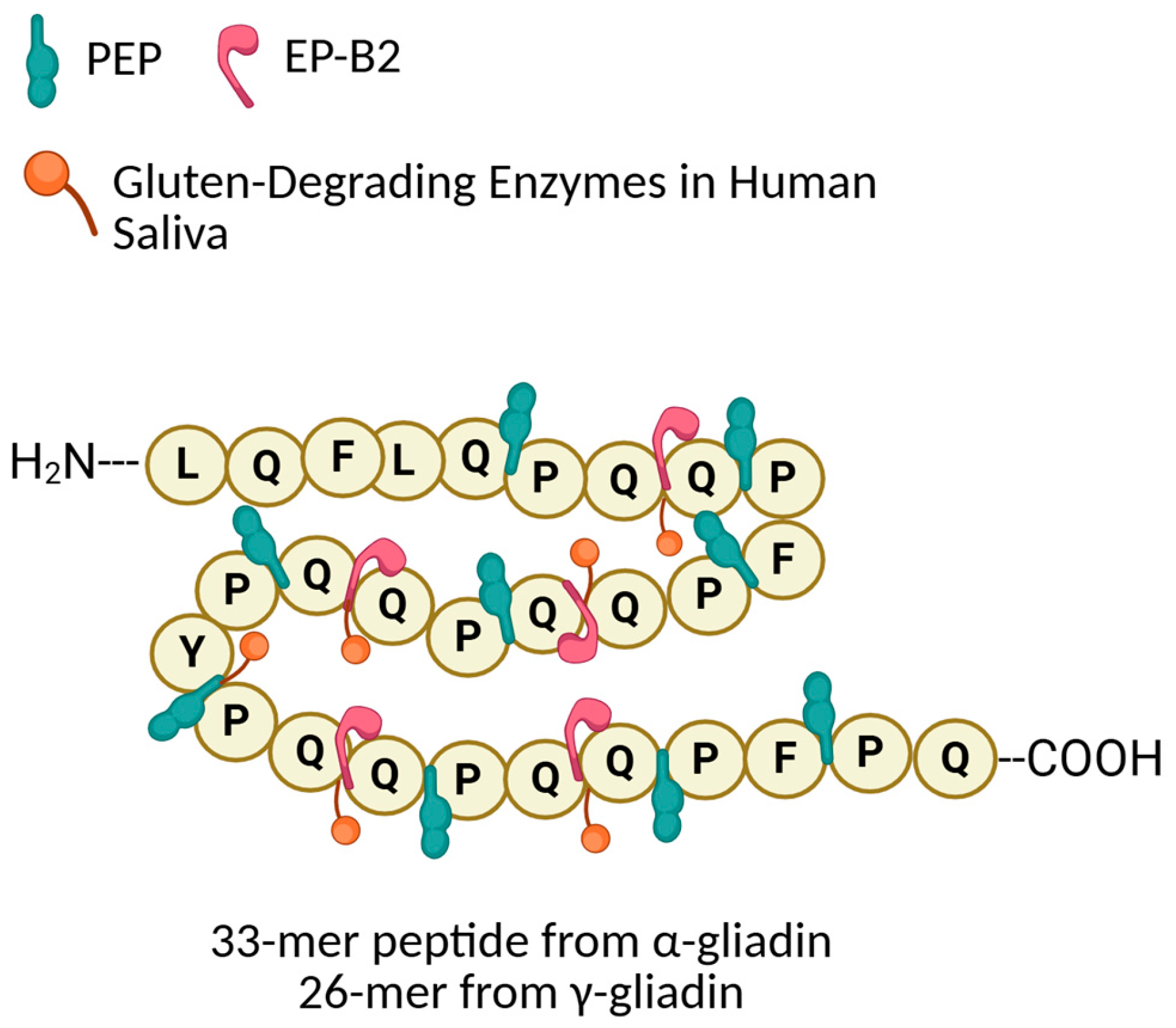
2. Gluten, Gliadin, and Immunogenic Peptides: Characteristics

Gluten is a composite protein predominantly found in cereals such as wheat, barley, and rye, comprising two main fractions: gliadins and glutenins. These proteins are fundamental to the baking industry due to their unique physicochemical properties. Gliadins confer the dough’s viscosity and extensibility, while glutenins provide elasticity and strength, together creating the dough’s characteristic viscoelastic network [31]. Although these properties are essential for bread quality and texture, they pose significant challenges for individuals suffering from gluten-related disorders, including celiac disease CeD and NCGS [32].
Among gluten proteins, gliadin is regarded as playing a key role in immunogenicity [33]. This protein is composed of complex amino acid sequences, particularly those rich in proline (Pro) and glutamine (Gln). These amino acids are considered the main factors responsible for the immunogenicity of gluten-derived fragments, as their structure enables the peptides to reach the small intestine intact and bind to immune receptor [34]. This composition renders gliadin difficult to digest, as proline in particular hinders the activity of human digestive enzymes. Due to this resistance to digestion, partially undigested gliadin predisposed individual to developing an immune response [35]. Once in the intestinal lamina propria, these gliadin-derived peptides undergo posttranslational modification by tissue transglutaminase 2 (TG2), which deamidates specific glutamine residues into glutamic acid. This modification increases the peptides’ affinity for the major histocompatibility complex (MHC) class II molecules, namely HLA-DQ2.5, HLA-DQ8, or HLA-DQ2.2, which are strongly associated with CeD susceptibility [36] The 33-mer peptide fragment derived from α-gliadin is one of the most extensively studied immunogenic peptides. It contains multiple proline and glutamine-rich sequences, rendering it highly resistant to enzymatic digestion and particularly effective at eliciting a CD4+ T-cell mediated immune response [36,37] (Figure 1).
The immune activation initiated by these peptides leads to the recruitment and activation of various immune cells, including CD4+ T cells, B cells, and antigen-presenting cells (APCs). The B-cell response targets both the deamidated gluten peptides (DGP) and the autoantigen TG2, resulting in the production of disease-specific autoantibodies, against gliadin, DGP and TG2 [36]. This immune cascade causes chronic inflammation, epithelial cell damage, and disruption of the intestinal barrier, ultimately leading to villous atrophy and impaired nutrient absorption, characteristic hallmarks of celiac disease [38,39].
Figure 1. Gluten intake and peptide processing after partial digestion in the stomach. The peptides derived from gliadins and glutenins are rich in proline and glutamine residues, making them highly resistant to gastrointestinal enzymatic digestion. When they cross the gut epithelium, increasing the membrane permeability, they are recognized by tissular TG2, leading to the formation of deamidated gluten peptides that interact with APC carrying HLA-DQ2/DQ8 receptors, triggering a cascade of gluten-specific immune response characterized by a release of certain cytokines, with the overexpression of IL-15 in epithelium and lamina propria responsible of chronic inflammation and even epithelial damage [40]. IEC means intestinal epithelial cells and IEL, intraepithelial lymphocytes. Figure prepared in Biorender.com.
Applsci 15 08111 g001
Although gliadin is the most extensively studied fraction for its immunogenicity, glutenins also contribute to the generation of immunogenic peptides. Glutenins, despite being larger and structurally more complex than gliadins, contain similarly proline and glutamine-rich sequences that resist digestion and contribute to immune activation in CeD [18]. Together, peptides derived from both gliadin and glutenin fractions initiate and perpetuate the immune response in susceptible individuals.
The resistance of these peptides to enzymatic digestion and their central role in CeD pathogenesis have made them a prime target for therapeutic intervention. Enzymatic therapies have been developed to degrade these immunogenic peptides before they can activate the immune system. Prolyloligopeptidase (POP) and prolylendopeptidase (PEP) are two classes of enzymes capable of cleaving the proline-rich peptide bonds within gluten fragments, significantly reducing their immunogenic potential [41]. These enzymes have potential applications both in the food industry for the production of safer gluten-free products and as therapeutic supplements for affected individuals.

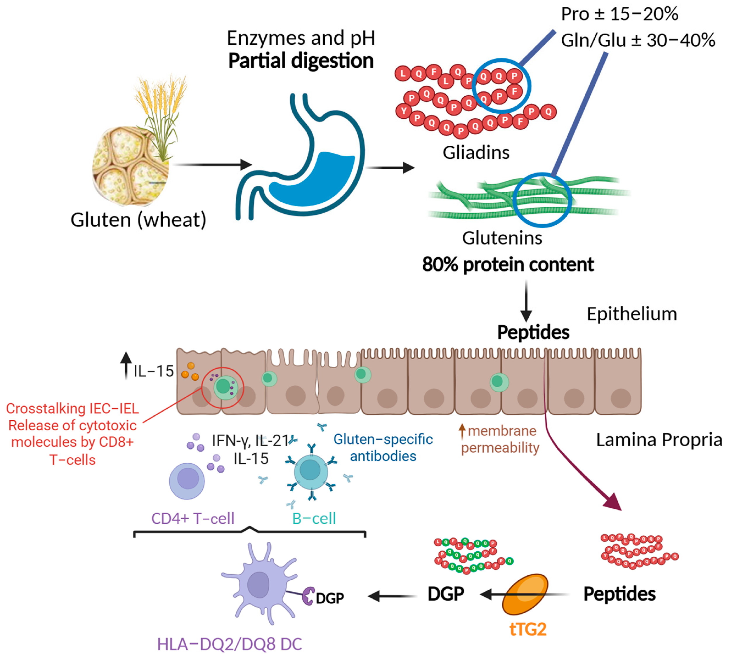
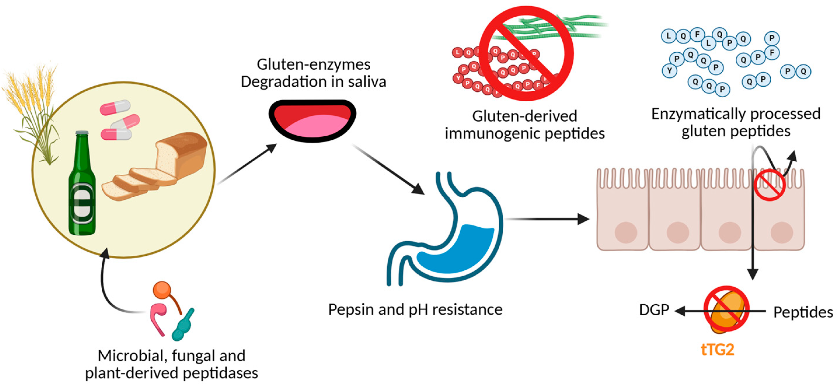
3. Treatment for Gluten Elimination: Pros and Cons

Gluten-related disorders, particularly celiac disease (CeD) and non-celiac gluten sensitivity (NCGS), represent a significant clinical challenge due to the persistent exposure to gluten peptides that provoke immune activation in susceptible individuals [42,43]. Although the cornerstone of treatment remains a strict gluten-free diet (GFD), adherence is challenging due to the ubiquity of gluten in many food products and the risk of crosscontamination [44]. Therefore, alternative and complementary treatment approaches aimed at reducing gluten immunogenicity have been actively researched, including enzymatic therapies, fermentation strategies, and digestive supplements.
Enzymatic degradation of gluten peptides, as discussed, offers a promising adjunct to dietary management. These enzymes are designed to cleave resistant proline-rich sequences that human digestive enzymes cannot process [45]. However, despite encouraging in vitro and early clinical results, enzymatic treatments have limitations that must be carefully considered [46]. One major challenge lies in ensuring sufficient enzyme activity throughout the gastrointestinal tract, which presents a highly variable environment. The acidic pH of the stomach, presence of endogenous proteases, and changing pH gradients in the small intestine can reduce enzyme stability and effectiveness [41,47]. Formulation strategies such as microencapsulation or use of enzyme cocktails combining different peptidases are under investigation to overcome these barriers and enhance therapeutic efficacy [47].
Clinically, enzymatic therapies have shown variable success. While they may reduce the immunogenic gluten load and mitigate symptoms caused by accidental gluten exposure, they have not been demonstrated to fully prevent mucosal damage in patients with active celiac disease [21,48]. This is particularly relevant for individuals with severe intestinal lesions or refractory celiac disease, where enzyme supplementation alone is insufficient. Furthermore, enzymatic treatments are generally unable to reverse existing tissue damage, emphasizing their role as supportive rather than curative [49].
The development and commercialization of enzymatic supplements such as latiglutenase and Tolerase G have expanded treatment options for patients. However, the high costs associated with production, quality control, and regulatory compliance can limit their accessibility, particularly in resource limited settings [50,51]. Regulatory frameworks for enzyme-based therapeutics also vary globally, complicating widespread adoption and standardization [52]. These challenges highlight the importance of continued investment in research, manufacturing innovation, and policy harmonization.
In addition to enzymatic approaches, advancements in fermentation processes offer promising routes to reduce gluten content in food products. Sourdough fermentation using specific lactic acid bacteria strains has been shown to partially hydrolyze gluten proteins, improving digestibility and reducing immunogenicity [9,45,53]. Optimization of such bioprocesses could provide a broader range of safer bakery products, enhancing dietary diversity and compliance for gluten-sensitive populations.
Beyond enzymatic and fermentation strategies, patient education and awareness remain critical components of successful gluten management. The risk of inadvertent gluten exposure persists due to the prevalence of gluten in processed foods, crosscontact during food preparation, and unclear labeling [9]. Enzymatic treatments should therefore be viewed as complementary tools rather than replacements for strict gluten avoidance. Overreliance on these supplements may engender a false sense of security, potentially leading to increased gluten ingestion and exacerbation of symptoms [45].
Looking forward, the future of gluten elimination therapies lies in the integration of multiple strategies. Combined enzyme formulations, improved delivery systems, and synergistic use of fermentation and biotechnological tools could enhance treatment outcomes. Additionally, personalized medicine approaches, considering individual genetic, immunological, and microbiome profiles, may allow tailored interventions that optimize efficacy and safety. The ultimate goal remains the development of safe, effective, and accessible therapies that improve the quality of life for individuals with gluten-related disorders, reducing the burden of strict dietary restrictions while preventing disease progression.

4. Enzymes Capable of Degrading Immunogenic Peptides and Their Industrial Applications from Microorganisms

Gluten-related disorders, such as CeD and NCGS, stem from the inability of the human digestive system to fully degrade certain proline and glutamine-rich peptides derived from gluten proteins, leading to the characteristic gluten immune responses in genetically predisposed individuals [42,43]. Consequently, microbial and plant-derived peptidases that can target these resistant sequences have been extensively investigated for their potential to detoxify gluten and mitigate disease symptoms.
Gluten-degrading enzymes are generally classified into three main groups according to their substrate specificity and catalytic mechanisms (Figure 2):
  • Prolyl Endopeptidases (PEP):
PEPs specifically cleave peptide bonds at the carboxyl side of proline residues, which are highly abundant in immunogenic gluten peptides. Bacterial PEPs have been isolated from species such as Flavobacterium meningosepticum (FM-PEP), Sphingomonas capsulata (SC-PEP, also known as ALV002), and Myxococcus xanthus (MX-PEP [54,55,56]). These enzymes exhibit robust proteolytic activity against immunotoxic sequences like the 33-mer from α-gliadin, which is resistant to mammalian gastrointestinal proteases. PEPs from F. meningosepticum and S. capsulata belong to the S9A family of serine proteases and possess a conserved catalytic triad composed of Ser-His-Asp (Ser554, His660, and Asp627 in FM-PEP) [57]. This triad forms a charge relay system critical for the nucleophilic attack on peptide bonds adjacent to internal proline residues. Structural analyses have further revealed that the substrate binding pocket of PEPs accommodates the pyrrolidine ring of proline through hydrophobic interactions, contributing to their substrate specificity [58].
However, their activity is optimal at neutral to slightly alkaline pH and they are sensitive to pepsin degradation, limiting their effectiveness in the acidic stomach environment [38]. Recently, engineered PEP variants with enhanced acid stability and broader substrate specificity have been developed through protein engineering and directed evolution techniques. For example, modified SC-PEP mutants show increased catalytic efficiency in simulated gastric fluid, improving in vivo gluten detoxification potential [59]. Fungal PEPs such as the Aspergillus niger prolyl endopeptidase (AN-PEP) have the advantage of acidic pH optima (around pH 4–5), enabling activity in the stomach before gluten peptides reach the small intestine [60]. Clinical trials with AN-PEP revealed safety and partial gluten degradation, though efficacy as a monotherapy was limited [60,61]. New formulations combining AN-PEP with other proteases have shown synergistic effects in reducing gluten immunogenicity in human gastrointestinal simulations [62].
2.
Glutamine-Specific Cysteine Endoprotease (EP-B2):
EP-B2 is a glutamine-specific cysteine protease that cleaves peptide bonds adjacent to glutamine residues in gliadin peptides [63,64]. Its catalytic mechanism is mediated by a Cys-His-Asn triad (Cys25, His159, and Asn175), analogous to that found in papain-like cysteine proteases [65]. EP-B2 specifically cleaves peptide bonds following glutamine residues, which are abundant in toxic gliadin peptides. Kinetic analyses using chromogenic and fluorogenic gliadin substrates revealed Michaelis constants (Km) of 30–100 µM and turnover numbers (kcat) indicative of efficient cleavage under gastric pH conditions. These findings, together with detailed structural and kinetic characterizations, provide crucial insights into the enzyme’s functional performance and support its potential application in oral enzyme therapy for CeD and related disorders.
EP-B2’s mechanism complements PEPs by targeting peptide bonds not cleavable by prolyl-specific enzymes, facilitating more thorough gluten degradation. Oral EP-B2 administration reduced gluten-induced pathology in gluten-sensitive macaques, demonstrating therapeutic promise [66]. Heterologous expression and fermentation production of EP-B2 have enabled scalable enzyme supply for clinical and industrial applications [67]. The studies have identified novel EP-B2 isoforms with altered substrate specificity and improved stability under gastrointestinal conditions, expanding the toolbox for enzyme therapy in CeD [68].
3.
Gluten-Degrading Enzymes in Human Saliva:
Human saliva contains microbially derived gluten-degrading enzymes, including subtilisin-like serine proteases from Rothia species, such as Rothia aeria and Rothia mucilaginosa [69,70,71]. These enzymes initiate gluten degradation during oral processing by cleaving immunodominant epitopes like the 33-mer and 26-mer peptides [69,72]. The presence of these enzymes contributes to the partial detoxification of gluten in the oral cavity and opens possibilities for novel probiotic or enzymatic therapies harnessing oral microbiota [70,73]. Emerging metaproteomic analyses have revealed a broader diversity of oral microbial glutenases, including metalloproteases and additional serine proteases, with potential for enhanced gluten degradation. Manipulation of oral microbiota composition is being explored to increase endogenous glutenase activity [73]. Beyond their initial hydrolytic action, the complex interplay between salivary glutenases, particularly those of microbial origin, and the oral microbiota warrants attention. These microbial peptidases can influence the oral and gut flora composition and metabolic activity, for instance, by altering peptide availability [74,75]. Conversely, metabolites produced by the oral microbiota may modulate the expression or efficiency of these glutenases. Understanding these bidirectional interactions within the oral cavity is essential for a comprehensive view of early gluten processing and its potential implications downstream in the gastrointestinal tract.
Beyond these groups, insect-derived peptidases, such as prolyloligopeptidases from Tenebrio molitor and postglutamine peptidases from Tribolium castaneum, have demonstrated the ability to degrade gluten peptides effectively, offering alternative enzyme sources for industrial or therapeutic applications [76,77]. Plant-derived enzymes, particularly cysteine proteases like EP-B2, have also been explored for gluten degradation. The EP-B2 from barley is a prime example of an enzyme adapted to degrade gliadin peptides in the acidic gastric environment (Table 1).
The food industry leverages these enzymes to produce gluten-reduced or gluten-free products. In baking, prolonged fermentation with lactic acid bacteria such as Lactobacillus plantarum and Lactobacillus sanfranciscensis decreases immunogenic gluten peptides, enabling the production of breads with lower gluten content without sacrificing quality [78]. In brewing, enzymes like AN-PEP and prolyl endopeptidases from Flammulina velutipes facilitate gluten degradation during fermentation, allowing the production of beers suitable for celiac individuals [79]. Digestive supplements containing AN-PEP (e.g., Tolerase G) have been marketed to aid gluten digestion in gluten-sensitive individuals, though they are considered adjuncts rather than replacements for gluten-free diets [80].
Table 1. Microbial peptidases with the ability to degrade immunogenic peptides derived from gluten and/or gluten.
EnzymeOrganismDegraded SubstrateProbioticDegradation EfficiencypH Stability RangeCommercial or Clinical UseReference
Prolyloligopeptidase (POP)Myxococcus xanthusProline-rich peptidesNoHigh activity against 33-mer; no reported kinetic constantsNeutral to slightly alkalineResearch stage[81]
Prolyloligopeptidase (POP)Sphingomonas capsulataProline-rich peptidesNoKm ≈ 82 µM; effective in combination therapy (ALV003)pH 6–8Approved for clinical testing
Prolyloligopeptidase (POP)Flavobacterium meningosepticumImmunogenic gliadin peptidesNoEffective in simulated digestion; structural features describedNeutralResearch stage[43]
Dipeptidylpeptidase IV (DPP IV)Lactobacillus caseiProline-rich peptidesYesModerate cleavage activity; kinetics not reported~7Over the counter supplement[43]
Prolylendopeptidase (PEP)Aspergillus nigerImmunogenic gluten peptidesNo>80% 33-mer degradation in vitro; Km ≈ 170 µMpH 3–5Available as Tolerase G[48,82]
Prolylendopeptidase (PEP)Flammulina velutipesImmunogenic gliadin peptidesNoSpecific activity = 56.7 U/mg (Z-Gly-Pro-pNA)NeutralUsed in industrial gluten removal[43]
Glutamine-specific cysteine endoprotease EP-B2Hordeum vulgareImmunogenic gliadin peptidesNoKm = 30–100 µM; kcat ≈ 3.2–5.4 s−1pH 3–5Combined with SC-PEP in ALV003[48,64]
ProlyloligopeptidaseTenebrio molitorProline-rich peptidesNoIn vitro activity confirmed; no kinetic dataAcidic to neutralPotential for food processing[48]
Postglutamine peptidaseTribolium castaneumGlutamine-rich peptidesNoKm ≈ 25 µM (QPQLPYPQPQ); effective on immunogenic targetsAcid stableResearch stage[48]
Prolyloligopeptidase (POP)Rothia sp.Immunogenic gliadin peptidesYesComparable to AN-PEP in oral gluten hydrolysispH 5–7Experimental probiotic use[70,83,84]
PeptidaseActinomyces odontolyticus33-mer peptideYesSelective degradation shown in vitroNeutralInvestigational oral application[84,85]

5. Future Perspectives on the Application of Enzymes for Celiac Disease Treatment

Enzyme therapy based on gluten-degrading proteases (glutenases) has long been regarded as a promising yet still limited complementary strategy for the treatment of CeD. Enzymes capable of degrading immunogenic gluten peptides must overcome several physiological and technological barriers before being considered safe and effective therapeutic agents. One critical concern lies in the potential increase in immunogenicity caused by the generation of smaller peptide fragments that can traverse the intestinal epithelial barrier more readily than the larger peptides produced by human gastrointestinal proteases. This phenomenon has been demonstrated in vitro using proline-specific endopeptidases derived from commensal microorganisms such as Pseudomonas, while the addition of Lactobacillus-derived enzymes has been shown to generate non-immunogenic fragments [86].
Beyond enzymatic specificity and catalytic efficiency, a major obstacle to the pharmacological application of glutenases is their rapid inactivation and structural instability under acidic gastric conditions [48,69]. These enzymes are susceptible to degradation by endogenous digestive proteases such as pepsin, trypsin, and chymotrypsin, potentially preventing them from remaining active until reaching the small intestine, the site where gluten-derived peptides trigger T-cell-mediated inflammatory responses. Although protective pharmaceutical strategies such as PEGylation and microencapsulation have been extensively investigated to improve the pharmacokinetics, stability, and bioavailability of therapeutic agents, clinical success has remained limited due to several persistent challenges. PEGylation, which involves the covalent attachment of polyethylene glycol chains to therapeutic proteins or peptides, has been shown to extend circulation time and reduce immunogenicity; however, its clinical effectiveness has been compromised by the emergence of anti-PEG antibodies in a significant portion of patients, leading to reduced efficacy and increased risk of hypersensitivity reactions [69,87]. Furthermore, PEGylation can negatively impact the biological activity of the modified drug due to conformational alterations. Similarly, microencapsulation has been explored to protect active pharmaceutical ingredients and allow for controlled drug release, but clinical translation has been impeded by inconsistent release kinetics, potential immunological reactions to encapsulating materials, and challenges in large-scale manufacturing [88]. These limitations highlight the need for continued innovation in drug delivery systems to achieve more reliable and effective therapeutic outcomes.
Two principal approaches have been proposed to address this limitation: (1) employing acid-stable peptidases and altering their substrate specificity toward prolamins, or (2) engineering existing glutenases to improve their acid stability [89].
These strategies have recently been expanded through site-directed mutagenesis and active site modification. For instance, the specificity of the glutenase Bga1903 was successfully enhanced by structural engineering to improve its activity against immunogenic peptides implicated in CD [90]. Such efforts have furthered the design of enzymes with both improved substrate specificity and greater resilience in gastrointestinal conditions.
Clinical evaluation of enzyme cocktails remains a key area of translational research. ALV003 (also known as Latiglutenase), which combines EP-B2 (ALV001) and SC-PEP (ALV002), has shown significant mucosal protection and a reduction in intraepithelial lymphocytosis in phase 1b–2a trials with CeD patients in remission who were exposed to gluten. Mucosal injury was assessed by calculating the villus height to crypt depth ratio in biopsies and by measuring epithelial lymphocyte density before and after treatment. Using the same enzyme cocktail, other researchers have also evaluated the expression of gluten-specific antibodies and T cells [91,92].
However, a larger multicenter phase 2b study in patients strictly adhering to a gluten-free diet failed to show significant histological improvement over placebo, likely due to enhanced dietary compliance under clinical supervision [93]. Nonetheless, recent trials involving controlled gluten challenges confirmed that Latiglutenase (IMGX003) reduced both mucosal injury and symptom severity in seropositive patients, reinforcing its potential as a personalized adjuvant therapy [94]. ALV003, administered daily for up to 42 days to seronegative patients undergoing a daily oral gluten challenge while on a strict gluten-free diet, was well tolerated. Discontinuations were mainly due to moderate to severe nausea, vomiting, and abdominal pain, largely attributed to gluten exposure. The study reported no serious adverse events and no clinically significant changes in vital signs, safety laboratory tests, or electrocardiogram results.
An additional promising development is TAK-062 (zamaglutenase), a recombinant enzyme engineered for high activity and stability in gastric conditions. In a recent phase 1 clinical study, TAK-062 exhibited robust gluten-degrading capacity, high oral bioavailability, and favorable safety and tolerability profiles, suggesting strong therapeutic potential for future clinical application [95]. In clinical evaluations, TAK-062 demonstrated a favorable safety profile. The highest dose administered (900 mg, liquid formulation) was well tolerated, with no treatment emergent adverse events exceeding Grade 1 severity. Critically, no adverse events were attributed to the study drug, nor were any serious adverse events or fatalities observed throughout the trial. Furthermore, routine safety assessments, including clinical laboratory parameters, vital signs, physical examinations, electrocardiograms, and antidrug antibody levels, revealed no clinically significant trends.
Moreover, enzyme combinations involving Aspergillus niger, derived aspergillopepsin (ASP), Aspergillus oryzae, and derived dipeptidyl peptidase IV (DPP-IV) have shown limited but notable synergistic effects in degrading immunogenic peptides in vitro [96]. Although such combinations have not yet demonstrated consistent in vivo efficacy, they continue to serve as valuable models for evaluating enzymatic synergy and formulation.
These strategies are summarized in Figure 3, which illustrates the current and emerging biotechnological approaches aimed at enhancing the stability, specificity, and delivery of glutenases for therapeutic and industrial use.
Recent evidence has highlighted the potential role of the human microbiota in modulating the activity of glutenase. Specifically, microbial communities in the oral cavity, such as Rothia and Actinomyces spp., have been shown to produce subtilisin-like proteases capable of initiating gluten degradation during mastication [62,74]. Moreover, metaproteomic and microbiome analyses suggest that the composition and metabolic activity of the oral and intestinal microbiota may influence both the expression and efficiency of microbial glutenases [74,75]. These findings point to a dynamic interplay between microbial populations and enzymatic function, potentially opening new avenues for probiotic or microbiota-targeted therapeutic strategies.
Additionally, although this review focuses on enzymes with gluten-degrading activity, it is worth noting that dipeptidyl peptidase IV (DPP-IV) is also a target of pharmacological inhibitors used in type 2 diabetes therapy to prolong incretin activity. These DPP-IV inhibitors may potentially reduce the enzymatic breakdown of gluten-derived peptides by both human and microbial DPP-IV–like enzymes [97,98,99]. While further research is needed, this interaction could represent a relevant factor to consider in patients undergoing DPP-IV inhibitor therapy who are also affected by gluten-related disorders.

6. Conclusions

The application of enzymatic treatment for the degradation of immunogenic gluten peptides has been extensively investigated as a strategy to mitigate the effects of gluten-related disorders rather than to prevent their initial development. Various microbial, fungal, and plant-derived peptidases have demonstrated efficacy in hydrolyzing proline and glutamine-rich peptides, such as gliadins and glutenins, that resist digestion in the human gastrointestinal tract. These enzymes have been incorporated into different industrial processes, including food production, brewing, and dietary supplements, aiming to reduce gluten immunogenicity before consumption and thereby alleviate symptoms in affected individuals.
Despite significant advancements, major challenges remain. A critical obstacle to the pharmacological application of glutenases is their rapid inactivation and structural instability under the acidic conditions of the stomach. These enzymes are susceptible to degradation by endogenous digestive proteases such as pepsin, trypsin, and chymotrypsin, often preventing them from remaining active until reaching the small intestine, where gluten-derived peptides trigger T-cell-mediated inflammatory responses. Protective pharmaceutical strategies like PEGylation and microencapsulation have been explored to improve enzyme stability and bioavailability; however, clinical success has been limited due to issues including immune reactions, altered enzymatic activity, inconsistent drug release, and manufacturing challenges. In addition, new enzyme variants with improved gastric stability, such as TAK-062 (zamaglutenase) and latiglutenase (ALV003), have demonstrated promising clinical results, showing both mucosal protection and symptom reduction in seropositive patients.
An emerging area of interest is the interaction between glutenases and the host microbiota. Specifically, oral and intestinal microorganisms such as Rothia and Actinomyces have been shown to contribute to gluten degradation during digestion, and recent studies suggest that microbial composition may influence the activity and expression of these enzymes. These findings open avenues for microbiota-targeted therapies that could enhance enzyme-based treatments.
Future research should also focus on standardizing evaluation protocols to compare enzyme efficacy across food processing and clinical settings. The development of personalized therapeutic approaches integrating enzyme cocktails, delivery systems, and microbiome modulation represents a promising direction for advancing non-dietary interventions in gluten-related disorders.
Potential outcomes of enzymatic interventions include enhanced safety of gluten-containing foods for individuals with gluten sensitivity, improved therapeutic management of CeD symptoms, and the development of novel enzyme formulations with optimized performance. Future research should focus on enzyme engineering, innovative drug delivery systems, combination therapies, and rigorous clinical validation to advance the reliability and effectiveness of enzymatic treatments aimed at alleviating the impact of gluten exposure in affected patients.

Author Contributions

A.S., M.I.S.-M., N.N., H.M.-B., M.Á.F., L.B.R.-A. and A.C.-A.; writing—original draft preparation: A.S., M.I.S.-M., N.N., H.M.-B., M.Á.F., L.B.R.-A. and A.C.-A.; writing—review and editing: M.I.S.-M., A.S. and A.C.-A.; supervision: A.S., L.B.R.-A. and A.C.-A. All authors have read and agreed to the published version of the manuscript.

Funding

The APC was funded by the University of León. We acknowledge the received support from the Spanish Ministerio de Ciencia e Innovación, grant number PID2020-119044GB-I00, and the Junta de Castilla y León, grant number LE015P20.

Institutional Review Board Statement

Not applicable.

Informed Consent Statement

Not applicable.

Data Availability Statement

No new data have been created for this review article.

Acknowledgments

We thank Eduardo Fernández for providing technical assistance.

Conflicts of Interest

The authors declare no conflicts of interest.

References

  1. Scherf, K.A. Gluten and Wheat-Related Disorders. In Handbook of Food Allergen Detection and Control; Elsevier: Amsterdam, The Netherlands, 2025; pp. 369–387. [Google Scholar]
  2. Catassi, G.; Marmo, C.; Gasbarrini, A.; Riccioni, M.E. Role of Device-Assisted Enteroscopy in Crohn’s Disease. J. Clin. Med. 2024, 13, 3919. [Google Scholar] [CrossRef]
  3. Rahmani, S.; Galipeau, H.J.; Clarizio, A.V.; Wang, X.; Hann, A.; Rueda, G.H.; Kirtikar, U.N.; Constante, M.; Wulczynski, M.; Su, H.-M.; et al. Gluten-Dependent Activation of CD4+ T Cells by MHC Class II–Expressing Epithelium. Gastroenterology 2024, 167, 1113–1128. [Google Scholar] [CrossRef]
  4. San-Martin, M.I.; Chamizo-Ampudia, A.; Sanchiz, Á.; Ferrero, M.Á.; Martínez-Blanco, H.; Rodríguez-Aparicio, L.B.; Navasa, N. Microbiome Markers in Gastrointestinal Disorders: Inflammatory Bowel Disease, Colorectal Cancer, and Celiac Disease. Int. J. Mol. Sci. 2025, 26, 4818. [Google Scholar] [CrossRef]
  5. Caio, G.; Volta, U.; Sapone, A.; Leffler, D.A.; De Giorgio, R.; Catassi, C.; Fasano, A. Celiac Disease: A Comprehensive Current Review. BMC Med. 2019, 17, 142. [Google Scholar] [CrossRef] [PubMed]
  6. Poddighe, D.; Abdukhakimova, D. Celiac Disease in Asia beyond the Middle East and Indian Subcontinent: Epidemiological Burden and Diagnostic Barriers. World J. Gastroenterol. 2021, 27, 2251–2256. [Google Scholar] [CrossRef] [PubMed]
  7. Catassi, C.; Alaedini, A.; Bojarski, C.; Bonaz, B.; Bouma, G.; Carroccio, A.; Castillejo, G.; De Magistris, L.; Dieterich, W.; Di Liberto, D.; et al. The Overlapping Area of Non-Celiac Gluten Sensitivity (NCGS) and Wheat-Sensitive Irritable Bowel Syndrome (IBS): An Update. Nutrients 2017, 9, 1268. [Google Scholar] [CrossRef] [PubMed]
  8. Rubin, E.B.; Viscuso, M.R.; Moleski, S.M. Maintaining a Balanced Diet While Gluten-Free: Treatment Options. Curr. Treat. Options Gastroenterol. 2020, 18, 699–717. [Google Scholar] [CrossRef]
  9. Wieser, H.; Segura, V.; Ruiz-Carnicer, Á.; Sousa, C.; Comino, I. Food Safety and Cross-Contamination of Gluten-Free Products: A Narrative Review. Nutrients 2021, 13, 2244. [Google Scholar] [CrossRef]
  10. Liu, S.; El Khoury, D.; Joye, I.J. Gluten-Free Product Recalls and Their Impact on Consumer Trust. Nutrients 2023, 15, 4170. [Google Scholar] [CrossRef]
  11. Catassi, C.; Monachesi, C.; Catassi, G.N.; Lionetti, E. Lessons from Low-Gluten Challenge Studies in Celiac Disease: Is It Time to Reconsider the Gluten Threshold in Gluten-Free Food? Dig. Liver Dis. 2025, 57, 1105–1106. [Google Scholar] [CrossRef]
  12. Mandal, S.; Verma, A.K. Wheat Breeding, Fertilizers, and Pesticides: Do They Contribute to the Increasing Immunogenic Properties of Modern Wheat? Gastrointest. Disord. 2021, 3, 247–264. [Google Scholar] [CrossRef]
  13. Ali, A.; AlHussaini, K.I. Pesticides: Unintended Impact on the Hidden World of Gut Microbiota. Metabolites 2024, 14, 155. [Google Scholar] [CrossRef]
  14. Sawant, S.S.; Park, H.-Y.; Sim, E.-Y.; Kim, H.-S.; Choi, H.-S. Microbial Fermentation in Food: Impact on Functional Properties and Nutritional Enhancement—A Review of Recent Developments. Fermentation 2025, 11, 15. [Google Scholar] [CrossRef]
  15. Fasano, A.; Catassi, C. Current Approaches to Diagnosis and Treatment of Celiac Disease: An Evolving Spectrum. Gastroenterology 2001, 120, 636–651. [Google Scholar] [CrossRef] [PubMed]
  16. Zhang, Y.; Wu, H.; Huilian, C.; Fu, L. Detoxification Mechanism and the Impact of Transamidation-Modified Gliadin on Celiac-Based Gluten Sensitivity: The Potential of Unlocking Gluten Tolerance in Functional Food. J. Agric. Food Chem. 2025, 73, 1682–1693. [Google Scholar] [CrossRef] [PubMed]
  17. Gélinas, P.; Théolier, J. How to Reduce Gluten in Foods: A Critical Review of Patents. Int. J. Food Sci. Technol. 2024, 59, 8069–8081. [Google Scholar] [CrossRef]
  18. Mamone, G.; Di Stasio, L.; Vitale, S.; Picascia, S.; Gianfrani, C. Analytical and Functional Approaches to Assess the Immunogenicity of Gluten Proteins. Front. Nutr. 2023, 9, 1049623. [Google Scholar] [CrossRef]
  19. Pourmohammadi, K.; Abedi, E. Hydrolytic Enzymes and Their Directly and Indirectly Effects on Gluten and Dough Properties: An Extensive Review. Food Sci. Nutr. 2021, 9, 3988–4006. [Google Scholar] [CrossRef]
  20. Scherf, K.A.; Catassi, C.; Chirdo, F.; Ciclitira, P.J.; Feighery, C.; Gianfrani, C.; Koning, F.; Lundin, K.E.A.; Schuppan, D.; Smulders, M.J.M.; et al. Recent Progress and Recommendations on Celiac Disease From the Working Group on Prolamin Analysis and Toxicity. Front. Nutr. 2020, 7, 29. [Google Scholar] [CrossRef]
  21. Zhang, Y.; Wu, H.; Fu, L. A Review of Gluten Detoxification in Wheat for Food Applications: Approaches, Mechanisms, and Implications. Crit. Rev. Food Sci. Nutr. 2025, 65, 2100–2116. [Google Scholar] [CrossRef]
  22. Xu, K.; Kuang, J. Effects of Thermal Treatment on the Physicochemical and Structural Properties of Wheat Gluten Proteins: Insights from Gluten, Glutenin, and Gliadin Fractions. Int. J. Food Sci. Technol. 2024, 59, 2275–2285. [Google Scholar] [CrossRef]
  23. Ogilvie, O.; Roberts, S.; Sutton, K.; Gerrard, J.; Larsen, N.; Domigan, L. The Effect of Baking Time and Temperature on Gluten Protein Structure and Celiac Peptide Digestibility. Food Res. Int. 2021, 140, 109988. [Google Scholar] [CrossRef] [PubMed]
  24. Frick, R.; Høydahl, L.S.; Petersen, J.; du Pré, M.F.; Kumari, S.; Berntsen, G.; Dewan, A.E.; Jeliazkov, J.R.; Gunnarsen, K.S.; Frigstad, T.; et al. A High-Affinity Human TCR-like Antibody Detects Celiac Disease Gluten Peptide–MHC Complexes and Inhibits T Cell Activation. Sci. Immunol. 2021, 6, eabg4925. [Google Scholar] [CrossRef] [PubMed]
  25. Cabanillas, B. Gluten-Related Disorders: Celiac Disease, Wheat Allergy, and Nonceliac Gluten Sensitivity. Crit. Rev. Food Sci. Nutr. 2020, 60, 2606–2621. [Google Scholar] [CrossRef]
  26. Abbasi, A.; Bazzaz, S.; Ibrahim, S.A.; Hekmatdoost, A.; Hosseini, H.; Sabahi, S.; Sheykhsaran, E.; Rahbar Saadat, Y.; Asghari Ozma, M.; Lahouty, M. A Critical Review on the Gluten-Induced Enteropathy/Celiac Disease: Gluten-Targeted Dietary and Non-Dietary Therapeutic Approaches. Food Rev. Int. 2024, 40, 883–923. [Google Scholar] [CrossRef]
  27. Baharin, A.; Ting, T.-Y.; Goh, H.-H. Post-Proline Cleaving Enzymes (PPCEs): Classification, Structure, Molecular Properties, and Applications. Plants 2022, 11, 1330. [Google Scholar] [CrossRef]
  28. Pinto-Sanchez, M.I.; Silvester, J.A.; Lebwohl, B.; Leffler, D.A.; Anderson, R.P.; Therrien, A.; Kelly, C.P.; Verdu, E.F. Society for the Study of Celiac Disease Position Statement on Gaps and Opportunities in Coeliac Disease. Nat. Rev. Gastroenterol. Hepatol. 2021, 18, 875–884. [Google Scholar] [CrossRef]
  29. de Lourdes Moreno, M.; Segura, V.; Ruiz-Carnicer, Á.; Nájar, A.M.; Comino, I.; Sousa, C. Oral Enzyme Strategy in Celiac Disease. In Biotechnological Strategies for the Treatment of Gluten Intolerance; Elsevier: Amsterdam, The Netherlands, 2021; pp. 201–220. [Google Scholar]
  30. Cristofori, F.; Francavilla, R.; Capobianco, D.; Dargenio, V.N.; Filardo, S.; Mastromarino, P. Bacterial-Based Strategies to Hydrolyze Gluten Peptides and Protect Intestinal Mucosa. Front. Immunol. 2020, 11, 567801. [Google Scholar] [CrossRef]
  31. Gasparre, N.; Rosell, C.M. Wheat Gluten: A Functional Protein Still Challenging to Replace in Gluten-free Cereal-based Foods. Cereal Chem. 2023, 100, 243–255. [Google Scholar] [CrossRef]
  32. Stanciu, D.; Staykov, H.; Dragomanova, S.; Tancheva, L.; Pop, R.; Ielciu, I.; Crișan, G. Gluten Unraveled: Latest Insights on Terminology, Diagnosis, Pathophysiology, Dietary Strategies, and Intestinal Microbiota Modulations—A Decade in Review. Nutrients 2024, 16, 3636. [Google Scholar] [CrossRef]
  33. Woldemariam, K.Y.; Yuan, J.; Wan, Z.; Yu, Q.; Cao, Y.; Mao, H.; Liu, Y.; Wang, J.; Li, H.; Sun, B. Celiac Disease and Immunogenic Wheat Gluten Peptides and the Association of Gliadin Peptides with HLA DQ2 and HLA DQ8. Food Rev. Int. 2022, 38, 1553–1576. [Google Scholar] [CrossRef]
  34. Rani, M.; Sogi, D.S.; Gill, B.S. Characterization of Gliadin, Secalin and Hordein Fractions Using Analytical Techniques. Sci. Rep. 2021, 11, 23135. [Google Scholar] [CrossRef]
  35. Voisine, J.; Abadie, V. Interplay Between Gluten, HLA, Innate and Adaptive Immunity Orchestrates the Development of Coeliac Disease. Front. Immunol. 2021, 12, 674313. [Google Scholar] [CrossRef]
  36. Lindstad, C.B.; Dewan, A.E.; Stamnaes, J.; Sollid, L.M.; du Pré, M.F. TG2-Gluten Complexes as Antigens for Gluten-Specific and Transglutaminase-2 Specific B Cells in Celiac Disease. PLoS ONE 2021, 16, e0259082. [Google Scholar] [CrossRef] [PubMed]
  37. Dodero, V.I.; Herrera, M.G. Oligomerization of 33-mer Gliadin Peptides: Supramolecular Assemblies in Celiac Disease. ChemMedChem 2025, 20, e202400789. [Google Scholar] [CrossRef] [PubMed]
  38. Dunne, M.R.; Byrne, G.; Chirdo, F.G.; Feighery, C. Coeliac Disease Pathogenesis: The Uncertainties of a Well-Known Immune Mediated Disorder. Front. Immunol. 2020, 11, 1374. [Google Scholar] [CrossRef] [PubMed]
  39. Cardo, A.; Churruca, I.; Lasa, A.; Navarro, V.; Vázquez-Polo, M.; Perez-Junkera, G.; Larretxi, I. Nutritional Imbalances in Adult Celiac Patients Following a Gluten-Free Diet. Nutrients 2021, 13, 2877. [Google Scholar] [CrossRef]
  40. Jabri, B.; Abadie, V. IL-15 Functions as a Danger Signal to Regulate Tissue-Resident T Cells and Tissue Destruction. Nat. Rev. Immunol. 2015, 15, 771–783. [Google Scholar] [CrossRef]
  41. Kõiv, V.; Tenson, T. Gluten-Degrading Bacteria: Availability and Applications. Appl. Microbiol. Biotechnol. 2021, 105, 3045–3059. [Google Scholar] [CrossRef]
  42. Shan, L.; Molberg, Ø.; Parrot, I.; Hausch, F.; Filiz, F.; Gray, G.M.; Sollid, L.M.; Khosla, C. Structural Basis for Gluten Intolerance in Celiac Sprue. Science 2002, 297, 2275–2279. [Google Scholar] [CrossRef]
  43. Hausch, F.; Shan, L.; Santiago, N.A.; Gray, G.M.; Khosla, C. Intestinal Digestive Resistance of Immunodominant Gliadin Peptides. Am. J. Physiol. Gastrointest. Liver Physiol. 2002, 283, G996–G1003. [Google Scholar] [CrossRef]
  44. Pinto-Sanchez, M.I.; Blom, J.-J.; Gibson, P.R.; Armstrong, D. Nutrition Assessment and Management in Celiac Disease. Gastroenterology 2024, 167, 116–131.e1. [Google Scholar] [CrossRef]
  45. Wei, G.; Helmerhorst, E.J.; Darwish, G.; Blumenkranz, G.; Schuppan, D. Gluten Degrading Enzymes for Treatment of Celiac Disease. Nutrients 2020, 12, 2095. [Google Scholar] [CrossRef]
  46. Freitas, D.; Gómez-Mascaraque, L.G.; Brodkorb, A. Digestion of Protein and Toxic Gluten Peptides in Wheat Bread, Pasta and Cereal and the Effect of a Supplemental Enzyme Mix. Front. Nutr. 2022, 9, 986272. [Google Scholar] [CrossRef]
  47. Segura, V.; Ruiz-Carnicer, Á.; Sousa, C.; Moreno, M.D.L. New Insights into Non-Dietary Treatment in Celiac Disease: Emerging Therapeutic Options. Nutrients 2021, 13, 2146. [Google Scholar] [CrossRef] [PubMed]
  48. Dunaevsky, Y.E.; Tereshchenkova, V.F.; Belozersky, M.A.; Filippova, I.Y.; Oppert, B.; Elpidina, E.N. Effective Degradation of Gluten and Its Fragments by Gluten-Specific Peptidases: A Review on Application for the Treatment of Patients with Gluten Sensitivity. Pharmaceutics 2021, 13, 1603. [Google Scholar] [CrossRef]
  49. Vaquero, L.; Rodríguez-Martín, L.; León, F.; Jorquera, F.; Vivas, S. New Coeliac Disease Treatments and Their Complications. Gastroenterol. Hepatol. (Engl. Ed.) 2018, 41, 191–204. [Google Scholar] [CrossRef] [PubMed]
  50. Palaiodimou, L.; Kokotis, P.; Zompola, C.; Papagiannopoulou, G.; Bakola, E.; Papadopoulou, M.; Zouvelou, V.; Petras, D.; Vlachopoulos, C.; Tsivgoulis, G. Fabry Disease: Current and Novel Therapeutic Strategies. A Narrative Review. Curr. Neuropharmacol. 2023, 21, 440–456. [Google Scholar] [CrossRef]
  51. Hossain, I.; Mitu, I.J.; Hasan, M.R.; Saha, S.R. Industrial Enzyme Production in Bangladesh: Current Landscape, Scope, and Challenges. Asian J. Med. Biol. Res. 2023, 9, 145–159. [Google Scholar] [CrossRef]
  52. Ianiro, G.; Pecere, S.; Giorgio, V.; Gasbarrini, A.; Cammarota, G. Digestive Enzyme Supplementation in Gastrointestinal Diseases. Curr. Drug Metab. 2016, 17, 187–193. [Google Scholar] [CrossRef]
  53. Brzozowski, B.; Stasiewicz, K.; Adamczak, M. Biotransformation of Wheat Proteins with Lactobacillus Sp. Following High Hydrostatic Pressure Pre-Treatment to Reduce Gluten Immunoreactivity. J. Cereal Sci. 2023, 114, 103787. [Google Scholar] [CrossRef]
  54. Gass, J.; Ehren, J.; Strohmeier, G.; Isaacs, I.; Khosla, C. Fermentation, Purification, Formulation, and Pharmacological Evaluation of a Prolyl Endopeptidase from Myxococcus Xanthus: Implications for Celiac Sprue Therapy. Biotechnol. Bioeng. 2005, 92, 674–684. [Google Scholar] [CrossRef]
  55. Kabashima, T.; Fujii, M.; Meng, Y.; Ito, K.; Yoshimoto, T. Prolyl Endopeptidase FromSphingomonas Capsulata:Isolation and Characterization of the Enzyme and Nucleotide Sequence of the Gene. Arch. Biochem. Biophys. 1998, 358, 141–148. [Google Scholar] [CrossRef]
  56. Diefenthal, T.; Dargatz, H.; Witte, V.; Reipen, G.; Svendsen, I. Cloning of Proline-Specific Endopeptidase Gene from Flavobacterium Meningosepticum: Expression in Escherichia Coli and Purification of the Heterologous Protein. Appl. Microbiol. Biotechnol. 1993, 40, 90–97. [Google Scholar] [CrossRef]
  57. Ekici, Ö.D.; Paetzel, M.; Dalbey, R.E. Unconventional Serine Proteases: Variations on the Catalytic Ser/His/Asp Triad Configuration. Protein Sci. 2008, 17, 2023–2037. [Google Scholar] [CrossRef]
  58. Pijning, T.; Vujičić-Žagar, A.; van der Laan, J.; de Jong, R.M.; Ramirez-Palacios, C.; Vente, A.; Edens, L.; Dijkstra, B.W. Structural and Time-resolved Mechanistic Investigations of Protein Hydrolysis by the Acidic Proline-specific Endoprotease from Aspergillus Niger. Protein Sci. 2024, 33, e4856. [Google Scholar] [CrossRef] [PubMed]
  59. Xiao, B.; Zhang, C.; Zhou, J.; Wang, S.; Meng, H.; Wu, M.; Zheng, Y.; Yu, R. Design of SC PEP with Enhanced Stability against Pepsin Digestion and Increased Activity by Machine Learning and Structural Parameters Modeling. Int. J. Biol. Macromol. 2023, 250, 125933. [Google Scholar] [CrossRef] [PubMed]
  60. Stefanolo, J.P.; Segura, V.; Grizzuti, M.; Heredia, A.; Comino, I.; Costa, A.F.; Puebla, R.; Temprano, M.P.; Niveloni, S.I.; de Diego, G.; et al. Effect of Aspergillus Niger Prolyl Endopeptidase in Patients with Celiac Disease on a Long-Term Gluten-Free Diet. World J. Gastroenterol. 2024, 30, 1545–1555. [Google Scholar] [CrossRef] [PubMed]
  61. König, J.; Holster, S.; Bruins, M.J.; Brummer, R.J. Randomized Clinical Trial: Effective Gluten Degradation by Aspergillus Niger-Derived Enzyme in a Complex Meal Setting. Sci. Rep. 2017, 7, 13100. [Google Scholar] [CrossRef]
  62. Shukla, M.; Gharote, A.; Muchahary, S. Industrial Scope of Cysteine Protease and Its Anti-Gluten Property: A Review. Food Nutr. 2025, 1, 100006. [Google Scholar] [CrossRef]
  63. Gass, J.; Vora, H.; Bethune, M.T.; Gray, G.M.; Khosla, C. Effect of Barley Endoprotease EP-B2 on Gluten Digestion in the Intact Rat. J. Pharmacol. Exp. Ther. 2006, 318, 1178–1186. [Google Scholar] [CrossRef]
  64. Bethune, M.T.; Strop, P.; Tang, Y.; Sollid, L.M.; Khosla, C. Heterologous Expression, Purification, Refolding, and Structural-Functional Characterization of EP-B2, a Self-Activating Barley Cysteine Endoprotease. Chem. Biol. 2006, 13, 637–647. [Google Scholar] [CrossRef]
  65. Denessiouk, K.; Uversky, V.N.; Permyakov, S.E.; Permyakov, E.A.; Johnson, M.S.; Denesyuk, A.I. Papain-like Cysteine Proteinase Zone (PCP-Zone) and PCP Structural Catalytic Core (PCP-SCC) of Enzymes with Cysteine Proteinase Fold. Int. J. Biol. Macromol. 2020, 165, 1438–1446. [Google Scholar] [CrossRef]
  66. Bethune, M.T.; Ribka, E.; Khosla, C.; Sestak, K. Transepithelial Transport and Enzymatic Detoxification of Gluten in Gluten-Sensitive Rhesus Macaques. PLoS ONE 2008, 3, e1857. [Google Scholar] [CrossRef]
  67. Vora, H.; McIntire, J.; Kumar, P.; Deshpande, M.; Khosla, C. A Scaleable Manufacturing Process for Pro-EP-B2, a Cysteine Protease from Barley Indicated for Celiac Sprue. Biotechnol. Bioeng. 2007, 98, 177–185. [Google Scholar] [CrossRef]
  68. Yoosuf, S.; Makharia, G.K. Evolving Therapy for Celiac Disease. Front. Pediatr. 2019, 7, 193. [Google Scholar] [CrossRef]
  69. Darwish, G.; Helmerhorst, E.J.; Schuppan, D.; Oppenheim, F.G.; Wei, G. Pharmaceutically Modified Subtilisins Withstand Acidic Conditions and Effectively Degrade Gluten in Vivo. Sci. Rep. 2019, 9, 7505. [Google Scholar] [CrossRef] [PubMed]
  70. Wei, G.; Darwish, G.; Oppenheim, F.G.; Schuppan, D.; Helmerhorst, E.J. Commensal Bacterium Rothia Aeria Degrades and Detoxifies Gluten via a Highly Effective Subtilisin Enzyme. Nutrients 2020, 12, 3724. [Google Scholar] [CrossRef] [PubMed]
  71. Wei, G.; Tian, N.; Siezen, R.; Schuppan, D.; Helmerhorst, E.J. Identification of Food-Grade Subtilisins as Gluten-Degrading Enzymes to Treat Celiac Disease. Am. J. Physiol. Gastrointest. Liver Physiol. 2016, 311, G571–G580. [Google Scholar] [CrossRef] [PubMed]
  72. Zamakhchari, M.; Wei, G.; Dewhirst, F.; Lee, J.; Schuppan, D.; Oppenheim, F.G.; Helmerhorst, E.J. Identification of Rothia Bacteria as Gluten-Degrading Natural Colonizers of the Upper Gastro-Intestinal Tract. PLoS ONE 2011, 6, e24455. [Google Scholar] [CrossRef]
  73. Tian, N.; Faller, L.; Leffler, D.A.; Kelly, C.P.; Hansen, J.; Bosch, J.A.; Wei, G.; Paster, B.J.; Schuppan, D.; Helmerhorst, E.J. Salivary Gluten Degradation and Oral Microbial Profiles in Healthy Individuals and Celiac Disease Patients. Appl. Environ. Microbiol. 2017, 83, e03330-16. [Google Scholar] [CrossRef]
  74. Pryor, J.C.; Nieva, C.; Talley, N.J.; Eslick, G.D.; Duncanson, K.; Burns, G.L.; Hoedt, E.C.; Keely, S. Microbial-Derived Peptidases Are Altered in Celiac Disease, Non-Celiac Gluten Sensitivity, and Functional Dyspepsia: A Systematic Review and Re-Analysis of the Duodenal Microbiome. Gut Microbes 2025, 17, 2500063. [Google Scholar] [CrossRef]
  75. Kriaa, A.; Jablaoui, A.; Rhimi, S.; Soussou, S.; Mkaouar, H.; Mariaule, V.; Gruba, N.; Gargouri, A.; Maguin, E.; Lesner, A.; et al. SP-1, a Serine Protease from the Gut Microbiota, Influences Colitis and Drives Intestinal Dysbiosis in Mice. Cells 2021, 10, 2658. [Google Scholar] [CrossRef] [PubMed]
  76. Maciejewska, M.; Dąbrowska, A.; Cano-Lamadrid, M. Sustainable Protein Sources: Functional Analysis of Tenebrio Molitor Hydrolysates and Attitudes of Consumers in Poland and Spain Toward Insect-Based Foods. Foods 2025, 14, 333. [Google Scholar] [CrossRef] [PubMed]
  77. Dvoryakova, E.A.; Klimova, M.A.; Simonyan, T.R.; Dombrovsky, I.A.; Serebryakova, M.V.; Tereshchenkova, V.F.; Dunaevsky, Y.E.; Belozersky, M.A.; Filippova, I.Y.; Elpidina, E.N. Recombinant Cathepsin L of Tribolium Castaneum and Its Potential in the Hydrolysis of Immunogenic Gliadin Peptides. Int. J. Mol. Sci. 2022, 23, 7001. [Google Scholar] [CrossRef]
  78. Francavilla, R.; De Angelis, M.; Rizzello, C.G.; Cavallo, N.; Dal Bello, F.; Gobbetti, M. Selected Probiotic Lactobacilli Have the Capacity To Hydrolyze Gluten Peptides during Simulated Gastrointestinal Digestion. Appl. Environ. Microbiol. 2017, 83, e00376-17. [Google Scholar] [CrossRef] [PubMed]
  79. Schulz, K.; Giesler, L.; Linke, D.; Berger, R.G. A Prolyl Endopeptidase from Flammulina Velutipes for the Possible Degradation of Celiac Disease Provoking Toxic Peptides in Cereal Proteins. Process Biochem. 2018, 73, 47–55. [Google Scholar] [CrossRef]
  80. Bonner, E.R.; Tschollar, W.; Anderson, R.; Mourabit, S. Review Article: Novel Enzyme Therapy Design for Gluten Peptide Digestion Through Exopeptidase Supplementation. Aliment. Pharmacol. Ther. 2025, 61, 1123–1139. [Google Scholar] [CrossRef]
  81. SHAN, L.; MARTI, T.; SOLLID, L.M.; GRAY, G.M.; KHOSLA, C. Comparative Biochemical Analysis of Three Bacterial Prolyl Endopeptidases: Implications for Coeliac Sprue. Biochem. J. 2004, 383, 311–318. [Google Scholar] [CrossRef]
  82. Mitea, C.; Havenaar, R.; Drijfhout, J.W.; Edens, L.; Dekking, L.; Koning, F. Efficient Degradation of Gluten by a Prolyl Endoprotease in a Gastrointestinal Model: Implications for Coeliac Disease. Gut 2007, 57, 25–32. [Google Scholar] [CrossRef]
  83. Helmerhorst, E.J.; Zamakhchari, M.; Schuppan, D.; Oppenheim, F.G. Discovery of a Novel and Rich Source of Gluten-Degrading Microbial Enzymes in the Oral Cavity. PLoS ONE 2010, 5, e13264. [Google Scholar] [CrossRef]
  84. Fernandez-Feo, M.; Wei, G.; Blumenkranz, G.; Dewhirst, F.E.; Schuppan, D.; Oppenheim, F.G.; Helmerhorst, E.J. The Cultivable Human Oral Gluten-Degrading Microbiome and Its Potential Implications in Coeliac Disease and Gluten Sensitivity. Clin. Microbiol. Infect. 2013, 19, E386–E394. [Google Scholar] [CrossRef]
  85. Li, J.; Li, Y.; Zhou, Y.; Wang, C.; Wu, B.; Wan, J. Actinomyces and Alimentary Tract Diseases: A Review of Its Biological Functions and Pathology. Biomed. Res. Int. 2018, 2018, 3820215. [Google Scholar] [CrossRef]
  86. Krishnareddy, S.; Stier, K.; Recanati, M.; Lebwohl, B.; Green, P.H. Commercially Available Glutenases: A Potential Hazard in Coeliac Disease. Ther. Adv. Gastroenterol. 2017, 10, 473–481. [Google Scholar] [CrossRef] [PubMed]
  87. Fuhrmann, G.; Leroux, J.-C. In Vivo Fluorescence Imaging of Exogenous Enzyme Activity in the Gastrointestinal Tract. Proc. Natl. Acad. Sci. USA 2011, 108, 9032–9037. [Google Scholar] [CrossRef]
  88. Ma, J.; Yan, L.; Yang, J.; He, Y.; Wu, L. Effect of Modification Strategies on the Biological Activity of Peptides/Proteins. ChemBioChem 2024, 25, e202300481. [Google Scholar] [CrossRef]
  89. Chugunov, A.O.; Dvoryakova, E.A.; Dyuzheva, M.A.; Simonyan, T.R.; Tereshchenkova, V.F.; Filippova, I.Y.; Efremov, R.G.; Elpidina, E.N. Fighting Celiac Disease: Improvement of PH Stability of Cathepsin L In Vitro by Computational Design. Int. J. Mol. Sci. 2023, 24, 12369. [Google Scholar] [CrossRef]
  90. Liu, Y.-Y.; Ye, R.-L.; Meng, M. Specificity Enhancement of Glutenase Bga1903 toward Celiac Disease-Eliciting Pro-Immunogenic Peptides via Active-Site Modification. Int. J. Mol. Sci. 2023, 25, 505. [Google Scholar] [CrossRef]
  91. Lähdeaho, M.-L.; Kaukinen, K.; Laurila, K.; Vuotikka, P.; Koivurova, O.-P.; Kärjä-Lahdensuu, T.; Marcantonio, A.; Adelman, D.C.; Mäki, M. Glutenase ALV003 Attenuates Gluten-Induced Mucosal Injury in Patients With Celiac Disease. Gastroenterology 2014, 146, 1649–1658. [Google Scholar] [CrossRef]
  92. Tye-Din, J.A.; Anderson, R.P.; Ffrench, R.A.; Brown, G.J.; Hodsman, P.; Siegel, M.; Botwick, W.; Shreeniwas, R. The Effects of ALV003 Pre-Digestion of Gluten on Immune Response and Symptoms in Celiac Disease in Vivo. Clin. Immunol. 2010, 134, 289–295. [Google Scholar] [CrossRef]
  93. Syage, J.A.; Green, P.H.R.; Khosla, C.; Adelman, D.C.; Sealey-Voyksner, J.A.; Murray, J.A. Latiglutenase Treatment for Celiac Disease: Symptom and Quality of Life Improvement for Seropositive Patients on a Gluten-free Diet. GastroHep 2019, 1, 293–301. [Google Scholar] [CrossRef]
  94. Murray, J.A.; Syage, J.A.; Wu, T.-T.; Dickason, M.A.; Ramos, A.G.; Van Dyke, C.; Horwath, I.; Lavin, P.T.; Mäki, M.; Hujoel, I.; et al. Latiglutenase Protects the Mucosa and Attenuates Symptom Severity in Patients With Celiac Disease Exposed to a Gluten Challenge. Gastroenterology 2022, 163, 1510–1521.e6. [Google Scholar] [CrossRef] [PubMed]
  95. Pultz, I.S.; Hill, M.; Vitanza, J.M.; Wolf, C.; Saaby, L.; Liu, T.; Winkle, P.; Leffler, D.A. Gluten Degradation, Pharmacokinetics, Safety, and Tolerability of TAK-062, an Engineered Enzyme to Treat Celiac Disease. Gastroenterology 2021, 161, 81–93.e3. [Google Scholar] [CrossRef] [PubMed]
  96. Ehren, J.; Morón, B.; Martin, E.; Bethune, M.T.; Gray, G.M.; Khosla, C. A Food-Grade Enzyme Preparation with Modest Gluten Detoxification Properties. PLoS ONE 2009, 4, e6313. [Google Scholar] [CrossRef] [PubMed]
  97. Mulvihill, E.E.; Drucker, D.J. Pharmacology, Physiology, and Mechanisms of Action of Dipeptidyl Peptidase-4 Inhibitors. Endocr. Rev. 2014, 35, 992–1019. [Google Scholar] [CrossRef]
  98. Gupta, S.; Sen, U. More than Just an Enzyme: Dipeptidyl Peptidase-4 (DPP-4) and Its Association with Diabetic Kidney Remodelling. Pharmacol. Res. 2019, 147, 104391. [Google Scholar] [CrossRef]
  99. Li, D.; Silvester, J.A.; Crowley, M.J.; Yang, J.Y.; Alexopoulos, A.-S.; Xu, Y.; Zhan, S.; Wang, T. Assessing the Association between Dipeptidyl Peptidase-4 Inhibitors Use and Celiac Disease through Drug Adverse Event Reporting. Ther. Adv. Chronic Dis. 2020, 11, 2040622320904301. [Google Scholar] [CrossRef]
Figure 2. Classification of gluten-degrading enzymes according to their substrate. Figure prepared in Biorender.com.
Applsci 15 08111 g002
Figure 3. Peptidases from microorganisms or plants (individually or in combination) might show significant capacity for gluten degradation. They can be used in the food industry, brewing, or as dietary supplements. New biotechnological strategies consist in modifications of enzymes to improve their resistance to gastric conditions, degrading gluten before remaining peptides reach the gut epithelium. Microencapsulation of peptidases is another effective strategy to protect them from degradation, enabling their intact delivery to the intestine. Once there, those peptides can cross the mucosal barrier but with no capacity to trigger any immune response. Complementary, modulation of intestinal permeability or inhibition of tissular TG2 are being considered as CeD treatments for patients. Figure prepared in Biorender.com.
Applsci 15 08111 g003
Disclaimer/Publisher’s Note: The statements, opinions and data contained in all publications are solely those of the individual author(s) and contributor(s) and not of MDPI and/or the editor(s). MDPI and/or the editor(s) disclaim responsibility for any injury to people or property resulting from any ideas, methods, instructions or products referred to in the content.

