Article

Functional Evaluation of Fucus vesiculosus Extract: Bioactivity Retention After In Vitro Digestion and Anti-Inflammatory Effects on Murine Peritoneal Macrophages

1
Department of Veterinary Medicine and Animal Sciences—DIVAS, University of Milan, Via dell’Università 6, 26900 Lodi, Italy
2
Animal Care Sector—Research Services Division, University of Milan, Via dell’Università 6, 26900 Lodi, Italy
3
Biotecnologie BT, Frazione Pantalla, 06059 Todi, Italy
4
Department of Agricultural and Environmental Sciences-Production Landscape and Agroenergy, University of Milano, Via Celoria 2, 20133 Milan, Italy
*
Author to whom correspondence should be addressed.
Appl. Sci. 2025, 15(14), 7911; https://doi.org/10.3390/app15147911
Submission received: 25 June 2025 / Revised: 14 July 2025 / Accepted: 14 July 2025 / Published: 16 July 2025

Abstract

Featured Application

This study highlights the potential industrial application of Fucus vesiculosus extract as a bioactive ingredient in the formulation of functional feed products for livestock. Its proven antioxidant, antimicrobial, and anti-inflammatory activities, along with the demonstrated stability of key bioactive compounds after simulated digestion, support its integration into commercial feed additives. The use of Fucus vesiculosus may contribute to improving animal health and resilience, offering a sustainable and natural alternative to conventional additives, including antibiotics, in intensive farming systems.

Abstract

Background: Nowadays, to improve animal production sustainably, the zootechnical sector is exploring novel, functional ingredients, such as seaweed. This study investigated the functional properties of Fucus vesiculosus and their persistence after simulated digestion. Methods: F. vesiculosus was nutritionally characterized (AOAC methods) and digested in vitro through the INFOGEST protocol. The polyphenol, flavonoid, and phlorotannin contents of the samples were analyzed through colorimetric assays. The antioxidant properties were evaluated using ABTS assay and the growth inhibition capacity against Escherichia coli using the microdilution method. The cytotoxic activity and anti-inflammatory properties were evaluated on mouse peritoneal macrophages using crystal violet assay and the gene expression of IL-1β, IL-6, TNF-α, and iNOS. Results: F. vesiculosus demonstrated high levels of dietary fiber (47.36%) and protein (13.99%). Significant levels of polyphenols (6428.98 µg TAE/g), flavonoids (5171.31 µg CE/g), and phlorotannins (2.10 mg PGE/g) were detected. These bioactive compounds allowed for strong antioxidant activity (85.96% ABTS+ scavenging) and E. coli growth inhibition (17%). Simulated digestion minimally impacted the content of bioactive compounds and their associated functional properties. F. vesiculosus exhibited a protective effect against oxidative stress in macrophages, downregulating pro-inflammatory cytokines (IL-1β, IL-6, and TNF-α). Conclusions: These findings support the potential of F. vesiculosus as a functional feed ingredient for livestock, maintaining its beneficial properties even after digestion.

1. Introduction

The importance of alternatives in livestock feed is multifaceted, addressing economic, environmental, and sustainability concerns. Rising commercial feed costs and competition between human and animal grain consumption necessitate the exploration of alternative feed sources to reduce production costs and increase farmers’ profitability [1,2]. On the contrary, there is a need for the introduction of innovative additives in animal nutrition that can enhance animal health, growth, and productivity while addressing environmental and ethical concerns [3]. Feed additives can be classified into various categories, including metabolic modifiers, antibiotics, growth promoters, enzymes, probiotics, prebiotics, and minerals. These additives serve multiple purposes, such as enhancing nutrient utilization, controlling pathogens, stimulating immune responses, and improving digestibility [4]. In this context, among all the possible resources, seaweed plays a significant role [5,6]. Seaweeds have emerged as a promising component in livestock feed due to their nutritional benefits and potential to mitigate environmental impacts. Historically, various types of seaweeds have been incorporated into the diets of livestock such as ruminants, pigs, horses, and poultry, with practices varying by region [7]. The nutritional profile of seaweeds, which includes essential amino acids, minerals, polyunsaturated fatty acids, antioxidants, and pigments, contributes to the health, growth, and performance of animals, making them a valuable addition to animal feed [8]. Additionally, the antioxidant properties of seaweeds can improve animal health and meat quality by enhancing the redox status and immune response, although overconsumption, particularly of brown seaweeds, should be avoided due to a high iodine content [9]. The cost of seaweed production and the need for sustainable harvesting practices are significant challenges that must be addressed to make seaweed-based feeds viable on a larger scale [10]. Across the different algal species, Fucus vesiculosus, commonly known as bladderwrack, is a brown macroalga with significant ecological, pharmaceutical, and industrial applications. It thrives in temperate rocky shores and exhibits substantial seasonal variations in its ecophysiology, influenced by factors such as temperature and macronutrient availability, which affect its photosynthetic efficiency and elemental composition [11]. This seaweed is rich in bioactive compounds, including phlorotannins, peptides, fucoxanthin, and fucoidans, which contribute to its health-promoting effects [12,13]. One of the primary functional properties of F. vesiculosus is its potential to modulate lipid metabolism and reduce hyperlipidemia. Studies have shown that extracts rich in phlorotannins from F. vesiculosus can significantly lower serum cholesterol, triglycerides, and free fatty acids while increasing high-density lipoprotein cholesterol (HDL-C) levels in animal models [14]. These findings suggest that F. vesiculosus could be beneficial in managing lipid profiles in animals, potentially reducing the risk of atherosclerosis. Additionally, F. vesiculosus exhibits anti-inflammatory and antioxidant properties, primarily due to its fucoidan content. Fucoidan has been shown to modulate immune responses, inhibit inflammation, and scavenge free radicals, which could be advantageous in preventing chronic inflammation-related disorders in animals [15,16]. Moreover, F. vesiculosus has demonstrated potential in improving gut health and metabolic functions. Its polysaccharides can remodel gut microbiota and regulate glycolipid metabolism, as evidenced by studies on diabetic rats where F. vesiculosus polysaccharides alleviated hyperglycemia, insulin resistance, and dyslipidemia [17]. This suggests that F. vesiculosus could enhance nutrient absorption and metabolic efficiency in animals, contributing to better overall health and growth performance. The antioxidant properties of F. vesiculosus are also noteworthy. The presence of phlorotannins and flavonoids correlates with high antioxidant activity, which can protect against oxidative stress and improve the health status of animals [18]. This is particularly relevant in animal nutrition, where oxidative stress can impact growth and productivity. Considering this evidence, F. vesiculosus offers multiple functional benefits that can be harnessed in animal nutrition. Its ability to improve lipid metabolism, modulate immune responses, enhance gut health, and provide antioxidant protection makes it a valuable addition to animal diets, potentially leading to improved health outcomes and productivity. This aligns with sustainable and health-oriented agricultural practices. However, further research is needed to determine the most effective dosages for different animal species, thereby maximizing these benefits. Despite the limited number of studies currently available on the use of F. vesiculosus in animal nutrition, this macroalga was specifically selected for its high potential and the promising bioactive profile it exhibits. The rationale behind focusing on F. vesiculosus, rather than on more extensively studied algal species, lies precisely in its underexplored nature. Its richness in bioactive compounds together with its demonstrated anti-inflammatory, antioxidant, hypolipidemic, and gut-modulating properties make it a valuable candidate for further investigation. Exploring lesser-known species like F. vesiculosus is essential to broaden the spectrum of functional ingredients available for animal nutrition, especially in the context of sustainable and innovative feeding strategies. Therefore, the aim of this study was to perform a comprehensive chemical and functional characterization of Fucus vesiculosus to assess its nutritional potential and properties for use in animal nutrition. Moreover, to be able to best translate observable in vitro results with future in vivo tests, our goal was also to evaluate the permanence of the bioactive components and related functional properties following the simulated in vitro digestive process. This approach contributes to filling a gap in the current literature and potentially supports the development of novel, evidence-based applications of F. vesiculosus in animal health and productivity.

2. Materials and Methods

2.1. Seaweed Biomass

Fucus vesiculosus was purchased from Sevecom S.p.a (Milan, Italy) as dried whole seaweed with a 90% dry matter content. Ten subsamples of 5 g each were taken and ground to a particle size of 0.05 mm using a mill (Retsch, Bergamo, Italy). These subsamples were then mixed to obtain a representative sample of the whole seaweed, which was used in the subsequent analyses.

2.2. Chemical Composition

The chemical analyses were conducted on the samples in accordance with the “Official Methods of Analysis”, as outlined by AOAC [19], to determine the main nutritional components (ash, crude protein, and ether extract). The content of dietary fiber was assessed using a Total Dietary Fiber Assay Kit (Megazyme, Wicklow, Ireland). The mineral content of Fucus vesiculosus was evaluated after mineralization using inductively coupled plasma mass spectrometry (ICP-MS), according to Frazzini et al. [20].

2.3. Extraction Procedure

Solid–liquid extraction was employed to extract Fucus vesiculosus using cold water [21]. Briefly, freeze-dried macroalgal material was mixed with the cold water at a ratio of 1:20 w/v. The extraction was carried out for 24 h at room temperature under continuous agitation. During extraction, the solution was filtered twice, and the solvent of both was refreshed each time. The extract was stored at −20 °C until further analysis could be undertaken.

2.4. In Vitro Digestion

In vitro digestion was performed based on the standardized INFOGEST protocol [22] with specific modifications introduced to better simulate the gastrointestinal conditions of the weaned pig. The digestion process was conducted in three successive stages (oral, gastric, and intestinal) while considering the entire sequence as a continuous system to reflect the physiological progression of digestion. During the oral phase, 2 g of algae powder was combined with 2 mL of simulated saliva fluid stock solution and 200 µL of α-amylase solution (1500 U/mL; Sigma–Aldrich A3176–500KU, (Merck, St. Louis, MO, USA)). The pH was then adjusted to 7 using 1 mol/L NaOH, followed by incubation in a water bath at 37 °C with continuous shaking for 2 min. For the gastric phase, 3.2 mL of simulated gastric fluid stock solution was added, along with 200 µL of pepsin solution (25,000 U/mL; Sigma–Aldrich P7000 (Merck, St. Louis, MO, USA)). The pH was lowered to 3 by adding 1 mol/L HCl, and the mixture was incubated at 37 °C for 2 h. In the final stage, representing the intestinal phase, 3.4 mL of simulated intestinal fluid stock solution, 2 mL of pancreatin solution (800 U/mL; Sigma–Aldrich P1625 (Merck, St. Louis, MO, USA)), and 1 mL of bile (Sigma–Aldrich B8631 (Merck, St. Louis, MO, USA)) were introduced. The pH was readjusted to 7 using NaOH before incubating the mixture at 37 °C for an additional 2 h.

2.5. Evaluation of the Total Polyphenol Content (TPC), Total Flavonoid Content (TFC), and Total Phlorotannin Content (TPhC)

The bioactive compounds present in the Fucus vesiculosus extract, as well as in the products obtained from the various stages of the simulated in vitro digestive process applied to the dried seaweed, were evaluated. Specifically, the total contents of phenols, flavonoids, and phlorotannins were quantified. Total phenol content (TPC) was measured using the Folin–Ciocalteu assay [23], with quantification based on a tannic acid calibration curve ranging from 60 µg/mL to 960 µg/mL. The TPC values were expressed as milligrams of tannic acid equivalents (TAE) per 50 mg of sample (mg TAE/50 mg). Total flavonoid content (TFC) was determined via the aluminum chloride (AlCl3) colorimetric method [24]. The calibration curve for TFC quantification was prepared through serial 1:2 dilutions of catechin standard, spanning concentrations from 250 µg/mL to 7 µg/mL. Results were reported as milligrams of catechin equivalents (CE) per 50 mg of sample (mg CE/50 mg). Lastly, total phlorotannin content (TPhC) was assessed using the 2,4-dimethoxybenzaldehyde (DMBA) colorimetric assay [25]. The quantification relied on a phloroglucinol calibration curve within the range of 0.06 to 0.1 mg/mL, and values were expressed as milligrams of phloroglucinol equivalents (PGE) per 50 mg of sample (mg PGE/50 mg). All measurements were performed in technical triplicate with three biological replicates.

2.6. Evaluation of Functional Properties

2.6.1. Evaluation of ABTS Radical Scavenging Activity

The scavenging activity was evaluated using an ABTS assay as carried out in previous studies [26]. The sample was reacted with the ABTS working solution, and after 6 min of incubation, the absorbance was read at 734 nm. The result, expressed as the percentage inhibition of radical scavenging activity (PI%), was calculated. The experiment was carried out in technical triplicate with three biological replicates.

2.6.2. Evaluation of Growth Inhibitory Activity

The antimicrobial activity was assessed following a protocol previously established by our group [27]. Specifically, growth inhibition was assessed against Escherichia coli F18+. Briefly, 100 µL of the sample concentrated at 50 mg/mL was added to a 96-well plate along with 30 µL of E. coli inoculum. The samples were then incubated at 37 °C under shaking condition for 6 h at 37 °C. Absorbance readings were taken every 60 min using a microplate reader spectrophotometer (ScanReady P-800, Life Real, Hangzhou, China) at an optical density (OD) of 620 nm. The absorbance data were subsequently converted to the log10 of cells/mL. The experiment was carried out in technical triplicate with three biological replicates.

2.6.3. Evaluation of Minimal Inhibitory Concentration (MIC)

The minimum inhibitory concentration (MIC) was determined following the broth microdilution protocol described in our previous study [28]. Briefly, Fucus vesiculosus extracts and its digested products were incubated with E. coli O138 in 96-well microplates at 37 °C for 20 h. Bacterial growth was monitored by measuring absorbance at 620 nm, and the MIC was defined as the lowest extract concentration showing no visible turbidity compared to the control. All analyses were conducted in technical triplicate with three biological replicates.

2.6.4. Evaluation of Anti-Inflammatory Bioactivity

Collection and Treatment of Peritoneal Macrophages (PMs)
In compliance with the 3Rs principles (Replace, Reduce, and Refine) and the Legislative Decree number 26 of 2014, no animals were sacrificed for experimental purposes. Mouse peritoneal macrophages were collected from 14 female mice that were not part of the breeding programs for experimental purposes at the University of Milan Animal Facility. Mouse peritoneal cells were collected according to Pepe et al. [29]. Briefly, peritoneal lavage was performed with 5 mL of 0.9% NaCl injected into the peritoneal cavity using a 21 G needle. The cell suspension was centrifuged at 1500 rpm at 4 °C for 5 min. The pellet was resuspended in ACK lysing buffer (Thermo Fisher, Waltham, MA, USA) and incubated for 5 min at 4 °C. Following incubation, the sample was centrifuged (1500 rpm at 4 °C for 5 min). Then, the cell pellet was resuspended in complete DMEM (10% FBS, 1% penicillin/streptomycin, and 2 mM L-glutamine). The cells were counted and seeded at a concentration of 106 cells/mL in a 24-well plate in complete DMEM (10% FBS, 1% penicillin/streptomycin, and 2 mM L-glutamine). Then, the plates were incubated at 37 °C with 5% CO2 for 45 min to allow the adhesion of the macrophages. After the incubation, the medium was aspirated, the plate washed three times with PBS, and the complete DMEM containing the different treatments was added. The plates were incubated at 37 °C with 5% CO2 for 24 h then used to assess viability and for RNA extraction. As treatments, the Fucus vesiculosus extract was tested with the same experimental design as a 2 (without or with 0.125 mM H2O2) × 5 (0, 25, 50, 100, and 250 µg/mL of seaweed extract) factorial arrangement in a randomized complete block design. The negative control was the treatment without either seaweed or H2O2, and the positive control was the treatment without seaweed but with H2O2.
Cell Viability
Cell viability was assessed using a crystal violet assay, as described by Khatua et al., with some modifications [30]. Briefly, the PMs were washed with PBS and incubated with 0.2% (w/v) crystal violet containing 2% (v/v) ethanol in PBS at 37 °C for 30 min under agitation. Then, the PMs were washed with PBS and allowed to dry, following which the crystal violet was solubilized with 200 µL of 33% (v/v) acetic acid in water. Absorbance at 540 nm was measured in a microplate reader (BioTek Epoch, Agilent, Santa Clara, CA, USA). The percentage of viability was calculated according to the following formula:
O D   s a m p l e O D   c o n t r o l × 100
RNA Extraction Procedure, Reverse-Transcription and Quantitative RT-PCR (qRT-PCR)
Total RNA was obtained from the PMs using an RNeasy Mini Kit (Qiagen, Hilden, Germany) in accordance with the manufacturer’s protocol. Once RNA yield and purity were assessed, the samples were stored at −80 °C until use. For cDNA synthesis, 1 μg of total RNA from each sample was reverse transcribed using an iScript™ cDNA Synthesis Kit (Bio-Rad Laboratories, Hercules, CA, USA). The reverse transcription was performed in 20 μL under the following thermal conditions: 5 min at 25 °C, followed by 20 min at 46 °C, and a final step of 1 min at 95 °C. The qRT-PCR was carried out using the CFX Opus 96 (BioRAD, Richmond, CA, USA). The reaction mixes were optimized in a total of 20 µL containing 10 µL of 2X SsoAdvanced Universal SYBR Green Supermix (BioRad, Richmond, CA, USA), 500 nM of each primer (Table 1), and 10 ng of cDNA template. The qPCR cycling protocol included an initial denaturation at 95 °C for 30 s, followed by 40 cycles of denaturation at 95 °C for 15 s and combined annealing/extension at 60 °C for 30 s. Relative gene expression levels were calculated using the 2−ΔΔCT method. GADPH was employed as the reference gene according to Bao et al. [30].

2.7. Statistical Analysis

All the data were analyzed using GraphPad Prism (version 9.0.0) (GraphPad Software, Boston, MA, USA). The normality of the data distribution and residuals was evaluated using D’Agostino–Pearson tests. For the content of bioactive molecules and the evaluation of antioxidant activity, the data were analyzed using one-way analysis of variance (ANOVA). For the growth inhibition assay, the data were analyzed using a two-way analysis of variance (two-way ANOVA), which included the effects of treatment, time, and their interaction. Additionally, the data concerning vitality and anti-inflammatory activity were analyzed using two-way ANOVA. Post hoc pairwise comparisons were performed using Bonferroni Sidak’s test. The data are reported as the mean ± standard deviation, and differences were considered statistically significant at p ≤ 0.05.

3. Results

3.1. Chemical Analysis

The bromatological analysis disclosed that dry seaweed with a moisture content of 9.73 ± 0.06 had a high content of dietary fiber (47.36 ± 0.54%), followed by a moderate content of ashes (18.71 ± 0.01%) and protein (13.99 ± 0.21%) and a low content of lipids (3.26 ± 0.02%). The analysis using ICP/MS enabled the determination of the microelement profile presented in Table 2. In particular, it was disclosed that F. vesiculosus was rich in boron (B), manganese (Mn), aluminum (Al), iron (Fe), and strontium (Sr) equal to 163.27 ± 4.15, 265.28 ± 20.13, 380.38 ± 73.36, 761.82 ± 20.13, and 823.84 ± 24.33 mg/kg, respectively.

3.2. Evaluation of Bioactive Compounds

The evaluation of bioactive molecules, such as polyphenols, flavonoids, and phlorotannins, disclosed that the in vitro simulated digestion process does not affect the release of these molecules. In fact, the values found for the as-is extract and the digestion product did not differ significantly. Specifically, for the TPC, the values achieved were 321.40 ± 7.37 and 319.00 ± 1.14 mg TAE/ 50 mg of sample, respectively, for the F. vesiculosus extract and the digested F. vesiculosus. Flavonoids were also present at a concentration of 244.40 ± 8.45 and 234.0 ± 3.64 mg CE/50 mg of sample, respectively. Finally, the TPhC turned out to be equal to 0.105 ± 0.016 and 0.118 ± 0.009 mg PGE/50 mg of sample. Moreover, as shown in Figure 1, the analysis of the release of bioactive molecules in the individual digestive phases inferred that polyphenols and flavonoids were released in higher concentrations during the gastric phase. In fact, the values obtained in this phase (195.1 ± 6.50 mg TEA/50 mg of sample for TPC and 105.60 ± 8.56 mg CE/50 mg of sample for TFC) were statistically significant compared to those of the other digestion phases. On the contrary, the concentration of phlorotannins was not significantly different between the different digestion phases, even if the value obtained after the gastric phase was higher than those of the oral and intestinal phases.

3.3. Evaluation of Antioxidant Capacity

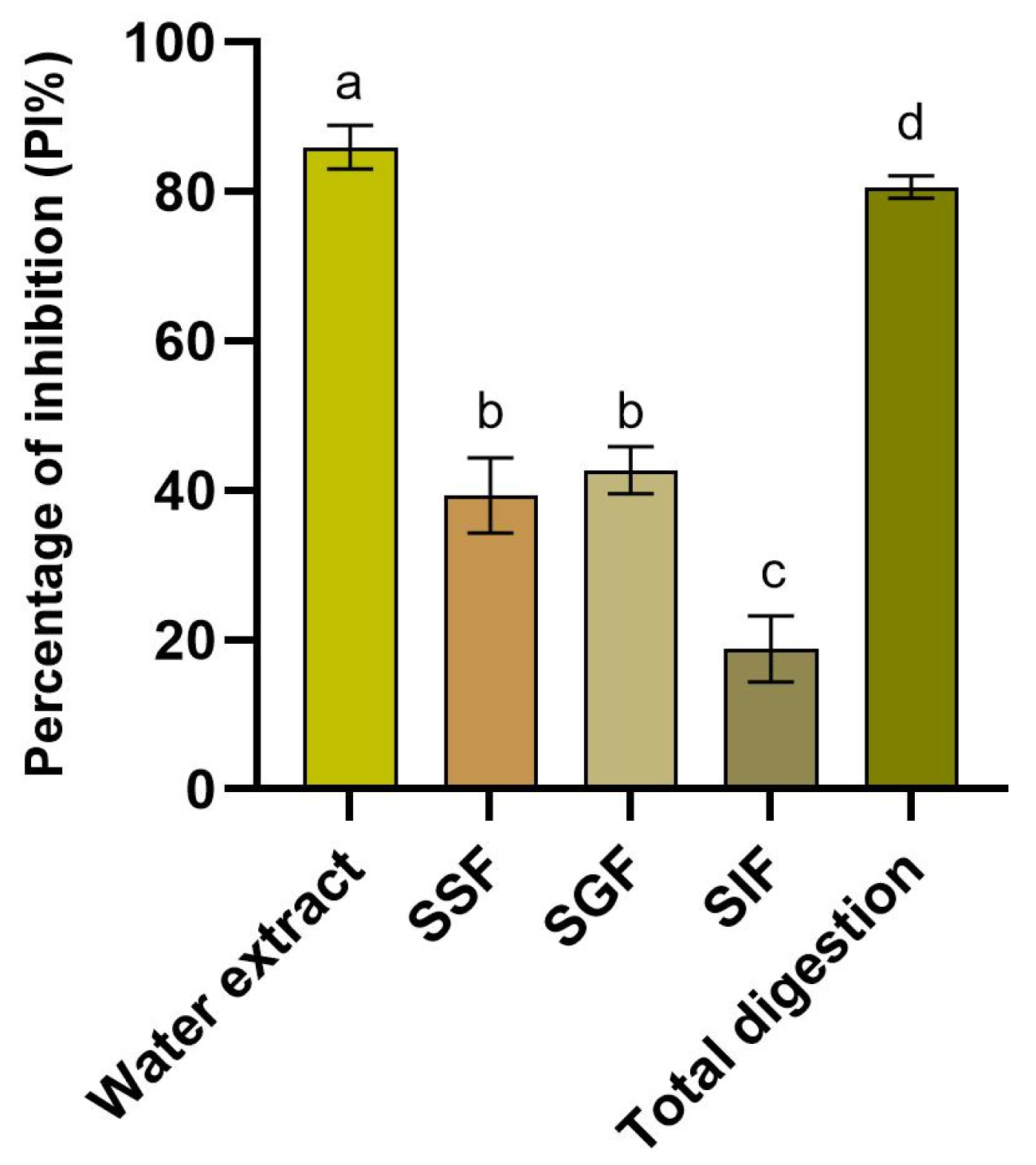
Evaluation of the antioxidant capacity carried out using an ABTS assay disclosed that the water extract of F. vesiculosus had a higher inhibition potential (p = 0.0275) than that shown by the digested product, showing inhibition rates of free radicals equal to 85.96 ± 2.95% and 80.66 ± 1.50%, respectively (Figure 2).

3.4. Evaluation of the Growth Inhibitory Activity of Escherichia coli F18+

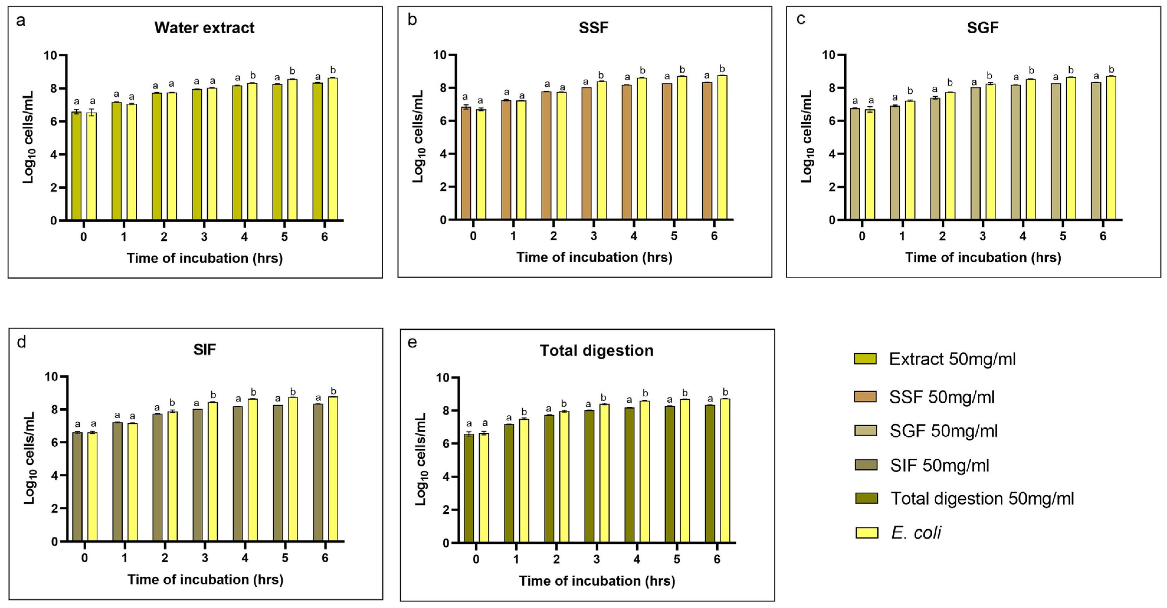
Evaluation of the growth inhibition of Escherichia coli F18+ by Fucus vesiculosus revealed that the water extract of the alga had the capacity to inhibit bacterial growth starting from the fourth hour of incubation, with a percentage of 14%. In contrast, the digested product demonstrated an inhibition ability of 34% from the first hour of incubation to the end of the incubation period for the assay. Specifically, by analyzing the different phases of digestion, it was possible to observe that the growth inhibitory capacity is more evident during the intermediate phase (SGF), where starting from the first hour of incubation the Fucus vesiculosus digested product was able to reduce the growth of Escherichia coli passing from 7.22 ± 0.036 log10 cell/mL of the positive control to 6.91 ± 0.049 log10 cell/mL of the sample with F. vesiculosus (Figure 3).
Furthermore, to assess the antimicrobial potential of the Fucus vesiculosus extract and its digested fractions, the minimum inhibitory concentration (MIC) was determined, as reported in Table 3.

3.5. Cytotoxicity on Mice Peritoneal Macrophages (PMs)

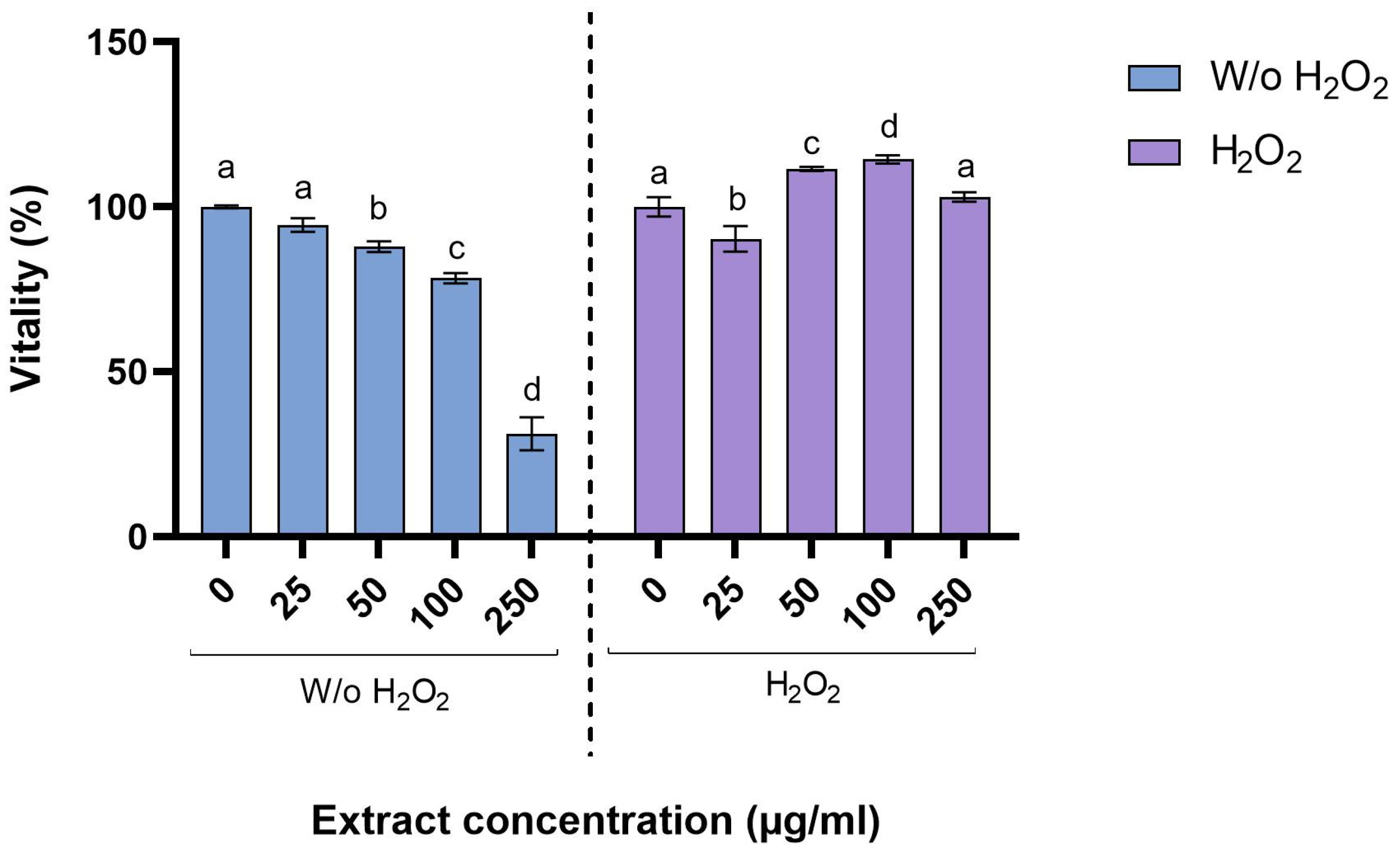
The treatment with H2O2 at a concentration of 0.125 mM was able to reduce the vitality of the peritoneal macrophages by 20.46%, demonstrating that the addition of H2O2 solution could be a valuable stressor in an in vitro assay. The inclusion of algal extracts in the PM culture disclosed that the presence of F. vesiculosus in healthy cell cultures reduced the vitality of the PMs in a manner directly proportional to the concentration used in the culture, achieving a decrease in viability of 68.84% when the concentration used was 250 µg/mL. On the other hand, when macrophages are subjected to stressors, such as H2O2 treatment, the introduction of algal extracts preserves their viability. In particular, the presence of Fucus vesiculosus extract at concentrations of 50 and 100 µg/mL is able to significantly improve the viability of PMs (p = 0.0031 and p = 0.0006, respectively). A higher concentration (250 µg/mL) did not disclose any significant improvement in the vitality, while a low concentration such as 25 µg/mL was not sufficient to protect the macrophages from the effect of H2O2, showing a significant reduction in viability of 9.67% (p = 0.01) (Figure 4).

3.6. Evaluation of Anti-Inflammatory Properties

Different concentrations of Fucus vesiculosus extract were evaluated for their effect on the expression of certain pro-inflammatory cytokines (IL-1β, IL-6, and TNF-α) and enzymes (iNOS) (Figure 5). The relative expression values revealed that two main interleukins (IL-1β and IL-6), which are involved in the inflammatory process, were significantly downregulated following treatment with F. vesiculosus extract. Specifically, it was observed that for both IL-1β and IL-6, the greatest downregulation was achieved when the extract was added at a concentration of 50 µg/mL, yielding fold-change values of 0.040 ± 0.014 and 0.285 ± 0.021, respectively, for IL-1β and IL-6. Instead, by increasing the concentration of the extract, the downregulatory effect decreases proportionally, reaching, at a 250 µg/mL extract concentration, a fold-change value of 0.130 ± 0.001 for IL-1, while for IL-6 at the highest concentration tested, there was a slight upregulation that led to a fold-changes value equal to 1.075 ± 0.021 (p = 0.0321). On the contrary, the expression of TNF-α showed a downregulation once the macrophages were treated with the lowest concentration of extract (25 µg/mL), with a fold-change equal to 0.280 ± 0.028, while it was then significantly (p < 0.001) up-regulated once treated with the other concentration, reaching a fold-change value of 3.845 ± 0.021 at a concentration of 250 µg/mL. In addition to the cytokines mentioned above, the gene expression of a pro-inflammatory enzyme (iNOS) was also evaluated. The results showed that regardless of the concentration of the algal extract used, it underwent an upregulation with an up-regulatory peak at a concentration of 100 µg/mL where the fold-change value was equal to 2.800 ± 0.014.

4. Discussion

4.1. Bromatological Composition

Fucus vesiculosus, commonly known as bladderwrack, is a brown seaweed that boasts a rich nutritional profile, making it a valuable addition to diets. This alga, as reported in the literature, consists mostly of water, which is crucial for maintaining its structural integrity and facilitating biochemical reactions. The dry matter of this seaweed is particularly noted for its high content of dietary fibers, proteins, vitamins, and essential minerals. Our study disclosed a high content of dietary fiber, which aligned with the data available in the literature that reported a percentage of dietary fiber ranging from 45% to 59% in raw forms [36]. This high fiber content is particularly beneficial, as it aids digestion, improves lipid profiles, and promotes gut health, maintaining a healthy gut microbiome. Additionally, the protein content could be considered good from a nutritional point of view. In fact, the protein content was around 13% of the dry matter. This makes it a valuable source of essential amino acids that are vital for various metabolic processes, including glutamic acid, aspartic acid, and alanine [37]. Furthermore, the presence of microelements adds to the nutritional value of Fucus vesiculosus, making it a valuable dietary supplement [38]. In fact, the richness in minerals such as potassium, calcium, magnesium, manganese, and iron contributes to the seaweed’s nutritional value since these minerals are crucial for various physiological functions [37,39]. On the contrary, the lipid content is relatively low, highlighting values around 3.0% of dry weight, which, as reported in the literature, were dominated by polyunsaturated fatty acids (PUFAs) [40]. Together, these results highlight how F. vesiculosus appears nutritionally, competitive when compared to other brown seaweeds such as S. latissima and U. rigida, which have also shown promising bioactive potential [40].

4.2. Evaluation of Polyphenols, Flavonoids, and Phlorotannins During the Simulated In Vitro Digestion Process

Additionally, in conjunction with the main nutritional sources, Fucus vesiculosus, as reported in our results, is known for its richness in polyphenols, such as flavonoids and phlorotannins, bioactive molecules with antioxidant, antimicrobial, and anti-inflammatory properties [36,40]. Since the bioactive compounds found in Fucus vesiculosus play a crucial role in its health-promoting properties and potential therapeutic applications, it becomes essential that these bioactive molecules remain intact even following the digestive process. Our results showed that after the simulated in vitro digestion process, the total content of polyphenols, including those of flavonoids and phlorotannins, was maintained. These results are in line with our previous study [41], where the retention of the polyphenol class present in the hemp seed extract following the digestive process was observed, a result supported by several studies showing the stability of polyphenol molecules after simulating the digestive process [42,43,44]. Similar findings were reported for brown seaweeds, where polyphenol compounds remained stable through simulated digestion, further supporting the resilience of brown algal bioactives under gastrointestinal conditions [45]. Analyzing the release of bioactive molecules through the digestive process more deeply, we found that regardless of the type of molecule, the major release occurred after the gastric phase. These results are reflected in the literature. In fact, the pH of the gastric tract is around 2; this, in addition to the action promoted by the enzymes, can help the release of polyphenols. Polyphenols are a diverse group of secondary plant metabolites characterized by the presence of one or more hydroxyl groups (-OH) directly attached to aromatic rings. These phenolic hydroxyl groups confer to the molecules’ acid–base properties, allowing them to act as weak acids by donating protons (H+) in alkaline conditions. In aqueous solution, the degree of ionization of polyphenols depends on the pH of the surrounding environment: at neutral or basic pH, the hydroxyl groups are more likely to dissociate, resulting in the formation of phenolate anions. This dissociated form is generally less stable, more reactive, and prone to oxidation, especially in the presence of dissolved oxygen or trace amounts of transition metal ions (e.g., Fe2+, Cu2+), which can catalyze the formation of quinones and other degradation products through redox cycling mechanisms [46,47]. Conversely, under acidic conditions—such as those present in the gastric environment (pH ~ 2)—the high concentration of H+ ions suppresses the ionization of the hydroxyl groups, favoring the formation of protonated species. These are typically more water-soluble and chemically stable, which facilitates both their desorption from the food matrix and their resistance to oxidative degradation during digestion [48]. Furthermore, the acidic environment and pepsin activity can promote the hydrolysis of glycosidic or ester bonds that bind polyphenols to complex food structures (e.g., polysaccharides, proteins), enhancing their release and bioaccessibility [49]. In contrast, the reduced recovery of polyphenols after the oral and intestinal phases may reflect their chemical instability at near-neutral pH. At pH 7, polyphenolic compounds tend to be more chemically reactive, undergoing autoxidation, polymerization, or structural rearrangements, which not only reduce their quantifiable concentration but may also alter their biological activity [50]. Moreover, in the intestinal phase, the presence of bile salts and pancreatin may influence the solubility and partitioning behavior of phenolics, potentially leading to their precipitation, degradation, or transformation into less detectable forms [51]. These underline the pH-dependent nature of polyphenol stability and release and help explain the enhanced liberation observed during the gastric phase as compared to the oral and intestinal phases.
Although the results disclosed a precise trend for the three molecules analyzed, precisely determining the behavior of the polyphenolic compounds through the different phases of the digestive process is complex, closely related to the methodology employed, and could give us just an idea of the real release of these compounds [52]. In real physiological conditions, additional variables, such as gut microbiota metabolism, mucosal absorption, and host enzymatic diversity, can significantly affect the fate of these molecules. For instance, microbial fermentation in the colon can lead to further biotransformation of polyphenols into smaller, bioactive metabolites that may exert systemic effects. These aspects are not fully captured in static in vitro models, underscoring the need for complementary in vivo studies to confirm the actual bioavailability and functionality of these compounds after ingestion [53].

4.3. Evaluation of the Antioxidant and Growth Inhibition Capacity During the Simulated In Vitro Digestion Process

The results obtained in this study showed the significant antioxidant properties of Fucus vesiculosus, primarily attributed to its high content of polyphenolic compounds, particularly phlorotannins [54,55]. These compounds, as reported in the literature, exhibit strong radical scavenging activity, which is crucial for neutralizing reactive oxygen species (ROS) that contribute to oxidative stress [56]. Although the antioxidant capacity of F. vesiculosus is well established and has been demonstrated by several researchers [51], the maintenance of this property, even after the digestive process, remains poorly understood to date. The results obtained by simulating the digestive process in vitro demonstrated the maintenance of this characteristic, highlighting the close relationship between radical scavenging activities and the release of polyphenolic components during the three main stages of digestion. Although there are few studies in this perspective, a comparable trend was observed with Sargassum fusiforme, where the in vitro digestion process modulated antioxidant activity while retaining functional efficacy [57]. The permanence of antioxidant capacity, following the digestive process, is also made possible by the presence of sulfated polysaccharides in F. vesiculosus, such as fucoidan, that, together with the permanence of phlorotannin, contribute to neutralizing ROS, enhancing the antioxidant bioactivity [54,58,59]. In addition, it was reported that polyphenolic compounds undergo a chemical transformation in response to the alkaline pH of the intestinal milieu. The shift from an acidic to an alkaline environment has been shown to correlate with an increase in phenolic and flavonoid compounds, which in turn are associated with enhanced antioxidant activity [41]. This phenomenon is attributed to the deprotonation of hydroxyl groups within aromatic rings [60,61]. Furthermore, the interaction of polyphenolic compounds with other substances released during digestion, such as minerals or dietary fiber, can influence their solubility and bioavailability [62]. These results are also supported by the findings obtained in various biological systems, including activated macrophages and ex vivo assays in plasma and erythrocytes, where the extracts from Fucus vesiculosus have shown promising results in maintaining antioxidant activity [55]. The findings, therefore, suggest the synergistic effects of polyphenols and sulfated polysaccharides, underscoring the potential of this seaweed as a natural source of antioxidants, which could be beneficial for animal and human health [63]. In addition to its antioxidant activity, as reported in our results, the aqueous extract of Fucus vesiculosus exhibits a notable growth inhibition capacity against E. coli. The antibacterial properties of F. vesiculosus are widely reported in the literature and are primarily attributed to various bioactive compounds present in the alga, including fucoidans, polyhydroxylated fucophlorethols, phlorotannins, and polyphenolic compounds [64,65,66]. If the growth inhibition capacity of F. vesiculosus against E. coli is known, an interesting yet unexplored area of study concerns the evaluation of this activity following the digestive process. The present study highlights that the growth inhibition capacity is also maintained after the digestion process, and in particular, the product derived from the gastric phase was the one most able to inhibit the growth of E. coli. Although knowledge of the effect of digestion on the functional properties of algae is currently limited, it is well established that the effectiveness of bioactive compounds can be altered during the digestive process. Research indicates that the bioavailability and efficacy of these compounds can be influenced by their structural integrity post-digestion [67,68]. The results achieved by our study can be explained by the conditions of digestion, such as pH and enzymatic activity, which, as shown above in our experiment, lead to the increased release of bioactive compounds, including polyphenols, flavonoids, and phlorotannins, compounds closely related to antimicrobial capacity. Although these results are still preliminary, and kinetics studies should be conducted to assess the exact behavior during digestion, the data obtained can be considered a good starting point for considering Fucus vesiculosus as a functional ingredient with antimicrobial power.

4.4. Evaluation of the Effect of Fucus vesiculosus Extract on Peritoneal Macrophages Vitality

The effect of F. vesiculosus extract on cell viability is a topic of interest due to the potential therapeutic applications and safety concerns associated with its use. Our results on mouse peritoneal macrophages disclosed that exposure to different concentrations of algal extract reduces viability as the dose tested increases. The cytotoxic effects of these extracts may vary depending on the specific compounds present and their concentrations, particularly the fucoidan molecule, which is closely related to this effect. Research indicates that low-molecular-weight (LMW) fucans derived from Fucus vesiculosus can inhibit the growth of certain cell lines. For instance, studies have shown that these fucans can inhibit the proliferation of Chinese hamster fibroblasts by up to 80% at a concentration of 1000 μg/mL while exhibiting less than 50% inhibition on human colon adenocarcinoma cells at the same concentration [69]. Additionally, other studies have reported dose-dependent cytotoxic effects of fucoidan, particularly in the 100–1000 µg/mL concentration range, on macrophage cell lines, such as RAW 264.7 cells [70,71]. This evidence suggests that the cytotoxic effect is context-dependent and can vary based on factors such as concentration, highlighting, however, that Fucus vesiculosus extract may be beneficial in targeting specific cell types. In the context of peritoneal macrophages, the extract’s influence on cell viability and function is crucial, and it is essential to consider the balance between its cytotoxic and protective effects under stressed conditions. In fact, our study also highlighted that when macrophages are pre-treated with H2O2, leading to oxidative stress, concentrations of 50 and 100 µg/mL of F. vesiculosus extract protect macrophages from cellular damage, increasing their vitality. As reported before, Fucus vesiculosus exhibits notable antioxidant properties, which can mitigate the damaging effects of oxidative stress. The extracts from this seaweed have demonstrated the ability to enhance the antioxidant status in various biological systems, including activated macrophages [72]. Specifically, studies have shown that Fucus vesiculosus extracts can scavenge free radicals and reduce oxidative damage in cellular models, suggesting a protective role against H2O2-induced injury [73,74]. The mechanism by which Fucus vesiculosus exerts its protective effects may involve modulating inflammatory responses and enhancing cellular antioxidant defenses. Peritoneal macrophages, which are crucial in the immune response, can release H2O2 as part of their antimicrobial activity [75]. However, excessive H2O2 can lead to cellular dysfunction and apoptosis. The bioactive compounds found in Fucus vesiculosus are believed to play a significant role in counteracting this oxidative stress by neutralizing reactive oxygen species and supporting the activity of endogenous antioxidant enzymes [76,77,78]. Furthermore, the extracts have been shown to enhance the activity of key antioxidant enzymes, such as superoxide dismutase and catalase, which are vital for maintaining cellular integrity in the face of oxidative challenges [38]. Thus, our results, supported by those found in the literature, highlight that the protective role of Fucus vesiculosus extract on peritoneal macrophages stressed with H2O2 is supported by its antioxidant properties, which help mitigate oxidative damage and modulate inflammatory responses, suggesting that Fucus vesiculosus could be a valuable dietary supplement for enhancing cellular resilience against oxidative stress.

4.5. Evaluation of the Anti-Inflammatory Properties of Fucus vesiculosus Extract

Among the various bioactive components, Fucus vesiculosus is especially known for its fucoidan content, a molecule recognized for its anti-inflammatory power [79]. The present study, which focused on the overall extract of Fucus vesiculosus, highlights the anti-inflammatory properties of this brown seaweed. Specifically, it can downregulate pro-inflammatory mediators such as TNF-α, IL-1β, and IL-6. These cytokines play pivotal roles in orchestrating the inflammatory response. They are not only crucial in the initiation and propagation of inflammation but also in the resolution phase. Their roles are diverse, ranging from modulating immune cell activity to influencing systemic responses, such as fever and the production of acute-phase proteins [80,81,82]. As our study revealed the anti-inflammatory capacity of Fucus vesiculosus, likely due to its fucoidan content, various studies have highlighted the anti-inflammatory properties of fucoidan from different algae in murine RAW 264.7 macrophage cell lines. For example, Jeong et al. [83] reported that fucoidan from Fucus vesiculosus (50 and 100 µg/mL) diminishes the secretion of TNF-α and IL-1β in RAW 264.7 macrophages, highlighting its potential to suppress the early stages of the inflammation. In line with this and with the present study, other researchers have disclosed a down-regulation in the levels of TNF-α, IL-1β, and IL-6 in comparison to nontreated LPS-stimulated RAW 264.7 cells once macrophages are treated with fucoidan derived from different algae species, such as Chnoospora minima, Ecklonia cava, Sargassum horneri, and Laminaria japonica [79,84,85]. It is important to emphasize that the response to fucoidan is often concentration-dependent and influenced by the structural characteristics of the polysaccharide, such as sulfation degree, molecular weight, and algal source. These factors can modulate receptor binding and cellular uptake mechanisms in macrophages, thereby altering the downstream signaling pathways involved in cytokine expression [57].
If the downregulation of interleukins IL-1β and IL-6 occurs for all concentrations tested, the downregulatory effect of TNF-α was observed only in the lowest concentration tested. This outcome might initially appear counterintuitive, as higher concentrations are typically expected to exert stronger effects. However, TNF-α is a cytokine primarily involved in the early stages of inflammation, initiating a cascade that leads to the expression of other cytokines such as IL-1β and IL-6 [86,87]. Thus, at lower concentrations, fucoidan may effectively inhibit this initial response. Moreover, increased concentration could paradoxically activate stress response pathways or lead to receptor desensitization, thereby attenuating the inhibitory effect.
Additionally, this study considered the expression of pro-inflammatory cytokines and the pro-inflammatory enzyme nitric oxide synthase 2 (iNOS). The results showed that, regardless of the concentration of the algal extract used, it undergoes upregulation. This seems to be in contradiction with other studies found in the literature. Fernando et al. found dose-dependent inhibition of NO production (IC50 27.82 µg/mL), iNOS, and COX-2 expression in LPS-induced RAW 264.7 macrophages treated with fucoidan fractions derived from Chnoospora minima [88]. Similarly, Ni and colleagues reported decreased NO production, iNOS, and COX-2 expression in LPS-stimulated RAW 264.7 macrophage cells following fucoidan application [79]. The discrepancy with our findings could be attributed to differences in experimental setup, such as the use of whole algal extracts versus purified fucoidan fractions, and the specific conditions of the in vitro assay, including duration of exposure and LPS stimulation intensity. Furthermore, iNOS is a late-stage inflammatory marker, and its expression is known to be highly inducible and persistent under oxidative stress conditions. Therefore, the concentrations of Fucus vesiculosus extract employed in our study may not have been sufficient to counteract the strong pro-inflammatory stimulus, or the extract matrix may contain other compounds that interfered with the inhibitory activity of fucoidan [89,90].

5. Conclusions

The present study investigated the functional properties of Fucus vesiculosus, focusing on the evaluation of bioactive molecules and their associated bioactivity. Specifically, our analysis enabled the determination of molecules, such as polyphenols, flavonoids, and phlorotannins, that are strictly related to functional properties such as antioxidant, microbial growth inhibition, and anti-inflammatory effects. Additionally, our results highlighted that the functional properties of Fucus vesiculosus are maintained throughout the different phases of the digestion process, further reinforcing the beneficial effects of incorporating this ingredient into the diet. Nonetheless, some limitations should be acknowledged. Despite adaptations to better simulate porcine physiology, the in vitro digestion model cannot fully reproduce the complexity of in vivo systems, particularly in terms of absorption, microbiota interactions, and systemic effects. Moreover, bioactivity assessments were conducted exclusively through in vitro assays, which may not directly reflect in vivo efficacy. Lastly, the findings are specific to a single algal species and target species, limiting their generalizability. Further in vivo studies are needed to validate these results, define optimal inclusion levels, and assess long-term effects on animal health and performance. Despite these limitations, given the need to develop an increasingly sustainable livestock sector, the present work provides a solid foundation for future research on the use of marine-derived functional ingredients in animal nutrition.

Author Contributions

Conceptualization, S.F. and L.R.; methodology, S.F., N.R. and S.R.P.; software, S.F. and A.P.F.; validation, S.F. and L.R.; formal analysis, S.F.; investigation, S.F., N.R. and A.P.F.; resources, L.R.; data curation, S.F. and S.R.P.; writing—original draft preparation, S.F.; writing—review and editing, S.F., N.R., E.F. and L.R.; visualization, S.F. and E.F.; supervision, E.F., S.R.P. and L.R.; project administration, L.R. All authors have read and agreed to the published version of the manuscript.

Funding

This research received no external funding.

Institutional Review Board Statement

Not applicable.

Informed Consent Statement

Not applicable.

Data Availability Statement

The original contributions presented in this study are included in the article. Further inquiries can be directed to the corresponding author.

Acknowledgments

The authors thank Sevecom S.p.a. for providing the sample of Fucus vesiculosus.

Conflicts of Interest

A.P.F. was employed by Biotecnologie B.T. Srl. No commercial manufacturing products of Biotecnologie B.T. Srl. were used in this study. The remaining authors declare that the research was conducted in the absence of any commercial or financial relationships that could be construed as conflicting.

References

  1. Jata, N.; Swain, R.; Raula, A.; Priyadarshini, N.P. Alternative feed ingredients for sustainable livestock production. In Futuristic Trends in Agriculture Engineering & Food Sciences Volume 3 Book 24; Iterative International Publishers, Selfypage Developers Pvt Ltd.: Karnataka, India, 2024; pp. 194–204. [Google Scholar]
  2. Popoola, Y.A.; Idowu, A.B.; Ajijola, S.; Popoola, K.A. Economic Importance of Kenaf Seed as Alternative Feed Resource in Micro Livestock Production: A Review. J. Agric. Ecol. Res. Int. 2024, 25, 45–51. [Google Scholar] [CrossRef]
  3. Akintan, O.; Gebremedhin, K.G.; Uyeh, D.D. Animal Feed Formulation—Connecting Technologies to Build a Resilient and Sustainable System. Animals 2024, 14, 1497. [Google Scholar] [CrossRef] [PubMed]
  4. Pandey, A.K.; Kumar, P.; Saxena, M.J. Feed Additives in Animal Health. In Nutraceuticals in Veterinary Medicine; Springer International Publishing: Cham, Switzerland, 2019; pp. 345–362. [Google Scholar]
  5. Alimi, N.; Assani, A.S.; Sanni Worogo, H.; Baco, N.M.; Traoré, I.A. Livestock Feed Resources Used as Alternatives during Feed Shortages and Their Impact on the Environment and Ruminant Performance in West Africa: A Systematic Review. Front. Vet. Sci. 2024, 11, 1352235. [Google Scholar] [CrossRef] [PubMed]
  6. Boudalia, S.; Smeti, S.; Dawit, M.; Senbeta, E.K.; Gueroui, Y.; Dotas, V.; Bousbia, A.; Symeon, G.K. Alternative Approaches to Feeding Small Ruminants and Their Potential Benefits. Animals 2024, 14, 904. [Google Scholar] [CrossRef] [PubMed]
  7. Fleurence, J. Feeding of Livestock. In Algae in Agrobiology; Wiley: Hoboken, NJ, USA, 2023; pp. 69–133. [Google Scholar]
  8. González-Meza, G.M.; Elizondo-Luevano, J.H.; Cuellar-Bermudez, S.P.; Sosa-Hernández, J.E.; Iqbal, H.M.N.; Melchor-Martínez, E.M.; Parra-Saldívar, R. New Perspective for Macroalgae-Based Animal Feeding in the Context of Challenging Sustainable Food Production. Plants 2023, 12, 3609. [Google Scholar] [CrossRef]
  9. Michalak, I.; Tiwari, R.; Dhawan, M.; Alagawany, M.; Farag, M.R.; Sharun, K.; Emran, T.B.; Dhama, K. Antioxidant Effects of Seaweeds and Their Active Compounds on Animal Health and Production—A Review. Vet. Q. 2022, 42, 48–67. [Google Scholar] [CrossRef]
  10. De Bhowmick, G.; Hayes, M. Potential of Seaweeds to Mitigate Production of Greenhouse Gases during Production of Ruminant Proteins. Glob. Chall. 2023, 7, 2200145. [Google Scholar] [CrossRef]
  11. Takolander, A. Seasonal Ecophysiology of Fucus vesiculosus (Phaeophyceae) in the Northern Baltic Sea. Eur. J. Phycol. 2023, 58, 300–314. [Google Scholar] [CrossRef]
  12. Circuncisão, A.R.; Ferreira, S.S.; Silva, A.M.S.; Coimbra, M.A.; Cardoso, S.M. Fucus vesiculosus-Rich Extracts as Potential Functional Food Ingredients: A Holistic Extraction Approach. Foods 2024, 13, 540. [Google Scholar] [CrossRef]
  13. André, R.; Pacheco, R.; Alves, A.C.; Santos, H.M.; Bourbon, M.; Serralheiro, M.L. The Hypocholesterolemic Potential of the Edible Algae Fucus vesiculosus: Proteomic and Quantitative PCR Analysis. Foods 2023, 12, 2758. [Google Scholar] [CrossRef]
  14. Yu, J.; Geng, Y.; Xia, H.; Ma, D.; Liu, C.; Wu, R.; Wu, J.; You, S.; Bi, Y. LAB Fermentation Improves Production of Bioactive Compounds and Antioxidant Activity of Withania somnifera Extract and Its Metabolic Signatures as Revealed by LC-MS/MS. J. Microbiol. Biotechnol. 2022, 32, 473–483. [Google Scholar] [CrossRef] [PubMed]
  15. Augustyniak, A.; Przyborska, J.; McMahon, H. Differential Effects of Fucus vesiculosus Fucoidan on Fibroblast and Macrophage Cell Lines Inflammatory Activation. Results Chem. 2024, 7, 101443. [Google Scholar] [CrossRef]
  16. Wang, L.; Oliveira, C.; Li, Q.; Ferreira, A.S.; Nunes, C.; Coimbra, M.A.; Reis, R.L.; Martins, A.; Wang, C.; Silva, T.H.; et al. Fucoidan from Fucus vesiculosus Inhibits Inflammatory Response, Both In Vitro and In Vivo. Mar. Drugs 2023, 21, 302. [Google Scholar] [CrossRef] [PubMed]
  17. Zheng, Q.; Zheng, Y.; Jia, R.-B.; Luo, D.; Chen, C.; Zhao, M. Fucus vesiculosus Polysaccharide Alleviates Type 2 Diabetes in Rats via Remodeling Gut Microbiota and Regulating Glycolipid Metabolism-Related Gene Expression. Int. J. Biol. Macromol. 2023, 248, 126504. [Google Scholar] [CrossRef]
  18. Obluchinskaya, E.D.; Pozharitskaya, O.N.; Zakharov, D.V.; Flisyuk, E.V.; Terninko, I.I.; Generalova, Y.E.; Smekhova, I.E.; Shikov, A.N. The Biochemical Composition and Antioxidant Properties of Fucus vesiculosus from the Arctic Region. Mar. Drugs 2022, 20, 193. [Google Scholar] [CrossRef]
  19. Association of Official Analytical Chemists. Official Methods of Analysis, 22nd ed.; Association of Official Analytical Chemists: Washington, DC, USA, 2023. [Google Scholar]
  20. Frazzini, S.; Reggi, S.; Dell’Anno, M.; Fifi, A.P.; Scaglia, E.; Ferri, I.; Rossi, L. Chemical-Functional Characterization of Ascophyllum nodosum and Phymatolithon calcareum and Dietary Supplementation in Post-Weaning Pigs. Front. Vet. Sci. 2024, 11, 1431091. [Google Scholar] [CrossRef]
  21. Tierney, M.S.; Smyth, T.J.; Rai, D.K.; Soler-Vila, A.; Croft, A.K.; Brunton, N. Enrichment of Polyphenol Contents and Antioxidant Activities of Irish Brown Macroalgae Using Food-Friendly Techniques Based on Polarity and Molecular Size. Food Chem. 2013, 139, 753–761. [Google Scholar] [CrossRef]
  22. Brodkorb, A.; Egger, L.; Alminger, M.; Alvito, P.; Assunção, R.; Ballance, S.; Bohn, T.; Bourlieu-Lacanal, C.; Boutrou, R.; Carrière, F.; et al. INFOGEST Static in Vitro Simulation of Gastrointestinal Food Digestion. Nat. Protoc. 2019, 14, 991–1014. [Google Scholar] [CrossRef]
  23. Attard, E. A Rapid Microtitre Plate Folin-Ciocalteu Method for the Assessment of Polyphenols. Open Life Sci. 2013, 8, 48–53. [Google Scholar] [CrossRef]
  24. Herald, T.J.; Gadgil, P.; Tilley, M. High-throughput Micro Plate Assays for Screening Flavonoid Content and DPPH-scavenging Activity in Sorghum Bran and Flour. J. Sci. Food Agric. 2012, 92, 2326–2331. [Google Scholar] [CrossRef]
  25. Lopes, G.; Sousa, C.; Silva, L.R.; Pinto, E.; Andrade, P.B.; Bernardo, J.; Mouga, T.; Valentão, P. Can Phlorotannins Purified Extracts Constitute a Novel Pharmacological Alternative for Microbial Infections with Associated Inflammatory Conditions? PLoS ONE 2012, 7, e31145. [Google Scholar] [CrossRef]
  26. Frazzini, S.; Zuorro, A.; Panseri, S.; Pavlovic, R.; Sgoifo Rossi, C.A.; Rossi, L. Repurposing Hazelnut Waste Products for a Sustainable Economy: A Metabolomic Analysis of Cuticles and Shells to Highlight Their Antioxidant Potential and Inhibitory Activity against Verocytotoxic Escherichia Coli. Sustainability 2023, 15, 3268. [Google Scholar] [CrossRef]
  27. Frazzini, S.; Torresani, M.C.; Hejna, M.; Di Dio, M.; Rossi, L. Ascophillum nodosum and Lithothamnium calcareum and Their Prebiotic Potential on Lactobacillus Strains. J. Funct. Foods 2024, 118, 106257. [Google Scholar] [CrossRef]
  28. Rossi, L.; Canala, B.; Fifi, A.P.; Frazzini, S. In Vitro Evaluation of Functional Properties of Extracts of Fucus vesiculosus Obtained with Different Conventional Solvents. Algal Res. 2024, 84, 103787. [Google Scholar] [CrossRef]
  29. Pepe, G.; Braga, D.; Renzi, T.A.; Villa, A.; Bolego, C.; D’Avila, F.; Barlassina, C.; Maggi, A.; Locati, M.; Vegeto, E. Self-Renewal and Phenotypic Conversion Are the Main Physiological Responses of Macrophages to the Endogenous Estrogen Surge. Sci. Rep. 2017, 7, 44270. [Google Scholar] [CrossRef]
  30. Bao, Z.; Huang, Y.; Chen, J.; Wang, Z.; Qian, J.; Xu, J.; Zhao, Y. Validation of Reference Genes for Gene Expression Normalization in RAW264.7 Cells under Different Conditions. Biomed. Res. Int. 2019, 2019, 6131879. [Google Scholar] [CrossRef]
  31. Khatua, S.; Simal-Gandara, J.; Acharya, K. Understanding Immune-Modulatory Efficacy in Vitro. Chem. Biol. Interact. 2022, 352, 109776. [Google Scholar] [CrossRef]
  32. Ulett, G.C.; Ketheesan, N.; Hirst, R.G. Cytokine Gene Expression in Innately Susceptible BALB/c Mice and Relatively Resistant C57BL/6 Mice during Infection with Virulent Burkholderia pseudomallei. Infect. Immun. 2000, 68, 2034–2042. [Google Scholar] [CrossRef]
  33. da Silva, J.B.; Carvalho, E.; Covarrubias, A.E.; Ching, A.T.C.; Mattaraia, V.G.M.; Paiva, D.; de Franco, M.; Fávaro, R.D.; Pereira, M.M.; Vasconcellos, S.; et al. Induction of TNF-Alfa and CXCL-2 MRNAs in Different Organs of Mice Infected with Pathogenic Leptospira. Microb. Pathog. 2012, 52, 206–216. [Google Scholar] [CrossRef]
  34. Mandal, M.; Rakib, A.; Kiran, S.; Al Mamun, M.A.; Raghavan, S.; Kumar, S.; Singla, B.; Park, F.; Leo, M.D.; Singh, U.P. Inhibition of MicroRNA-34c Reduces Detrusor ROCK2 Expression and Urinary Bladder Inflammation in Experimental Cystitis. Life Sci. 2024, 336, 122317. [Google Scholar] [CrossRef]
  35. Ruiz-Villalba, A.; Mattiotti, A.; Gunst, Q.D.; Cano-Ballesteros, S.; van den Hoff, M.J.B.; Ruijter, J.M. Reference Genes for Gene Expression Studies in the Mouse Heart. Sci. Rep. 2017, 7, 24. [Google Scholar] [CrossRef]
  36. Díaz-Rubio, M.E.; Pérez-Jiménez, J.; Saura-Calixto, F. Dietary Fiber and Antioxidant Capacity in Fucus vesiculosus Products. Int. J. Food Sci. Nutr. 2009, 60, 23–34. [Google Scholar] [CrossRef] [PubMed]
  37. Lorenzo, J.; Agregán, R.; Munekata, P.; Franco, D.; Carballo, J.; Şahin, S.; Lacomba, R.; Barba, F. Proximate Composition and Nutritional Value of Three Macroalgae: Ascophyllum nodosum, Fucus vesiculosus and Bifurcaria bifurcata. Mar. Drugs 2017, 15, 360. [Google Scholar] [CrossRef] [PubMed]
  38. Drygaś, B.; Piechowiak, T.; Kreczko, J.; Matłok, N.; Saletnik, B.; Balawejder, M. The Utilisation of Fucus vesiculosus Algae Extracts in the Production of Microgreens Hordeum vulgare L. with an Increased Content of Selected Bioactive Compounds. Plants 2024, 13, 2871. [Google Scholar] [CrossRef] [PubMed]
  39. Nova, P.; Pimenta-Martins, A.; Maricato, É.; Nunes, C.; Abreu, H.; Coimbra, M.A.; Freitas, A.C.; Gomes, A.M. Chemical Composition and Antioxidant Potential of Five Algae Cultivated in Fully Controlled Closed Systems. Molecules 2023, 28, 4588. [Google Scholar] [CrossRef]
  40. Neto, R.T.; Marçal, C.; Queirós, A.S.; Abreu, H.; Silva, A.M.S.; Cardoso, S.M. Screening of Ulva rigida, Gracilaria Sp., Fucus vesiculosus and Saccharina latissima as Functional Ingredients. Int. J. Mol. Sci. 2018, 19, 2987. [Google Scholar] [CrossRef]
  41. Frazzini, S.; Torresani, M.C.; Roda, G.; Dell’Anno, M.; Ruffo, G.; Rossi, L. Chemical and Functional Characterization of the Main Bioactive Molecules Contained in Hulled Cannabis sativa L. Seeds for Use as Functional Ingredients. J. Agric. Food Res. 2024, 16, 101084. [Google Scholar] [CrossRef]
  42. Cavia, M.M.; Arlanzón, N.; Busto, N.; Carrillo, C.; Alonso-Torre, S.R. The Impact of In Vitro Digestion on the Polyphenol Content and Antioxidant Activity of Spanish Ciders. Foods 2023, 12, 1861. [Google Scholar] [CrossRef]
  43. Odriozola-Serrano, I.; Nogueira, D.P.; Esparza, I.; Vaz, A.A.; Jiménez-Moreno, N.; Martín-Belloso, O.; Ancín-Azpilicueta, C. Stability and Bioaccessibility of Phenolic Compounds in Rosehip Extracts during In Vitro Digestion. Antioxidants 2023, 12, 1035. [Google Scholar] [CrossRef]
  44. Ma, Y.-L.; Wang, Y.; Wu, Z.-F.; Mei, J.; Zhang, W.-Q.; Shang, Y.-F.; Thakur, K.; Wei, Z.-J. Exploring the Effect of in Vitro Digestion on the Phenolics and Antioxidant Activity of Lycium barbarum Fruit Extract. Food Biosci. 2023, 51, 102255. [Google Scholar] [CrossRef]
  45. Corona, G.; Coman, M.M.; Guo, Y.; Hotchkiss, S.; Gill, C.; Yaqoob, P.; Spencer, J.P.E.; Rowland, I. Effect of Simulated Gastrointestinal Digestion and Fermentation on Polyphenolic Content and Bioactivity of Brown Seaweed Phlorotannin-rich Extracts. Mol. Nutr. Food Res. 2017, 61, 1700223. [Google Scholar] [CrossRef]
  46. Tomás-Barberán, F.A.; Andrés-Lacueva, C. Polyphenols and Health: Current State and Progress. J. Agric. Food Chem. 2012, 60, 8773–8775. [Google Scholar] [CrossRef]
  47. Verzelloni, E.; Tagliazucchi, D.; Conte, A. Relationship between the Antioxidant Properties and the Phenolic and Flavonoid Content in Traditional Balsamic Vinegar. Food Chem. 2007, 105, 564–571. [Google Scholar] [CrossRef]
  48. Tagliazucchi, D.; Verzelloni, E.; Bertolini, D.; Conte, A. In Vitro Bio-Accessibility and Antioxidant Activity of Grape Polyphenols. Food Chem. 2010, 120, 599–606. [Google Scholar] [CrossRef]
  49. McDougall, G.J.; Dobson, P.; Smith, P.; Blake, A.; Stewart, D. Assessing Potential Bioavailability of Raspberry Anthocyanins Using an in Vitro Digestion System. J. Agric. Food Chem. 2005, 53, 5896–5904. [Google Scholar] [CrossRef] [PubMed]
  50. de Ferrars, R.M.; Czank, C.; Zhang, Q.; Botting, N.P.; Kroon, P.A.; Cassidy, A.; Kay, C.D. The Pharmacokinetics of Anthocyanins and Their Metabolites in Humans. Br. J. Pharmacol. 2014, 171, 3268–3282. [Google Scholar] [CrossRef]
  51. Bohn, T.; McDougall, G.J.; Alegría, A.; Alminger, M.; Arrigoni, E.; Aura, A.; Brito, C.; Cilla, A.; El, S.N.; Karakaya, S.; et al. Mind the Gap—Deficits in Our Knowledge of Aspects Impacting the Bioavailability of Phytochemicals and Their Metabolites—A Position Paper Focusing on Carotenoids and Polyphenols. Mol. Nutr. Food Res. 2015, 59, 1307–1323. [Google Scholar] [CrossRef]
  52. Wojtunik-Kulesza, K.; Oniszczuk, A.; Oniszczuk, T.; Combrzyński, M.; Nowakowska, D.; Matwijczuk, A. Influence of In Vitro Digestion on Composition, Bioaccessibility and Antioxidant Activity of Food Polyphenols—A Non-Systematic Review. Nutrients 2020, 12, 1401. [Google Scholar] [CrossRef]
  53. Mateo Anson, N.; van den Berg, R.; Havenaar, R.; Bast, A.; Haenen, G.R.M.M. Bioavailability of Ferulic Acid Is Determined by Its Bioaccessibility. J. Cereal Sci. 2009, 49, 296–300. [Google Scholar] [CrossRef]
  54. Rupérez, P.; Ahrazem, O.; Leal, J.A. Potential Antioxidant Capacity of Sulfated Polysaccharides from the Edible Marine Brown Seaweed Fucus vesiculosus. J. Agric. Food Chem. 2002, 50, 840–845. [Google Scholar] [CrossRef]
  55. Zaragozá, M.C.; López, D.; Sáiz, M.P.; Poquet, M.; Pérez, J.; Puig-Parellada, P.; Màrmol, F.; Simonetti, P.; Gardana, C.; Lerat, Y.; et al. Toxicity and Antioxidant Activity in Vitro and in Vivo of Two Fucus vesiculosus Extracts. J. Agric. Food Chem. 2008, 56, 7773–7780. [Google Scholar] [CrossRef]
  56. Dzah, C.S.; Zhang, H.; Gobe, V.; Asante-Donyinah, D.; Duan, Y. Anti- and pro-Oxidant Properties of Polyphenols and Their Role in Modulating Glutathione Synthesis, Activity and Cellular Redox Potential: Potential Synergies for Disease Management. Adv. Redox Res. 2024, 11, 100099. [Google Scholar] [CrossRef]
  57. Wang, Y.; Xing, M.; Cao, Q.; Ji, A.; Liang, H.; Song, S. Biological Activities of Fucoidan and the Factors Mediating Its Therapeutic Effects: A Review of Recent Studies. Mar. Drugs 2019, 17, 183. [Google Scholar] [CrossRef]
  58. Costa, L.S.; Fidelis, G.P.; Telles, C.B.S.; Dantas-Santos, N.; Camara, R.B.G.; Cordeiro, S.L.; Costa, M.S.S.P.; Almeida-Lima, J.; Melo-Silveira, R.F.; Oliveira, R.M.; et al. Antioxidant and Antiproliferative Activities of Heterofucans from the Seaweed Sargassum filipendula. Mar. Drugs 2011, 9, 952–966. [Google Scholar] [CrossRef] [PubMed]
  59. Veena, C.K.; Josephine, A.; Preetha, S.P.; Varalakshmi, P. Beneficial Role of Sulfated Polysaccharides from Edible Seaweed Fucus vesiculosus in Experimental Hyperoxaluria. Food Chem. 2007, 100, 1552–1559. [Google Scholar] [CrossRef]
  60. Dutra, R.L.T.; Dantas, A.M.; Marques, D.d.A.; Batista, J.D.F.; Meireles, B.R.L.d.A.; de Magalhães Cordeiro, Â.M.T.; Magnani, M.; Borges, G.d.S.C. Bioaccessibility and Antioxidant Activity of Phenolic Compounds in Frozen Pulps of Brazilian Exotic Fruits Exposed to Simulated Gastrointestinal Conditions. Food Res. Int. 2017, 100, 650–657. [Google Scholar] [CrossRef]
  61. Dantas, A.M.; Mafaldo, I.M.; Oliveira, P.M.d.L.; Lima, M.d.S.; Magnani, M.; Borges, G.d.S.C. Bioaccessibility of Phenolic Compounds in Native and Exotic Frozen Pulps Explored in Brazil Using a Digestion Model Coupled with a Simulated Intestinal Barrier. Food Chem. 2019, 274, 202–214. [Google Scholar] [CrossRef]
  62. Sęczyk, Ł.; Gawlik-Dziki, U.; Świeca, M. Influence of Phenolic-Food Matrix Interactions on In Vitro Bioaccessibility of Selected Phenolic Compounds and Nutrients Digestibility in Fortified White Bean Paste. Antioxidants 2021, 10, 1825. [Google Scholar] [CrossRef]
  63. Wang, K.; Hu, S. The Synergistic Effects of Polyphenols and Intestinal Microbiota on Osteoporosis. Front. Immunol. 2023, 14, 1285621. [Google Scholar] [CrossRef]
  64. Sandsdalen, E.; Haug, T.; Stensvåg, K.; Styrvold, O.B. The Antibacterial Effect of a Polyhydroxylated Fucophlorethol from the Marine Brown Alga, Fucus vesiculosus. World J. Microbiol. Biotechnol. 2003, 19, 777–782. [Google Scholar] [CrossRef]
  65. Buedenbender, L.; Astone, F.A.; Tasdemir, D. Bioactive Molecular Networking for Mapping the Antimicrobial Constituents of the Baltic Brown Alga Fucus vesiculosus. Mar. Drugs 2020, 18, 311. [Google Scholar] [CrossRef]
  66. Ayrapetyan, O.N.; Obluchinskaya, E.D.; Zhurishkina, E.V.; Skorik, Y.A.; Lebedev, D.V.; Kulminskaya, A.A.; Lapina, I.M. Antibacterial Properties of Fucoidans from the Brown Algae Fucus vesiculosus L. of the Barents Sea. Biology 2021, 10, 67. [Google Scholar] [CrossRef] [PubMed]
  67. Capuano, E.; Pellegrini, N. An Integrated Look at the Effect of Structure on Nutrient Bioavailability in Plant Foods. J. Sci. Food Agric. 2019, 99, 493–498. [Google Scholar] [CrossRef] [PubMed]
  68. Rein, M.J.; Renouf, M.; Cruz-Hernandez, C.; Actis-Goretta, L.; Thakkar, S.K.; da Silva Pinto, M. Bioavailability of Bioactive Food Compounds: A Challenging Journey to Bioefficacy. Br. J. Clin. Pharmacol. 2013, 75, 588–602. [Google Scholar] [CrossRef] [PubMed]
  69. Ellouali, M.; Boisson-Vidal, C.; Jozefonvicz, J. Antiproliferative Effect and Interaction of Fucans with Cells. Colloids Surf. B Biointerfaces 1994, 2, 305–314. [Google Scholar] [CrossRef]
  70. Yoo, Y.-C.; Kim, W.-J.; Kim, S.-Y.; Kim, S.-M.; Chung, M.-K.; Park, J.-W.; Suh, H.-H.; Lee, K.-B.; Park, Y.-I. Immunomodulating Activity of a Fucoidan Isolated from Korean Undaria Pinnatifida Sporophyll. Algae 2007, 22, 333–338. [Google Scholar] [CrossRef]
  71. Nishiguchi, T.; Jiang, Z.; Ueno, M.; Takeshita, S.; Cho, K.; Roh, S.W.; Kang, K.-H.; Yamaguchi, K.; Kim, D.; Oda, T. Reevaluation of Bactericidal, Cytotoxic, and Macrophage-Stimulating Activities of Commercially Available Fucus vesiculosus Fucoidan. Algae 2014, 29, 237–247. [Google Scholar] [CrossRef]
  72. Catarino, M.D.; Silva, A.; Cruz, M.T.; Mateus, N.; Silva, A.M.S.; Cardoso, S.M. Phlorotannins from Fucus vesiculosus: Modulation of Inflammatory Response by Blocking NF-ΚB Signaling Pathway. Int. J. Mol. Sci. 2020, 21, 6897. [Google Scholar] [CrossRef]
  73. Pinteus, S.; Silva, J.; Alves, C.; Horta, A.; Thomas, O.; Pedrosa, R. Antioxidant and Cytoprotective Activities of Fucus spiralis Seaweed on a Human Cell in Vitro Model. Int. J. Mol. Sci. 2017, 18, 292. [Google Scholar] [CrossRef]
  74. O’Sullivan, A.M.; O’Callaghan, Y.C.; O’Grady, M.N.; Queguineur, B.; Hanniffy, D.; Troy, D.J.; Kerry, J.P.; O’Brien, N.M. Assessment of the Ability of Seaweed Extracts to Protect against Hydrogen Peroxide and Tert-Butyl Hydroperoxide Induced Cellular Damage in Caco-2 Cells. Food Chem. 2012, 134, 1137–1140. [Google Scholar] [CrossRef]
  75. Herb, M.; Schramm, M. Functions of ROS in Macrophages and Antimicrobial Immunity. Antioxidants 2021, 10, 313. [Google Scholar] [CrossRef]
  76. Vo, T.-S.; Kim, S.-K. Fucoidans as a Natural Bioactive Ingredient for Functional Foods. J. Funct. Foods 2013, 5, 16–27. [Google Scholar] [CrossRef]
  77. Golshany, H.; Siddiquy, M.; Elbarbary, A.; Seddiek, A.S.; Kamal, A.; Yu, Q.; Fan, L. Exploring Fucus vesiculosus Phlorotannins: Insights into Chemistry, Extraction, Purification, Identification and Bioactivity. Food Biosci. 2024, 61, 104769. [Google Scholar] [CrossRef]
  78. Lomartire, S.; Gonçalves, A.M.M. An Overview of Potential Seaweed-Derived Bioactive Compounds for Pharmaceutical Applications. Mar. Drugs 2022, 20, 141. [Google Scholar] [CrossRef] [PubMed]
  79. Sanjeewa, K.K.A.; Fernando, I.P.S.; Kim, E.-A.; Ahn, G.; Jee, Y.; Jeon, Y.-J. Anti-Inflammatory Activity of a Sulfated Polysaccharide Isolated from an Enzymatic Digest of Brown Seaweed Sargassum horneri in RAW 264.7 Cells. Nutr. Res. Pract. 2017, 11, 3. [Google Scholar] [CrossRef]
  80. Hirano, T. IL-6 in Inflammation, Autoimmunity and Cancer. Int. Immunol. 2021, 33, 127–148. [Google Scholar] [CrossRef]
  81. Tomaszek, Ł. The Biological Role of IL-1, IL-6 and CRP and Their Application in the Diagnosis of the Inflammatory Process. Diagn. Lab. 2022, 58, 66–73. [Google Scholar] [CrossRef]
  82. Latifynia, A.; Khamesipour, A.; Khansari, N. Proinflammatory Cytokines (TNF-a, IL-1B, IL-6) and Antioxidant in Different Stages of Patients with Cutaneous Leishmaniasis. World J. Adv. Res. Rev. 2023, 17, 825–835. [Google Scholar] [CrossRef]
  83. Jeong, J.-W.; Hwang, S.J.; Han, M.H.; Lee, D.-S.; Yoo, J.S.; Choi, I.-W.; Cha, H.-J.; Kim, S.; Kim, H.-S.; Kim, G.-Y.; et al. Fucoidan Inhibits Lipopolysaccharide-Induced Inflammatory Responses in RAW 264.7 Macrophages and Zebrafish Larvae. Mol. Cell Toxicol. 2017, 13, 405–417. [Google Scholar] [CrossRef]
  84. Lee, S.-H.; Ko, C.-I.; Ahn, G.; You, S.; Kim, J.-S.; Heu, M.S.; Kim, J.; Jee, Y.; Jeon, Y.-J. Molecular Characteristics and Anti-Inflammatory Activity of the Fucoidan Extracted from Ecklonia cava. Carbohydr. Polym. 2012, 89, 599–606. [Google Scholar] [CrossRef]
  85. Ni, L.; Wang, L.; Fu, X.; Duan, D.; Jeon, Y.-J.; Xu, J.; Gao, X. In Vitro and in Vivo Anti-Inflammatory Activities of a Fucose-Rich Fucoidan Isolated from Saccharina japonica. Int. J. Biol. Macromol. 2020, 156, 717–729. [Google Scholar] [CrossRef]
  86. Zhao, S.; Jiang, J.; Jing, Y.; Liu, W.; Yang, X.; Hou, X.; Gao, L.; Wei, L. The Concentration of Tumor Necrosis Factor-α Determines Its Protective or Damaging Effect on Liver Injury by Regulating Yap Activity. Cell Death Dis. 2020, 11, 70. [Google Scholar] [CrossRef]
  87. Bhol, N.K.; Bhanjadeo, M.M.; Singh, A.K.; Dash, U.C.; Ojha, R.R.; Majhi, S.; Duttaroy, A.K.; Jena, A.B. The Interplay between Cytokines, Inflammation, and Antioxidants: Mechanistic Insights and Therapeutic Potentials of Various Antioxidants and Anti-Cytokine Compounds. Biomed. Pharmacother. 2024, 178, 117177. [Google Scholar] [CrossRef]
  88. Fernando, I.P.S.; Sanjeewa, K.K.A.; Samarakoon, K.W.; Lee, W.W.; Kim, H.-S.; Kang, N.; Ranasinghe, P.; Lee, H.-S.; Jeon, Y.-J. A Fucoidan Fraction Purified from Chnoospora minima; a Potential Inhibitor of LPS-Induced Inflammatory Responses. Int. J. Biol. Macromol. 2017, 104, 1185–1193. [Google Scholar] [CrossRef]
  89. Jung, D.H.; Kim, K.-H.; Byeon, H.E.; Park, H.J.; Park, B.; Rhee, D.-K.; Um, S.H.; Pyo, S. Involvement of ATF3 in the Negative Regulation of INOS Expression and NO Production in Activated Macrophages. Immunol. Res. 2015, 62, 35–45. [Google Scholar] [CrossRef]
  90. Prestes-Carneiro, L.; Shio, M.; Fernandes, P.; Jancar, S. Cross-Regulation of INOS and COX-2 by Its Products in Murine Macrophages Under Stress Conditions. Cell. Physiol. Biochem. 2007, 20, 283–292. [Google Scholar] [CrossRef] [PubMed]
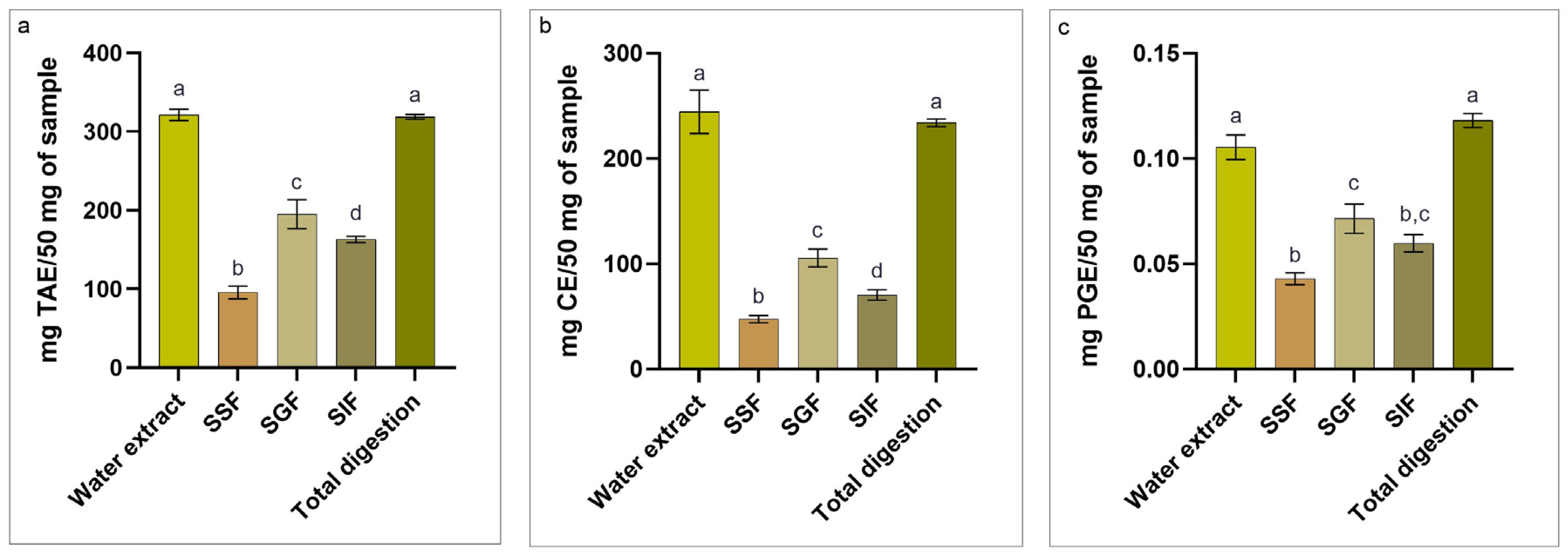
Figure 1. Major bioactive compounds in Fucus vesiculosus extract, with total phenolic content (TPC, panel (a)); total flavonoid content (TFC, panel (b)); total phlorotannin content (TPhC, panel (c)). TAE: tannic acid equivalent; CE: catechin equivalent; PGE: phloroglucinol equivalent. The data are shown as the means ± standard deviations (SDs). a–d Means with different superscripts are significantly different (p < 0.05).
Applsci 15 07911 g001
Figure 2. Percentage inhibition of radical scavenging activity (PI%) of Fucus vesiculosus before, during, and after in vitro digestion. The data are shown as the means ± standard deviations (SDs). a–d Means with different superscripts are significantly different (p < 0.05).
Applsci 15 07911 g002
Figure 3. Growth inhibition against Escherichia coli F18+. (a) Growth inhibition of Fucus vesiculosus water extract; (b) growth inhibition of Fucus vesiculosus after oral phase; (c) growth inhibition of Fucus vesiculosus after gastric phase; (d) growth inhibition of Fucus vesiculosus after intestinal phase; (e) growth inhibition of Fucus vesiculosus after in vitro digestion. The data are shown as the means ± standard deviations (SDs). a,b Means with different superscripts are significantly different (p < 0.05).
Applsci 15 07911 g003
Figure 4. Percentage of vitality of murine peritoneal macrophages after treatment with different concentration of Fucus vesiculosus extract. The data are shown as the means ± standard deviations (SDs). a–d Means with different superscripts are significantly different (p < 0.05).
Applsci 15 07911 g004
Figure 5. Representation of the expression of cytokines (IL-1β; IL-6; TNF-α) and enzymes (iNOS) involved in the inflammatory process.
Applsci 15 07911 g005
Table 1. Oligonucleotide sequences used for the qRT-PCR assay.
Gene 1Nucleotide SequenceAccession NumberReference
IL-1βF: TCATGGGATGATGATGATAACCTGCTNM_008361[31]
R: CCCATACTTTAGGAAGACACGGATT
IL-6F: CGTGGAAATGAGAAAAGAGTTGTGCNM_001314054[32]
R: ATGCTTAGGCATAACGCACTAGGT
TNF-αF: CACAAGATGCTGGGACAGTGANM_013693[33]
R: TCCTTGATGGTGGTGCATGA
iNOSF: GAGACAGGGAAGTCTGAAGCACNM_010927[34]
R: CCAGCAGTAGTTGCTCCTCTTC
GAPDHF: CTCCCACTCTTCCACCTTCGAC166162.6[35]
R: GCCTCTCTTGCTCAGTGTCC
1 IL-1β: interleukin-1 β; IL-6: interleukin-6; TNF-α: tumor necrosis factor-α; iNOS: Nitric Oxide Synthase 2; GAPDH: glyceraldehyde-3-phosphate dehydrogenase.
Table 2. Microelement composition of Fucus vesiculosus.
Atomic Mass (u)Concentration (mg/kg)
Be9.0121831n.d.
B10.81163.27 ± 4.15
Al26.9815384380.38 ± 73.36
Ti46.951765.47 ± 1.17
Ti47.94723.42 ± 3.29
V50.99612.93 ± 0.07
Cr51.99614.87 ± 0.41
Mn54.938045265.28 ± 20.13
Fe55.845761.82 ± 12.88
Co58.9331951.28 ± 0.05
Ni58.69345.51 ± 0.38
Cu63.5463.05 ± 0.02
Zn65.3815.11 ± 0.52
As74.9159539.83 ± 0.26
Se78.9710.52 ± 0.08
Sr87.62823.84 ± 24.33
Mo95.950.93 ± 0.07
Ag107.87n.d.
Cd112.4410.84 ± 0.02
Sb121.76n.d.
Ba137.32710.24 ± 0.23
Tl204.3833n.d.
Pb205.974465n.d.
Pb206.9758969n.d.
U238.031.36 ± 0.01
n.d.: not detectable.
Table 3. Minimal inhibitory concentration (MIC) of Fucus vesiculosus extract and its digested fraction on E. coli F18+.
Extract Concentration (mg/mL)Inhibition Rate (%)
Extract11.586.15
SSF13.583.98
SGF1087.94
SIF1285.63
Total digestion9.588.56
Disclaimer/Publisher’s Note: The statements, opinions and data contained in all publications are solely those of the individual author(s) and contributor(s) and not of MDPI and/or the editor(s). MDPI and/or the editor(s) disclaim responsibility for any injury to people or property resulting from any ideas, methods, instructions or products referred to in the content.