Article

Cellular Entry, Cytotoxicity, and Antifungal Activity of Newly Synthesized Dendrimers

1
Institute of Biophysics and Biomedical Engineering, Bulgarian Academy of Sciences, Acad. G. Bonchev Str., Bl. 21, 1113 Sofia, Bulgaria
2
Faculty of Biology, Sofia University ‘St. Kliment Ohridski’, 8 Dragan Tzankov Blvd., 1164 Sofia, Bulgaria
3
Institute of Microbiology, Bulgarian Academy of Sciences, Akad. Georgi Bonchev Str., Bl. 26, 1113 Sofia, Bulgaria
4
Faculty of Medicine, Sofia University “St. Kliment Ohridski”, 1 Koziak Str., 1407 Sofia, Bulgaria
*
Author to whom correspondence should be addressed.
Appl. Sci. 2025, 15(14), 7764; https://doi.org/10.3390/app15147764
Submission received: 19 June 2025 / Revised: 5 July 2025 / Accepted: 9 July 2025 / Published: 10 July 2025
(This article belongs to the Section Nanotechnology and Applied Nanosciences)

Abstract

Dendrimers, 4-dimethylamino-1,8-naphthalimide (DAB) and its halogenated analog 3-bromo-4-dimethylamino-1,8-naphthalimide (DAB-Br), were evaluated on eukaryotic cells, human HFF-1 fibroblast cells, and five fungal species. Although both dendrimers have demonstrated antibacterial and antiviral potential, thus far, their effects on eukaryotic cells, particularly human and fungal cells, have not been investigated. For this purpose, their cytotoxicity, mechanisms of cellular entry, and antifungal activity were studied. Dynamic light scattering measurements revealed that both dendrimers exhibited positive surface charges (+28 to +35 mV), good colloidal stability, and nanoscale dimensions (117–234 nm), facilitating interactions with target cells. The MTT assay showed that DAB was more cytotoxic toward HFF-1 cells (IC50 = 27 µg/mL) compared to DAB-Br (IC50 = 68 µg/mL). In contrast, the resazurin-based antifungal assay demonstrated that DAB-Br had superior antifungal activity, achieving a lower minimum inhibitory concentration (0.148 µg/µL), compared to DAB (0.295 µg/µL). A trypan blue exclusion test revealed that both dendrimers entered cells through membrane permeabilization, either temporarily or permanently, depending on the concentration and exposure time. At concentrations above 30 µg/mL, irreversible permeabilization was observed within two hours of treatment, accompanied by a decrease in membrane lipid order, indicating altered membrane integrity and permeability. Conversely, at lower concentrations (7.5–15 µg/mL), dendrimers induced only temporary membrane permeabilization, with membranes remaining intact, suggesting a reversible interaction with the lipid bilayer. Conducting thorough and systematic research to fully explore their biological activities could provide valuable insight for future applications.

1. Introduction

Modern nanomaterials exhibit a broad spectrum of applications, ranging from biosensors to nutritional supplements [1,2]. Dendrimer-based compounds and their derivatives have gained increasing attention in the fields of medicine and pharmaceuticals, including targeted drug delivery, diagnostic imaging, gene therapy, and the development of antibacterial, antiviral, and anti-amyloid agents [3,4,5,6].
Dendrimers are polymeric molecules with highly branched structures. They are composed of layers of dendrons (concentrated branched entities) from a central initiating core, and each subsequent layer is referred to as a generation (G) [7]. The selection of a core directly determines the structure of the dendrimer, including the total number of branches of the dendron, the size, and the number of cavities within the dendrimer. Dendrimers possess nanoscale dimensions, ranging from 1 to 100 nm, and exhibit a low polydispersity index, which enhances their ability to penetrate cell membranes and facilitates efficient cellular uptake [8].
The size, topology, and viscosity of dendrimers can be precisely controlled during their synthesis, making them highly versatile nanostructures. Currently, approximately 100 distinct dendrimer families have been identified, each defined by its core structure, such as carbon, nitrogen, or phosphorus, as well as its branching units and terminal functional groups, which can carry a variety of charges [9]. This structural complexity enables dendrimers to exhibit an effect closely resembling the polyvalent interactions commonly observed in biological systems [10]. Such molecular diversity allows for extensive chemical modifications and broadens their potential applications, ranging from drug delivery and gene therapy to antimicrobial and antiviral therapies [11,12,13,14].
Many dendrimers are known to exhibit antimicrobial activity. The antimicrobial effect of dendrimers depends on their concentration and surface charge: cationic dendrimers tend to exhibit stronger effects compared to anionic and neutral ones due to the electrostatic interactions with the negatively charged microbial membranes, the progressive permeabilization of the bacterial membranes, and the subsequent disruption of membrane integrity [15,16]. The two newly synthesized dendrimers composed of 4-dimethylamino-1,8-naphthalimide (DAB) and 3-bromo-4-dimethylamino-1,8-naphthalimide (DAB-Br), used in our work, have demonstrated antibacterial and antiviral potential in a previous study [17]. In parallel, the growing resistance of pathogenic fungi continues to pose a serious public health challenge. Compared to antibacterial agents, the number of antifungal compounds under development or approved for clinical use remains disproportionately low. Fungal diseases currently affect nearly a quarter of the global population, with superficial infections of the skin, hair, nails, and mucous membranes representing the most prevalent mycoses. These infections, impacting approximately 20–25% of people worldwide, are predominantly caused by dermatophytes belonging to the genera Trichophyton, Microsporum, Alternaria, Aspergillus, and Candida spp., particularly C. albicans. Given these challenges, there is an urgent need to explore alternative therapeutic strategies and novel compounds with innovative mechanisms of action and new therapeutic applications. Dendrimers emerge as promising candidates in this context as they offer significant potential both as efficient carriers of antifungal agents and as active therapeutic agents [18].
Cell membrane integrity is a vital feature when evaluating the mode of entry and potential cytotoxicity of dendrimers, which can be specifically engineered to efficiently penetrate cellular membranes [19,20,21,22,23]. The lipid bilayer of the cell membrane is a dynamic, two-dimensional fluid, with its most essential properties being fluidity and molecular order. These characteristics are fundamental for maintaining membrane integrity, permeability, and membrane adaptation. Membrane fluidity and order are determined by the phase behavior of lipid molecules, particularly glycerophospholipids, sphingolipids, and cholesterol content. In living cells, two primary lipid phase states are observed: the liquid-disordered (Lα or Ld) phase, characterized by loosely packed lipid molecules and high fluidity; and the liquid-ordered (Lo) phase, enriched in cholesterol and sphingolipids, with tightly packed molecules and reduced fluidity [24,25]. The degree of alterations in the bilayer fluidity and order caused by nanomaterials is directly related to their mechanism of cellular entry and could serve as an indicator for potential cytotoxicity. Permanent alterations are often associated with membrane permeabilization and cytotoxicity, whereas temporary changes may facilitate efficient cellular uptake without compromising cell viability [19].
This study aims to evaluate the biological activities of two newly synthesized dendrimers, DAB and DAB-Br, both of which have demonstrated antibacterial and antiviral potential. In this work, we emphasize their unexplored effects on fibroblast cells and fungal species, specifically the mechanisms of cellular entry, potential cytotoxicity, and antifungal properties, to determine their potential biomedical applications.

2. Materials and Methods

2.1. Chemicals

Culture media, salt buffers, fluorescent markers, such as Laurdan, and MTT cytotoxicity assay reagents were purchased from Sigma-Aldrich, Saint Louis, MO, USA. The fetal serum and the antibiotic were sourced from BioWhittaker TM, Walkersville, MD, USA.

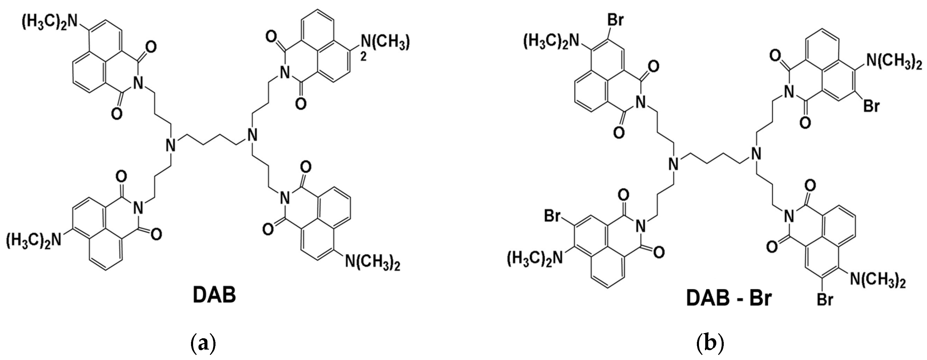
2.2. Synthesis of Dendrimers

Newly synthesized dendrimers containing 4-dimethylamino-1,8-naphthalimide (DAB) (Figure 1a) and 3-bromo-4-dimethyl-1,8-naphthalimide (DAB-Br) (Figure 1b) were obtained via peripheral modification of the first-generation (G1) polypropylene amine dendrimer, as described [17]. The initial zero-generation dendrimer, containing 4 primary amino groups, was reacted with 4-nitro-naphthalic anhydride in a methanol solution at 50 °C. The nucleophilic substitution of the nitro group with dimethylamino group was carried out in a N,N-dimethylformamide medium at 30 °C for 24 h. The dendrimer DAB was obtained after pouring the reaction mixture into ice, filtering the precipitate formed, and drying under a vacuum. The dendrimer DAB-Br was obtained by bromination of DAB in a dichloromethane medium at 25 °C for 60 min.

2.3. Cell Models

2.3.1. Cell Culturing of Fibroblasts from HSF Cell Line

The human fibroblast HFF-1 cell line (HFF-1; fibroblast; Human foreskin. Product Code ATCC-SCRC-1041, LGC Standards Sp. z.o.o., Kielpin Lomianki, Poland) was cultured under standard physiological conditions in a humidified atmosphere with 5% CO2 at 37 °C. The cells were cultured in Dulbecco’s Modified Eagle Medium (DMEM), supplemented with 10% fetal calf serum (FBS) and 1% (v/v) antibiotic–antimycotic solution containing penicillin (100 U/mL), streptomycin (100 µg/mL), and amphotericin B (0.25 µg/mL).

2.3.2. Fungal Cultures

The fungal strains Aspergillus fumigatus, Alternaria alternata, Aspergillus niger, Penicillium griseofulvum, and Candida albican belong to the Mycological Collection of the Institute of Microbiology, BAS. The fungal cultures were cultured in potato dextrose medium (HiMedia, Mumbai, India) for 96 h at 28 °C.

2.4. Dynamic Light Scattering

Dynamic light scattering (DLS) was used to determine the average size and apparent zeta potential values of the dendrimers. The measurements were conducted using a Zetasizer Nano ZS analyzer (Malvern Instruments, Malvern, UK) equipped with a U-type cell containing gold electrodes. The DLS profiles of DAB and DAB-Br were obtained at a concentration of 40 µg/mL at 25 °C, on the 3rd, 5th, and 7th day after preparing the dispersions. Each sample was measured three times per experiment.

2.5. Cell Cytotoxicity

2.5.1. MTT-Test

MTT (3-(4,5-dimethylthiazol-2-yl)-2,5-diphenyltetrazolium bromide) analysis was utilized to assess metabolic activity as an indicator of cell viability, proliferation, and cytotoxicity [26]. Dendrimer dispersions at concentrations of 3 µg/mL, 7.5 µg/mL, 15 µg/mL, 30 µg/mL, 45 µg/mL, 60 µg/mL, and 120 µg/mL were added to a 96-well plate, containing HFF-1 cells. The plate was then incubated in a CO2 incubator at 37 °C for 2, 24, and 48 h. Following the incubation periods, the medium containing the dendrimers was removed, and the MTT reagent was added to each well. The plate was further incubated in darkness at 37 °C for 4 h. Subsequently, the formazan was dissolved, and the optical density was measured at 570 nm using a spectrofluorometer (Epoch Microplate Reader Bio BioTek/USA, Shoreline, WA, USA). The experimental setup was performed in three repetitions.

2.5.2. Antifungal Test

The antifungal properties of both dendrimers were evaluated using the resazurin method on a 96-well plate [27]. Each row of the plate contained decreasing dilutions of either Dab or Dab-Br dendrimer: the first well was loaded with the highest tested concentration of 1.179 µg/µL, whereas the 6th well contained the lowest concentration of 0.0369 µg/µL. Two additional wells served as control variants: the 7th well was designed as a positive control, containing one fungal culture and nystatin as the antifungal agent; the 8th well contained only pure medium (potato dextrose liquid medium) and the corresponding fungal culture, serving as the negative control. The wells were inoculated with a 10 μL fungal spore suspension of each tested fungal strain at a concentration of 1 × 10⁸ cells/mL, and 30 µL of 0.02% resazurin solution was dripped into all wells. This experimental setup was performed for each fungal strain with both types of dendrimers, with three repetitions per experiment.

2.6. Evaluation of the Cell Morphology and Cell Membrane Permeabilization by TBE Test

The trypan blue exclusion (TBE) test was used for the evaluation of cell membrane integrity as described [28]. Cells were cultured in a well plate at a concentration of 1 × 10⁵ cells/well. Subsequently, the cells were treated with both dendrimers at concentrations of 3 µg/mL, 7.5 µg/mL, 15 µg/mL, 30 µg/mL, 60 µg/mL, and 120 µg/mL. The plate was then incubated in a thermostat at 37 °C for 2, 24, and 48 h. Untreated cells served as control variants. After incubation, the medium with the dendrimers was pipetted, and the cells were washed with PBS. Microscopic observation and photography were performed using a phase-contrast microscope (Nikon Eclipse TS 100, Tokyo, Japan). The images of the cells treated with DAB, DAB-Br, and the control variants were taken at the 2nd, 24th, and 48th h, with 10 images taken per well. Cell morphology and characteristics of the monolayer were assessed by direct observation of the wells by phase-contrast microscopy (Leica DM5500 B., Leica Microsystems GmbH, Wetzlar, Germany).

2.7. Laurdan Fluorescence Spectroscopy

HFF-1 cells were cultured in a 6-well plate at a density of 1 × 10⁵ cells/mL. The cells were treated with DAB and DAB-Br dispersions at concentrations of 3 and 30 µg/mL and further incubated for 24 h in DMEM. After that, 0.25% trypsin–EDTA was added to each well to facilitate the detachment of the cells from the culture dish. The samples were centrifuged for 5 min at 2500 rpm, the supernatant was removed, and PBS was added to the cells. This procedure was repeated two times. The fluorescent marker Laurdan (2 µL of a 350 µM stock solution) was added to the samples in a 96-well plate to achieve a final concentration of 3.5 µM. The emission spectrum was measured at physiological temperature (37 °C). The samples were excited at 355 nm, and emission spectra were recorded in the range from 390 to 600 nm in 0.5 nm steps (bandwidth of 5 nm for both excitation and emission monochromators) using a fluorescence plate reader (Tecan Infinite 200 Pro, Tecan, Grödig, Austria). The values of the intensities at 440 and 490 nm were used to calculate the general polarization (GP), which serves as a mathematical measure of the lipid order in the cell membranes by the following formula:
GP = (I440I490)/(I440 + I490)
where I440 and I490 are the intensities at 440 and 490 nm, respectively.
GP values range from −1 to +1, indicating highly fluid to highly ordered membranes, respectively [25].

2.8. Statistics

Experimental data were processed statistically using Excel and Origin programs. Statistics were obtained by one-way ANOVA, R 4.0.

3. Results

3.1. Size and Zeta (ζ) Potential of DAB and DAB-Br

The colloidal characteristics of both DAB and DAB-Br dispersions were assessed. According to the obtained DLS profiles, DAB-Br exhibited a larger average size of 222 nm and a higher ζ-potential of +35.2 mV compared to DAB, which had an average size of 118 nm and a ζ-potential of +28.7 mV (Table 1).
To evaluate dendrimer stability, the colloidal properties were monitored on the 3rd and 7th days after preparation. By day 7, the dispersions demonstrated very good colloidal stability over the one-week period. No statistically significant differences were observed in the properties of DAB between days 1, 3, and 7. A similar trend was observed for DAB-Br. Based on these results, all subsequent experiments were conducted using dendrimer solutions no older than one week.

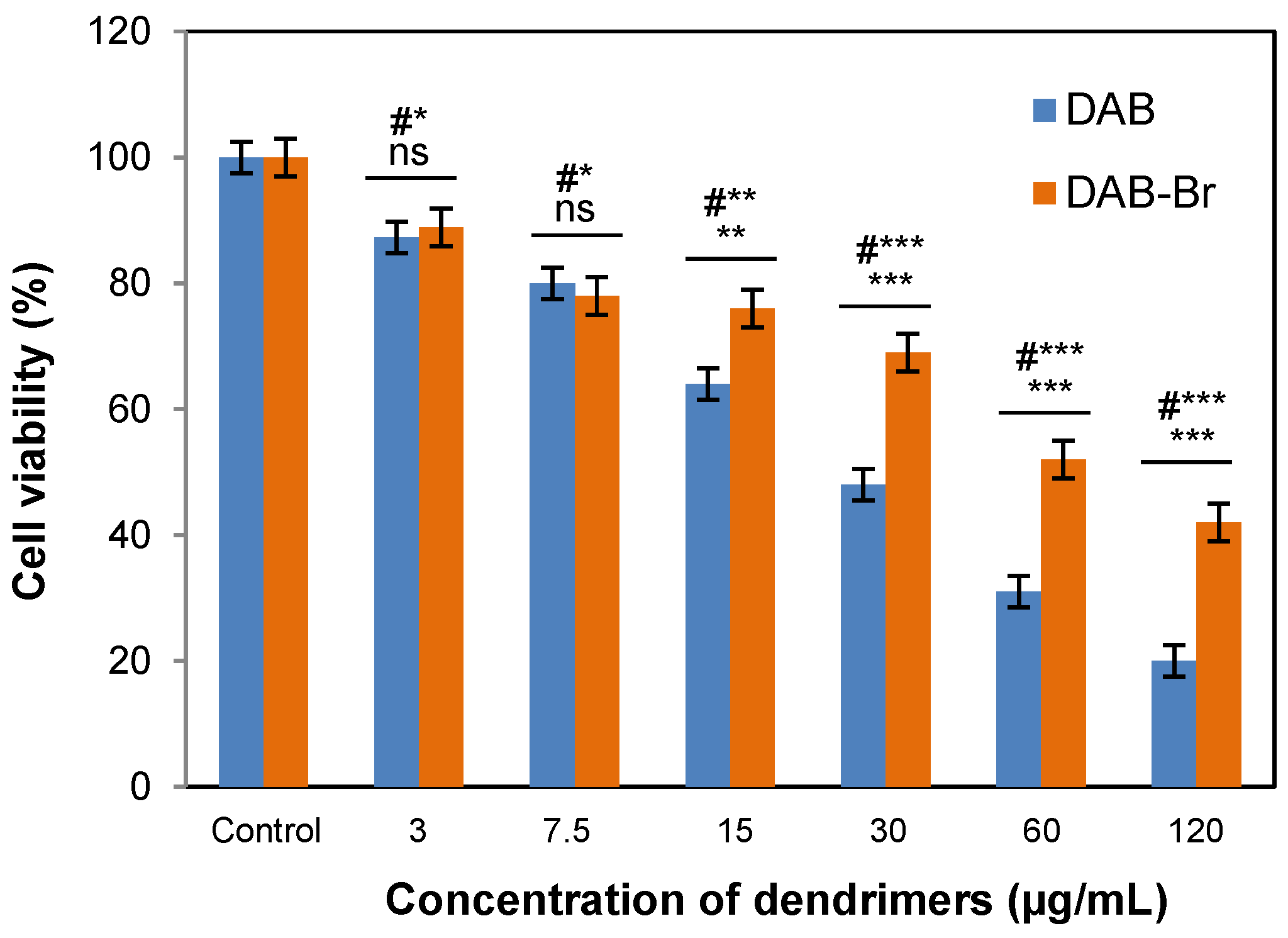
3.2. Cell Cytotoxicity

3.2.1. MTT Assay

A cytotoxicity assessment was carried out on the HFF-1 cell line using an MTT assay. The signs of cellular adaptation or cellular stress were checked at the 24th hour after treatment with both dendrimers. At concentrations of 3 μg/mL and 7.5 μg/mL, cell survival remained high (80–90%) with no significant differences between the effects of both dendrimers (Figure 2). After treatment with concentrations above 15 μg/mL with both dendrimers, the survival of HFF-1 significantly reduced in a dose-dependent manner, especially for DAB: the IC50 value was at 27 μg/mL, and for DAB-Br, the IC50 value was at 68 μg/mL.

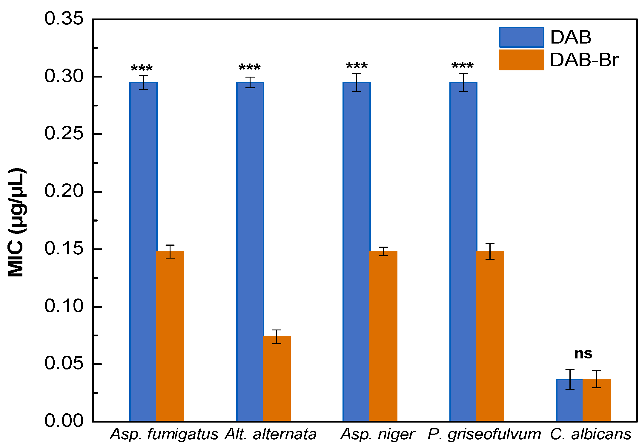
3.2.2. Antifungal Activity

Antifungal activity was evaluated against the fungal strains Aspergillus fumigatus, Alternaria alternata, Aspergillus niger, Penicillium griseofulvum, and Candida albicans using the resazurin method on a 96-well plate. Both dendrimers demonstrated potent antifungal activities against all five tested fungal strains. At the highest concentrations of 0.589 and 1.179 µg/µL, DAB exhibited a strong antifungal effect on all fungal strains, effectively suppressing fungal growth for up to 96 h from the start of micromycete cultivation (Table 2). The lower concentration of 0.295 µg/µL inhibited Alt. alternata growth for up to 72 h and the growth of the other four strains for up to 24 h post treatment. Among the fungal strains, C. albicans was the most sensitive to DAB dendrimers. Even the lowest concentration of 0.0369 µg/µL inhibits its growth for up to 24 h.
DAB-Br exhibited significantly stronger antifungal activity than DAB: DAB-Br had a lower minimum inhibitory concentration (0.148 µg/µL) compared to DAB (0.295 µg/µL) against all tested fungal strains (Table 3). At 0.148 µg/µL DAB-Br suppressed the growth of Asp. fumigatus, Asp. niger, and P. griseofulvum for 24 h; C. albicans for 48 h; and Alt. alternata for up to 72 h. Moreover, DAB-Br sustained a potent inhibitory effect for 96 h at just 0.295 µg/µL, a period at which DAB required 0.589 µg/µL to achieve comparable activity. At an even lower concentration of 0.074 µg/µL, DAB-Br suppressed A. alternata spore germination for up to 24 h. Notably, C. albicans was sensitive to the lowest tested concentration of DAB-Br (0.0369 µg/µL), with growth inhibition observed for up to 24 h.
Figure 3 presents the minimum inhibitory concentrations (MICs) of the two dendrimers against the five different fungal strains. The results indicate that DAB-Br exhibits stronger antifungal activity compared to DAB, as evidenced by its lower MIC values ranging from 0.074 to 0.147 µg/µL, whereas DAB shows a MIC of 0.295 µg/µL. Among the five tested fungal strains, Candida albicans demonstrated the highest sensitivity to both dendrimers, with equal MIC values of 0.0369 µg/µL.

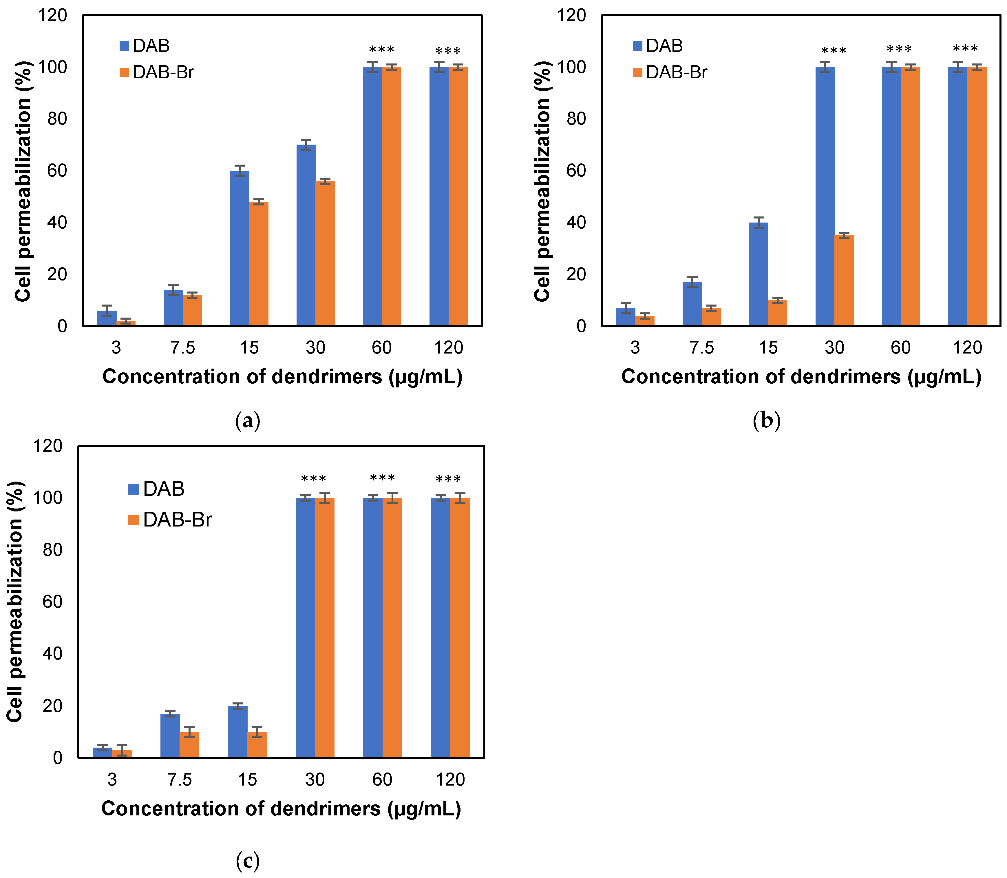
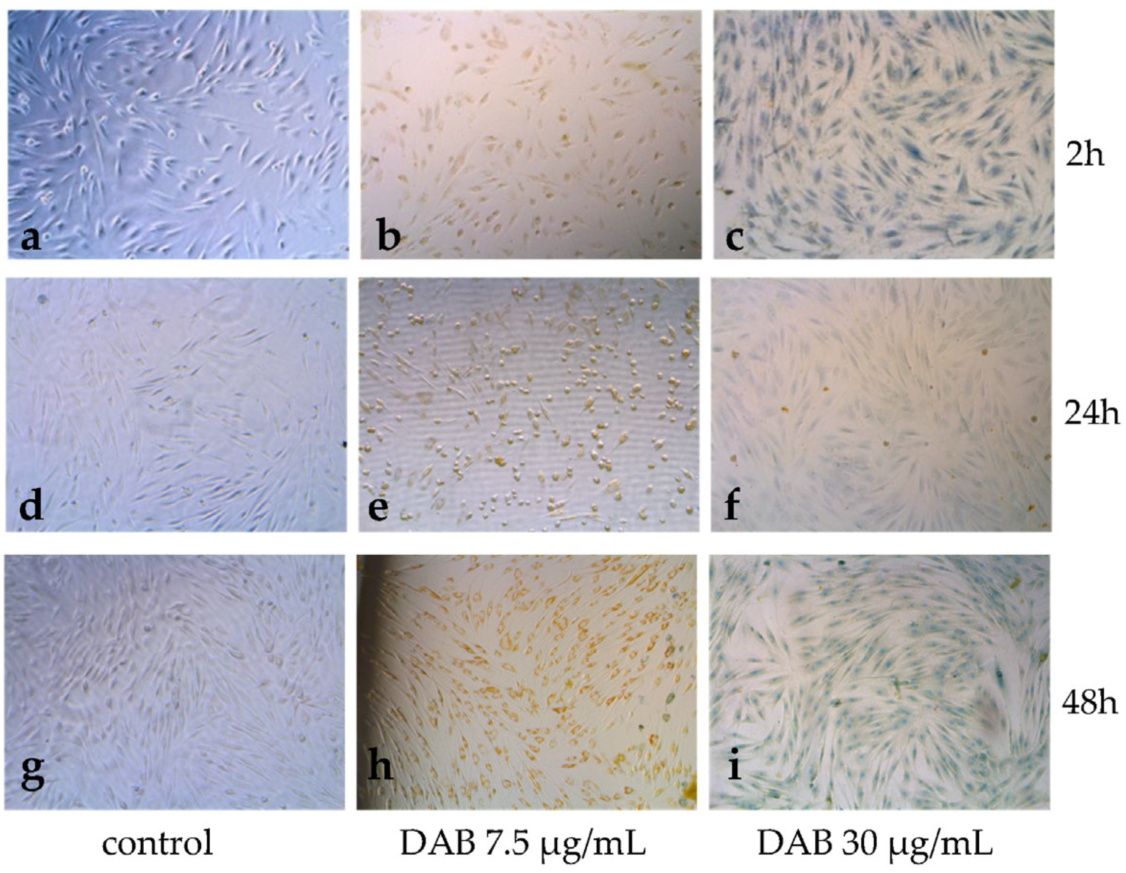
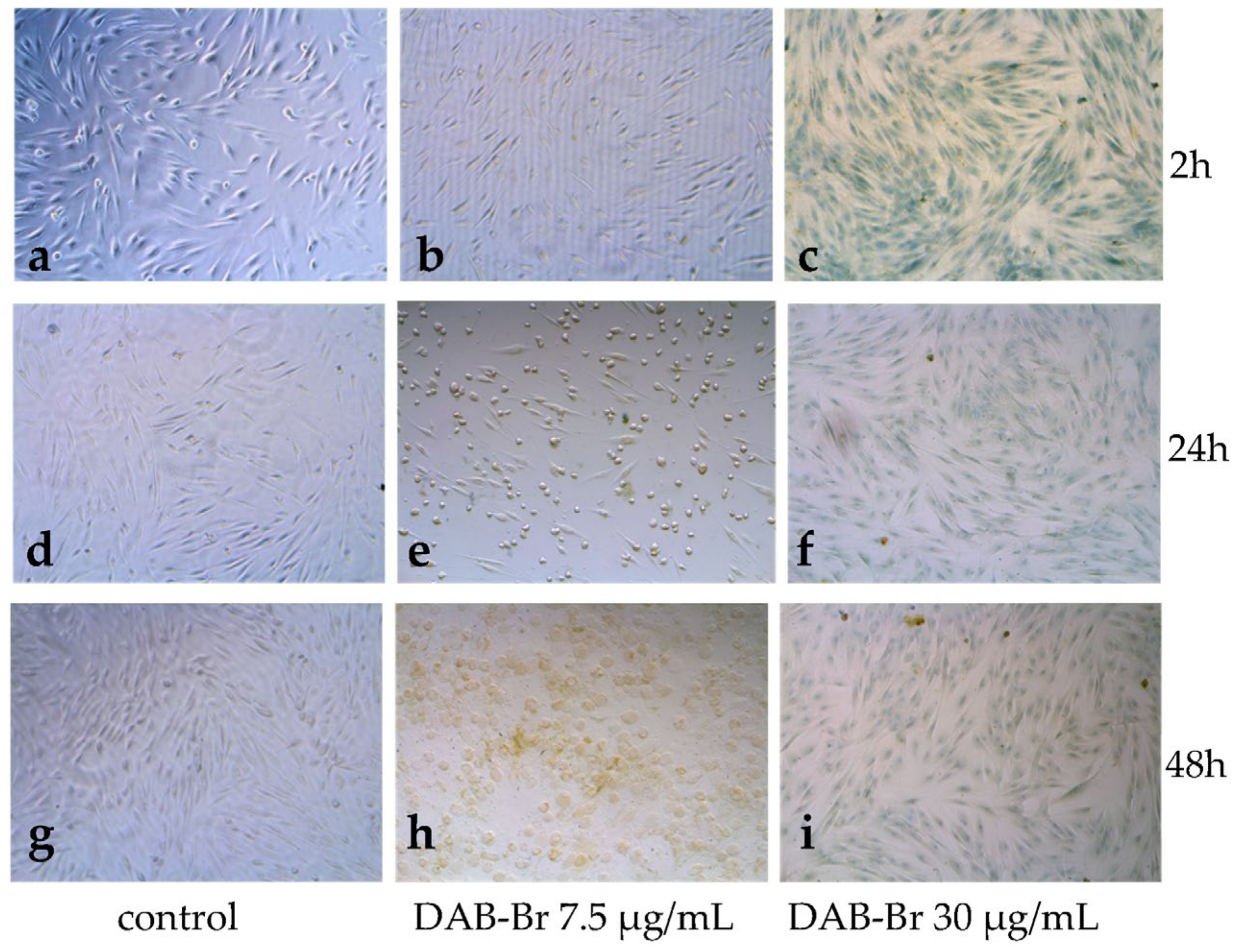
3.3. Permeabilization of Cell Membranes of HFF-1 Cells and Changes in Cell Morphology

The evaluation of cell membrane integrity by the TBE test revealed that both dendrimers induced permeabilization of the cell membrane in a dose-dependent manner (Figure 4). Insignificant permeabilization was observed at the lowest tested concentration of 3 μg/mL (2–3%) at the 2nd, 24th, and 48th hour after treatment with both dispersions of dendrimers. However, a noticeable effect on permeabilization was observed at the highest tested concentrations. At the 2nd hour, permeabilization reached 100% at a concentration of 60 μg/mL for both dendrimers (Figure 4a). At the 24th hour, DAB exhibited a significant effect, reaching 100% permeabilization at 30 μg/mL (Figure 4b), while for DAB-Br this parameter at the same concentration was detected at the 48th hour (Figure 4c). A noticeable dynamic in cell permeabilization was observed at lower concentrations, 7.5 and 15 μg/mL, suggesting the occurrence of reverse processes in the cell membranes. In both cases, a stronger permeabilization was noted by DAB: at 15 μg/mL at the 2nd hour, permeabilization was increased to 60% (Figure 4a). However, at the same concentration at the 24th hour, permeabilization dropped to 40% and further decreased to 20% by the 48th hour (Figure 4b,c). A similar effect was observed with DAB-Br, with a significant decrease in membrane permeabilization after the 24th hour post treatment.
Following the evaluation of cell membrane permeabilization, alterations in the cell morphology were monitored, as these changes indicate adaptation and stress responses. The morphology of the untreated adherent cells reveals spindle-shaped and highly elongated forms, typical for fibroblasts, which were closely spaced one next to the other (Figure 5a and Figure 6a). At the 24th and 48th hour, the fibroblasts spread onto the Petri dish and formed a thick layer due to extracellular interactions (Figure 5d,g and Figure 6d,g). However, after treatment with DAB and DAB-Br, noticeable morphological changes occurred. At the lower concentration of 7.5 µg/mL, both dendrimers induced similar cell morphological changes at the 24th and 48th hour post treatment (Figure 5e,h and Figure 6e,h). The cells appeared more rounded, with accumulated brown particles (internalized dendrimers) evident in the cytoplasm. However, cell viability was not affected. The cells continued to spread and formed a thick layer (Figure 5h and Figure 6h).
At the higher concentration of 30 µg/mL 2 h post treatment, all recorded cells appeared blue under the microscope, indicating compromised membrane integrity and dead or dying cells (Figure 5c and Figure 6c). At the 24th and 48th hour, brown clusters of dendrimers in the medium surrounding the cells were observed (Figure 5f,i and Figure 6f,i). Presumably, the dendrimers passed through the cell membrane when cell death occurred. However, apart from the coloring, the cells did not show alterations in their morphology.

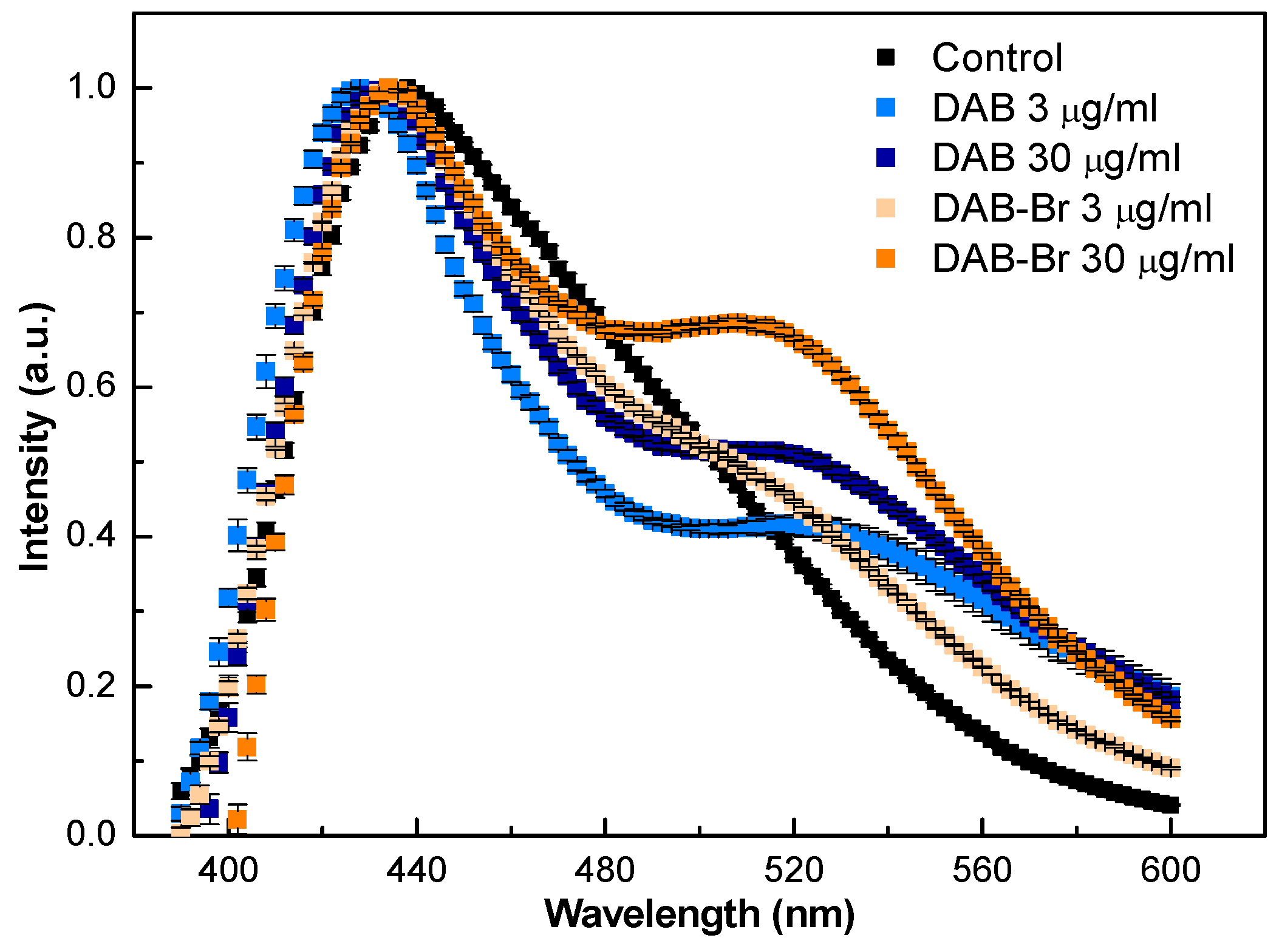
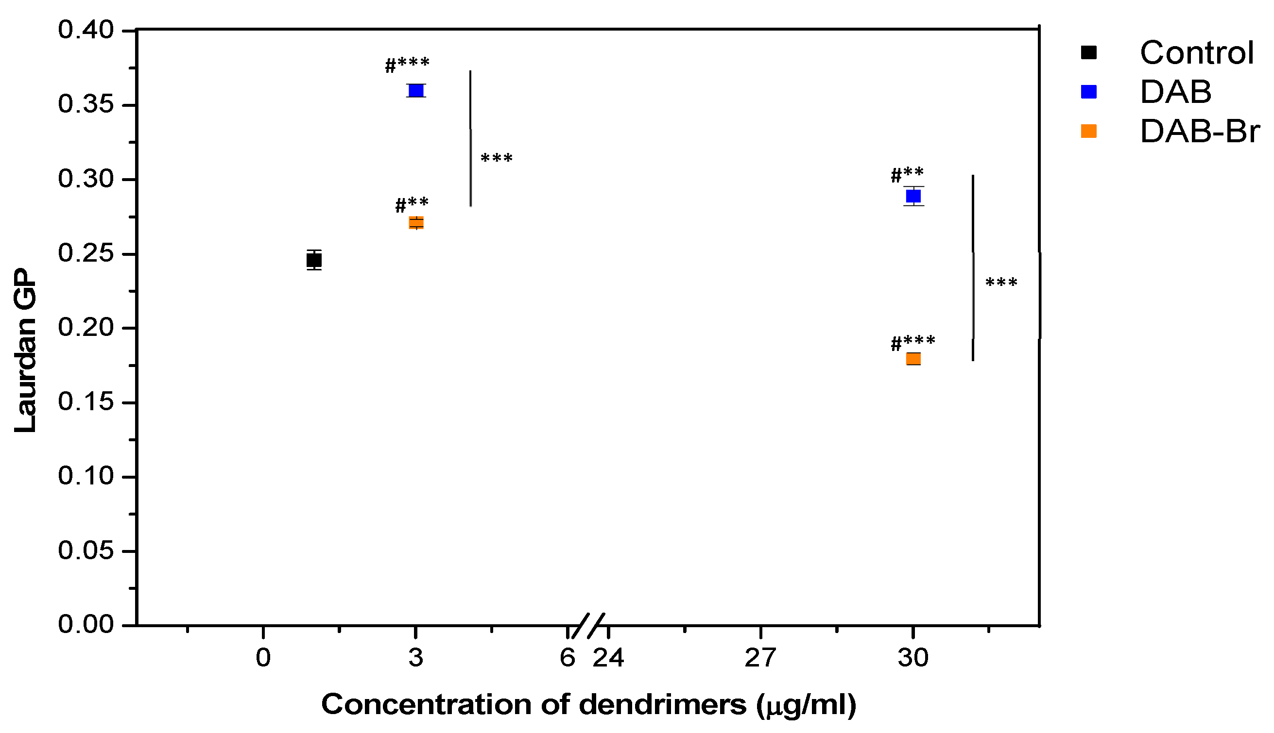
3.4. Laurdan Fluorescence Spectroscopy

In order to assess the changes in membrane lipid order following treatment with the tested dendrimers, Laurdan fluorescence spectroscopy was applied (Figure 7). The emission spectrum of the control HFF-1 cells exhibited an asymmetric shape, characterized by a main emission peak centered around 440 nm, suggesting a high degree of lipid order in the cell membranes. Upon treatment with both dendrimers, alterations in the emission spectra of the cells were observed. Two emission peaks were detected at both tested concentrations of 3 and 30 µg/mL. The first, larger peak was noticed around the 420–440 nm range, indicating a shift in the Laurdan emission spectra in the cells after treatment with the dendrimers, more prominent for DAB. This shift toward lower wavelengths suggests an increase in the lipid order of the cell membranes. Additionally, a second peak appeared in the 490–560 nm range, with its intensity increasing in response to higher dendrimer concentrations (Figure 7).
Laurdan’s general polarization was calculated from the emission intensities using the equation outlined in the Materials and Methods Section, adapted from Parasassi et al. [29]. The GP of the control (untreated cells) exhibited a high and positive value of 0.245 (Figure 8). This indicates a significant degree of membrane order and reflects the normal membrane integrity of the living cells. Upon treatment with dendrimers at the lower concentration of 3 µg/mL, the GP values increased compared to the control. This effect was particularly pronounced in cells treated with DAB, with GP values exceeding 1.5 times that of the control. The observed increase in GP values suggests an increased lipid order in the cell membranes and reduced fluidity. However, at the higher concentration of 30 µg/mL, the GP values were relatively lower: notably, the DAB treatment maintained higher GP values than the control. In contrast, DAB-Br treatment resulted in significantly lower GP value than the control, suggesting a membrane decrease in the lipid order.

4. Discussion

In this study, newly synthesized dendrimers, DAB and DAB-Br, were thoroughly evaluated for their biological activities, particularly their potential cytotoxicity, mechanism of cellular entry, and antifungal efficacy. Prior to this research it has been demonstrated that both dendrimers exhibit potent antibacterial activity against Gram-positive and Gram-negative bacteria. Additionally, they showed promising inhibitory effects on herpes simplex virus (HSV) production, indicating their potential application in combating herpes infection [17]. However, thus far, their effects on eukaryotic cells, particularly human and fungal cells, have not been investigated. Conducting thorough and systematic research to fully explore their biological activities could provide valuable insight for future applications.

4.1. Cellular Entry, Cytotoxicity, and Interactions of DAB and DAB-Br with HFF-1 Cells

The Human Foreskin Fibroblast (HFF-1 SCRC-1041) cell line originates from human skin fibroblasts and is an appropriate model for the in vitro testing of novel nanomaterials with potential medical applications. These cells possess a normal diploid karyotype, making them particularly valuable for testing the biological activity and potential cytotoxicity of dendrimers. It is well-documented that dendrimers’ characteristics—such as generation, size, number of surface groups, and the nature of terminal moieties (anionic, neutral, or cationic)—play a crucial role in modulating their biological effect on target mammalian cells [19,30].
In the presented study, the zeta potential measurements obtained via DLS showed that both dendrimers possess noticeable positive surface charges (+28 mV for DAB, and +35 mV for DAB-Br). This suggests good colloidal stability of the dispersion, as higher zeta potential values promote repulsion between particles, preventing their aggregation. The observed cationic surface charges of both dendrimers are attributed to the presence of tertiary amines in their internal structures and the terminal amine functional groups [26]. Notably, DAB-Br exhibited a higher zeta potential value compared to the non-halogenic DAB dendrimer due to surface bromination, which enhances the positive charge. Additionally, the hydrodynamic diameters of the dendrimers were determined to be 117 nm for DAB and 234 nm for DAB-Br. These nanoscale dimensions fall within the optimal range for cellular uptake and facilitate effective interactions with plasma membranes, cell organelles (endosomes, mitochondria, and nucleus), and cellular proteins and enzymes [31,32].
The MTT assay revealed that both tested dendrimers elicited a dose-dependent decrease in cell viability. The cytotoxicity was evident at concentrations exceeding 30 µg/mL. DAB dendrimers exhibited stronger cytotoxicity (IC50 = 27 µg/mL), whereas DAB-Br dendrimers had a much weaker effect (IC50 = 68 µg/mL). These findings highlight the influence of dendrimer structure and surface chemistry on cytotoxic outcomes. Cationic dendrimers, while often effective at cellular internalization, are commonly associated with higher cytotoxic potential. In support of this, a previous study has reported that positively charged PPI dendrimers induce pronounced dose-dependent cytotoxicity on target cells [33]. The positive charge is considered a primary contributor to the cytotoxic effects of the dendrimers because it promotes electrostatic interactions with cell components, such as proteins and cell membranes [34]. These interactions can disrupt normal cellular processes and ultimately can lead to cellular impairment and cellular damage. A study conducted by Yang and colleagues has revealed that cationic amino-terminated G2 dendrimers internalize in epidermal and dermal cells, contrary to anionic and uncharged ones [35]. However, the mechanism underlying the cellular uptake of the dendrimers is still debatable and there are various hypotheses regarding their mode of cellular entry. Some authors consider endocytosis as the primary pathway for internalization, while others propose temporary or permanent membrane permeabilization [36,37]. Based on the results from the TBE test, it can be stated that both DAB and DAB-Br dendrimers enter cells through cell membrane permeabilization, either temporary or permanent, depending on the tested concentrations and duration of exposure. Irreversible membrane permeabilization was observed at cytotoxic concentrations (>30 µg/mL), indicating strong disruptive interactions with the lipid bilayer. Notably, DAB exerts a stronger and more rapid membrane-disrupting effect compared to the halogenated DAB-Br, consistent with the MTT results. No changes in cellular morphology were detected, implying that at high concentrations, both dendrimers may function similarly to chemical fixatives, preserving cell structure despite inducing membrane permeabilization. At sub-toxic concentrations (<30 µg/mL) both dendrimers accumulated in the cells by temporary and reversible membrane permeabilization. The integrity of the cell membranes remained intact, indicating an occurrence of reversible processes within the cell membranes, likely occurring after transient pore formation. The nanoscale size and cationic surface charge of both dendrimers facilitate their permeation through the lipid bilayer by promoting the formation of nanoscale pores, particularly within the more fluid membrane regions. These pores lead to a significant increase in membrane permeability. At higher concentrations, this enhanced permeability culminates in membrane disruption [36,38,39].
To exert cytotoxicity and membrane permeabilization, dendrimers interact with cell membranes mainly through electrostatic forces. Cationic dendrimers are attracted to the slightly negatively charged phospholipids in the membrane bilayer, facilitating their binding, adhesion, and accumulation on the membrane surface [40,41]. Our previous studies on the molecular mechanisms of interaction between DAB, DAB-Br, and POPC model membranes (monolayers and liposomes) demonstrated that dendrimers interact with hydrophilic head groups of phospholipids, leading to their incorporation into the lipid bilayers [42]. Due to their cationic surface charge and nanoscale dimensions, dendrimers are electrostatically attracted to the surfaces of cell membranes. This attraction causes them to adhere to the membrane and penetrate the bilayer to some extent, substituting water molecules around the glycerol backbone of the lipids, and consequently, this leads to an increase in the lipid order [41].
Laurdan fluorescence spectroscopy provided valuable insight into the interaction of the tested dendrimers with cell membranes by assessing changes in the lipid order [43], an important parameter influencing membrane fluidity, permeability, and functions. A blue shift in the Laurdan emission spectra (420–440 nm) was observed at 3 µg/mL for DAB and DAB-Br, indicating increased membrane lipid order, likely due to dendrimer incorporation into the bilayer. This effect was more pronounced for DAB, which also exhibited higher generalized polarization values, suggesting a tighter lipid packing and reduced hydration at the membrane interface. This was further supported by experiments with model vesicles (POPC), where DAB induced greater ordering than DAB-Br, pointing to structural differences in membrane interaction [42]. Interestingly, the greater ordering effect of DAB may be associated with its smaller particle size (118 nm vs. 221 nm for DAB-Br), which may allow more efficient insertion into the lipid bilayer. In contrast, DAB-Br, despite its higher positive ζ-potential (+35.2 mV vs. +28.7 mV for DAB), induced a less marked increase in the lipid order. While a higher surface charge typically enhances electrostatic interactions with negatively charged membranes, the larger size of DAB-Br may limit its ability to integrate into the membrane as effectively as DAB. This suggests that particle size, not just surface charge, plays a critical role in determining how dendrimers modulate membrane structure. At the higher concentration of 30 µg/mL, both dendrimers caused a decrease in the lipid order, consistent with Laurdan’s sensitivity to water penetration and indicative of increased membrane permeability. This threshold concentration also corresponded with the results from the TBE assay, where both dendrimers caused irreversible membrane permeabilization. Overall, the data point to a concentration-dependent dual effect: membrane stabilization at lower concentrations and increased permeability at higher dendrimer doses. These observations underscore the importance of dendrimer physicochemical properties, such as size and surface charge, in governing their interactions with cell membranes, cell viability, and their potential applications in drug delivery.

4.2. Antifungal Activities of DAB and DAB-Br

Many polymers are known to exhibit antimicrobial activity, but much less attention is paid to their specific antifungal properties. The mechanisms underlying microbial cell death induced by various polymers include cytotoxicity mediated by reactive oxygen species (ROS), the disruption of the microbial cell envelope, and the inhibition of nucleic acid and protein synthesis. Polymers with antimicrobial activity often possess cationic surface groups, which facilitate electrostatic interactions with the negatively charged microbial membranes, leading to membrane destabilization and cell death [44]. Hoque et al. [45] explored the antibacterial and antifungal activities of polymeric materials concerning their structure–activity relationships and membrane-active mechanisms of action. The researchers demonstrated that cationic polymers effectively inactivated Candida spp. and Cryptococcus spp., achieving up to a 5-log reduction in viable cells compared to the control. These cationic polymer coatings act via a membrane-active mechanism, disrupting the lipid membranes of both bacterial and fungal cells.
In a study by Bahar and Ren [46] investigating the antifungal activity of antimicrobial peptides, several fungal cellular components were identified as primary targets, including the chitin-rich cell wall, the cell membrane, and various intracellular structures. Membrane-active antifungal peptides exert their effects through diverse mechanisms, such as cilium-like disruption, membrane thinning, aggregation, toroidal pore formation, and barrel-stave (barrelation) models of membrane penetration [46,47].
Evidence in the literature shows that dendrimers share a similar mode of action to cationic antimicrobial peptides [48,49]. According to recent findings, the antifungal activity of dendrimers includes four main mechanisms [18]:
(1)
Electrostatic interaction between the cationic terminal groups of the dendrimer and the anionic cell membranes, leading to an increase in membrane permeability;
(2)
A “carpet” mechanism in which dendrimers form small pores in the cell membrane, compromising membrane integrity and membrane repair mechanisms;
(3)
Inhibition of 1,3-β-D-glucan synthase, which disrupts cell wall synthesis and leads to leakage of cellular components;
(4)
Chelation, by which microbial enzymes are inhibited.
The degree of dendrimer toxicity is influenced by both their concentration and the presence of positively charged functional groups. Higher concentrations of antimicrobial compounds generally enhance their ability to inhibit fungal growth. In addition, several other physicochemical properties, such as molecular structure, dipole moment, solubility, molecular geometry, and electrical conductivity, also contribute significantly to their antifungal activity [50]. Anionic and neutral dendrimers are less toxic to fungal cells than cationic ones. Studies of the antifungal activity of cationic dendrimers suggest an interaction with the negatively charged cell membrane, leading to pore formation, increased permeability, and impaired membrane integrity. As a result, membrane depolarization can be induced and ultimately collapse and dead cells. Moreover, dendrimers may penetrate into the cell and may affect organelles and vital cell processes [51,52,53].
Our investigation of the antifungal activity of DAB and DAB-Br demonstrated potent inhibitory effects against all five fungal strains (Asp. fumigatus, Alt. alternata, Asp. niger, P. griseofulvum, and C. albicans) with their effectiveness being both time- and concentration-dependent. DAB-Br showed greater antifungal potency than DAB. Notably, DAB-Br achieved complete growth inhibition of all tested fungal strains at a lower minimum inhibitory concentration (0.148 µg/µL) compared to DAB (0.295 µg/µL). Furthermore, DAB-Br maintained a strong antifungal effect for up to 96 h at 0.295 µg/µL, whereas DAB required twice the higher concentration of 0.589 µg/µL for a similar response. C. albicans was the most sensitive strain, with growth inhibition observed for up to 24 h at the lowest tested concentration of both dendrimers (0.0369 µg/µL). The stronger antifungal activity of DAB-Br is attributed to the bromine atom in the dendrimer structure. Halogenation is commonly employed to improve the antimicrobial properties of natural compounds [54,55] and is widely recognized as an effective strategy to enhance the biological activity of drug candidates [56]. In particular, bromination can affect biological activity in different ways: (1) increases the affinity for drug targets by creating halogen bonds [57], improves the pharmacokinetic parameters of a molecule, and (2) optimizes or stabilizes biomolecular conformations [58]. Molchanova et al. [54] have reported that the incorporation of bromine atoms into metabolites significantly modifies their physicochemical and antibacterial properties [59]. In a related study, brominated furanones demonstrated stronger antifungal activity against Candida albicans than their non-brominated counterparts, suggesting that the bromine moiety is critical for the inhibitory effects. Specifically, monosubstituted exocyclic methyl group bromides exhibited potent inhibition of C. albicans growth [60]. Additionally, naphthalamide azoles synthesized through multistep reactions from 4-bromo-1,8-naphthalic anhydride have shown promising antibacterial and antifungal efficacy. The conversion of these compounds into azolium salts using various halogen-substituted aryl groups resulted in strong and comparable antimicrobial activities, indicating that strategic structural modifications can lead to the development of highly effective antimicrobial agents [61].

5. Conclusions

The biological activities of newly synthesized dendrimers, 4-dimethylamino-1,8-naphthalimide (DAB) and its halogenated analog 3-bromo-4-dimethylamino-1,8-naphthalimide (DAB-Br), specifically their cytotoxicity, mechanisms of cellular entry, and antifungal properties were investigated on eukaryotic cells. Dynamic light scattering measurements revealed that both dendrimers carried strong positive surface charges and different nanoscale hydrodynamic diameters. These size dimensions, along with their cationic nature, facilitate effective interactions with cellular membranes and promote internalization into target cells.
The treatment of the HFF-1 cell line and five fungal strains with the dendrimers tested revealed that DAB dendrimers were more cytotoxic to human cells, whereas DAB-Br exhibited greater antifungal potency. The MTT assay using the HFF-1 cell line showed that DAB had a lower IC50 value, indicating stronger cytotoxicity, while DAB-Br was less toxic. In contrast, antifungal testing using the resazurin method showed that DAB-Br induced complete growth inhibition of all tested fungal strains at a lower minimum inhibitory concentration than DAB. Furthermore, DAB-Br maintained its antifungal activity for up to 96 h at lower concentration, whereas DAB required a twofold higher concentration to produce a comparable effect.
The TBE assay indicated that both DAB and DAB-Br dendrimers enter HFF-1 cells via temporary or permanent membrane permeabilization. At the threshold concentration of 30 µg/mL, both dendrimers induced irreversible, time-dependent membrane permeabilization, as higher concentrations accelerated this effect. Microscopic observations and Laurdan fluorescence spectroscopy confirmed compromised membrane integrity, as treated cells exhibited a predominantly blue fluorescence and a decreased lipid order, which is indicative of disrupted membrane integrity and permeability. At non-toxic concentrations below 30 µg/mL, dendrimers crossed the cell membrane via temporary permeabilization. Treated cells showed dendrimer accumulation in the cytoplasm, while membrane integrity remained intact, suggesting the formation of transient nanoholes and reversible membrane processes under these conditions.

Supplementary Materials

The following supporting information can be downloaded at: https://www.mdpi.com/article/10.3390/app15147764/s1, Supplement S1. Cell morphology after treatment with dendrimers (a) at low concentration of 7.5 µg/mL and (b) high concentration of 30 µg/mL. The arrows point the dendrimers in cell periphery and the triangles point the dendrimers outside of the cells. The cells appeared more rounded, with accumulated brown particles (internalized dendrimers) evident in the cytoplasm (a, arrows). However, cell viability was not affected. The cells continued to spread and formed a thick layer. At the higher concentration of 30 µg/mL 2 h post-treatment, all recorded cells appeared blue under the microscope, indicating compromised membrane integrity and dead or dying cells, and brown clusters of dendrimers in the medium surrounding the cells were observed (b).

Author Contributions

Conceptualization, A.K., T.T.-H., A.T. and A.J.; methodology, A.K., T.T.-H., G.S. and I.G.; validation, T.T.-H., M.M. and G.S.; formal analysis, D.B.; investigation, A.K., E.G., T.T.-H., D.B., G.S., E.K., R.H., M.M. and I.G.; data curation, A.K.; writing—original draft preparation, A.K., T.T.-H., D.B., E.K. and M.M.; writing—review and editing, A.T. and A.J.; visualization, D.B. and A.T.; supervision, A.J.; project administration, A.J.; funding acquisition, A.J. All authors have read and agreed to the published version of the manuscript.

Funding

This research was funded by the Scientific Research Fund of the Ministry of Education and Science, project No. KП-06-H51/15, dated 19 November 2021, and by the European Union Next Generation EU, through the National Recovery and Resilience Plan of the Republic of Bulgaria, project № BG-RRP-2.004-0008-C01.

Institutional Review Board Statement

Not applicable.

Informed Consent Statement

Not applicable.

Data Availability Statement

The original contributions presented in the study are included in the article and Supplementary Materials, and further inquiries can be directed at the corresponding author.

Acknowledgments

The authors thank the Bulgarian Ministry of Education and Science, project No. KП-06-H51/15, dated 19 November 2021, and the European Union Next Generation EU, through the National Recovery and Resilience Plan of the Republic of Bulgaria, project № BG-RRP-2.004-0008-C01. The authors are also grateful to the Faculty of Medicine, Sofia University, “St. Kliment Ohridski”, for the overall support. The authors thank the Bulgarian Ministry of Education and Science for support—the Scientific Infrastructure on Cell Technologies in Biomedicine (SICTB) D01-361/2023 (Spectroscopy facilities) and the National Center for Biomedical Photonics D01-352/2023 (Malvern Panalytical Zetasizer Nano)—part of the Bulgarian National Roadmap for Scientific Infrastructures 2020–2027.

Conflicts of Interest

The authors declare no conflicts of interest.

References

  1. Malik, S.; Singh, J.; Goyat, R.; Saharan, Y.; Chaudhry, V.; Umar, A.; Ibrahim, A.A.; Akbar, S.; Ameen, S.; Baskoutas, S. Nanomaterials-based biosensor and their applications: A review. Heliyon 2023, 9, e19929. [Google Scholar] [CrossRef]
  2. Altemimi, A.B.; Farag, H.A.M.; Salih, T.H.; Awlqadr, F.H.; Al-Manhel, A.J.A.; Vieira, I.R.S.; Conte-Junior, C.A. Application of nanoparticles in human nutrition: A review. Nutrients 2024, 16, 636. [Google Scholar] [CrossRef] [PubMed]
  3. Contin, M.; Garcia, C.; Dobrecky, C.; Lucangioli, S.; D’Accorso, N. Advances in drug delivery, gene delivery and therapeutic agents based on dendritic materials. Future Med. Chem. 2019, 11, 1791–1810. [Google Scholar] [CrossRef] [PubMed]
  4. Huang, Y.M.; Hong, X.Z.; Shen, J.; Geng, L.J.; Pan, Y.H.; Ling, W.; Zhao, H.L. Amyloids in site-specific autoimmune reactions and inflammatory responses. Front. Immunol. 2020, 10, 2980. [Google Scholar] [CrossRef] [PubMed]
  5. Fateh, S.T.; Aghaii, A.H.; Aminzade, Z.; Shahriari, E.; Roohpour, N.; Koosha, F.; Dezfuli, A.S. Inorganic nanoparticle-cored dendrimers for biomedical applications: A review. Heliyon 2024, 10, e29726. [Google Scholar] [CrossRef]
  6. Aminian, F.; Hemmati, A. Dendrimers as versatile platforms for advanced biosensor development: A review on chemistry, synthesis, and performance enhancements. JECE 2025, 13, 116365. [Google Scholar] [CrossRef]
  7. Jain, K.; Jain, N.K.; Kesharwani, P. Types of dendrimers. In Dendrimer-Based Nanotherapeutics, 1st ed.; Kesharwani, P., Ed.; Academic Press: Cambridge, MA, USA, 2021; pp. 95–123. [Google Scholar] [CrossRef]
  8. Wang, J.; Li, B.; Qiu, L.; Qiao, X.; Yang, H. Dendrimer-based drug delivery systems: History, challenges, and latest developments. J. Biol. Eng. 2022, 16, 18. [Google Scholar] [CrossRef]
  9. Fox, L.J.; Richardson, R.M.; Briscoe, W.H. PAMAM dendrimer—Cell membrane interactions. Adv. Colloid Interface Sci. 2018, 257, 1–18. [Google Scholar] [CrossRef]
  10. Caminade, A.M.; Turrin, C.O. Dendrimers for drug delivery. J. Mater. Chem. B 2014, 2, 4055–4066. [Google Scholar] [CrossRef]
  11. Kesharwani, P.; Gothwal, A.; Iyer, A.K.; Jain, K.; Chourasia, M.K.; Gupta, U. Dendrimer nanohybrid carrier systems: An expanding horizon for targeted drug and gene delivery. Drug Discov. Today 2018, 23, 300–314. [Google Scholar] [CrossRef]
  12. Chis, A.A.; Dobrea, C.; Morgovan, C.; Arseniu, A.M.; Rus, L.L.; Butuca, A.; Juncan, A.M.; Totan, M.; Vonica-Tincu, A.L.; Cormos, G.; et al. Applications and limitations of dendrimers in biomedicine. Molecules 2020, 25, 3982. [Google Scholar] [CrossRef] [PubMed]
  13. Hoyos, P.; Perona, A.; Juanes, O.; Rumbero, Á.; Hernáiz, M. Synthesis of glycodendrimers with antiviral and antibacterial activity. Chem. A Eur. J. 2021, 27, 7593–7624. [Google Scholar] [CrossRef] [PubMed]
  14. Chis, A.A.; Dobrea, C.M.; Rus, L.L.; Frum, A.; Morgovan, C.; Butuca, A.; Totan, M.; Juncan, A.M.; Gligor, F.G.; Arseniu, A.M. Dendrimers as non-viral vectors in gene-directed enzyme prodrug therapy. Molecules 2021, 26, 5976. [Google Scholar] [CrossRef] [PubMed]
  15. Gholami, M.; Mohammadi, R.; Arzanlou, M.; Akbari Dourbash, F.; Kouhsari, E.; Majidi, G.; Mohseni, S.M.; Nazari, S. In vitro antibacterial activity of poly (amidoamine)-G7 dendrimer. BMC Infect. Dis. 2017, 17, 395. [Google Scholar] [CrossRef]
  16. Falanga, A.; Del Genio, V.; Galdiero, S. Peptides and dendrimers: How to combat viral and bacterial infections. Pharmaceutics 2021, 13, 101. [Google Scholar] [CrossRef]
  17. Staneva, D.; Vasileva-Tonkova, E.; Grozdanov, P.; Vilhelmova-Ilieva, N.; Nikolova, I.; Grabchev, I. Synthesis and photophysical characterisation of 3-bromo-4-dimethylamino-1, 8-naphthalimides and their evaluation as agents for antibacterial photodynamic therapy. J. Photochem. Photobiol. A Chem. 2020, 401, 112730. [Google Scholar] [CrossRef]
  18. Mlynarczyk, D.T.; Dlugaszewska, J.; Kaluzna-Mlynarczyk, A.; Goslinski, T. Dendrimers against fungi—A state of the art review. J. Control. Release 2021, 330, 599–617. [Google Scholar] [CrossRef]
  19. Janaszewska, A.; Lazniewska, J.; Trzepiński, P.; Marcinkowska, M.; Klajnert-Maculewicz, B. Cytotoxicity of dendrimers. Biomolecules 2019, 9, 330. [Google Scholar] [CrossRef]
  20. Khan, M.A.; Peng, R.; Liu, C.L.; Chen, Z. Synthesis, dynamics and applications (cytotoxicity and biocompatibility) of dendrimers: A mini-review. Eur. Polym. J. 2022, 181, 111708. [Google Scholar] [CrossRef]
  21. Baecker, D.; Kapp, T.; Schumacher, P.; Gust, R.; Kircher, B. Cell death-inducing properties of selected dendrimers against different breast cancer and leukemia cell lines. Arch. Pharm. 2020, 353, e2000209. [Google Scholar] [CrossRef]
  22. Žmudová, Z.; Šanderová, Z.; Liegertová, M.; Vinopal, S.; Herma, R.; Sušický, L.; Müllerová, M.; Strašák, T.; Malý, J. Biodistribution and toxicity assessment of methoxyphenyl phosphonium carbosilane dendrimers in 2D and 3D cell cultures of human cancer cells and zebrafish embryos. Sci. Rep. 2023, 13, 15477. [Google Scholar] [CrossRef] [PubMed]
  23. Li, L.; Deng, Y.; Zeng, Y.; Yan, B.; Deng, Y.; Zheng, Z.; Li, S.; Yang, Y.; Hao, J.; Xiao, X.; et al. The application advances of dendrimers in biomedical field. VIEW 2023, 4. [Google Scholar] [CrossRef]
  24. Ipsen, J.H.; Karlström, G.; Mouritsen, O.G.; Wennerström, H.; Zuckermann, M.J. Phase equilibria in the phosphatidylcholine-cholesterol system. BBA-Biomembr. 1987, 905, 162–172. [Google Scholar] [CrossRef] [PubMed]
  25. Ammendolia, D.A.; Bement, W.M.; Brumell, J.H. Plasma membrane integrity: Implications for health and disease. BMC Biol. 2021, 19, 71. [Google Scholar] [CrossRef]
  26. Mosmann, T. Rapid colorimetric assay for cellular growth and survival: Application to proliferation and cytotoxicity assays. J. Immunol. Meth. 1983, 65, 55–63. [Google Scholar] [CrossRef]
  27. Elshikh, M.; Ahmed, S.; Funston, S.; Dunlop, P.; McGaw, M.; Marchant, R.; Banat, I.M. Resazurin-based 96-well plate microdilution method for the determination of minimum inhibitory concentration of biosurfactants. Biotechnol. Lett. 2016, 38, 1015–1019. [Google Scholar] [CrossRef]
  28. Strober, W. Trypan blue exclusion test of cell viability. Curr. Protoc. Immunol. 2001, 111, A3.B.1–A3.B.3. [Google Scholar] [CrossRef]
  29. Parasassi, T.; De Stasio, G.; d’Ubaldo, A.; Gratton, E. Phase fluctuation in phospholipid membranes revealed by Laurdan fluorescence. Biophys. J. 1990, 57, 1179–1186. [Google Scholar] [CrossRef]
  30. Gothwal, A.; Malik, S.; Gupta, U.; Jain, N.K.. Toxicity and biocompatibility aspects of dendrimers. In Pharmaceutical Applications of Dendrimers (Micro and Nano Technologies); Chauhan, A., Kulhari, H., Eds.; Elsevier: Amsterdam, The Netherlands, 2020; pp. 251–274. [Google Scholar] [CrossRef]
  31. Lee, H.; Larson, D.R.; Lawrence, D.S. Illuminating the chemistry of life: Design, synthesis, and applications of ‘caged’ and related photoresponsive compounds. ACS Chem. Biol. 2008, 4, 409–427. [Google Scholar] [CrossRef]
  32. Choi, Y.; Thomas, T.; Kotlyar, A.; Islam, M.T.; Baker, J.R. Synthesis and functional evaluation of DNA-assembled polyamidoamine dendrimer clusters for cancer cell-specific targeting. Chem. Biol. 2006, 12, 35–43. [Google Scholar] [CrossRef]
  33. Kuo, J.H.; Jan, M.S.; Chiu, H.W. Mechanism of cell death induced by cationic dendrimers in RAW 264.1 murine macrophage-like cells. J. Pharm. Pharmacol. 2005, 57, 489–495. [Google Scholar] [CrossRef] [PubMed]
  34. Ziemba, B.; Halets, I.; Shcharbin, D.; Appelhans, D.; Voit, B.; Pieszynski, I.; Bryszewska, M.; Klajnert, B. Influence of fourth generation poly (propylene imine) dendrimers on blood cells. J. Biomed. Mater. Res. A 2012, 100A, 2870–2880. [Google Scholar] [CrossRef] [PubMed]
  35. Yang, Y.; Sunoqrot, S.; Stowell, C.; Ji, J.; Lee, C.W.; Kim, J.W.; Khan, S.A.; Hong, S. Effect of size, surface charge, and hydrophobicity of poly(amidoamine) dendrimers on their skin penetration. Biomacromolecules 2012, 13, 2154–2162. [Google Scholar] [CrossRef] [PubMed]
  36. Kheraldine, H.; Rachid, O.; Habib, A.M.; Moustafa, A.A.; Benter, I.F.; Akhtar, S. Emerging innate biological properties of nano-drug delivery systems: A focus on PAMAM dendrimers and their clinical potential. Adv. Drug Deliv. Rev. 2021, 178, 113908. [Google Scholar] [CrossRef]
  37. Zhang, J.; Liu, D.; Zhang, M.; Sun, Y.; Zhang, X.; Guan, G.; Zhao, X.; Qiao, M.; Chen, D.; Hu, H. The cellular uptake mechanism, intracellular transportation, and exocytosis of polyamidoamine dendrimers in multidrug-resistant breast cancer cells. Int. J. Nanomed. 2016, 11, 3677–3690. [Google Scholar] [CrossRef]
  38. de Luca, S.; Seal, P.; Parekh, H.S.; Tupally, K.R.; Smith, S.C. Cell Membrane Penetration without Pore Formation: Chameleonic Properties of Dendrimers in Response to Hydrophobic and Hydrophilic Environments. Adv. Theory Simul. 2020, 3, 1900152. [Google Scholar] [CrossRef]
  39. Qian, Y.; Yang, D.; Lin, X.; Shen, C.; Zhang, J.; Xu, J.; Zhao, Y.; Zhu, L.; Kong, H.; Zhang, M.; et al. Dendrimer-Derived Mimics of Host Defense Peptides Selectively Disrupt Cancer Cell Membranes for Melanoma Therapy. Pharmaceutics 2025, 17, 361. [Google Scholar] [CrossRef]
  40. Lombardo, D.; Calandra, P.; Bellocco, E.; Laganà, G.; Barreca, D.; Magazù, S.; Wanderlingh, U.; Kiselev, M.A. Effect of anionic and cationic polyamidoamine (PAMAM) dendrimers on a model lipid membrane. BBA-Biomembr. 2016, 1858, 2769–2777. [Google Scholar] [CrossRef]
  41. Metwally, S.; Stachewicz, U. Surface potential and charges impact on cell responses on biomaterials interfaces for medical applications. Mater. Sci. Eng. C 2019, 104, 109883. [Google Scholar] [CrossRef]
  42. Jordanova, A.; Tsanova, A.; Stoimenova, E.; Minkov, I.; Kostadinova, A.; Hazarosova, R.; Angelova, R.; Antonova, K.; Vitkova, V.; Staneva, G.; et al. Molecular Mechanisms of Action of Dendrimers with Antibacterial Activities on Model Lipid Membranes. Polymers 2025, 17, 929. [Google Scholar] [CrossRef]
  43. Parasassi, T.; Krasnowska, E.; Bagatolli, L.; Gratton, E. Laurdan and Prodan as Polarity-Sensitive Fluorescent Membrane Probes. J. Fluoresc. 1998, 8, 365–373. [Google Scholar] [CrossRef]
  44. Santos, A.; Veiga, F.; Figueiras, A. New advances in general biomedical applications of PAMAM dendrimers. Molecules 2016, 23, 2849. [Google Scholar] [CrossRef]
  45. Hoque, J.; Akkapeddi, P.; Yadav, V.; Manjunath, G.B.; Uppu, D.S.; Konai, M.M.; Yarlagadda, V.; Sanyal, K.; Haldar, J. Broad spectrum antibacterial and antifungal polymeric paint materials: Synthesis, structure-activity relationship, and membrane-active mode of action. ACS Appl. Mater. Interfaces. 2015, 7, 1804–1815. [Google Scholar] [CrossRef] [PubMed]
  46. Bahar, A.A.; Ren, D. Antimicrobial Peptides. Pharmaceuticals 2013, 6, 1543–1575. [Google Scholar] [CrossRef]
  47. Shai, Y. Mechanism of the binding, insertion and destabilization of phospholipid bilayer membranes by α-helical antimicrobial and cell non-selective membrane-lytic peptides. BBA-Biomembr. 1999, 1462, 55–70. [Google Scholar] [CrossRef]
  48. Powers, K.W.; Brown, S.C.; Krishna, V.B.; Wasdo, S.C.; Moudgil, B.M.; Roberts, S.M. Dendrimers as potential drug carriers; encapsulation of acidic hydrophobic drugs. Tetrahedron 2003, 59, 5285–5290. [Google Scholar] [CrossRef]
  49. Galanakou, C.; Dhumal, D.; Peng, L. Amphiphilic dendrimers against antibiotic resistance: Light at the end of the tunnel? Biomater. Sci. 2023, 11, 3379–3393. [Google Scholar] [CrossRef]
  50. Nouman; Rana, M.; Ahmedi, S.; Arif, R.; Manzoor, N.; Rahisuddin. 4-bromo-1, 8-naphthalimide derivatives as antifungal agents: Synthesis, characterization, DNA binding, molecular docking, antioxidant and ADMET studies. Polycycl. Aromat. Compd. 2024, 44, 748–772. [Google Scholar] [CrossRef]
  51. Pesee, S.; Angkananuwat, C.; Tancharoensukjit, S.; Muanmai, S.; Sirivan, P.; Bubphawas, M.; Tanarerkchai, N. In vitro activity of Caspofungin combined with Fluconazole on mixed Candida albicans and Candida glabrata biofilm. Med. Mycol. 2016, 54, 384–393. [Google Scholar] [CrossRef]
  52. Grela, E.; Zdybicka-Barabas, A.; Pawlikowska-Pawlega, B.; Cytrynska, M.; Wlodarczyk, M.; Grudzinski, W.; Luchowski, R.; Gruszecki, W.I. Modes of the antibiotic activity of amphotericin B against Candida albicans. Sci. Rep. 2019, 9, 17029. [Google Scholar] [CrossRef]
  53. Heredero-Bermejo, I.; Gómez-Casanova, N.; Quintana, S.; Soliveri, J.; de la Mata, F.J.; Pérez-Serrano, J.; Sánchez-Nieves, J.; Copa-Patiño, J.L. In Vitro Activity of Carbosilane Cationic Dendritic Molecules on Prevention and Treatment of Candida albicans Biofilms. Pharmaceutics 2020, 12, 918. [Google Scholar] [CrossRef] [PubMed]
  54. Molchanova, N.; Nielsen, J.E.; Sørensen, K.B.; Prabhala, B.K.; Hansen, P.R.; Lund, R.; Barron, A.E.; Jenssen, H. Halogenation as a tool to tune antimicrobial activity of peptoids. Sci. Rep. 2020, 10, 14805. [Google Scholar] [CrossRef] [PubMed]
  55. Raimondi, M.T.; Moretti, M.; Cioffi, M.; Giordano, C.; Boschetti, F.; Lagana, K.; Pietrabissa, R. The effect of hydrodynamic shear on 3D engineered chondrocyte systems subject to direct perfusion. Biorheology 2006, 43, 215–222. [Google Scholar] [CrossRef] [PubMed]
  56. Benedetto Tiz, D.; Bagnoli, L.; Rosati, O.; Marini, F.; Santi, C.; Sancineto, L. FDA-Approved Small Molecules in 2022: Clinical Uses and Their Synthesis. Pharmaceutics 2022, 14, 2538. [Google Scholar] [CrossRef]
  57. Xu, Z.; Yang, Z.; Liu, Y.; Lu, Y.; Chen, K.; Zhu, W. Halogen bond: Its role beyond drug-target binding affinity for drug discovery and development. J. Chem. Inf. Model. 2014, 54, 69–78. [Google Scholar] [CrossRef]
  58. Zhou, P.; Lv, J.; Zou, J.; Tian, F.; Shang, Z. Halogen–water–hydrogen bridges in biomolecules. J. Struct. Biol. 2010, 169, 172–182. [Google Scholar] [CrossRef]
  59. Cochereau, R.; Maffeis, V.; dos Santos, E.C.; Lörtscher, E.; Palivan, C.G. Polymeric giant unilamellar vesicles with integrated DNA-origami nanopores: An efficient platform for tuning bioreaction dynamics through controlled molecular diffusion. Adv. Funct. Mater. 2023, 33, 2304782. [Google Scholar] [CrossRef]
  60. Duo, M.; Zhang, M.; Luk, Y.Y.; Ren, D. Inhibition of Candida albicans growth by brominated furanone. Appl. Microbiol. Biotechnol. 2010, 85, 1551–1563. [Google Scholar] [CrossRef]
  61. Zhang, Y.Y.; Zhou, C.H. Synthesis and activities of naphthalimide azoles as a new type of antibacterial and antifungal agents. Bioorg. Med. Chem. Lett. 2011, 21, 4349–4352. [Google Scholar] [CrossRef]
Figure 1. Chemical structures of dendrimers: (a) 4-dimethylamino-1,8-naphthalimide (DAB) and (b) 3-bromo-4-dimethyl-1,8-naphthalimide (DAB-Br).
Applsci 15 07764 g001
Figure 2. MTT assay for cytotoxicity of HFF-1 cells at the 24th hour after treatment with different concentrations of DAB and DAB-Br. Statistical significance between the effects of DAB and DAB-Br at each tested concentration is denoted with *** (p < 0.001) and ** (p < 0.01), while between the control (untreated cells), DAB, and DAB-Br, at each tested concentration, it is indicated with #* (p < 0.05), #** (p < 0.01), and #*** (p < 0.001). The designation “ns” (not significant) shows statistically insignificant differences.
Applsci 15 07764 g002
Figure 3. Dendrimers’ minimum inhibitory concentrations (MICs) on five fungal strains. Statistical significance between the effects of DAB and DAB-Br on each fungal strain is denoted with *** (p < 0.001). The designation “ns” (not significant) shows statistically insignificant differences.
Applsci 15 07764 g003
Figure 4. Comparative quantitative analysis of cell membrane permeabilization following treatment of HFF-1 cells with DAB and DAB-Br at the (a) 2nd hour, (b) 24th hour, and (c) 48th hour. Statistical significance is denoted by *** (p < 0.001) when comparing effects of both dendrimers.
Applsci 15 07764 g004
Figure 5. Changes in cell morphology and cell permeabilization with trypan blue staining at the 2nd, 24th, and 48th hour post treatment with DAB at concentrations of 7.5 µg/mL and 30 µg/mL. For photo shooting, random fields were selected. Controls (untreated cells, a,d,g); cells treated with DAB at a concentration of 7.5 µg/mL (b,e,h) and 30 µg/mL (c,f,i). Magnification 200×.
Applsci 15 07764 g005
Figure 6. Changes in cell morphology and cell permeabilization with trypan blue staining at the 2nd, 24th, and 48th hour post treatment with DAB-Br at concentrations of 7.5 µg/mL and 30 µg/mL. For photo shooting, random fields were selected. Controls (untreated cells, a,d,g); cells treated with DAB-Br at a concentration of 7.5 µg/mL (b,e,h), and 30 µg/mL (c,f,i). Magnification 200×.
Applsci 15 07764 g006
Figure 7. Laurdan fluorescence emission spectra of HFF-1 cells upon treatment with DAB and DAB-Br at concentrations of 3 and 30 µg/mL at physiological temperature (37 °C).
Applsci 15 07764 g007
Figure 8. Laurdan general polarization (GP) of HFF-1 cells as a function of DAB and DAB-Br at concentrations of 3 and 30 µg/mL, measured at physiological temperature (37 °C). A statistical significance between the effects of DAB and DAB-Br at concentrations of 3 µg/mL to 30 µg/mL is denoted with *** (p < 0.001), and between the control (untreated cells), DAB, and DAB-Br, at each tested concentration, it is indicated with #** (p < 0.01) and #*** (p < 0.001).
Applsci 15 07764 g008
Table 1. Average size and apparent zeta potential values of DAB and DAB-Br at a concentration of 40 µg/mL on the 1st, 3rd, and 7th day after dispersion preparation. Two independent samples were measured, with three replicates per sample. (**) indicates a statistically significant difference between DAB and DAB-Br (p < 0.01).
Dendrimer Dispersions
(40 µg/mL)
Average Size
(nm)
Apparent ζ-Potential
(mV)
DAB (1st day)118 ± 66 **28.7 ± 0.4 **
DAB (3rd day)123 ± 45 **26.5 ± 1.6 **
DAB (7th day)123 ± 54 **26.5 ± 2.7 **
DAB-Br (1st day)222 ± 39 **35.2 ± 1.9 **
DAB-Br (3rd day)234 ± 82 **31.6 ± 2.4 **
DAB-Br (7th day)265 ± 72 **34.5 ± 1.7 **
Table 2. Antifungal activity of DAB against five fungal strains.
DAB1.179 µg/µL0.589 µg/µL0.295 µg/µL0.148 µg/µL0.074 µg/µL0.0369 µg/µL
Asp. fumigatus96 h96 h24 h
Alt. alternata96 h96 h72 h
Asp. niger96 h96 h24 h
P. griseofulvum96 h96 h24 h
C. albicans96 h96 h24 h24 h24 h24 h
Table 3. Antifungal activity of DAB-Br against five fungal strains.
DAB-Br1.179 µg/µL0.589 µg/µL0.295 µg/µL0.148 µg/µL0.074 µg/µL0.0369 µg/µL
Asp. fumigatus96 h96 h96 h24 h
Alt. alternata96 h96 h96 h96 h24 h
Asp. niger96 h96 h96 h24 h
P. griseofulvum96 h96 h96 h24 h
C. albicans96 h96 h96 h48 h24 h24 h
Disclaimer/Publisher’s Note: The statements, opinions and data contained in all publications are solely those of the individual author(s) and contributor(s) and not of MDPI and/or the editor(s). MDPI and/or the editor(s) disclaim responsibility for any injury to people or property resulting from any ideas, methods, instructions or products referred to in the content.

