Article

Randomized Controlled Clinical Trial to Evaluate Skeletal and Dental Treatment Effects of Rapid Maxillary Expansion in Children: Comparison Between Two-Band Expander and Bonded Palatal Expander

1
Dentistry Unit, Management Innovations, Diagnostics and Clinical Pathways, Bambino Gesù Children’s Hospital, IRCCS, 00165 Rome, Italy
2
Dentistry Unit, AORN Santobono-Pausilipon, 80100 Naples, Italy
3
Department of Neurosciences, Reproductive Sciences and Oral Sciences, School of Orthodontics, University of Naples “Federico II”, 80138 Naples, Italy
4
UN-EU International Research Project on Human Health–Oral Health Section, 1200 Geneva, Switzerland
*
Author to whom correspondence should be addressed.
Appl. Sci. 2025, 15(13), 7187; https://doi.org/10.3390/app15137187
Submission received: 29 May 2025 / Revised: 19 June 2025 / Accepted: 24 June 2025 / Published: 26 June 2025
(This article belongs to the Special Issue Advances in Orthodontic Treatment)

Abstract

Featured Application

This research paper aims to encourage specialists in orthodontics to customize their treatments of palatal expansion by choosing the appropriate device for the expected results.

Abstract

The rapid maxillary expander is one of the most widely used devices in orthodontics, and this study analyzes the skeletal and dental effects of a two-band rapid maxillary expander (RME) and a splint resin palatal expander (SRPE) in growing children with skeletal maxillary contraction. Seventy-four subjects with palatal skeletal contraction and unilateral or bilateral posterior crossbite were treated using maxillary expander devices. The sample was made up of two different randomly assigned groups: RME (21 females, 17 males; mean age ± SD 7.7 ± 1.1 years) and SRPE (24 females, 15 males; mean age ± SD 7.6 ± 1.0 years). The effects of these two different devices were evaluated based on lateral cephalograms and measurements of digital models before and after treatment (7.0 ± 1.0 months). Longitudinal changes in the different groups were evaluated statistically using Student’s t-test (p < 0.05). No significant differences in treatment effects were found for any vertical or sagittal skeletal variables in the groups. However, there was a significantly increased maxillary intercanine distance in the SRPE group (36 patients; mean ± SD = 6.0 ± 4.8 mm) compared to the RME group (38 patients; mean ± SD = 3.1 ± 2.9 mm). The results of this study showed an increase in vertical skeletal dimensions in more patients treated using SRPE than RME. Moreover, the SRPE device was shown to be better at increasing the intercanine distance, and it could therefore be preferred in children with anterior dental crowding. An evaluation of long-term treatment stability would be useful to confirm the study results.

1. Introduction

Transverse discrepancy between the maxilla and the mandible is a common orthodontic malocclusion that is often associated with mono- or bilateral posterior crossbite [1]. Posterior crossbite is a discrepancy in the buccolingual relationship between the upper and lower teeth, when the lower teeth—unilaterally or bilaterally—are in a buccal or labial position in relation to the upper teeth. The nature of the crossbite might be dental, skeletal, or functional alone, or in combination [2].
The etiology of lateral posterior crossbite could be related to genetic components and/or to environmental factors, such as thumb and/or pacifier sucking, mouth breathing, and low tongue posture [2,3]. The prevalence of this malocclusion in children is between 1% and 16%, and it is more common in Caucasian ethnic groups, which generally exhibit a higher incidence than African and Asian populations [2]. The early treatment of lateral posterior crossbite is a widely accepted practice, since this condition is related to the onset of temporomandibular disorders [4] and to adverse effects on the development of teeth and jaws, including disturbances in muscle activity [5,6]. In growing patients, the orthodontic approach to palatal contraction involves an orthopedic procedure, rapid maxillary expansion, that involves the opening of the palatal median suture [3,7]. Various types of orthodontic devices can be used to expand the upper maxilla, and the initial data systematically collected have shown that there is not sufficient scientific evidence to prove the most effective treatment method [3]. Some studies published over the years have recorded an insufficient sample size, bias and confounding variables, lack of analysis of measurement errors, and measurements obtained without the use of blinding techniques. Furthermore, most of the studies have presented deficient statistical methods [3,8,9]. Some authors [6,10] have reported short-term side effects after the use of palatal expanders, such as an increase in vertical dimensions due to post-mandibular rotation and an increase in the angle of the mandibular plane. On the other hand, some long-term studies suggest that increases in the angle of the mandibular plane could be merely a transitory effect of expansion [11,12]. Therefore, the effects of maxillary expansion on the vertical plane are still controversial and not easily predictable. In recent times, advances in traditional maxillary expansion approaches have included bone-anchored palatal expanders (bone-borne expanders) as a valid option in growing patients, usually adolescents or children exhibiting a good level of cooperation [13]. In pre-pubertal age patients, the circummaxillary sutures have not fused and temporary anchorage devices are unnecessary in achieving expansion [14]. So, despite the popularity of the bone-borne approach, traditional expanders still remain the gold standard for treatment as an early orthodontic intervention when rapid maxillary expansion is indicated. However, current evidence is insufficient to support their use in young adolescents, as outcomes are not sufficiently superior compared to other expanders [15].
The aim of this study was to compare two different maxillary expander devices—a two-band expander (RME) and a splint resin palatal expander (SRPE)—to evaluate the vertical and sagittal cephalometric variations in the upper and lower jaw and the effects on the dental arches.

2. Materials and Methods
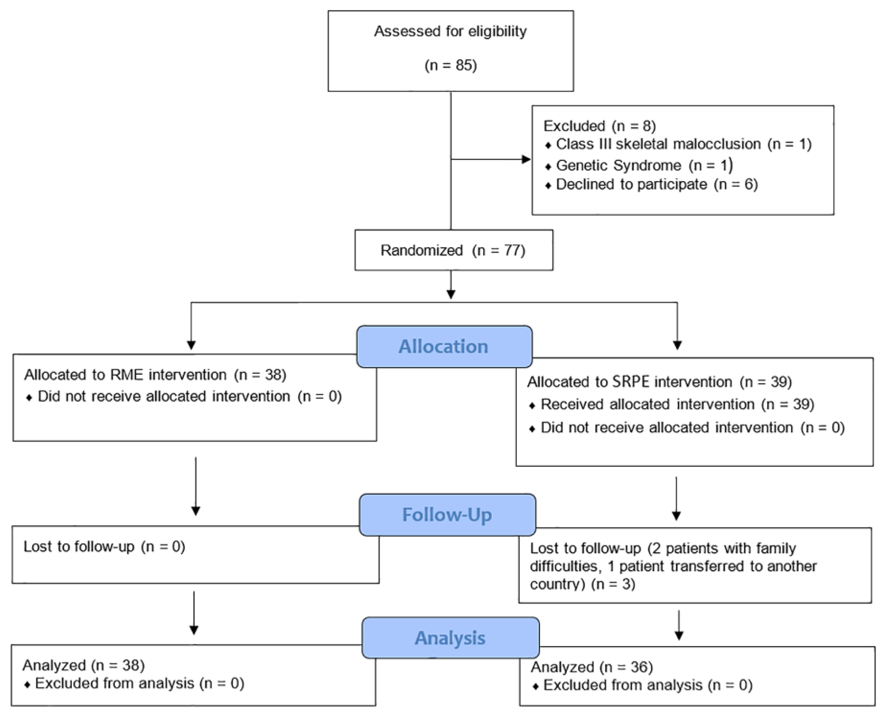
2.1. Study Design

The study protocol was approved by the Ethics Committee of Bambino Gesù Children’s Hospital (protocol number 286/2013). The study was designed as a randomized controlled trial (RCT) and followed the CONSORT checklist for RCT studies (Figure 1).
Sample size calculation: Sample size was calculated using G-Power© (Version 3.1.8, Heinrich-Heine-Universität, Düsseldorf, Germany) software analysis, assuming a significance level of 0.05 with a power of 90%. A sample size of 34 participants makes it possible to detect a difference in terms of effect size of 0.9, as suggested by the software. Considering possible dropouts, the study sought to recruit at least 25 patients per group.
Randomization: Enrolled patients were allocated treatment with either a two-banded expander, RME (Group A), or a splint resin palatal expander, SRPE (Group B). The patients were allocated to the two groups with stratification according to gender. Two randomization lists were prepared using a manual assignment, one for males and one for females, and the patients were randomly allocated to one of the two groups using a balanced block randomization method. A single operator allocated the patients by means of a custom-made Java script and was responsible for the concealment of allocations. The allocation was disclosed only when a new patient was enrolled in the trial. The patients and their parents were provided with detailed information about the trial and signed informed consent forms.
Blinding: It was not possible to ensure the blinding of the patients or clinicians during the study. However, the operators who performed the cephalometric, model, and statistical analyses were blinded to the treatment allocation. One expert orthodontist with 10 years of experience performed as a single operator for the measurements. Intra-examiner reliability was assessed by repeating measurements on anonymized patients, randomly selected after 2 weeks, and then mean values were subjected to statistical analysis. The cephalograms were labeled using numbers and randomly evaluated. The researchers, therefore, also did not know about the time point analysis.

2.2. Subjects and Methods

This study included patients with transverse discrepancy treated at the Dentistry Unit of Bambino Gesù Children’s Hospital (Rome, Italy) over a four-year period. Children aged 5–9 years were screened. The following baseline conditions were considered as inclusion criteria:
  • Deciduous, early, or late mixed dentition;
  • Unilateral or bilateral posterior crossbite affecting at least one cusp of the first permanent molar and/or second deciduous molar due to skeletal etiology.
The following baseline conditions were considered as exclusion criteria:
  • Birth defects or genetic syndromes (such as cleft lip or palate);
  • Systemic diseases that can affect the normal growth pattern;
  • Previous orthodontic treatment;
  • Signs and/or symptoms of temporomandibular disorders;
  • Severe periodontal problems.
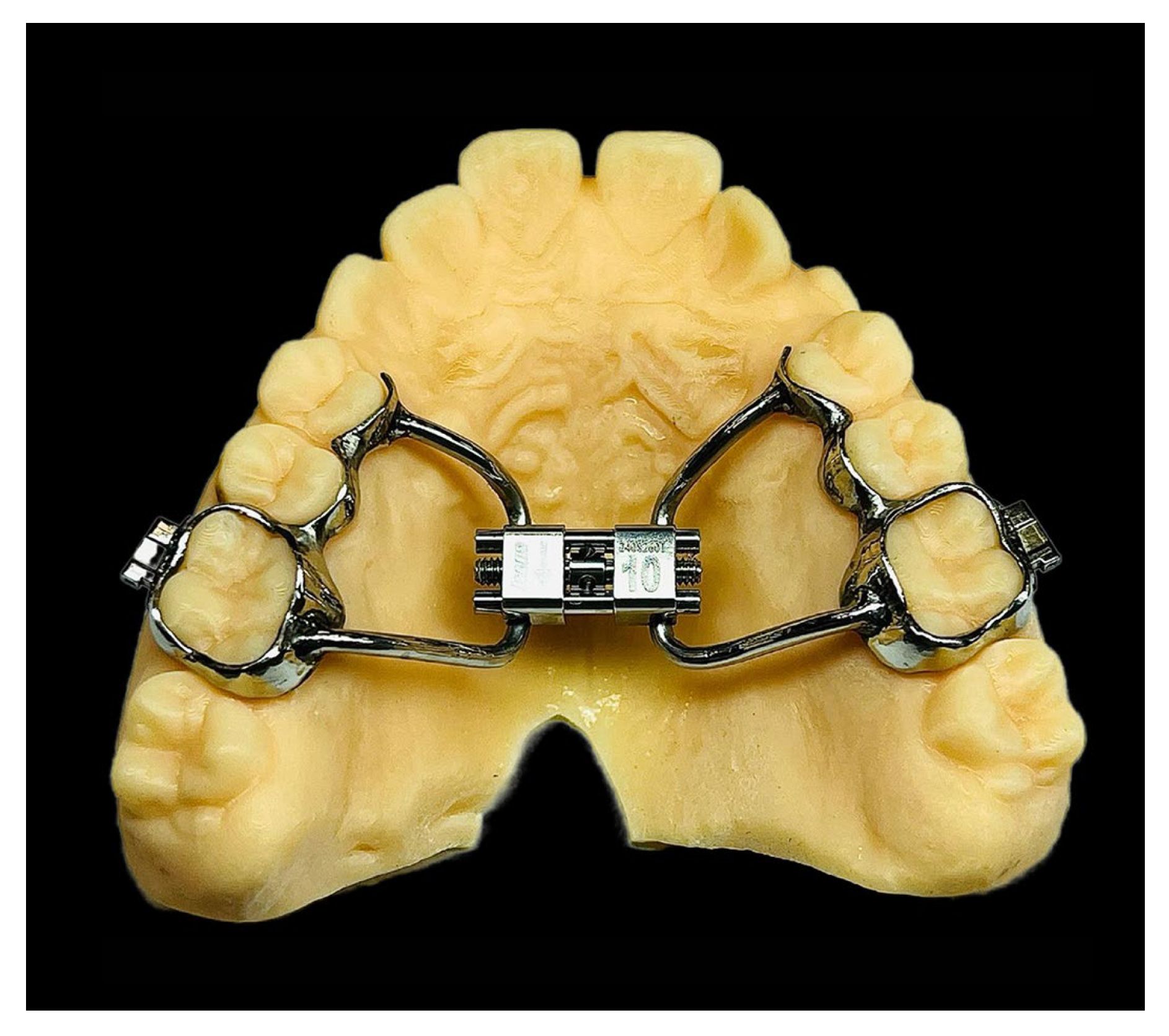
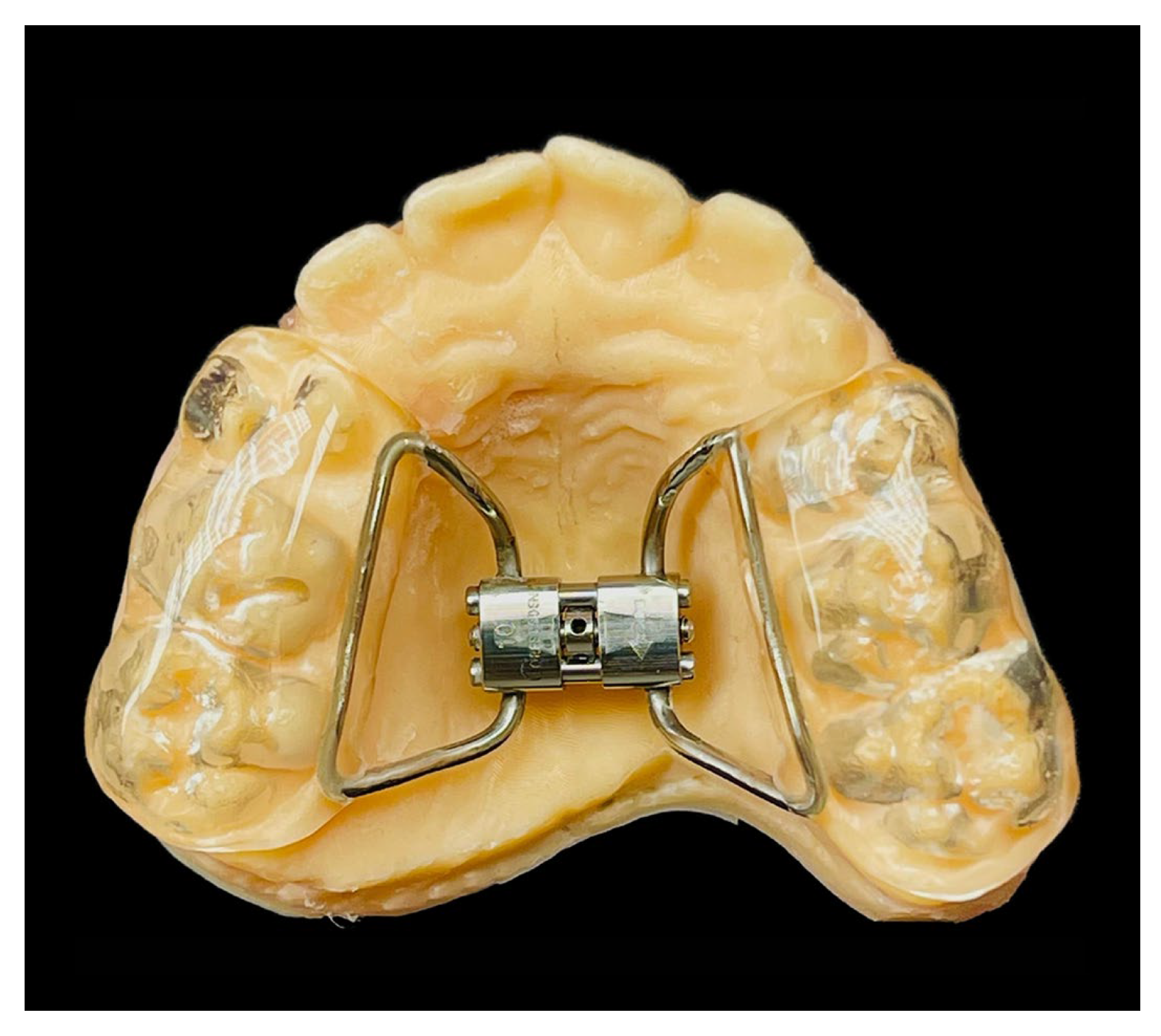
The patients and their parents were provided with detailed information about the trial and signed informed consent forms. For each enrolled patient, the following diagnostic data were collected: lateral cephalograms for cephalometric analysis, intraoral and extraoral photographs, and intraoral scans of upper and lower dental arches to obtain pre-treatment (T0) and post-treatment (T1) digital models.

2.3. Treatments

As mentioned above, the treatment consisted of the use of two different palatal expander devices: a two-band expander (RME) for Group A and a splint resin palatal expander (SRPE) for Group B. In both versions, the expander screw was positioned as close as possible to the palatal vault and very close to the posterior borders of the maxilla.
Group A (RME):
The two-band expander consisted of a central screw connected to two orthodontic bands using metal arms. The bands were positioned on two teeth, on the right and left sides (upper first molars or upper second deciduous molars), and from these two lateral arms extended up to the first deciduous molars (Figure 2).
Group B (SRPE):
The splint expander consisted of a central screw connected to two posterior bite-blocks (about 3 mm high) using metal arms. It was anchored on six teeth, three on the right and three on the left side: the first upper permanent molars and the first and second upper deciduous molars in mixed dentition, or the first and second upper deciduous molars and deciduous upper canines in deciduous dentition (Figure 3).
For both devices, the screw expands the transverse plane by 0.20 mm for a single activation. The total number of activations varied in each patient depending on the ideal correction.
In both groups, four screw activations (0.80 mm) were performed after the bonding of the device. Thereafter, the patient’s parents were instructed to turn the screw two times per day (0.40 mm activation per day). Each patient was provided with a custom-made diary and was instructed to report on the adjustments made to the device. The diary was checked at each visit by the clinical examiner to evaluate patient compliance.
In both groups, the jackscrew was activated until a 2 mm molar transverse overcorrection was achieved. After the active expansion phase, the screw was locked using brass wire and light-cure flow composite (Premise Flowable; Kerr Corporation, Orange, CA, USA). The palatal expander was removed six months after completion of the activations (T1), and final patient data were collected: intraoral and extraoral photographs, digital X-rays (provided by different radiographic units chosen by each patient’s parents or guardians), and digital models (3Shape© Intraoral Scanner, Copenhagen, Denmark) with superimposition (Figure 4).

2.4. Outcomes

The main outcome to be assessed was the success rate of crossbite correction. For both groups, digital measurements on virtual models were taken before (T0) and after treatment (T1) based on the following: maxillary and mandibular intercanine and intermolar width at the shortest linear distance at the gingival margins and the cusp tips of the teeth, overbite, and overjet (Figure 5). The secondary outcomes were skeletal changes evaluated on lateral cephalograms taken in the intercuspal position.

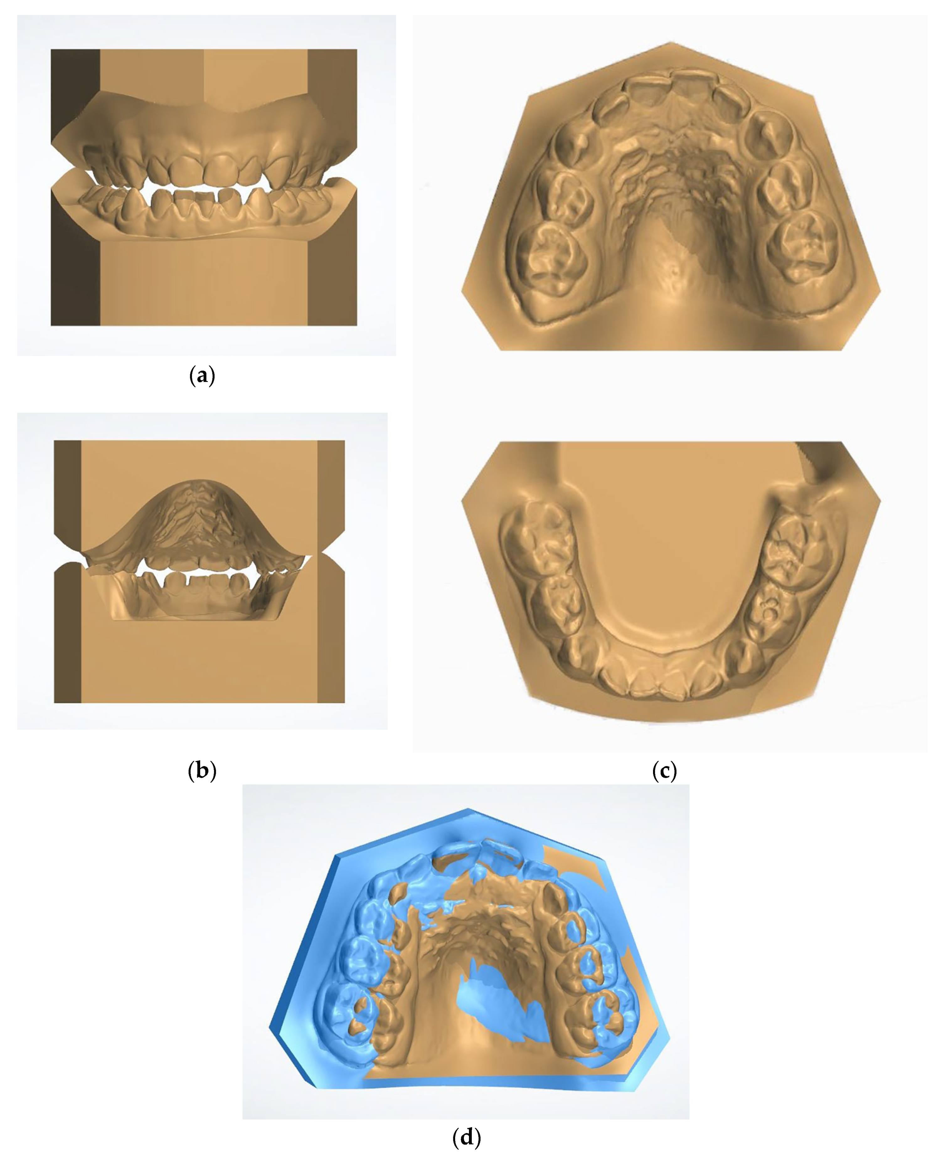
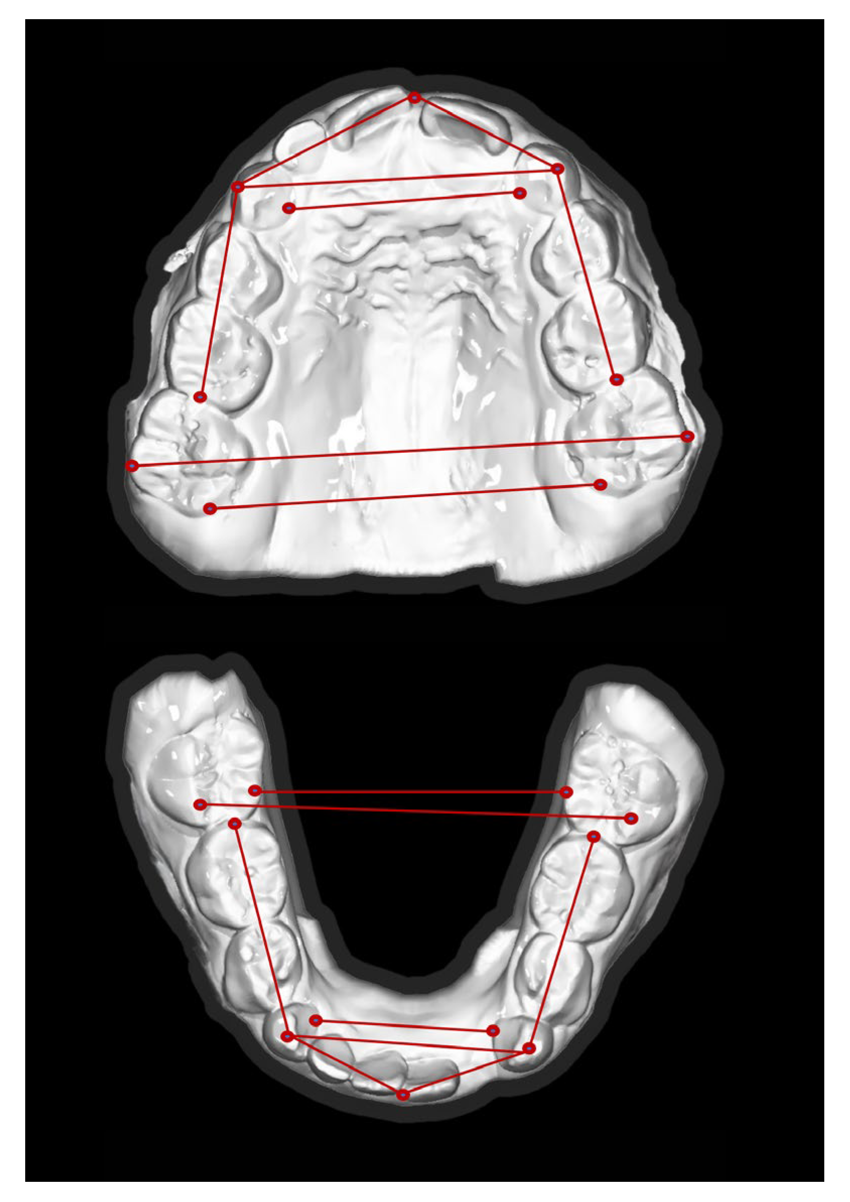
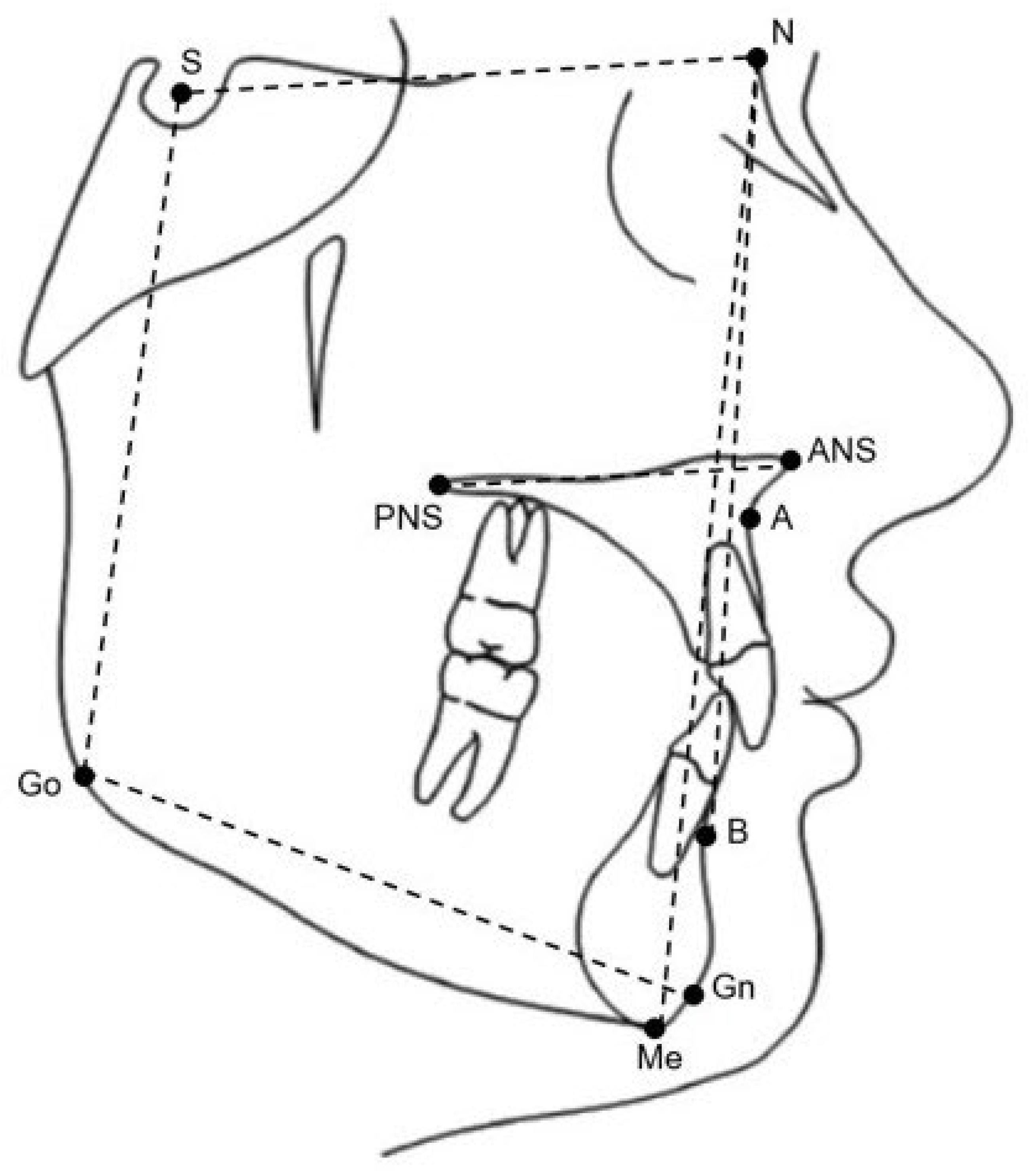
2.5. Cephalometric Analysis

Cephalometric measurements were taken before (T0) and after treatment (T1) using digital software (Dolphin Imaging 11.0 software, Chatsworth, CA, USA). The measuring points, reference points, and lines used are shown in Figure 6 and were defined based on the Pancherz method [16].
The following parameters were also considered: ANB angle, SNA angle and SNB angle, SN-ANS-PNS angle, SN-GoGn angle, ANS/PNS-GoGn angle, and P-A face height distance were used to evaluate the vertical skeletal relationship. The examiner had been extensively trained in digital cephalometric analysis and was unaware of the patients’ names and treatment allocations. The dates of the cephalometric X-rays were also concealed from the examiner when the measurements were taken. The T0 and T1 X-rays were randomly submitted to the examiner.

2.6. Statistical Analysis

The statistical analysis was based on raw scores. A single operator who was blinded to patient allocation (with treatment allocations therefore masked in the dataset) performed the statistical analysis. Data were analyzed using conventional descriptive statistics and inferential statistics. Means and standard deviations are presented for all variables, both cephalometric and dental measurements.
Statistical significance was set at p < 0.01, and significant results at p < 0.001 and p < 0.05 are alternatively reported when necessary. Analyses were performed using SPSS version 20.0 (SPSS IBM, New York, NY, USA). In order to accept or reject the null hypothesis H0: u1 = u2, where u1 is the mean of the population of patients with the two-banded expander and u2 the mean of the population with the splint resin expander, the t-test was applied to compare variations between initial cephalometric measurements and dental (maxillary and mandibular) measurements at baseline (T0). Subsequently, the paired t-test was applied to compare significant differences between the two groups related to cephalometric and dental measurements, both at T1. The t-test was also applied to compare the differences in cephalometric and dental measurements at T1 and T0 between the two groups.

3. Results

Eighty-five children with posterior crossbite were screened. Eight of these patients were excluded based on the inclusion/exclusion criteria and three patients were subsequently lost to follow-up (Figure 1). The age range of the patients was 5–9 years. The final group study therefore consisted of 74 patients (44 girls and 30 boys, mean age ± SD = 7.7 ± 1.1 years) divided into two groups: the first group (called Group A) consisted of 38 subjects (21 girls and 17 boys, mean age ± SD = 7.7 ± 1.1 years) treated with RME, and the second group (called Group B) consisted of 36 subjects (23 girls and 13 boys, mean age ± SD = 7.6 ± 1.0 years) treated with SRPE. The CONSORT flow diagram (Figure 1) demonstrates patient flow through the clinical trial. At baseline (T0), the two groups do not show significant differences for any of the skeletal variables (Table 1).
The mean (±SD) treatment duration was seven months (±one month). Dental baseline (T0) measurements for both the groups are not significantly different (Table 2).
The comparisons within both groups for the same variables at T0 and T1 are shown in Table 3 and Table 4.
The cephalometric measurements significantly changed in Group B from T0 to T1 for the SN-GoGn angle (increasing value) (t test = 0.009; p < 0.001) and for the P-A face height percentage (decreasing value) (t test = 0.008; p < 0.001), as shown in Table 3. The dental measurements significantly changed in Groups A and B from T0 to T1 (Table 4) for the following measurements: the maxillary intermolar and intercanine distance (Group A, t test = 0.000; Group B, t test = 0.000, both for p < 0.01); the mandibular intercanine distance in Group B (t test = 0.028, p < 0.028); the overjet value in Group A (t-test = 0.011, p < 0.05); and the overbite value in Group B (t-test, p < 0.05). There are no statistically significant differences between Group A and Group B in terms of sagittal and vertical skeletal variables (Table 5).
The difference in the maxillary intercanine distances between the groups (A and B) from T0 to T1 is statistically significant (t-test = 0.000, p < 0.001) (Table 6). The values for measurement of the widths of the mandible arch were unchanged after treatment in both groups. There were no statistically significant changes in the values of overjet and overbite in both groups.

4. Discussion

The purpose of this study was to compare the use of two different palatal expander designs to evaluate the difference in skeletal and dental outcomes in pediatric patients. The devices traditionally used to treat crossbites—and also used in this study—are the rapid palatal expander with metallic bands bonded onto the first molars and the median screw (RME), and the palatal expander equipped with resin splints (SRPE) as an alternative to the metallic bands, usually referred to as a McNamara expander [8,10]. Even though current therapeutic approaches in orthodontics encourage the use of “bone-borne” techniques to provide palatal vault expansion, especially in patients with increased maturation and interdigitation of the mid-palatal suture [13,14,15], the use of the abovementioned “tooth-borne” devices remains the primary therapeutic choice in growing patients affected by transverse deficiency [16,17]. This is also because of the limited degree of acceptance of temporary anchorage devices among both patients and dentists [18,19]. Dental side effects of traditional palatal expanders in the correction of posterior crossbite have been widely debated among experts. These relate mainly to the occurrence of buccal tipping of the upper molars, root resorption, and other side effects such as bone dehiscence and unstable results culminating in relapse [20]. This is of particular importance in all those cases with abnormal development of dentition, especially if misdiagnosis errors occur [21]. The findings of this study provide significant insights into the skeletal and dental effects of two different rapid maxillary expansion devices, the two-band expander (RME) and the splint resin palatal expander (SRPE), in children with maxillary contraction. Both devices effectively corrected posterior crossbite without inducing adverse vertical skeletal changes, making them suitable for use in hyperdivergent patients. The study demonstrated a significant increase in maxillary intercanine width with SRPE compared to RME (mean ± SD = 6.0 ± 4.8 mm vs. 3.1 ± 2.9 mm; p = 0.00), highlighting the advantage of using SRPE in expanding the anterior dental arch. This is consistent with previous findings [12], when similar increases in intercanine width were observed with the use of bonded palatal expanders. The difference in performance between SRPE and RME may be attributed to the lateral slides in the SRPE, which favor anterior expansion. Both devices showed a similar increase in maxillary intermolar distance (RME: 7.3 ± 3.4 mm; SRPE: 7.6 ± 5.1 mm), consistent with findings from Lione et al. [22], who reported no significant differences in intermolar expansion between bonded and banded expanders. The significant change in mandibular arch dimensions post-treatment aligns with the results found by Laganà et al. [23] only for the SRPE group, suggesting that the mandibular response to maxillary expansion can hopefully be achieved. Skeletal variables, including vertical dimensions, did not show significant changes between the two groups. The presence of significant effects on vertical skeletal variables (SN-GoGn) does not support previous studies [11,12,24], which reported no long-term changes in mandibular plane angle following RME. These findings could be crucial, as the vertical effects of maxillary expansion remain controversial, with some studies suggesting temporary increases in vertical height due to overexpansion impacts [6,10]. Our study found no significant changes in sagittal variables (ANB, SNA, and SNB) in either group, supporting the hypothesis that RME primarily affects the transverse plane, with minimal impact on anteroposterior skeletal relationships. However, the lack of long-term control must make us cautious about the stability of the vertical skeletal dimension provided by the two devices. These results are consistent with Solow and Kreiborg [25], who noted, in original first studies, that craniofacial growth and morphogenesis could be stabilized by soft tissue constraints, mitigating skeletal changes from RME. These observations are still valid if we also look at the effect on the sagittal airway changes induced by the rapid palatal expansion reported by other authors [26,27].
Despite the short-term benefits observed, the study acknowledges limitations, including the lack of long-term follow-up data to assess the stability of dental and skeletal changes. Future research should aim to evaluate the long-term retention of the effects of SRPE and RME, especially considering the potential for relapse in dental arch widths. The potential bias of single-operator measurements and the growth variability of our patient group could be further sources of research, also through the use of 3D images.

5. Conclusions

In conclusion, this randomized controlled trial demonstrates that both RME and SRPE are effective in treating maxillary contraction without significant adverse skeletal effects [26,27,28], even if vertical control is better with the RME device, to short-term observation. However, SRPE may be preferred in cases where increased anterior expansion is desired due to its superior performance in increasing intercanine width.

Author Contributions

Conceptualization, A.G. and V.D.; methodology, P.F.; software, G.G.; validation, A.G., V.V. and A.P.; formal analysis, G.B.; investigation, G.G.; resources, A.M.; data curation, G.B., P.F., G.G., A.P. and V.V.; writing—original draft preparation, V.V.; writing—review and editing, A.P.; visualization, A.G., V.D. and V.V.; supervision, A.G. All authors have read and agreed to the published version of the manuscript.

Funding

This work was also supported by the Italian Ministry of Health through “current research funds”.

Institutional Review Board Statement

The study was conducted in accordance with the Declaration of Helsinki and approved by Ethics Committee of Bambino Gesù Children’s Hospital (protocol code 622.13 and 24.04.13).

Informed Consent Statement

Informed consent was obtained from all subjects involved in the study.

Data Availability Statement

Research data are unavailable due to privacy provisions.

Acknowledgments

We would like to thank Patrizio Evangelista and the orthodontic laboratory team (Orthonet, Roma) for their support.

Conflicts of Interest

The authors declare no conflicts of interest.

References

  1. Iodice, G.; Danzi, G.; Cimino, R.; Paduano, S.; Michelotti, A. Association between posterior crossbite, skeletal, and muscle asymmetry: A systematic review. Eur. J. Orthod. 2016, 38, 638–651. [Google Scholar] [CrossRef]
  2. Malandris, M.; Mahoney, E.K. Aetiology, diagnosis and treatment of posterior crossbites in the primary dentition. Int. J. Paediatr. Dent. 2004, 14, 155–166. [Google Scholar] [CrossRef]
  3. Petrèn, S.; Bondemark, L.; Soderfeldt, B. A systematic review concerning early orthodontic treatment of unilateral posterior cross bite. Angle Orthod. 2003, 73, 588–596. [Google Scholar] [PubMed]
  4. Thilander, B.; Bjerklin, K. Posterior crossbite and temporomandibular disorders (TMDs): Need for orthodontic treatment? Eur. Orthod. 2012, 34, 667–673. [Google Scholar] [CrossRef]
  5. Veli, I.; Uysal, T.; Ozer, T.; Ucar, F.I.; Eruz, M. Mandibular asymmetry in unilateral and bilateral posterior crossbite patients using cone-beam computed tomography. Angle Orthod. 2011, 81, 966–974. [Google Scholar] [CrossRef] [PubMed]
  6. Lione, R.; Franchi, L.; Cozza, P. Does rapid maxillary expansion induce adverse effects in growing subjects? Angle Orthod. 2013, 83, 172–182. [Google Scholar] [CrossRef] [PubMed]
  7. Bistaffa, A.G.I.; Belomo-Yamaguchi, L.; de Castro Ferreira Conti, A.C.; Oltramari, P.V.P.; de Almeida, M.R.; de Almeida-Pedrin, R.R.; Fernandes, T.M.F. Immediate skeletal effects of rapid maxillary expansion at midpalatal suture opening with Differential, Hyrax and Haas expanders. Dent. Press J. Orthod. 2022, 27, e2220525. [Google Scholar] [CrossRef]
  8. Harrison, J.E.; Ashby, D. Orthodontic treatment for posterior crossbites. Cochrane Database Syst. Rev. 2000, 2, CD000979. [Google Scholar]
  9. Caroccia, F.; Moscagiuri, F.; Falconio, L.; Festa, F.; D’Attilio, M. Early Orthodontic Treatments of Unilateral Posterior Crossbite: A Systematic Review. J. Clin. Med. 2020, 10, 33. [Google Scholar] [CrossRef]
  10. Basciftci, F.A.; Karaman, A.I. Effects of a modified acrylic bonded rapid maxillary expansion appliance and vertical chin cap on dentofacial structures. Angle Orthod. 2002, 72, 61–71. [Google Scholar]
  11. Lineberger, M.W.; McNamara, J.A.; Baccetti, T.; Herberger, T.; Franchi, L. Effects of rapid maxillary expansion in hyperdivergent patients. Am. J. Orthod. Dentofac. Orthop. 2012, 142, 60–69. [Google Scholar] [CrossRef] [PubMed]
  12. Wendling, L.K.; McNamara, J.A., Jr.; Franchi, L.; Baccetti, T. A prospective study of the short-term treatment effects of the acrylic-splint rapid maxillary expander combined with the lower Schwarz appliance. Angle Orthod. 2005, 75, 7–14. [Google Scholar] [PubMed]
  13. Suzuki, H.; Moon, W.; Previdente, L.H.; Suzuki, S.S.; Garcez, A.S.; Consolaro, A. Miniscrew-assisted rapid palatal expander (BONE BORNE EXPANSION): The quest for pure orthopedic movement. Dent. Press J. Orthod. 2016, 21, 17–23. [Google Scholar] [CrossRef]
  14. Huang, X.; Han, Y.; Yang, S. Effect and stability of miniscrew-assisted rapid palatal expansion: A systematic review and meta-analysis. Korean J. Orthod. 2022, 52, 334–344. [Google Scholar] [CrossRef] [PubMed]
  15. Bazargani, F.; Lund, H.; Magnuson, A.; Ludwig, B. Skeletal and dentoalveolar effects using tooth-borne and tooth-bone-borne RME appliances: A randomized controlled trial with 1-year follow-up. Eur. J. Orthod. 2021, 43, 245–253. [Google Scholar] [CrossRef]
  16. Krüsi, M.; Eliades, T.; Papageorgiou, S.N. Are there benefits from using bone-borne maxillary expansion instead of tooth-borne maxillary expansion? A systematic review with meta-analysis. Prog. Orthod. 2019, 20, 9. [Google Scholar] [CrossRef]
  17. Vincenzo, G.; Fabrizia, D.; Abdolreza, J.; Felice, F.; Lorenzo, F.; Letizia, P. Comparison between rapid and mixed maxillary expansion through an assessment of arch changes on dental casts. Prog. Orthod. 2015, 16, 20. [Google Scholar] [CrossRef]
  18. Sampson, A.; Figueiredo, D.S.F.; Jeremiah, H.G.; Oliveira, D.D.; Freitas, L.R.P.; Chahoud, M.; Soares, R.V.; Cobourne, M.T. The effect of social media on patient acceptance of temporary anchorage devices. Angle Orthod. 2021, 91, 363–370. [Google Scholar] [CrossRef]
  19. Mehta, S.; Arqub, S.A.; Vishwanath, M.; Upadhyay, M.; Yadav, S. Biomechanics of conventional and miniscrew-assisted rapid palatal expansion. J. World Fed. Orthod. 2024, 13, 105–112. [Google Scholar] [CrossRef]
  20. Cheng, J.H.-C.; Chen, D.D.-S.; Tan, Y.; Hu, H.T. Factors associated with usage frequency and pricing of temporary anchorage devices among orthodontists. J. Dent. Sci. 2024, 19, 404–410. [Google Scholar] [CrossRef]
  21. Putrino, A.; Caputo, M.; Galeotti, A.; Marinelli, E.; Zaami, S. Type I Dentin Dysplasia: The Literature Review and Case Report of a Family Affected by Misrecognition and Late Diagnosis. Medicina 2023, 59, 1477. [Google Scholar] [CrossRef] [PubMed]
  22. Lione, R.; Huanca Ghislanzoni, L.T.; Defraia, E.; Franchi, L.; Cozza, P. Bonded versus banded rapid palatal expander followed by facial mask therapy: Analysis on digital dental casts. Eur. J. Orthod. 2016, 38, 217–222. [Google Scholar] [CrossRef] [PubMed]
  23. Laganà, G.; Paoloni, V.; Pavoni, C.; Palmacci, D.; Malara, A. Tridimensional Changes in Mandibular Arch after Rapid Maxillary Expansion Therapy: A Clinical Study. Children 2023, 10, 775. [Google Scholar] [CrossRef] [PubMed]
  24. Conroy-Piskai, C.; Galang-Boquiren, M.T.S.; Obrez, A.; Costa Viana, M.G.; Oppermann, N.; Sanchez, F.; Edgren, B.; Kusnoto, B. Assessment of vertical changes during maxillary expansion using quad helix or bonded rapid maxillary expander. Angle Orthod. 2016, 86, 925–933. [Google Scholar] [CrossRef]
  25. Solow, B.; Kreiborg, S. Soft-tissue stretching: A possible control factor in craniofacial morphogenesis. Scand J. Dent. Res. 1977, 85, 505–507. [Google Scholar] [CrossRef]
  26. Galeotti, A.; Viarani, V.; Franchi, L.; Martina, S.; Rongo, R.; D’Antò, V.; Uomo, R.; Aristei, F.; Festa, P. Cephalometric changes of pushing splints 3 compared to rapid maxillary expansion and facemask therapy on the airway space in class III growing patients: A randomized clinical trial. Orthod. Craniofac. Res. 2024, 27, 552–559. [Google Scholar] [CrossRef]
  27. Chang, J.Y.; McNamara, J.A., Jr.; Herberger, T.A. A longitudinal study of skeletal side effects induced by rapid maxillary expansion. Am. J. Orthod. Dentofac. Orthop. 1997, 112, 330–337. [Google Scholar] [CrossRef]
  28. O’Grady, P.W.; McNamara, J.A., Jr.; Baccetti, T.; Franchi, L. A long-term evaluation of the mandibular Schwarz appliance and the acrylic splint expander in early mixed dentition patients. Am. J. Orthod. Dentofac. Orthop. 2006, 130, 202–213. [Google Scholar] [CrossRef]
Figure 1. CONSORT 2010 flow diagram.
Applsci 15 07187 g001
Figure 2. Occlusal view of two-band expander (RME) device (Orthonet orthodontic laboratory, Rome, Italy).
Applsci 15 07187 g002
Figure 3. Occlusal view of splint resin palatal expander (SRPE) device.
Applsci 15 07187 g003
Figure 4. Digital model (in beige) taken by intraoral scanner and presented in the following views: (a) frontal; (b) posterior; (c) occlusal; and (d) superimposition of the upper arches (pre-treatment in beige and post-treatment in blue).
Applsci 15 07187 g004
Figure 5. Example of transverse linear measurement on the digital models reporting intercanine and intermolar distances.
Applsci 15 07187 g005
Figure 6. Cephalometric analysis and landmarks. Landmarks: A, most posterior point of the frontal concavity of the maxilla between the anterior nasal spine and the alveolar process; B, innermost point of the lower jaw region, between the alveolar process and pogonion; N (nasion), most anterior point of the junction of the nasal and frontal bone (frontonasal suture); S (sella), center of the hypophyseal fossa; Go (gonion), midpoint of the curvature at the angle of the mandible; Gn (gnathion), point of the mandibular symphysis on the facial axis; Me (menton), most inferior point of the mandibular symphysis; ANS (anterior nasal spine), the tip of anterior nasal spine; and PNS (posterior nasal spine), most posterior point of the bone palate. Reference: NA (nasion A line)—line through N and A; NB (nasion B line)—line through N and B; SN (sella-nasion line)—line through S and N; MP (mandibular plane)—line connecting the Go point to the Gn point; posterior facial height—line connecting the S point to the Go point; anterior facial height—line connecting the N point to the Me point; PP (palatal plane)—line connecting ANS and PNS. Linear distances/skeletal landmarks: the angular measurements are represented by: SNA (angle formed by the intersection of lines SN and NA); SNB (angle formed by the intersection of lines SN and NB); ANB (difference between angles SNA and SNB); SN—GoGn (craniomandibular angle)—indicates the degree of divergence of the mandible from the cranial base; SN^ANS-PNS—indicates the degree of divergence of the maxilla from the cranial base; MM (bimaxillary angle); ANS/PNS^GoGn—indicates the degree of divergence of the mandible from the maxilla; P-A face height (S-Go/N-Me).
Applsci 15 07187 g006
Table 1. Baseline cephalometric comparison of experimental groups.
Group A (n = 38)Group B (n = 36)
Cephalometric MeasurementsMeanSDMeanSD95% CI for Meanp Value (t Test)
ANB (degrees)4.62.442.10.570.1
SNB (degrees)76.33.175.73.10.530.3
SNA (degrees)80.83.479.731.10.2
SN-GoGn (degrees)32.84.333.85.2−10.5
SN_ANS_PNS (degrees)9.33.29.33.6−0.10.9
ANS_PNS_GoGn (degrees)26.24.227.14.8−0.80.8
P-A face height (percentage)62.93.862.24.10.70.5
Table 2. Baseline measurements (mm) for comparison of experimental groups.
Group A (n = 38)Group B (n = 36)
Baseline DataMeanSDMeanSD95% CI for Meanp Value (t Test)
Maxilla
Intermolar distance (mesiobuccal cusp tips)45.13.944.75.60.40.87
Intercanine distance (mesiobuccal cusp tips)29.63.728.451.20.51
Mandible
Intermolar distance (mesiobuccal cusp tips)45.34.245.33.2−0.10.65
Intercanine distance (mesiobuccal cusp tips)25.92.425.9300.92
Overjet2.32.12.42−0.10.96
Overbite1.91.81.51.60.450.14
Table 3. Cephalometric measurements: intra-group comparisons T0 and T1.
Group A (n = 38)Group B (n = 36)
Cephalometric MeasurementsMeanSDDifferencep Value (t Test)MeanSDDifferencep Value (t Test)
ANB (degrees)4.32.1−0.30.2643.81.8−0.20.253
SNB (degrees)762.900.41475.43.1−0.30.321
SNA (degrees)80.33.5−0.50.09979.13.3−0.70.137
SN-GoGn (degrees)32.94.10.10.8234.75.8−0.90.009 *
SN_ANS_PNS (degrees)9.43−0.10.787103.70.70.084
ANS_PNS_GoGn (degrees)26.13.6−0.20.76727.34.60.20.564
P-A face height (percentage)62.73.20.20.66761.14.81.10.008 *
* p < 0.01.
Table 4. Dental measurements (mm): intra-group comparisons T0 and T1.
Group A (n = 38)Group B (n = 36)
Baseline DataMeanSDDifferencep Value (t Test)MeanSDDifferencep Value (t Test)
Maxilla
Intermolar distance (mesiobuccal cusp tips)52.43.3−7.30.000 *52.32.8−7.60.000 *
Intercanine distance (mesiobuccal cusp tips)32.72.8−3.10.000 *34.42.8−5.90.000 *
Mandible
Intermolar distance (mesiobuccal cusp tips)45.72.7−0.40.40945.92.9−0.60.175
Intercanine distance (mesiobuccal cusp tips)25.92.200.994272.2−1.10.028 ‡
Overjet2.92.2−0.60.011 ‡2.61.6−0.20.47
Overbite2.41.6−0.50.052.11.6−0.60.010 ‡
* p < 0.01; ‡ p < 0.05.
Table 5. Cephalometric comparison of T1–T0 changes.
Group A (n = 38)Group B (n = 36)
Cephalometric MeasurementsMeanSDMeanSDDifferencep Value (t Test)
ANB (degrees)−0.31.6−0.21.2−0.10.67
SNB (degrees)−0.21.6−0.320.10.78
SNA (degrees)−0.51.8−0.72.70.20.62
SN-GoGn (degrees)0.12.30.92−0.80.06
SN_ANS_PNS (degrees)0.12.60.72.3−0.50.56
ANS_PNS_GoGn (degrees)−0.23.40.22.4−0.40.43
P-A face height (percentage)−0.22.51.12.3−1.30.1
Table 6. Baseline measurements (mm) for comparison of T1–T0 changes.
Group A (n = 38)Group B (n = 36)
Baseline DataMeanSDMeanSDDifferencep Value (t Test)
Maxilla
Intermolar distance (mesiobuccal cusp tips)7.33.47.65.1−0.30.54
Intercanine distance (mesiobuccal cusp tips)3.12.964.8−2.80.000 *
Mandible
Intermolar distance (mesiobuccal cusp tips)0.430.62.5−0.20.27
Intercanine distance (mesiobuccal cusp tips)01.41.12.9−1.1 *0.1
Overjet0.61.40.21.70.40.76
Overbite0.51.50.61.4−0.10.35
* p < 0.01.
Disclaimer/Publisher’s Note: The statements, opinions and data contained in all publications are solely those of the individual author(s) and contributor(s) and not of MDPI and/or the editor(s). MDPI and/or the editor(s) disclaim responsibility for any injury to people or property resulting from any ideas, methods, instructions or products referred to in the content.

