Article

Polysaccharide Hydrogels Based on Cellulose and Chitosan for Drug Sustained-Release Applications

1
A Key Lab of Science and Technology of Eco-Textile, Ministry of Education, College of Chemistry and Chemical Engineering, Donghua University, Shanghai 201620, China
2
Innovation Center for Textile Science and Technology of DHU, Donghua University, Shanghai 201620, China
3
Key Lab of Material & Technology of Textile, College of Fashion and Textiles, Tarim University, XP, Alaer 843300, China
*
Author to whom correspondence should be addressed.
Appl. Sci. 2025, 15(10), 5601; https://doi.org/10.3390/app15105601
Submission received: 11 April 2025 / Revised: 8 May 2025 / Accepted: 14 May 2025 / Published: 16 May 2025

Abstract

This study developed a novel water-soluble Cellulose Acetoacetate (CAA)-chitosan (CS) composite hydrogel drug delivery system. In this system, CAA and CS molecules are cross-linked via dynamic enamine bonds, forming a three-dimensional network structure suitable for drug encapsulation and controlled release. The primary objective was to address the challenges associated with the short half-life and significant fluctuations in therapeutic concentration of cytokine drugs, such as interleukin-2 (IL-2). A hydrogel system with a three-dimensional spatial network structure was successfully constructed via dynamic enamine bonds cross-linking between the acetoacetate groups in CAA molecules and the amino groups in CS. This system exhibits the following characteristics: (1) Dynamic covalent bonds impart adjustable mechanical properties to the hydrogel, enabling precise control over gelation time and mechanical performance; (2) A hierarchical pore structure (average pore size of 100–200 μm) provides a three-dimensional confined space for efficient drug encapsulation, achieving an IL-2 encapsulation efficiency of 83.3 ± 3.1%; (3) In vitro release studies demonstrated that the cumulative release of IL-2 within 72 h ranged from 18.4% to 34.7%, indicating sustained-release behavior. Cell viability assays confirmed that the hydrogel maintained the survival rate of L929 cells above 85% (as determined by the CCK-8 method), and live/dead staining revealed no apparent cytotoxicity. Overall, this three-dimensional network hydrogel based on dynamic covalent bonds represents a promising strategy for low-dose, long-lasting cytokine delivery.

Graphical Abstract

1. Introduction

Low-dose cytokine drugs have demonstrated promising therapeutic potential in the treatment of various autoimmune disease [1,2,3]. Given the short half-life of cytokines in vivo, frequent injections are necessary to maintain therapeutic concentrations, which can lead to a range of adverse reactions [2,4]. Therefore, developing injectable drug-loaded sustained-release materials represents an effective strategy to enhance therapeutic efficacy while mitigating side effects. Drug-loaded sustained-release materials can be categorized into organic polymer-based and inorganic non-metallic-based systems. Organic polymers, particularly hydrogels derived from natural sources (e.g., gelatin, cellulose, sodium alginate, hyaluronic acid, and chitosan) and synthetic polymers (e.g., polyacrylic acid [PAA], polyacrylamide [PAM]), dominate this field [5]. However, synthetic organic polymers often suffer from issues such as residual toxic cross-linking agents, poor biocompatibility, and lower drug loading capacity compared to their natural counterparts.
Hydrogels, as three-dimensional porous hydrophilic polymer networks, exhibit superior properties including high water content, flexibility, mechanical strength, biocompatibility, and degradability [6,7,8]. Injectable hydrogels, in particular, offer enhanced tissue and physiological adaptability, adjustable physical and chemical properties, low toxicity, good compatibility, and controllable degradation performance [9,10,11,12,13]. When used in drug delivery systems, injectable hydrogels enable localized drug release at the target site, thereby improving therapeutic outcomes and reducing systemic side effects [14,15,16]. Cellulose, rich in hydroxyl groups and hydrogen bonds [17,18,19], possesses excellent mechanical strength and properties. Cellulose hydrogels are advantageous for their hydrophilicity, biodegradability, biocompatibility, transparency, low cost, and non-toxicity [20], making them valuable for oral, ocular, and topical drug delivery systems [21]. Chitosan, a natural cationic polysaccharide with favorable biocompatibility and degradability, is an ideal material for hydrogel preparation [22,23,24]. Acetylated cellulose acetate (CAA), a derivative of cellulose acetylated with acetoacetic acid, features highly reactive acetyl groups that can form stable dynamic enamine bonds with amino groups under mild conditions [6,7,14]. The introduction of acetoacetyl groups into natural polymers significantly enhances the modification of polysaccharides and expands research possibilities.
In this study, water-soluble cellulose acetate acetoacetate (CAA) with dynamic enamine bonds was synthesized under mild conditions and cross-linked with chitosan to prepare CAA-CS polysaccharide hydrogels with a three-dimensional network structure. The morphological characteristics were analyzed by scanning electron microscopy, and rheological analysis was conducted to evaluate mechanical properties and stability. Under simulated physiological conditions, IL-2 was encapsulated, and the drug encapsulation rate, release rate, and biocompatibility were evaluated [25].

2. Experimental

2.1. Materials

Materials: wood pulp cellulose was provided by Xinxiang Natural Chemical Company (Xinxiang, China), 1-allyl-3-methylimidazolium chloride (AMIMCl) ionic liquid was purchased from the Green Chemistry and Catalysis Center of the Chinese Academy of Sciences (Lanzhou, China), chitosan (deacetylation degree ≥ 95%, molecular weight = 100,000), PBS buffer solution, and tert-butyl acetoacetate (t-BAA, 99%) were provided by China National Pharmaceutical Group Chemical Reagent Co., Ltd. (Beijing, China). IL-2 was provided by Beijing Sihuan Biotechnology Co., Ltd. (Beijing, China), source-refined grade bovine serum albumin (BSA) was purchased from Shanghai Aladdin Biochemical Technology Co., Ltd. (Shanghai, China). L929 mouse fibroblasts were purchased from KeyGEN BioTECH (Nanjing, China), and fetal bovine serum was purchased from Shanghai Nacang Biological Company (Shanghai, China). CCK-8 biological reagent, penicillin double-specific antibody solution, and DMEME culture medium were purchased from Beyotime Biotechnology Co., Ltd. (Shanghai, China). All other chemical reagents were of analytical purity and could be used without further purification.

2.2. Preparation of Hydrogel

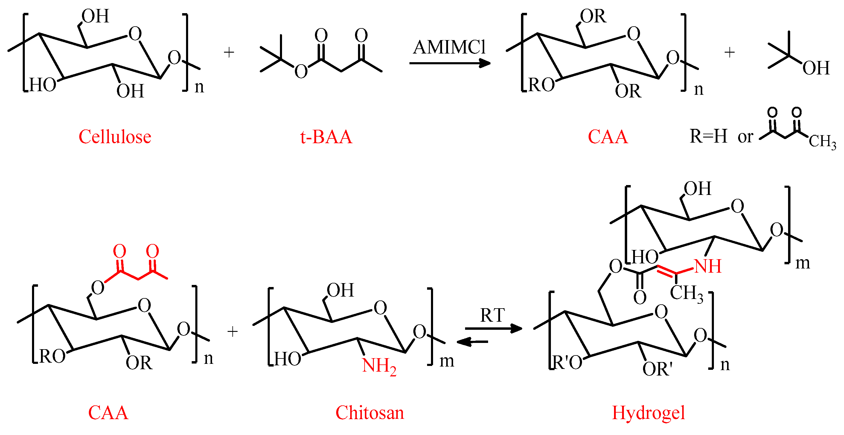
2.2.1. Synthesis of CAA

Soluble Cellulose Acetoacetate (CAA, DS = 0.85) was synthesized based on the established method [26] with certain improvements. Briefly, the pre-treated wood pulp cellulose was dissolved in 1-allyl-3-methylimidazolium chloride ionic liquid (90 °C), diluted with DMF, and then reacted with tert-butyl acetoacetate at 110 °C under nitrogen protection for 4 h (The molar ratio of t-BAA to cellulose is 4:1). The product was washed with ethanol and vacuum dried to obtain light yellow CAA powder. The reaction mechanism is depicted in Figure 1. The degree of substitution (DS) was determined to be 0.85 through 1H NMR analysis.

2.2.2. Synthesis of CAA-CS Hydrogels

The hydrogel was fabricated via a double-tube injection approach. The specific operation is detailed as follows: First, 2 mL medical syringes were separately loaded with the CAA-IL2 aqueous solution (with a CAA concentration ranging from 1.5 to 2.5%) and the chitosan solution (2%). The two syringes were connected by a three-way valve and propelled at a constant rate of 0.5 mL/min under room temperature, maintaining a 1:1 volume ratio of the mixture. The mixed solution was injected into a 2 mL centrifuge tube, inverted to observe the flow state, and the gelation time was recorded when the solution completely ceased flowing.

2.3. Physiochemical Characterization of CAA-CS Hydrogels

2.3.1. H NMR Spectroscopy

The 1H NMR spectra of CAA samples were recorded using an Avance 400 MHz NMR spectrometer (Bruker, Ettlingen, Germany) in the proton noise-decoupling mode with a standard 5 mm probe. The degree of substitution (DS) of the acetoacetate groups in CAA was calculated using Equation (1).
D S = I 1 × 7 I 2 × 3
where I1 is the integral area of the methyl protons in the acetoacetyl group (with three protons per methyl group), and I2 is the integral area of the anomeric glucoside protons in the AGU unit (with seven protons per AGU unit).

2.3.2. FT-IR Spectrometer

Fourier transform infrared (FTIR) spectroscopy was performed using a PerkinElmer Spectrum Two instrument (PerkinElmer, Waltham, MA, USA) equipped with an attenuated total reflectance (ATR) accessory. All spectra were recorded in the wavenumber range of 4000–400 cm−1 at a resolution of 4 cm−1.

2.3.3. Morphological Characterization of CAA-CS Polysaccharide Hydrogel

The microstructure characteristics of the hydrogel were jointly characterized via scanning electron microscopy (SEM) and porosity measurement. The hydrogel samples were treated with liquid nitrogen, then fixed with conductive adhesive and sputtered with gold under vacuum conditions. Subsequently, the morphology of the hydrogel was observed by SEM. To determine the porosity of the hydrogel, the samples were immersed in anhydrous ethanol for 48 h to reach equilibrium, and the volume of adsorbed ethanol was measured to calculate the average porosity. The porosity was calculated using Equation (2a,b) [27].
P o r o s i t y   ( % ) = v a v e × 100 %
V a = w 48 w 0 ρ
where w48 is the mass of the gel after soaking for 48 h, w0 is the initial mass of the gel sample, ρ is the density of ethanol, and ve is the external surface volume of the gel (three samples per group were tested, and the results were averaged).

2.3.4. The Stability Test of CAA-CS Composite Hydrogel

The swelling degree belongs to an important index of stability tests. The stability of hydrogels in physiological environments directly influences their drug sustained-release performance. Excessive swelling would result in structural collapse [28,29]. The swelling ratio of CAA-CS hydrogels was determined using a gravimetric method. Hydrogel samples were immersed in phosphate-buffered saline (PBS) and incubated in a constant-temperature water bath at 37 °C. At specific time intervals, the hydrogel samples were removed, excess surface water was blotted with filter paper, and the samples were weighed to observe the swelling behavior of the polysaccharide gels under physiological conditions. The swelling degree of the gel is described by Equation (3) [30]
S w e l l i n g   d e g r e e   ( % ) = w s w i w i × 100 %
where ws is the mass of the hydrogel after reaching swelling equilibrium, and wi is the mass of the dry gel before swelling.
There exists a close relationship between the degradation rate and stability of hydrogels. The degradation rate of the hydrogel was determined using a weight loss method. Dried hydrogel samples were immersed in phosphate-buffered saline (PBS, pH = 7.34) for 48 h to achieve swelling equilibrium. The swollen hydrogel samples were then transferred to a 37 °C PBS shaker operating at 60 rpm. At specific time intervals, the hydrogel samples were freeze-dried and weighed to calculate the degradation rate of the gel. The degradation rate (%) is described by Equation (4): [31]
D e g r a d a t i o n   d e g r e e   ( % ) = W t W d W d × 100 %
where wd is the dry weight of the degraded gel and wt is the initial dry weight of the gel.
The polysaccharide hydrogel was immersed in a phosphate-buffered saline (PBS, pH = 7.34) solution to ensure complete submersion. The hydrogel was then incubated at 37 °C, and its swelling behavior was monitored and documented through photography at immersion intervals of 24, 48, and 72 h. Additionally, a separate set of hydrogels was subjected to thermal treatment by placing them in an oven at 100 °C for 2 h. The morphological changes were subsequently examined to evaluate the thermal stability of the hydrogels.

2.3.5. Rheological Characterization of Polysaccharide Hydrogels

Rheological characterization of all hydrogel samples was conducted using a TA AR2000 rheometer (TA Instruments, New Castle, DE, USA). The instrument was configured with a rotor diameter of 25 mm and a plate spacing of 1 mm, operating at 37 °C. Oscillatory measurements were performed to determine the variation in storage modulus (G′) and loss modulus (G″) over time. The viscoelastic properties of the hydrogels were further evaluated using dynamic frequency sweep tests.

2.4. In Vitro Release Tests

2.4.1. Testing of Drug Loading Rate and Encapsulation Efficiency of Hydrogels

According to the Chinese Pharmacopoeia and the study by Dan Ding [32], encapsulation efficiency represents the percentage of the drug successfully encapsulated by the hydrogel in relation to the initial total drug dosage, reflecting the capture efficiency of the drug by the hydrogel. Drug loading capacity indicates the amount of drug carried per unit mass of the hydrogel, reflecting the loading ability of the gel. Specific operation of the drug loading process entailed the freeze-dried composite hydrogel CAA-CS (with the mass denoted as m) being immersed in the IL-2 solution (with the total drug dosage of m2) at room temperature and protected from light for 24 h of adsorption. The remaining drug amount in the supernatant was calculated using an ultraviolet spectrophotometer and the plotted standard curve. The calculations are described by Equations (5) and (6):
G e l   e n c a p s u l a t i o n   ( % ) = m 1 m 2 × 100 %
G e l   l o a d i n g   r a t e   % = m 1 m × 100 %
where m1 is the actual mass of IL-2 in the CAA-CS gel obtained through calculation based on the standard curve (m1 = m − m2), m2 is the initial total IL-2 dosage, and m is the mass of the freeze-dried hydrogel matrix (excluding the drug).

2.4.2. Study on the Release Performance of Polysaccharide Hydrogel

In accordance with the Chinese Pharmacopoeia and the study by Dan Ding et al. [32], CAA-CS polysaccharide hydrogel was immersed in various release media, including 0.9% (w/v) normal saline and phosphate-buffered saline (PBS, pH = 7.34), to evaluate drug release. By constructing standard curves for each release medium and measuring the release rate at different time points, the relationship between time and cumulative drug release rate was established. Additionally, a specific concentration of the drug was loaded into the CAA-CS hydrogel, and its release behavior was investigated in the corresponding medium.

2.4.3. Biocompatibility Testing of Hydrogels

The preparation of CAA-CS polysaccharide hydrogel followed the previously described method. The hydrogel was cut into discs with a diameter of approximately 2 cm, soaked in ethanol for 48 h, and subsequently washed with PBS (pH = 7.34). The gel discs were then incubated in complete culture medium for 48 h. The resulting gel extracts were mixed with complete medium at concentrations of 25%, 50%, 75%, and 100% extract. L929 fibroblasts were seeded in 24-well plates at a density of 2 × 10⁴ cells per well and cultured in these solutions. The complete medium consisted of DMEM supplemented with 10% fetal bovine serum (FBS), 1% penicillin-streptomycin, and 89% DMEM. The cultures were maintained in an incubator at 37 °C with 5% CO2, with media changes performed daily. Cell viability was assessed using the CCK-8 assay. Specifically, 500 μL of medium containing 50 μL of CCK-8 solution was added to each well and incubated at 37 °C for 4 h to allow formazan formation. The optical density (OD) of the solution was measured at 450 nm using a microplate reader, and cell viability was calculated accordingly. Each sample was tested in duplicate.

3. Results and Discussion

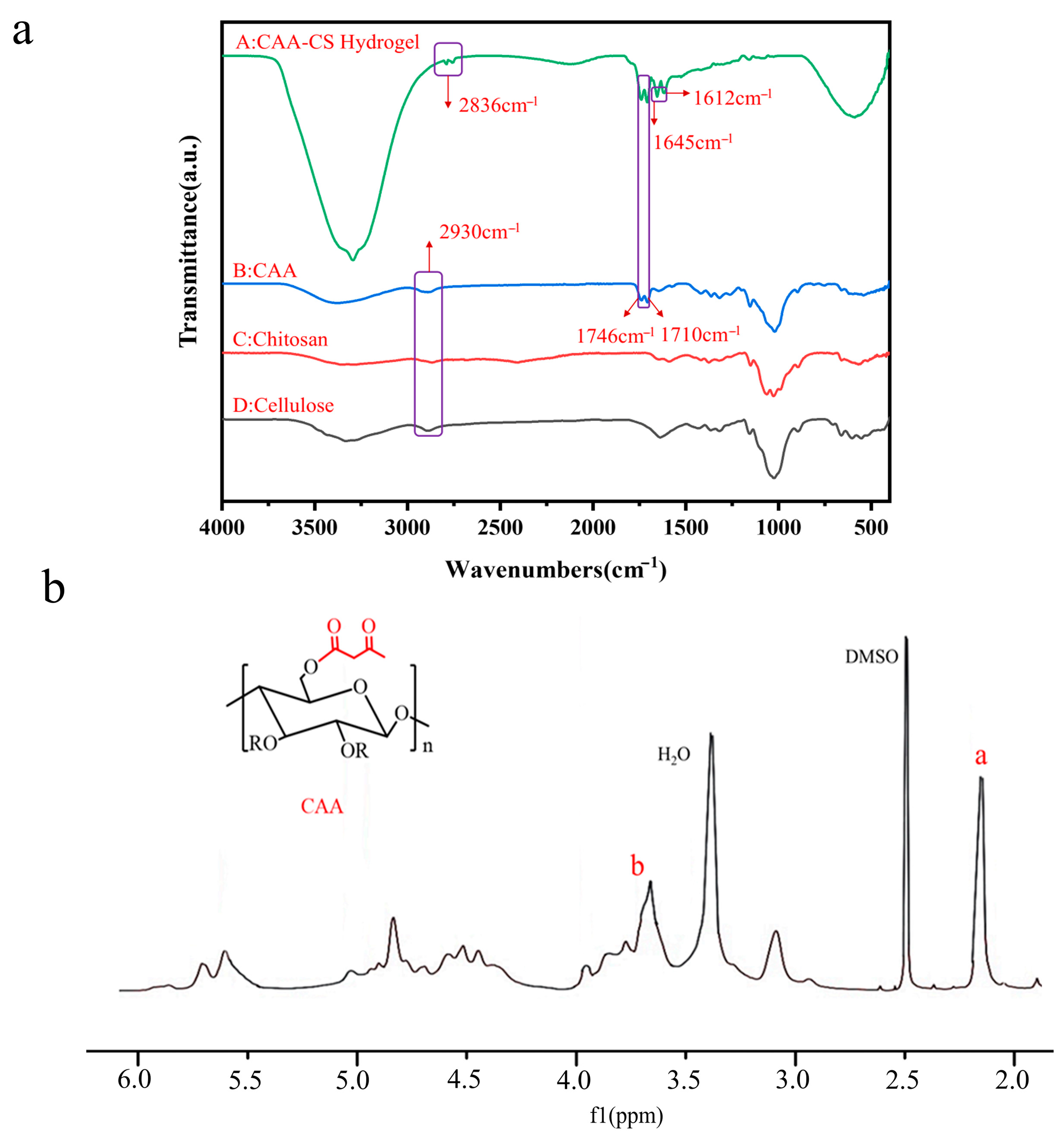
3.1. Structural Characterization of CAA Hydrogels

Since CAA samples with a degree of substitution (DS) of approximately 0.85 exhibit good water solubility and contain a large number of active acetoacetyl groups, they were selected for the relevant experiments in this study. Fourier transform infrared (FTIR) spectroscopy was used to characterize the chemical structure of the polysaccharide hydrogels with the FTIR spectrum of CAA as shown in Figure 2a. The absorption peak at 3400 cm−1 corresponds to the stretching vibration of −OH groups. The absorption peak at 2874 cm−1 is attributed to the stretching vibration of C−H bonds. The absorption peaks at 1710 cm−1 and 1746 cm−1 are characteristic bicarbonyl peaks of the acetoacetyl groups. In Figure 2b, the characteristic peak at 3.6 ppm in the NMR spectrum belongs to the methylene protons on the acetoacetyl group. The characteristic peak at 1.9–2.4 ppm corresponds to the methyl protons on the acetoacetyl group. The characteristic peak at 3.7–6.0 ppm is associated with the sugar ring of the cellulose backbone. In the FTIR spectrum of chitosan, the absorption peak at 1637 cm−1 corresponds to the stretching vibration of −NH2 groups on the six-membered ring of chitosan. The intensity of this absorption peak in the polysaccharide hydrogel is significantly lower than that in pure chitosan, and it overlaps with the absorption peak of the amide bond at 1645 cm−1. In the FTIR spectrum of the hydrogels, the characteristic bicarbonyl peaks of the acetoacetyl group at 1710 cm−1 and 1746 cm−1 are significantly reduced. New absorption peaks at 1645 cm−1 and 1612 cm−1 correspond to the stretching vibrations of enamine bonds as shown in Figure 2a.

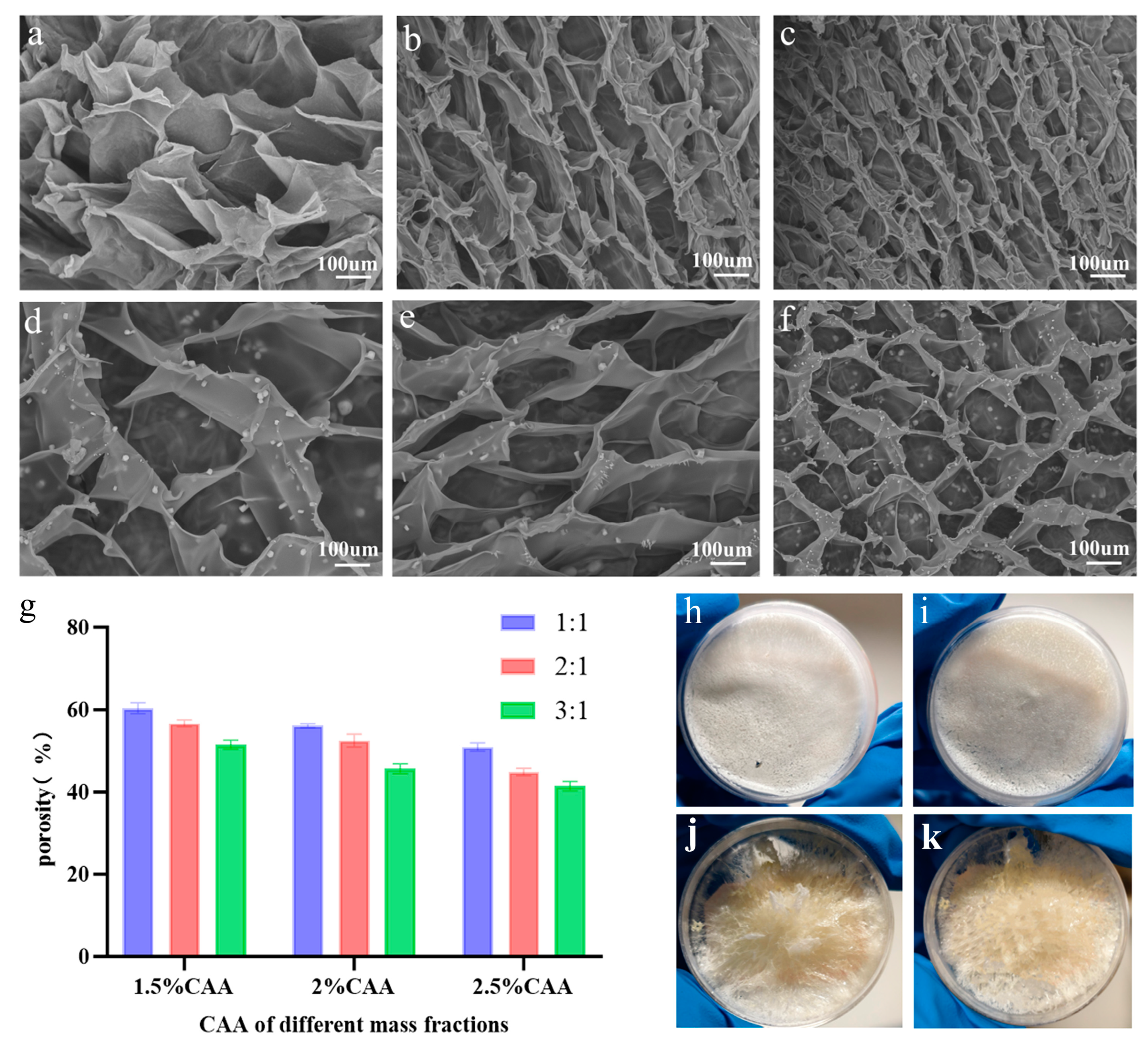
3.2. Morphology Analysis of CAA-CS Polysaccharide Hydrogel

Scanning electron microscopy (SEM) was utilized to characterize the microstructure of the CAA-CS series hydrogels; these hydrogels exhibit a continuous and porous three-dimensional network structure. The pore size of the CAA-CS polysaccharide hydrogel exhibits a certain positive correlation with the concentration of CAA. With the increase in the concentration of CAA (ranging from 1.5% to 2.5%), the cross-linking density of the hydrogel gradually elevates, leading to a decrease in pore size and a denser structure. This phenomenon might be ascribed to the fact that the increased concentration of CAA offers more acetoacetyl groups, thereby facilitating more efficient intermolecular cross-linking reactions. The SEM measurements in Figure 3a–f indicate that the average pore size of the polysaccharide hydrogels ranges from 100 to 200 um. This three-dimensional network structure not only provides a substantial specific surface area for drug loading but also offers an optimal spatial framework for cell scaffold applications. Moreover, the relatively large pore size facilitates cell growth and enhances the effective exchange of nutrients and gases [33]. Meanwhile, as the concentration of CAA increases, the porosity of the composite hydrogel decreases. The measured porosity of the CAA-CS series hydrogels ranged from 43% to 58% as shown in Figure 3g, indicating an enlargement of the internal pore size and a more abundant porous structure. Higher porosity indicates larger pore sizes or more developed pore structures. This trend is consistent with the results observed by scanning electron microscopy (SEM). A higher porosity indicates that a more abundant pore structure is formed inside the hydrogel, and the increase in CAA concentration promotes the crosslinking reaction between acetoacetyl groups and chitosan amino groups, thereby forming a more abundant three-dimensional network structure.

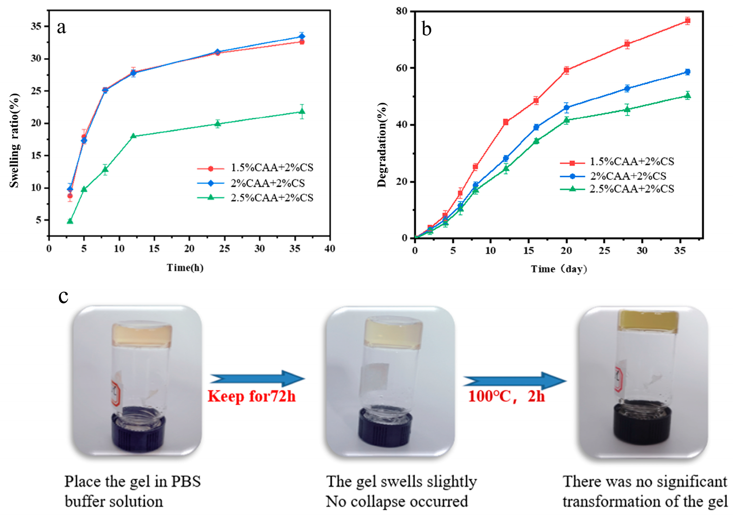
3.3. Stability Testing of CAA-CS Polysaccharide Hydrogel

Hydrogels prepared by blending different CAA concentrations (1.5%, 2 w%, 2.5 t%) with 2% CS were placed in PBS solution to analyze their swelling and degradation behaviors. The swelling property, which is the ability of hydrogels to absorb water and expand, is closely related to their crosslinking density and network structure. The swelling ratio of the CAA-CS composite hydrogels declined with the increase in CAA concentration, as depicted in Figure 4a. This phenomenon can be attributed to the fact that the increase in CAA concentration results in an elevated crosslinking density of the hydrogels. A higher concentration of CAA provides more acetoacetyl groups, which react with the amino groups in CS to generate more enamine bonds, thereby forming a denser three-dimensional network structure. Such a dense network structure inhibits the permeation of the PBS solution medium into the interior of the hydrogel, reducing the swelling extent of the hydrogel. Consequently, increasing the CAA concentration can simultaneously enhance the mechanical strength of the hydrogel and decrease its swelling property, enabling it to maintain superior morphological stability in a humid environment.
The degradation performance pertains to the ability of hydrogels to gradually disintegrate in a specific milieu, which directly influences their practical efficacy in applications. Figure 4b reveals that the degradation rate of CAA-CS hydrogels exhibits a downward trend with the escalation of CAA concentration. The experimental outcomes suggest that the degradation rate of hydrogels is negatively correlated with the CAA concentration. The hydrogel with a 2.5% CAA content demonstrates the lowest degradation rate, whereas the 1.5% CAA system undergoes degradation at the fastest pace. This phenomenon is in accordance with the results of the swelling performance, further validating the impact of CAA concentration on the crosslinking density and structural stability of hydrogels. Hydrogels formed by higher concentrations of CAA possess a denser crosslinking network, thereby manifesting a lower degradation rate and higher stability.
A further analysis was conducted on the stability of the CAA-CS hydrogel. The hydrogel was placed in PBS buffer for a certain period. After 72 h of observation, it was noted that the gel merely underwent slight expansion without collapsing. Subsequently, the gel was transferred to an oven and heated at 100 °C for 2 h (Figure 4c). During this period, no distinct morphological changes were observed in the gel. This indicates that the CAA-CS hydrogel possesses high structural stability and heat resistance. This stability is primarily attributed to the stable enamine bond (-C=N-) formed between CAA and CS through the Schiff base reaction, which can maintain a strong cross-linking intensity even in high-temperature and humid environments.

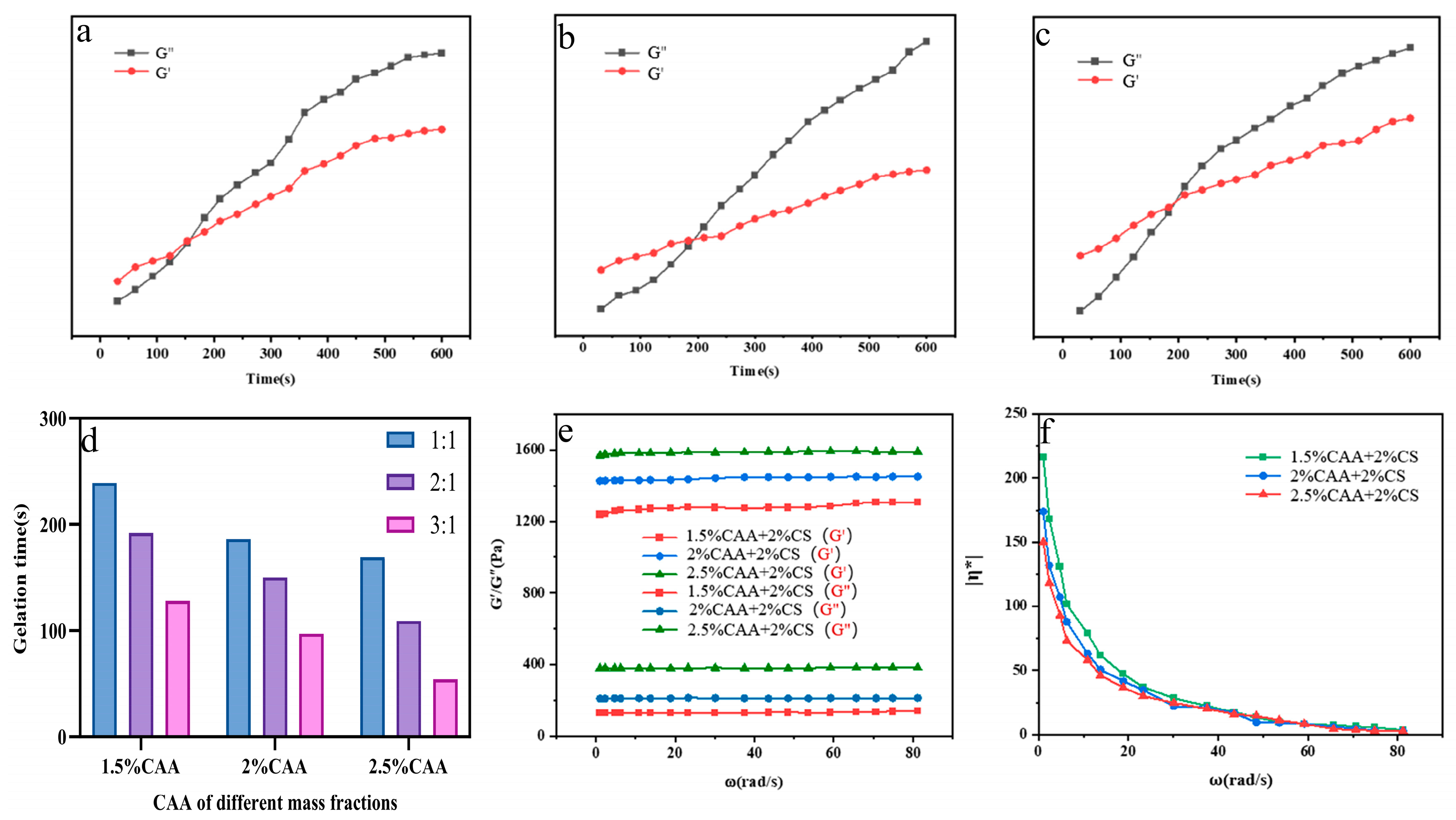
3.4. Rheological Performance Testing of CAA-CS Polysaccharide Hydrogel

In the initial stage of hydrogel formation, the energy storage modulus (G′) in the system is lower than the loss modulus (G″), indicating that the system remains in a solution state. As the reaction progresses, the acetoacetyl groups on CAA react more fully with the amino groups in chitosan, leading to an increase in the cross-linking degree of the three-dimensional network. Consequently, G′ of the gel increases, and the formation of enamine bonds causes G′ to rise faster than G″. Thus, when G′ exceeds G″, the system exhibits solid-like behavior. The intersection point where G′ equals G″ (G′ = G″) is defined as the gel point, and the corresponding time is referred to as the gel time as shown in Figure 5a–c. The gel time can be controlled by adjusting the concentration of CAA. Higher CAA concentrations result in faster gelation. For instance, as shown in Figure 5d, when the CAA concentration is 2 wt%, the gel time is 186 s, and increasing the CAA concentration to 2.5 wt% reduces the gel time to 169 s. The gel time decreases with increasing CAA concentration, while G′ increases. When both CAA and chitosan concentrations are 2 wt%, the gel time is 186 s, and the energy storage modulus is 1486 Pa. Appropriate gel times and energy storage moduli facilitate the thorough mixing of the hydrogel precursor solution with drugs and ensure proper formation and injectability of the hydrogel. The energy storage modulus (G′) and loss modulus (G″) of the hydrogel increase with the CAA mass fraction in Figure 5e. Additionally, G′ consistently exceeds G″, and G’’ remains at a low level, indicating that the hydrogel exhibits viscoelastic properties similar to those of a solid. As the CAA content increases, the number of acetoacetyl groups providing cross-linking sites also increases, forming a denser cross-linking structure. This results in enhanced gel strength within a certain range of CAA concentrations. In Figure 5f, the viscosity of the hydrogel as a function of angular frequency is shown. Similarly to most polymer materials, the viscosity of the hydrogel gradually decreases with increasing angular frequency, exhibiting shear-thinning behavior, which facilitates the injection of the hydrogel.

3.5. In Vitro Release of Hydrogel Loaded with IL-2

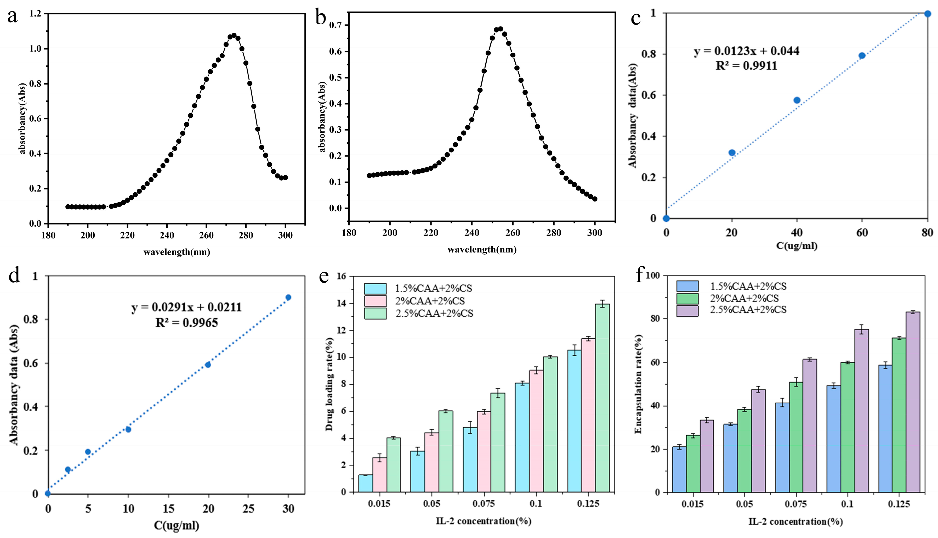
3.5.1. Analysis of Drug Loading Rate and Encapsulation Efficiency of Hydrogels

In the study by Antunes et al. [34,35,36], the relationship between IL-2 concentration and absorbance was established using the principle of ultraviolet spectrophotometry to create a standard curve. The maximum absorption wavelength (λmax) of IL-2 in phosphate-buffered solution within the range of 190–300 nm was determined to be 278.0 nm (Figure 6a). Based on this λmax, the standard curve equation y = 0.0123x + 0.044 was derived, with a high linear correlation coefficient (R2 = 0.9911) (Figure 6c). The corresponding IL-2 concentration was calculated from the absorbance values. For the CAA-IL-2 drug-containing solution, the maximum absorption wavelength (λmax) was found to be 252.0 nm (Figure 6b). Under this condition, the standard curve equation y = 0.0291x + 0.0211 was obtained, with a high linear correlation coefficient (R2 = 0.9965) (Figure 6d).
IL-2, as a cytokine abundant in amino and carboxyl groups, can form stable interactions with chitosan through hydrogen bonds, resulting in a homogeneous mixed system during the dissolution process. During the preparation of the CAA-CS drug-loaded composite gel, the acetoacetyl groups of CAA react with the amino groups of chitosan via a double-tube injection approach, forming stable enamine bonds (-C=N-), thereby establishing a three-dimensional cross-linked network structure. In this process, a portion of IL-2 forms complexes with chitosan and is effectively entrapped in the interlayer of the CAA-CS gel; another part is incorporated into the internal network of the gel, and a small quantity of IL-2 adheres to the surface of the gel.
The released IL-2 concentration was then calculated based on this standard curve. When the ratio of IL-2 to chitosan increased from 1.5 wt% to 10 wt%, the encapsulation efficiency of IL-2 initially increased and then decreased. At a ratio of 12.5 wt%, the maximum encapsulation efficiency reached 83.3% (Figure 6f). The drug loading rate of IL-2 increased as the proportion of IL-2 to chitosan gradually increased from 1.5 wt% to 12.5 wt%, reaching a maximum of 13.95% (Figure 6e). This indicates that the three-dimensional network structure of the CAA-CS hydrogel can provide a larger loading area for the drug, which is beneficial for drug encapsulation and loading.

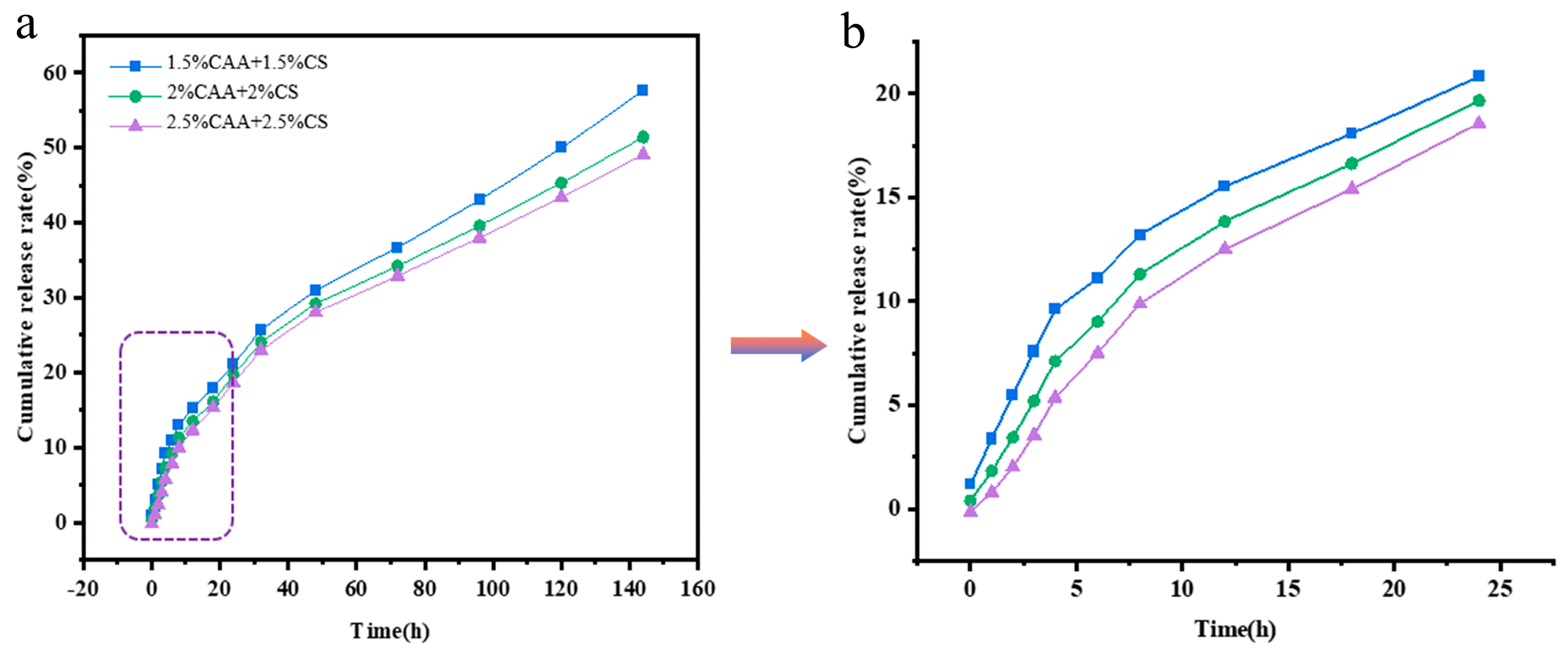
3.5.2. Release Test After Gel Loading with IL-2

To further study the release characteristics of different proportions of IL-2 in CAA-CS hydrogels, we immersed the drug-loaded gels in PS solution. The release behavior of IL-2 in CAA-CS hydrogels presented a distinct two-stage characteristic, as depicted in Figure 7. Within the initial 24 h, the release of 5% IL-2 exhibited an approximately linear relationship (Figure 7b), attaining 23.4%, indicating a relatively rapid initial release. As time elapsed, the cumulative release rate gradually decreased. Within 72 h, the cumulative release rate of IL-2 ranged from 18.4% to 34.7%, suggesting a gradually decelerating release rate over time, as illustrated in Figure 7a. Furthermore, with the increase in the CAA content, the cumulative release rate of IL-2 correspondingly decreased. This kinetic feature of transition from rapid release to sustained release reflects the controlled-release effect of the hydrogel network on the drug: initially, it is mainly the rapid release of the drugs adsorbed on the surface; later, it relies on the slow diffusion of the drugs due to the swelling and relaxation of the gel network.
Firstly, IL-2 loaded on the surface of CAA-CS hydrogels is rapidly released. This rapid release in the initial stage is mainly attributed to the high concentration of IL-2 on the surface of the hydrogels and the short contact time with the PBS solution, causing it to rapidly diffuse into the release medium in the early stage. As time progresses, the interaction between CAA-CS hydrogels and the PBS solution intensifies, and the hydrogels undergo significant swelling after absorbing water. This swelling process enables the release of IL-2 embedded within the CAA-CS hydrogels. The release of IL-2 is influenced by the interaction between CS and CAA, with the hydrogen bonding between hydroxyl and amino groups and the dynamic enamine bond between amino and acetoacetyl groups in the molecules jointly affecting the release rate of IL-2. PBS, as the release medium, gradually disrupts these interactions, allowing IL-2 to be gradually released from within the gel.

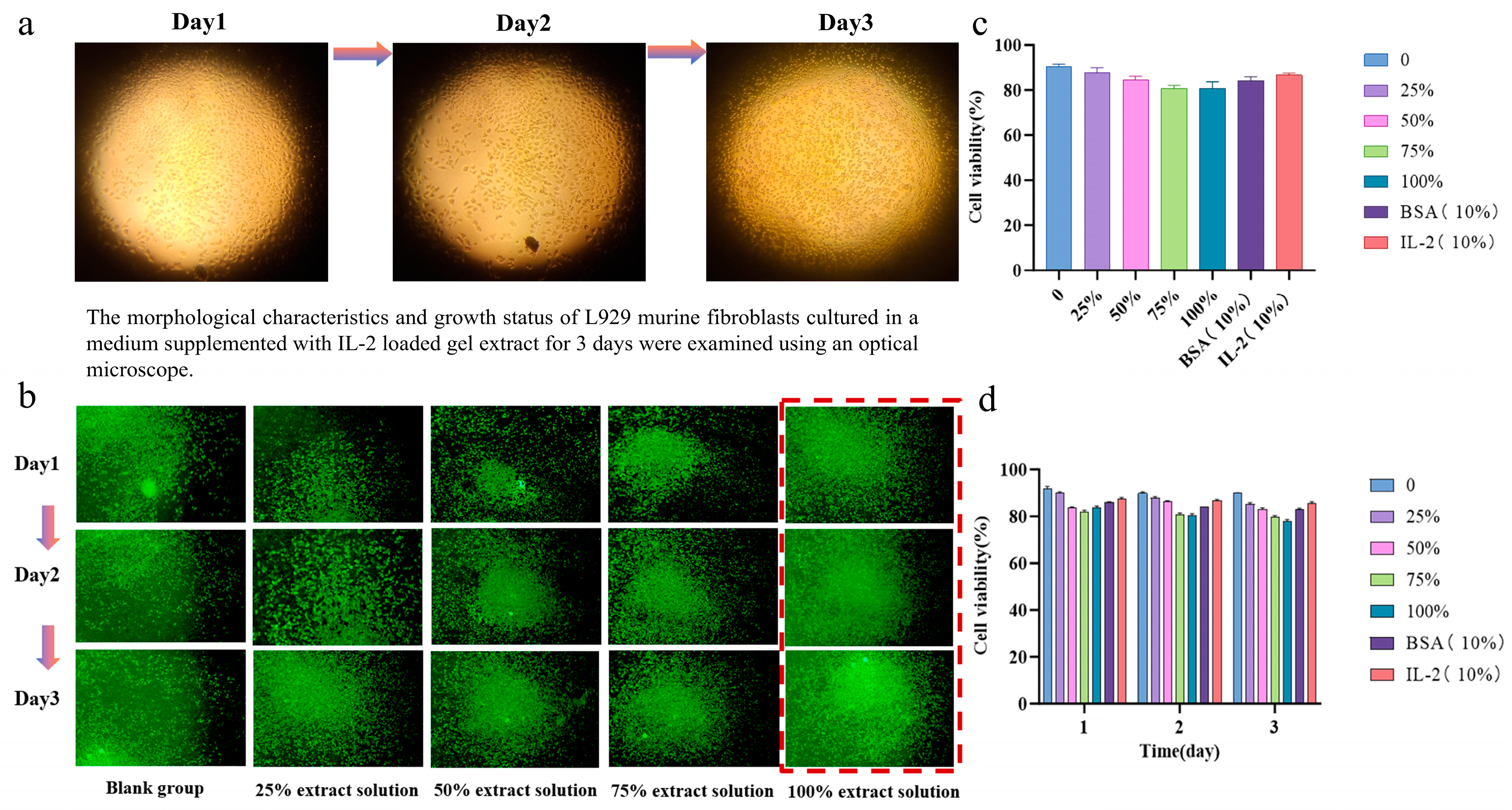
3.6. Biocompatibility of Hydrogels

The cytotoxicity and biocompatibility of the polysaccharide hydrogel were evaluated using the CCK-8 assay. L929 mouse fibroblasts were cultured with varying concentrations of CAA-CS gel extract for specific time intervals, and cell growth and viability were monitored and quantified. In Figure 8a, under an optical microscope, it was observed that the L929 cells cultured with the extract exhibited robust growth. Cell viability tests at designated time points revealed no significant differences in cell viability among the groups compared to the blank control group as the concentration of the extract increased as shown in Figure 8b. The cell viability across all groups remained comparable to that of the blank control, indicating that the gel extract did not adversely affect cell proliferation. Over time, there were no significant differences in OD values between the experimental groups and the blank control, further confirming that cell viability was maintained. After three days of culture, the results indicated that the polysaccharide hydrogel demonstrated excellent biocompatibility with L929 fibroblasts, with cell viability remaining above 85% (Figure 8c,d). These findings suggest that the CAA-CS system possesses good biocompatibility and is suitable for biomedical applications. For instance, these injectable hydrogels can serve as carriers for delivering precursor substances to target sites via injection.

4. Conclusions

Soluble cellulose acetoacetate (CAA) was synthesized via the acetoacetylation of modified cellulose, utilizing cellulose and chitosan as raw materials. Chitin (CS) was subsequently cross-linked within a three-dimensional network. By optimizing the concentration ratio of CAA and CS to determine the appropriate gelation time, an injectable drug-loaded polysaccharide hydrogel with covalent enamine bonds (CAA-CS) was prepared under mild conditions. The physicochemical properties of the drug-loaded polysaccharide hydrogel (CAA-CS) were systematically investigated, including the analysis of microscopic morphology, stability, and rheological properties. The results indicated that the CAA-CS series gels exhibited adjustable porous structures and favorable mechanical properties. Interleukin-2 (IL-2) was employed as a model drug to evaluate the encapsulation efficiency and drug release kinetics of the polysaccharide hydrogel. The findings revealed that the encapsulation efficiency of the CAA-CS hydrogel could reach up to 83.3%, demonstrating excellent sustained-release capability and drug encapsulation efficiency. Consequently, the polysaccharide hydrogels (CAA-CS) derived from CAA and CS hold significant potential as drug-loading materials with promising applications. Furthermore, CCK-8 assays demonstrated that the CAA-CS hydrogel exhibited excellent biocompatibility with L929 mouse fibroblasts, maintaining cell viability above 85%. The injectable CAA-CS polysaccharide hydrogel shows substantial potential for use in biomedical materials, such as drug delivery systems and tissue engineering.

Author Contributions

Design of the concept and study, Y.Z. and Z.M.; acquisition, analysis, and interpretation of data, Y.Z., X.J. and H.X.; drafting of the article, Y.Z. and X.J.; data curation and writing—review and editing, Y.Z. and X.J.; funding acquisition, Y.Z.; final approval of the version to be submitted, Z.M., Y.Z., H.X. and X.F. All authors have read and agreed to the published version of the manuscript.

Funding

The Science and Technology Plan Project of XPCC (Grant No. 2024AB060).

Institutional Review Board Statement

Not applicable.

Informed Consent Statement

Not applicable.

Data Availability Statement

The data used to support the findings of this study are available from the corresponding author upon request.

Acknowledgments

The authors are thankful to the Science and Technology Plan Project of XPCC (Grant No. 2024AB060). This research received support from Shanxi Medical University.

Conflicts of Interest

The authors declare no conflicts of interest.

References

  1. He, J.; Zhang, R.; Shao, M.; Zhao, X.; Miao, M.; Chen, J.; Liu, J.; Zhang, X.; Zhang, X.; Jin, Y.; et al. Efficacy and safety of low-dose IL-2 in the treatment of systemic lupus erythematosus: A randomised, double-blind, placebo-controlled trial. Ann. Rheum. Dis. 2020, 79, 141–149. [Google Scholar] [CrossRef]
  2. He, J.; Zhang, X.; Wei, Y.; Sun, X.; Chen, Y.; Deng, J.; Jin, Y.; Gan, Y.; Hu, X.; Jia, R.; et al. Low-dose interleukin-2 treatment selectively modulates CD4+ T cell subsets in patients with systemic lupus erythematosus. Nat. Med. 2016, 22, 991–993. [Google Scholar] [CrossRef] [PubMed]
  3. Jiang, T.; Zhou, C.; Ren, S. Role of IL-2 in cancer immunotherapy. Oncoimmunology 2016, 5, e1163462. [Google Scholar] [CrossRef]
  4. Rosenzwajg, M.; Lorenzon, R.; Cacoub, P.; Pham, H.P.; Pitoiset, F.; El Soufi, K.; Claire, R.I.; Bernard, C.; Aractingi, S.; Banneville, B.; et al. Immunological and clinical effects of low-dose interleukin-2 across 11 autoimmune diseases in a single, open clinical trial. Ann. Rheum. Dis. 2019, 78, 209–217. [Google Scholar] [CrossRef] [PubMed]
  5. Varghese, S.A.; Rangappa, S.M.; Siengchin, S.; Parameswaranpillai, J. Chapter 2-Natural polymers and the hydrogels prepared from them. In Hydrogels Based on Natural Polymers; Chen, Y., Ed.; Elsevier: Amsterdam, The Netherlands, 2020; pp. 17–47. [Google Scholar]
  6. Hennink, W.E.; van Nostrum, C.F. Novel crosslinking methods to design hydrogels. Adv. Drug Deliv. Rev. 2012, 64, 223–236. [Google Scholar] [CrossRef]
  7. Li, Y.; Rodrigues, J.; Tomas, H. Injectable and biodegradable hydrogels: Gelation, biodegradation and biomedical applications. Chem. Soc. Rev. 2012, 41, 2193–2221. [Google Scholar] [CrossRef]
  8. Tseng, T.C.; Tao, L.; Hsieh, F.Y.; Wei, Y.; Chiu, I.M.; Hsu, S.H. An Injectable, Self-Healing Hydrogel to Repair the Central Nervous System. Adv. Mater. 2015, 27, 3518–3524. [Google Scholar] [CrossRef] [PubMed]
  9. Guo, B.; Finne-Wistrand, A.; Albertsson, A.C. Facile synthesis of degradable and electrically conductive polysaccharide hydrogels. Biomacromolecules 2011, 12, 2601–2609. [Google Scholar] [CrossRef]
  10. Li, Y.; Liu, C.; Tan, Y.; Xu, K.; Lu, C.; Wang, P. In situ hydrogel constructed by starch-based nanoparticles via a Schiff base reaction. Carbohydr. Polym. 2014, 110, 87–94. [Google Scholar] [CrossRef]
  11. Liu, X.; Peng, W.; Wang, Y.; Zhu, M.; Sun, T.; Peng, Q.; Zeng, Y.; Feng, B.; Lu, X.; Weng, J.; et al. Synthesis of an RGD-grafted oxidized sodium alginate-N-succinyl chitosan hydrogel and an in vitro study of endothelial and osteogenic differentiation. J. Mater. Chem. B 2013, 1, 4484–4492. [Google Scholar] [CrossRef]
  12. Tsao, C.T.; Hsiao, M.H.; Zhang, M.Y.; Levengood, S.L.; Zhang, M. Chitosan-PEG hydrogel with sol-gel transition triggerable by multiple external stimuli. Macromol. Rapid Commun. 2015, 36, 332–338. [Google Scholar] [CrossRef] [PubMed]
  13. Yu, L.; Ding, J. Injectable hydrogels as unique biomedical materials. Chem. Soc. Rev. 2008, 37, 1473–1481. [Google Scholar] [CrossRef] [PubMed]
  14. Appel, E.A.; del Barrio, J.; Loh, X.J.; Scherman, O.A. Supramolecular polymeric hydrogels. Chem. Soc. Rev. 2012, 41, 6195–6214. [Google Scholar] [CrossRef] [PubMed]
  15. Cooper, R.C.; Yang, H. Hydrogel-based ocular drug delivery systems: Emerging fabrication strategies, applications, and bench-to-bedside manufacturing considerations. J. Control. Release 2019, 306, 29–39. [Google Scholar] [CrossRef]
  16. Zhao, D.; Feng, M.; Zhang, L.; He, B.; Chen, X.; Sun, J. Facile synthesis of self-healing and layered sodium alginate/polyacrylamide hydrogel promoted by dynamic hydrogen bond. Carbohyd. Polym. 2021, 256, 117580. [Google Scholar] [CrossRef]
  17. Klemm, D.; Heublein, B.; Fink, H.P.; Bohn, A. Cellulose: Fascinating biopolymer and sustainable raw material. Angew. Chem. Int. Ed. Engl. 2005, 44, 3358–3393. [Google Scholar] [CrossRef]
  18. Nishiyama, Y.; Langan, P.; Chanzy, H. Crystal Structure and Hydrogen-Bonding System in Cellulose Iβ from Synchrotron X-ray and Neutron Fiber Diffraction. J. Am. Chem. Soc. 2002, 124, 9074–9082. [Google Scholar] [CrossRef]
  19. Wang, H.; Gurau, G.; Rogers, R.D. Ionic liquid processing of cellulose. Chem. Soc. Rev. 2012, 41, 1519–1537. [Google Scholar] [CrossRef]
  20. Chang, C.; Zhang, L. Cellulose-based hydrogels: Present status and application prospects. Carbohyd. Polym. 2011, 84, 40–53. [Google Scholar] [CrossRef]
  21. Dai, L.; Si, C. Recent Advances on Cellulose-Based Nano-Drug Delivery Systems: Design of Prodrugs and Nanoparticles. Curr. Med. Chem. 2019, 26, 2410–2429. [Google Scholar] [CrossRef]
  22. Pillai, C.K.S.; Paul, W.; Sharma, C.P. Chitin and chitosan polymers: Chemistry, solubility and fiber formation. Prog. Polym. Sci. 2009, 34, 641–678. [Google Scholar] [CrossRef]
  23. Ravi Kumar, M.N.V. A review of chitin and chitosan applications. React. Funct. Polym. 2000, 46, 1–27. [Google Scholar] [CrossRef]
  24. Rinaudo, M. Chitin and chitosan: Properties and applications. Prog. Polym. Sci. 2006, 31, 603–632. [Google Scholar] [CrossRef]
  25. Sánchez, D.; Bastida, D.; Burés, J.; Isart, C.; Pineda, O.; Vilarrasa, J. Relative Tendency of Carbonyl Compounds To Form Enamines. Org. Lett. 2012, 14, 536–539. [Google Scholar] [CrossRef]
  26. Liu, H.; Sui, X.; Xu, H.; Zhang, L.; Zhong, Y.; Mao, Z. Self-Healing Polysaccharide Hydrogel Based on Dynamic Covalent Enamine Bonds. Macromol. Mater. Eng. 2016, 301, 725–732. [Google Scholar] [CrossRef]
  27. Wang, Y.; Ma, M.; Wang, J.; Zhang, W.; Lu, W.; Gao, Y.; Zhang, B.; Guo, Y. Development of a Photo-Crosslinking, Biodegradable GelMA/PEGDA Hydrogel for Guided Bone Regeneration Materials. Materials 2018, 11, 1345. [Google Scholar] [CrossRef]
  28. Holback, H.; Yeo, Y.; Park, K. 1-Hydrogel swelling behavior and its biomedical applications. In Biomedical Hydrogels; Rimmer, S., Ed.; Woodhead Publishing: Sawston, UK, 2011; pp. 3–24. [Google Scholar]
  29. Peppas, N.A.; Bures, P.; Leobandung, W.; Ichikawa, H. Hydrogels in pharmaceutical formulations. Eur. J. Pharm. Biopharm. 2000, 50, 27–46. [Google Scholar] [CrossRef]
  30. Liu, Z.; Liu, C.; Sun, X.; Zhang, S.; Yuan, Y.; Wang, D.; Xu, Y. Fabrication and characterization of cold-gelation whey protein-chitosan complex hydrogels for the controlled release of curcumin. Food Hydrocoll. 2020, 103, 105619. [Google Scholar] [CrossRef]
  31. Xu, Y.; Lu, G.; Chen, M.; Wang, P.; Li, Z.; Han, X.; Liang, J.; Sun, Y.; Fan, Y.; Zhang, X. Redox and pH dual-responsive injectable hyaluronan hydrogels with shape-recovery and self-healing properties for protein and cell delivery. Carbohydr. Polym. 2020, 250, 116979. [Google Scholar] [CrossRef]
  32. Ding, D.; Zhu, Z.; Liu, Q.; Wang, J.; Hu, Y.; Jiang, X.; Liu, B. Cisplatin-loaded gelatin-poly(acrylic acid) nanoparticles: Synthesis, antitumor efficiency in vivo and penetration in tumors. Eur. J. Pharm. Biopharm. 2011, 79, 142–149. [Google Scholar] [CrossRef]
  33. Loh, Q.L.; Choong, C. Three-dimensional scaffolds for tissue engineering applications: Role of porosity and pore size. Tissue Eng. Part B Rev. 2013, 19, 485–502. [Google Scholar] [CrossRef] [PubMed]
  34. Antunes, J.C.; Oliveira, J.M.; Reis, R.L.; Soria, J.M.; Gomez-Ribelles, J.L.; Mano, J.F. Novel poly(L-lactic acid)/hyaluronic acid macroporous hybrid scaffolds: Characterization and assessment of cytotoxicity. J. Biomed. Mater. Res. A 2010, 94, 856–869. [Google Scholar] [CrossRef] [PubMed]
  35. Li, L.; Wang, N.; Jin, X.; Deng, R.; Nie, S.; Sun, L.; Wu, Q.; Wei, Y.; Gong, C. Biodegradable and injectable in situ cross-linking chitosan-hyaluronic acid based hydrogels for postoperative adhesion prevention. Biomaterials 2014, 35, 3903–3917. [Google Scholar] [CrossRef]
  36. Prestwich, G.D.; Marecak, D.M.; Marecek, J.F.; Vercruysse, K.P.; Ziebell, M.R. Controlled chemical modification of hyaluronic acid: Synthesis, applications, and biodegradation of hydrazide derivatives. J. Control. Release 1998, 53, 93–103. [Google Scholar] [CrossRef] [PubMed]
Figure 1. Synthesis of cellulose acetoacetate (CAA) and preparation of polysaccharide hydrogel through the reaction of CAA with chitosan.
Applsci 15 05601 g001
Figure 2. (a) Infrared spectrum of cellulose acetoacetate (b) nuclear magnetic spectrum of cellulose acetoacetate. “a” represents the methylene peak on the acetoacetyl group, and “b” represents the methyl peak on the acetoacetyl group.
Applsci 15 05601 g002
Figure 3. Electron microscopy image of polysaccharide hydrogel; (a) 1.5% CAA + 2% CS; (b) 2% CAA + 2% CS; (c) 2.5% CAA + 1.5% CS; (d) 1.5% CAA (20 wiu IL-2) + 1.5% CS; (e) 2% CAA (20 wiu IL-2) + 2% CS; (f) 2.5% CAA (20 wiu IL-2) + 1.5% CS; (g) the porosity of CAA-CS hydrogels; (hk) freeze-dried CAA-CS hydrogel image.
Applsci 15 05601 g003
Figure 4. (a) Swelling degree of different polysaccharide hydrogels in PBS solution, (b) degradation degree of different polysaccharide hydrogels in PBS solution, (c) stability test of polysaccharide hydrogels in PBS (pH = 7.34) aqueous solution at 37 °C for 72 h and at 100 °C for 2 h.
Applsci 15 05601 g004
Figure 5. (ad) The gelation time of CAA-CS series hydrogels, (e) storage modulus and loss modulus of CAA-CS series hydrogels, (f) viscosity of CAA-CS Series Hydrogels.
Applsci 15 05601 g005
Figure 6. (a) Maximum absorption wavelength of IL-2 in PBS solution, (b) the maximum absorption wavelength of CAA-IL-2 drug-containing solution), (c) IL-2 phosphate buffer solution (pH = 7.2–7.4, PBS) standard curve, (d) the standard curve of IL-2 in CAA solution), (e) the drug loading rate of CAA-CS hydrogels loaded with the model drug IL-2, (f) the encapsulation efficiency of CAA-CS hydrogels loaded with the model drug IL-2.
Applsci 15 05601 g006
Figure 7. (a,b) Release profile of IL-2 from CAA-CS hydrogels in PBS.
Applsci 15 05601 g007
Figure 8. (a) The morphological characteristics and growth status of L929 murine fibroblasts cultured in a medium supplemented with IL-2 loaded gel extract for 3 days were examined using an optical microscope. (b) The fluorescence test images of L929 cells under different culture media (c). The cell survival rate of different extracts. (d) The cell survival rate at different times.
Applsci 15 05601 g008
Disclaimer/Publisher’s Note: The statements, opinions and data contained in all publications are solely those of the individual author(s) and contributor(s) and not of MDPI and/or the editor(s). MDPI and/or the editor(s) disclaim responsibility for any injury to people or property resulting from any ideas, methods, instructions or products referred to in the content.

