Article

Analysis of TihxOy Films Produced by Physical Vapor Deposition Method

1
Lithuanian Energy Institute, Breslaujos st. 3, LT-44403 Kaunas, Lithuania
2
Institute for Energy Technology, P.O. Box 40, NO-2027 Kjeller, Norway
*
Authors to whom correspondence should be addressed.
Appl. Sci. 2022, 12(21), 10811; https://doi.org/10.3390/app122110811
Submission received: 28 September 2022 / Revised: 20 October 2022 / Accepted: 24 October 2022 / Published: 25 October 2022

Abstract

:
For decades, partially oxidized hydrides were commonly considered as undesirably contaminated phases and were avoided by scientists. Nevertheless, more recently, it was realized that in some hydrides and oxides, partial substitution of dissimilar H and O2− anions allows one to obtain unique optical and electrical properties that might have appealing applications in commercial products. It was determined that specific properties of so called oxyhydride materials strongly depend on the used synthesis methods; therefore, there is a great interest in exploring various variants of oxyhydride formation. In the current study, TiHxOy films were deposited by a reactive magnetron sputtering process in Ar-O2-H2 gas mixtures. Color, transparency and crystal phase composition of the films coherently reacted to the Ar:O2:H2 gas ratio. Namely, the rise in partial hydrogen pressure promoted the formation of anatase phase TiO2 structure and darkening of the films. Interestingly, this had only minimal impact on the band gap values, but had a relatively strong negative effect on the photocatalytic activity of the films. The unaccustomed results stressed the difference between the partially reduced TiO2 with a significant amount of oxygen vacancies and synthesized TiHxOy films where some O2− ions are implicitly substituted by H ions.

1. Introduction

Oxides, hydrides and hydroxides are common material classes that have been known and used for ages. Oxyhydrides, on the other hand, make up a less known class of multi-anion materials where the oxide and hydride anions are sharing the same sites within the crystal lattice [1]. The first report on oxyhydride synthesis (namely LaH1+2xO1−x and LaH1+yO1−x (y < 2x) oxyhydrides) was published 40 years ago [2] and dealt with relatively high skepticism. At that time, researchers assumed that under normal conditions, the coexistence of dissimilarly charged hard O2− anions and soft, polarizable, H anions at the same sites of a crystalline solid was thermodynamically unlikely [3,4]. Direct experimental confirmation of hydrogen entering into the oxide structure was and still remains a challenging task, because most of the traditionally used material analysis methods such as X-ray diffraction, energy dispersive spectroscopy, X-ray photoelectron spectroscopy and others can not observe hydrogen directly. Nevertheless, credible crystal structure study by combining neutron and X-ray diffraction techniques [5] unambiguously confirmed the unusual type of oxygen and hydrogen anion site sharing and the existence of oxyhydrides was eventually accepted by the whole scientific community. Many of these materials were obtained by partially oxidizing hydrides and, based on their crystal structure, could be described as oxygen-containing hydrides [6]. However, hydrogen-containing oxides were reported as well [4]. More recently, some authors suggested adopting the usage of the -HO ending for the oxyhydrides [7], thus discriminating them from the hydroxides (i.e., materials with -OH groups).
Following the first reports of LaHO synthesis by J.F. Brice et al. [2,5], some more oxyhydrides were synthesized and identified as well [8,9,10]. However, in general, the reports of oxyhydride studies remained scarce, and for years these materials had no practical applications. Approximately one decade ago, researchers started to realize that unique properties of hydrogen can lead to the advanced functionality of oxyhydride materials, and the situation started to change drastically [3]. The ionic radius of hydrogen anion (139.9 pm) is relatively similar to the ionic radius of oxygen anion (145.2 pm) [11]; however, at the same time, H ion has two times smaller charge and is distinctly easier to polarize than the O2− ion [4]. Accordingly, it was assumed that if O2− were replaced by H (or vice versa), this would not induce large disturbances to the crystal lattice, but could create uncommon electron energy level structures and could lead to interesting magnetic and optical properties [3,4,12]. This, in turn, potentially might have appealing applications in commercial products.
Recently, it was reported that in some of the oxyhydrides, H and O2– ions might have the possibility of interacting via valence charge states of the metal center [1]. In partially oxidized yttrium hydride and several other rare-earth (Gd, Dy, and Er) hydrides, reversible switching of optical transmittance under intense irradiation was observed [13]. This feature of yttrium oxyhydride is currently being tested for application in smart window devices [14].
Titanium is also recognized for its potential to form corresponding oxyhydride phases [15]. However, up to now, most of the efforts to alter the TiO2 structure by hydrogen (both in powder and film form) have been focused on the reduction of TiO2 and the formation of oxygen vacancies that are beneficial for photocatalytic applications. Meanwhile, direct synthesis of ternary titanium oxyhydrides from the constituting elements (namely, titanium oxygen and hydrogen) have not yet been reported, and the practical value of such oxyhydrides has not yet been determined. Accordingly, in the current study, we focused on the controllable one-step synthesis of titanium oxyhydride films by a reactive magnetron sputtering technique. Structural, optical and photocatalytic properties of the as-deposited films were thoroughly analyzed and compared with the corresponding properties of black TiO2 films and powders, which were prepared by ex situ reduction of TiO2 (by interaction with hydrogen and other compounds).

2. Materials and Methods

TiHO samples were deposited using a custom modified Kurt J. Lesker PVD-75 physical vapor deposition system equipped with cryo pump and one unbalanced Torrus 3 magnetron. The magnetron had a titanium target (76 mm diameter, 6 mm thickness, 99.99% purity) and was powered by a 300 W DC power source. Ar, O2 and H2 gases (all 99.999% purity) were supplied by three mass flow controllers that were operated by a Nova Fabrica FloTron X multi-channel process control system. Three different Ar-O2-H2 gas mixtures were used to deposit titanium oxyhydride films with varying hydrogen concentrations. The hydrogen concentration was increased, going from sample B1 to sample B3 (more details are provided in Table 1). In addition, an Ar-O2 gas mixture was used to deposit conventional titanium dioxide films (samples A). For the deposition of later samples, the same experimental set-up was used. The only modifications to the process were the annulment of hydrogen gas supply and introduction of a pulsed direct-current (p-DC) power source. In the Ar-O2 gas mixture, the p-DC power source allowed us to prevent titanium target charging/poisoning and improved reactive magnetron sputtering process stability (in the Ar-O2-H2 gas mixture, the Ti target was continuously reduced by hydrogen; therefore, for TiHO deposition, the p-DC power source was not needed). Deposition time for all group B samples was the same—1 h, but their deposition rate varied significantly depending on the working gas mixture composition. Accordingly, the ultimate thickness of group B samples reached approximately 700 nm for B1, 900 nm for B2 and 1500 nm for B3. Group A samples were deposited for 2 h, and their thickness was approximately 1500 nm.
Crystal structures of the deposited films were determined by X-ray diffraction (XRD, Bruker D8). Elemental composition and chemical states of the TiHO films were analyzed by X-ray photoelectron spectroscopy (XPS, PHI 5000 Versaprobe). To minimize the extent of additional sample surface oxidation and contamination by adventitious hydrocarbons, XPS analysis was carried out straight after the samples were transferred from the deposition chamber to the XPS instrument. Optical transmission and reflectance of the films were acquired by UV–VIS spectrophotometer. To estimate band gap energies of the samples, corresponding Tauc plots were drawn. Since it was not easy to split the Kubelka–Munk spectrum into spectra of individual components (in this case, films and their substrates), a simplified band gap estimation method described in [16] was applied. By this method, at first, the traditional linear part of the plot was extrapolated to the x-axis, and an additional linear fit was applied for the slope below the fundamental absorption. Then, a band gap value was determined at the intersection of the corresponding two linear fittings. Photocatalytic activity of TiO2 and TiHO samples was estimated using methylene blue (MB) bleaching test (adopted version of ISO 10678:2010 standard [17]). For these tests, the MB solution volume was 15 mL, and initial concentration 15 mg/L. Photocatalysis was initiated with collimated 365 nm LED light source (Thorlabs M365LP1-C1).

3. Results

Reactive magnetron sputtering under Ar-O2 atmosphere produced a colorless transparent film (sample A) consisting of anatase and rutile TiO2 phases (Figure 1). When the gas mixture was supplemented with hydrogen, the crystallinity of the samples started to weaken, and they started to gain a noticeable tint. At the largest hydrogen concentration (sample B3), the effect was especially strong: the sample nearly lost the rutile phase and obtained a dark blue-grey shade. Traditionally, the appearance of blueish shade in titanium oxide samples is associated with Ti4+ reduction to Ti3+ and observation of Ti2O3 phase [18,19]. However, during close inspection of TiHO XRD patterns (Figure 1), we did not detect any features that could be attributed to Ti2O3 or any other titanium suboxides.
These results are interesting from several perspectives. First, earlier reports demonstrated that Ti-O-H interaction during TiH2 powder oxidation experiments nearly exclusively results in direct or indirect (i.e., through the intermediate phases of titanium suboxides) formation of the rutile TiO2 [10,15,20]. Naturally, the processes of TiH2 oxidation and titanium sputtering in a reactive Ar-O2-H2 atmosphere have fundamental differences and can not be compared directly. Still, the observed decline in the crystallinity of the thermodynamically most stable rutile TiO2 phase is not a trivial outcome and supports the indications by other groups [21,22], who suggested that products of Ti-O-H interaction can be method-specific.
Second, in defected TiO2 samples (including some “black TiO2” samples produced using hydrogen [23]) the dark color is commonly attributed to the presence of oxygen vacancies and Ti3+ species [24,25,26]. For example, a similar trend of TiO2 darkening and rutile phase depreciation from the initial anatase-rutile mixture was observed when standard white TiO2 powders were reduced by NaBH4 [27]. In that study, XPS analysis of reduced TiO2 powders confirmed that color and structural changes of the initial TiO2 powders can be attributed to the propagation of oxygen vacancies (indicated by the decrease in bulk O:Ti ratio) and partial reduction of Ti4+ to Ti3+ state (indicated by the appearance of the additional Ti 2p3/2 peak component at approximately 457.1 eV).
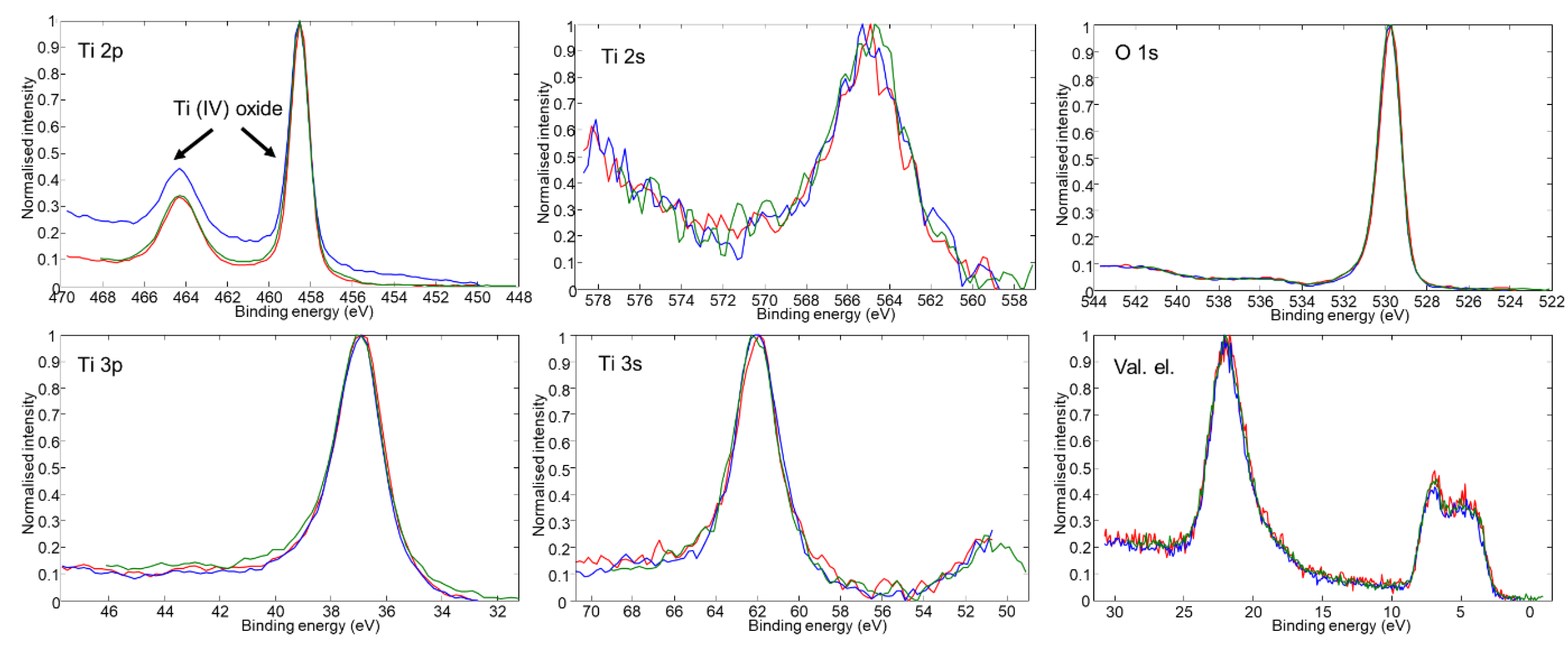
In order to resolve the discrepancy between the supposed (by the color) and observed crystal structure of TiHO samples, we also used the XPS technique and determined the chemical state of titanium. If it were present, Ti(III) oxide (i.e., Ti3+) should have an O 1s peak component at approximately 532.5 eV and Ti 2p peak components at 457.3 eV and 462.5 eV [28,29]. Moreover, looking at the color changes of the TiHO samples, one might expect that peak components related to Ti(III) oxide should become stronger proceeding from sample B1 to sample B3. Notwithstanding the initial assumptions and significantly different appearance of TiHO samples, all photoelectron spectra of titanium, oxygen and valence-band electrons were nearly identical (Figure 2). More specifically, Ti 2p photoelectron spectra for all TiHO samples had two almost fully symmetrical peaks at approximately 458.7 eV and 464.4 eV. These peaks fit well with the standard Ti 2p3/2 and Ti 2p1/2 peak positions of Ti(IV) oxide [28]. Similarly, all acquired O 1s spectra had a strong peak at 530.0 eV with a small shoulder at 531.0–531.5 eV. The main O 1s peak corresponded well with the oxygen from Ti(IV) oxide [28], whereas the higher energy shoulder was attributed to C-O [30] and O-H groups [31] presumably coming from the adventitious surface contamination and moisture.
Based solely on the XRD and XPS data, it might be assumed that the only form of titanium in TiHO samples is Ti4+ and it makes up only Ti(IV) oxide. But the evident tint of B2 and B3 samples suggest that this should not be the case. It is likely that other forms of titanium also exist, but they were not detected due to the limitations of the XRD and XPS methods. As mentioned above, the blue tint in Ti-O system traditionally is associated with the presence of Ti3+ [18,19]. Considering the limits of the XRD and XPS methods, it can be concluded that other forms of titanium (presumably Ti3+ next to the oxygen vacancies, or amorphous Ti2O3 regions at grain boundaries) should be present in the bulk of the film.
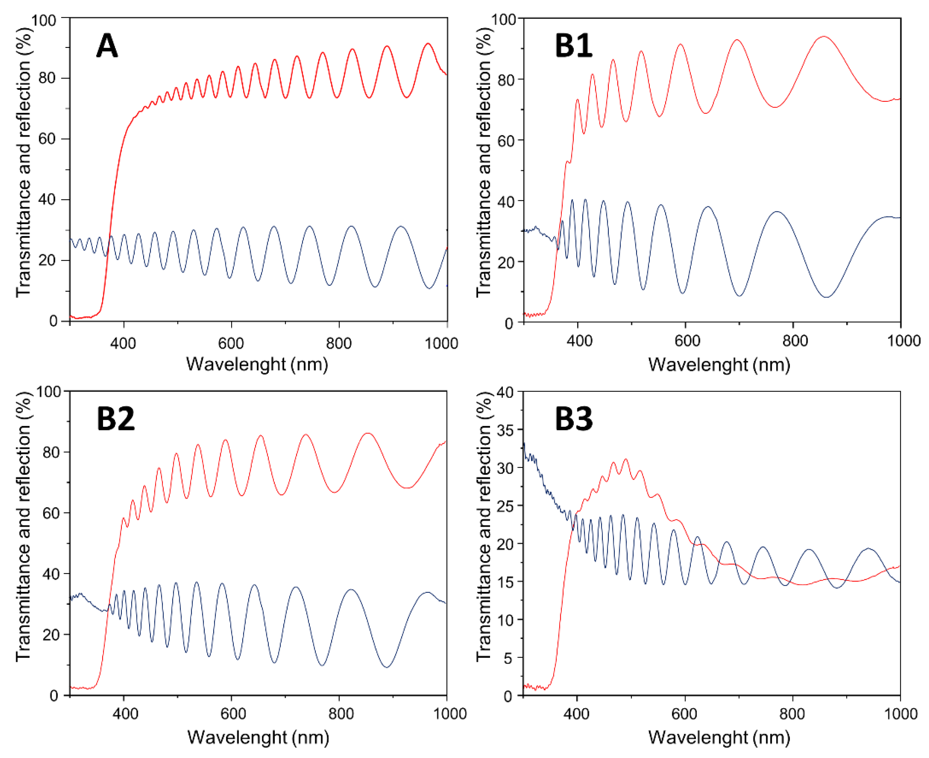
In defected TiO2 and mixed TiO2-Ti2O3 materials, Ti3+ cations introduce new electron levels just bellow the conduction band of TiO2 and result in the strong absorbance (mostly witnessed as a fall in light transmittance) above 500–600 nm [19,21,32]. At the same time, these materials demonstrate an increase in reflectance starting from 400–600 nm (the position of the lower end of reflectance increase depends on the oxygen content [21]). In order to test if as-deposited TiHO films have these optical characteristics of Ti3+ oxides, optical transmittance and reflectance measurements were performed (Figure 3). Typically for standard TiO2 films, above 400 nm, sample A had a stable transmittance level of nearly 80% and reflectance of approximately 20%. Films deposited with low and medium hydrogen concentrations (samples B1 and B2) had slightly lower optical transmittance, but it remained relatively stable over the whole tested range. Sample B3, on the other hand, had a considerably lower transmittance, which peaked out at 500 nm and reached just over 30%. Such transmittance spectra are typical for Ti(III) oxide [22], but there was no rise in reflectance spectra, and this feature distinguished the B3 samples from other reported blue tinted defected TiO2 and/or Ti2O3 films [4,21].
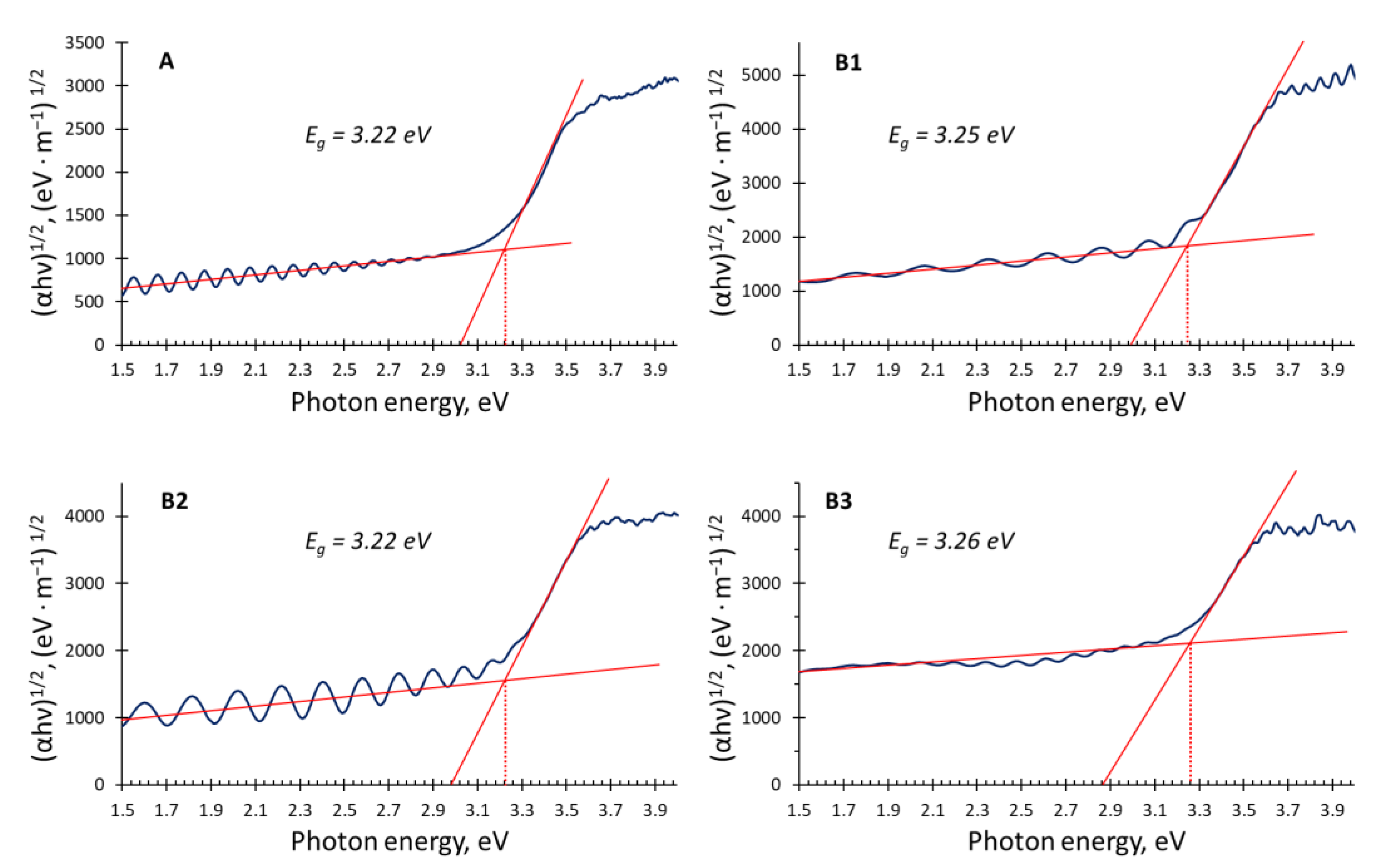
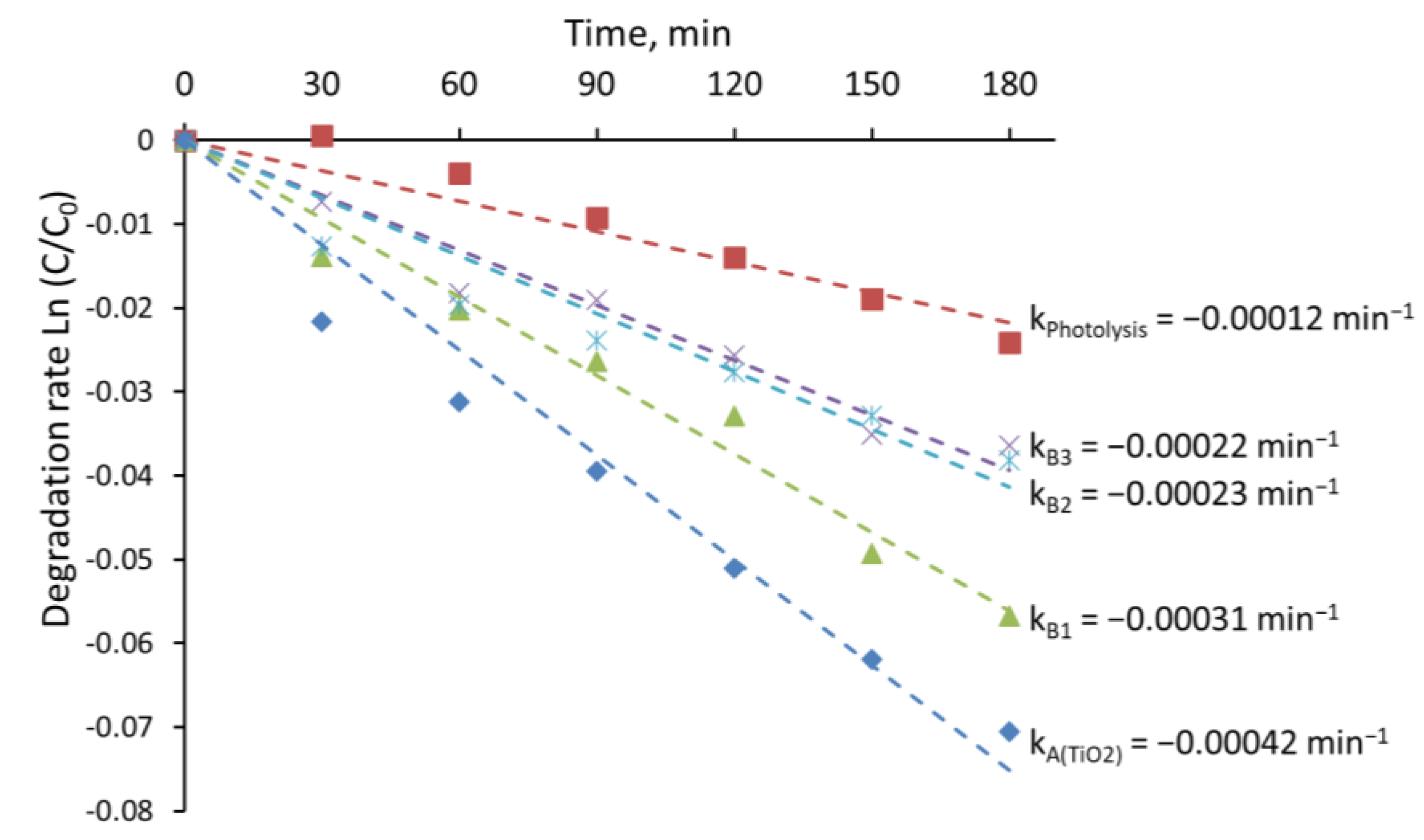
Another interesting characteristic of the current TiHO films is that higher hydrogen content resulted in slightly higher optical band gap values (samples B1 and B3 had band gap values of 3.25 and 3.26 eV, respectively, Figure 4) than that of standard TiO2 (sample A had a band gap of 3.22 eV). Meanwhile, the reported band gap values of reduced or defected TiO2 materials had a tendency to be lower [4]. Additionally, as a common trend, reduced or defected TiO2 oxides have higher photocatalytic activity [27]. However, in the current study, photocatalytic MB bleaching efficiency using tinted TiHO films (samples B2 and B3) was weaker in comparison to that with samples A and B1, which were deposited using lower hydrogen fractions in the gas mixture (Figure 5).

4. Discussion

Summing up the observed results, it can be concluded that reactive magnetron sputtering of titanium in Ar-O2-H2 gas mixtures with high hydrogen content allows the deposition of films whose visual appearance is similar to the titanium oxide with a significant amount of titanium in Ti3+ state. However, neither of the applied methods provided unambiguous evidence for the existence of Ti(III) oxide or oxygen vacancies. This suggests that during film deposition, hydrogen not only reduces TiO2 and stimulates the formation of oxygen vacancies, but also presumably creates bonds with titanium and occupies oxygen sites in crystal structures of Ti(IV) oxides. Earlier studies with various oxyhydrides [4] demonstrated that when hydrogen occupies oxygen sites, it can stabilize the crystal structure of the corresponding oxide and extend its stability to lower oxygen contents [4,21]. We assume that in currently investigated TiHO samples, hydrogen also stabilized the Ti(IV) crystal structure and averted the nucleation growth of crystalline Ti2O3 or other titanium suboxide phases.
The absence of Ti3+ component in XPS spectra also might be related to the fact that in TiHO samples, instead of oxygen vacancies, there are highly polarizable H anions. These anions can hinder the Ti3+ signal in two ways. First, although hydrogen has a lower charge than oxygen, it is still higher than the efficient charge of oxygen vacancy. Second, there is high thermodynamic potential for the hydrogen anions to combine and form neutral hydrogen molecules. Indeed, recently, researchers demonstrated that full oxidation of some oxyhydrides is even easier to achieve than oxidation of corresponding suboxides [4]. This means that in oxyhydride materials, the chemical bonds of hydrogen with the cations is not strong, and hydrogen might be relatively easily replaced by oxygen from the atmosphere. Accordingly, there is a probability that during TiHO sample transfer from the deposition chamber to XPS, hydrogen from the top layers of the TiHO samples was rapidly replaced by oxygen from the atmosphere, and after this oxidation, only Ti4+ peaks were acquired. Similar reasoning also can be used to explain the unexpectedly low photocatalytic activity of TiHO films. First, due to the relatively high reactivity of hydrogen in oxyhydrides, by placing TiHO samples into aqueous solutions, we might initiate additional TiHO sample oxidation by hydrolysis. This potentially may eliminate the beneficial Ti3+ cations from the sample and reduce the induction of electron-hole pairs. On the other hand, if hydrogen is not removed from the oxygen sites, its high polarizability might serve for the higher recombination rates of photoinduced charges.

5. Conclusions

The nature of oxyhydrides and technical challenges in direct observation of hydrogen anions make the identification and characterization of TiHO films complicated. Nevertheless, discrepancies between the structural and optical properties of the deposited films and previously reported Ti2O3 and other titanium suboxide materials (including reduced black TiO2) allow us to assume that reactive magnetron sputtering of titanium in an Ar-O2-H2 gas mixture forms TiHO films with the crystal structure of TiO2. When some O2− anions are replaced by H anions and hydrogen sits at oxygen sites, small and easily polarizable H ions stabilize the oxide crystal structure and halt the nucleation and/or growth of the Ti2O3 crystal phase. XPS analysis revealed that despite the different tints of TiHO films at the surface, all of them were composed almost solely of Ti(IV) oxide. The main reason for such composition may be related to the potentially high reactivity of hydrogen anions, which easily react with atmospheric oxygen. In order to test the provided assumptions, additional Raman spectroscopy and glow discharge optical emission spectroscopy measurements are planned to be taken in the future.

Author Contributions

Conceptualization and methodology: M.L.; experimental testing and validation M.U., M.L., S.K. and S.T.; analysis of the results and preparation of draft paper, M.L.; revision and edition, M.L., M.U., S.K. and S.T. All authors have read and agreed to the published version of the manuscript.

Funding

This study is a part of the “Theoretical and experimental study of transition metal oxyhydride nanomaterials for superconductivity and photocatalysis” project, acronym TESTIMONIES. The project has received financial support from the Research Council of Lithuania (LMTLT), agreement No S-M-ERA.NET-19-1/18-1924.19.22. Smagul Karazhanov has received funding from the Research Council Of Norway through Project No. 300107.

Institutional Review Board Statement

Not applicable.

Informed Consent Statement

Not applicable.

Data Availability Statement

Not applicable.

Conflicts of Interest

The authors declare no conflict of interest.

References

  1. Pishtshev, A.; Strugovshchikov, E.; Karazhanov, S. Conceptual Design of Yttrium Oxyhydrides: Phase Diagram, Structure, and Properties. Cryst. Growth Des. 2019, 19, 2574–2582. [Google Scholar] [CrossRef]
  2. Brice, J.F.; Moreau, A. Synthèse et conductivité anionique des hydruro-oxydes de lanthane de formule LaHO, LaH1+2xO1−x et LaH1+yO1−x (y < 2x). Ann. Chim. 1982, 7, 623–634. [Google Scholar]
  3. Norby, T.; Widerøe, M.; Glöckner, R.; Larring, Y. Hydrogen in oxides. Dalton Trans. 2004, 19, 3012–3018. [Google Scholar] [CrossRef] [PubMed]
  4. Kobayashi, Y.; Hernandez, O.; Tassel, C.; Kageyama, H. New chemistry of transition metal oxyhydrides. Sci. Technol. Adv. Mater. 2017, 18, 905–918. [Google Scholar] [CrossRef] [PubMed] [Green Version]
  5. Malaman, B.; Brice, J.F. Etude structurale de l’hydruro-oxyde LaHO par diffraction des rayons X et par diffraction des neutrons. J. Solid State Chem. 1984, 53, 44–54. [Google Scholar] [CrossRef]
  6. Montero, J.; Martinsen, F.A.; Lelis, M.; Karazhanov, S.Z.; Hauback, B.C.; Marstein, E.S. Preparation of yttrium hydride-based photochromic films by reactive magnetron sputtering. Sol. Energy Mater. Sol. Cells 2018, 177, 106–109. [Google Scholar] [CrossRef] [Green Version]
  7. Moldarev, D.; Moro, M.V.; You, C.C.; Baba, E.M.; Karazhanov, S.Z.; Wolff, M.; Primetzhofer, D. Yttrium oxyhydrides for photochromic applications: Correlating composition and optical response. Phys. Rev. Mater. 2018, 2, 115203. [Google Scholar] [CrossRef] [Green Version]
  8. Rotella, F.J.; Flotow, H.E.; Gruen, D.M.; Jorgensen, J.D. Deuterium site occupation in the oxygen-stabilized η-carbides Zr3V3ODx. I. Preparation and neutron powder diffraction. J. Chem. Phys. 1983, 79, 4522–4531. [Google Scholar] [CrossRef]
  9. Clark, N.J.; Wu, E. Hydrogen absorption by M5X3 phase Zr-Al compounds. J. Less Common Met. 1988, 142, 145–154. [Google Scholar] [CrossRef]
  10. Lavrenko, V.A.; Shemet, V.Z.; Petrov, L.A.; Teplov, O.A.; Dolukhanyan, S.K. High-temperature oxidation of titanium-hydride powders. Oxid. Met. 1990, 33, 177–189. [Google Scholar] [CrossRef]
  11. Lang, P.F.; Smith, B.C. Ionic radii for Group 1 and Group 2 halide, hydride, fluoride, oxide, sulfide, selenide and telluride crystals. Dalton Trans. 2010, 39, 7786–7791. [Google Scholar] [CrossRef] [PubMed]
  12. You, C.C.; Mongstad, T.; Maehlen, J.P.; Karazhanov, S. Engineering of the band gap and optical properties of thin films of yttrium hydride. Appl. Phys. Lett. 2014, 105, 31910. [Google Scholar] [CrossRef] [Green Version]
  13. Baba, E.M.; Montero, J.; Moldarev, D.; Moro, M.V.; Wolff, M.; Primetzhofer, D.; Sartori, S.; Zayim, E.; Karazhanov, S. Preferential Orientation of Photochromic Gadolinium Oxyhydride Films. Molecules 2020, 25, 3181. [Google Scholar] [CrossRef] [PubMed]
  14. Strugovshchikov, E.; Pishtshev, A.; Karazhanov, S. Theoretical Design of Effective Multilayer Optical Coatings Using Oxyhydride Thin Films. Phys. Status Solidi 2021, 258, 2100179. [Google Scholar] [CrossRef]
  15. Fokin, V.N.; Malov, Y.I.; Fokina, E.E.; Troitskaya, S.L.; Shilkin, S.P. Investigation of interactions in the TiH2-O2 system. Int. J. Hydrogen Energy 1995, 20, 387–389. [Google Scholar] [CrossRef]
  16. Makuła, P.; Pacia, M.; Macyk, W. How To Correctly Determine the Band Gap Energy of Modified Semiconductor Photocatalysts Based on UV–Vis Spectra. J. Phys. Chem. Lett. 2018, 9, 6814–6817. [Google Scholar] [CrossRef] [PubMed] [Green Version]
  17. ISO 10678:2010; Fine Ceramics (Advanced Ceramics, Advanced Technical Ceramics)—Determination of Photocatalytic Activity of Surfaces in an Aqueous Medium by Degradation of Methylene Blue. International Organization for Standardization: Geneva, Switzerland, 2010.
  18. Diebold, U. The surface science of titanium dioxide. Surf. Sci. Rep. 2003, 48, 53–229. [Google Scholar] [CrossRef]
  19. Cronemeyer, D.C. Infrared Absorption of Reduced Rutile TiO2 Single Crystals. Phys. Rev. 1959, 113, 1222–1226. [Google Scholar] [CrossRef]
  20. Kennedy, A.R.; Lopez, V.H. The decomposition behavior of as-received and oxidized TiH2 foaming-agent powder. Mater. Sci. Eng. A 2003, 357, 258–263. [Google Scholar] [CrossRef]
  21. Barros, H.W.S.; Duarte, D.A.; Sagás, J.C. Optical and electrical properties of Ti suboxides grown by reactive grid-assisted magnetron sputtering. Thin Solid Film. 2020, 696, 137762. [Google Scholar] [CrossRef]
  22. Stryhalski, J.; Duarte, D.A.; Rebouta, L.M.; Sagas, J.C.; Tavares, C.J.; Fontana, L.C. Nb-doped Ti2O3 Films Deposited through Grid-Assisted Magnetron Sputtering on Glass Substrate: Electrical and Optical Analysis. Mater. Res. 2019, 22, e20180524. [Google Scholar] [CrossRef]
  23. Godoy Junior, A.; Pereira, A.; Gomes, M.; Fraga, M.; Pessoa, R.; Leite, D.; Petraconi, G.; Nogueira, A.; Wender, H.; Miyakawa, W.; et al. Black TiO2 Thin Films Production Using Hollow Cathode Hydrogen Plasma Treatment: Synthesis, Material Characteristics and Photocatalytic Activity. Catalysts 2020, 10, 282. [Google Scholar] [CrossRef] [Green Version]
  24. Xiu, Z.; Guo, M.; Zhao, T.; Pan, K.; Xing, Z.; Li, Z.; Zhou, W. Recent advances in Ti3+ self-doped nanostructured TiO2 visible light photocatalysts for environmental and energy applications. Chem. Eng. J. 2020, 382, 123011. [Google Scholar] [CrossRef]
  25. Di Valentin, C.; Pacchioni, G.; Selloni, A. Reduced and n-type doped TiO2: Nature of Ti3+ species. J. Phys. Chem. C 2009, 113, 20543–20552. [Google Scholar] [CrossRef]
  26. Sarkar, A.; Khan, G.G. The formation and detection techniques of oxygen vacancies in titanium oxide-based nanostructures. Nanoscale 2019, 11, 3414–3444. [Google Scholar] [CrossRef]
  27. Andronic, L.; Lelis, M.; Enesca, A.; Karazhanov, S. Photocatalytic activity of defective black-titanium oxide photocatalysts towards pesticide degradation under UV/VIS irradiation. Surf. Interfaces 2022, 32, 102123. [Google Scholar] [CrossRef]
  28. Naumkin, A.V.; Kraut-Vass, A.; Gaarenstroom, S.W.; Powell, C.J. NIST X-Ray Photoelectron Spectroscopy Database, NIST Standard Reference Database 20; Version 4.1; On-Line Database, Updated on 2012; NIST: Gaithersburg, MD, USA, 2012.
  29. An, H.-R.; Park, S.Y.; Kim, H.; Lee, C.Y.; Choi, S.; Lee, S.C.; Seo, S.; Park, E.C.; Oh, Y.-K.; Song, C.-G.; et al. Advanced nanoporous TiO2 photocatalysts by hydrogen plasma for efficient solar-light photocatalytic application. Sci. Rep. 2016, 6, 29683. [Google Scholar] [CrossRef] [Green Version]
  30. Liu, H.; Liu, S.; Zhang, Z.; Dong, X.; Liu, T. Hydrothermal etching fabrication of TiO2@graphene hollow structures: Mutually independent exposed {001} and {101} facets nanocrystals and its synergistic photocaltalytic effects. Sci. Rep. 2016, 6, 33839. [Google Scholar] [CrossRef] [Green Version]
  31. Krishnan, P.; Liu, M.; Itty, P.A.; Liu, Z.; Rheinheimer, V.; Zhang, M.-H.; Monteiro, P.J.M.; Yu, L.E. Characterization of photocatalytic TiO2 powder under varied environments using near ambient pressure X-ray photoelectron spectroscopy. Sci. Rep. 2017, 7, 43298. [Google Scholar] [CrossRef] [Green Version]
  32. Ghosh, A.K.; Wakim, F.G.; Addiss, R.R. Photoelectronic Processes in Rutile. Phys. Rev. 1969, 184, 979–988. [Google Scholar] [CrossRef]
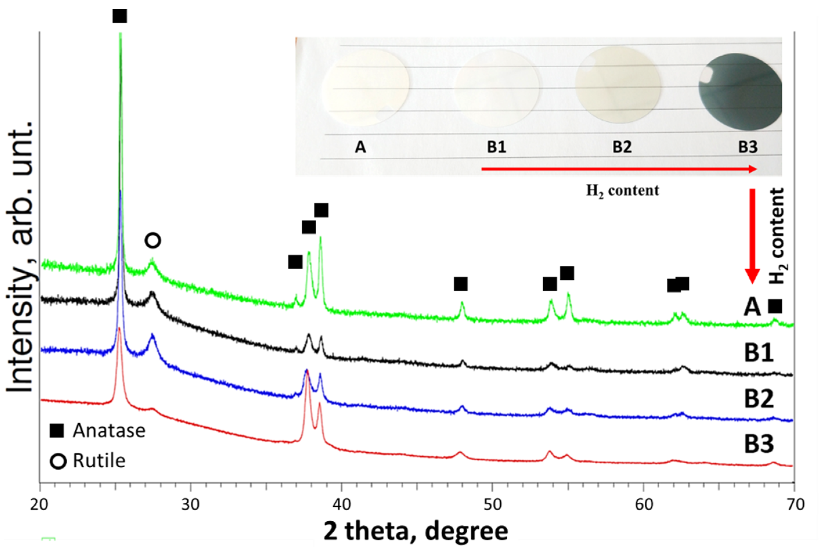
Figure 1. Visual appearance (insert) and XRD patterns of TiO2 (A) and TiHO (B1B3) samples.
Applsci 12 10811 g001
Figure 2. XPS spectra of the as-deposited TiHO films: B1—red, B2—blue, B3—green.
Applsci 12 10811 g002
Figure 3. Optical transmittance (red) and reflection (blue) of TiHO films.
Applsci 12 10811 g003
Figure 4. Tauc plots (blue curves), trendlines for the linear parts of the plots (solid red lines) and respective band gap values (dotted red lines) of TiHO films.
Applsci 12 10811 g004
Figure 5. Estimation of photocatalytic bleaching of MB solution using different TiHO films.
Applsci 12 10811 g005
Table 1. Main parameters of reactive magnetron sputtering deposition process.
SamplesAr FluxO2 FluxH2 FluxAr:O2:H2
Flux Ratio
Total PressurePower Source
A4.5 sccm1.2 sccm-3.78:1:-6 × 10⁻³ mbar300 W p-DC
B12.7 sccm6.4 sccm15.1 sccm0.42:1:2.366 × 10⁻³ mbar300 W DC
B22.8 sccm4.2 sccm20.1 sccm0.67:1:4.786 × 10⁻³ mbar300 W DC
B32.8 sccm3.6 sccm22.5 sccm0.78:1:6.256 × 10⁻³ mbar300 W DC
Publisher’s Note: MDPI stays neutral with regard to jurisdictional claims in published maps and institutional affiliations.